Note: Descriptions are shown in the official language in which they were submitted.
CA 0223~804 1998-04-24
W O 97/15691 PCTAUS96/17191
METHODS AND COMPOSITIONS FOR
DETECTION OF SPECIFIC NUCLEOTIDE
SEQUENCES
Cross-Reference to Related Application
This application claims priority to U.S.
Provisional Patent Application No . 60/005 ,93 8, filed
October 27, 1995, entitled Diagnostic Procedures and
Process for Detection of Specific DNA Sequences. This
provisional patent application is herein incorporated, in its
elltireLy, by reference.
Technical Field
The present invention comprises methods and
compositions for detecting nucleic acid sequences. More
particularly, the present invention comprises methods and
compositions for detection of specific genetic sequences
using nucleic acid target protection strategies. The methods
and compositions of the present invention can be used in the
detection of microorganisms, for diagnosis of infectious
diseases in humans, ~nim~ and plants; assays of blood
products, and for genetic analysis for use in such areas as
early detection of tumors, forensics, paternity
determin~tions, transplantation of tissues or organs and
genetic disease determinations.
CA 0223~804 1998-04-24
W O 97/15691 PCTAUS96/17191
;BackPround of the Invention
Many target and signal amplification methods have
s been described in the literature, but none are believed to
offer the combination of high specificity, simplicity, and
speed. General reviews of these methods have been
prepared by Landegren, U., et al., Science 242:229-237
(1988) and Lewis, R., Genetic Engineering News 10:1,
54-55 (1990). These methods include polymerase chain
reaction (PCR), PCR in situ, ligase amplification reaction
(LAR), ligase hybridization, Q13 bacteriophage replicase,
transcription-based amplification system (TAS), genomic
amplification with transcript sequencing (GAWTS), nucleic
acid sequence-based amplification (NASBA) and in situ
hybridization. Some of these various techniques are
described below.
Polymerase Chain Reaction (PCR)
PCR is the nucleic acid amplification method
described in U.S. Patent Nos. 4,683,195 and 4,683,202 to
Mullis. PCR consists of repeated cycles of DNA
polymerase generated primer extension reactions. The
target DNA is heat denatured and two oligonucleotides,
which bracket the target sequence on opposite strands of the
DNA to be amplified, are hybridized. These
oligonucleotides become primers for use with DNA
polymerase. The DNA is copied by primer extension to
make a second copy of both strands. By repeating the cycle
of heat denaturation, primer hybridization and extension,
the target DNA can be amplified a million fold or more in
about two to four hours. PCR is a molecular biology tool
which must be used in conjunction with a detection
technique to determine the results of amplification. The
3s advantage of PCR is that it may increase sensitivity by
CA 0223~804 1998-04-24
W O 97/15691 PCT~US96/17191
amplifying the amount of target DNA by 1 million to 1
billion fold in approxim~tely 4 hours. The disadvantage is
that cont~min~tion may cause false positive results, or
~ reduced specificity.
Transcription-based Amplification System (TAS)
TAS utilizes RNA transcription to amplify a DNA or
RNA target and is described by Kwoh et al. (1989) Proc.
Natl. Acad. Sci., USA 86:1173. TAS uses two phases of
amplification. In phase 1, a duplex cDNA is formed
cont~ining an overh~n~ing, single-stranded T7 transcription
promoter by hybridizing a polynucleotide to the target.
The DNA is copied by reverse transcriptase into a duplex
form. The duplex is heat denatured and a primer is
lS hybridized to the strand opposite that containing the T7
region. Using this prirner, reverse transcriptase is again
added to create a double stranded cDNA, which now has a
double stranded (active) T7 polymerase binding site. T7
~NA polymerase transcribes the duplex to create a large
quantity of single-stranded RNA.
In phase 2, the primer is hybridized to the new RNA
and again converted to duplex cDNA. The duplex is heat
denatured and the cycle is continued as before. The
advantage of TAS over PCR, in which two copies of the
target are generated during each cycle, is that between 10
and 100 copies of each target molecule are produced with
each cycle. This means that 106 fold amplification can be
achieved in only 4 to 6 cycles. However, this number of
amplification cycles requires approximately three to four
hours for completion. The major disadvantage of TAS is
that it requires numerous steps involving the addition of
enzymes and heat denaturation.
CA 0223~804 1998-04-24
W O 97/15691 PCT~US96/17191
Transcriptions Ampli~lcation (3SR)
In a modification of TAS, known as 3SR, enzymatic
degradation of the RNA of the RNA/DNA heteroduple~ is
used instead of heat denaturation, as described by Guatelli
s et al. (1990) Proc. Natl. Acad. Sci. USA 87:1874. RNAse
H and all other enzymes are added to the reaction and all
steps occur at the same temperature and without further
reagent additions. Following this process, amplifications of
106 to 109 have been achieved in one hour at 42~C.
Li ation Amplification (LAR/LAS)
Ligation amplification reaction or ligation
amplification system uses DNA ligase and four
oligonucleotides, two per target strand. This technique is
described by Wu, D.Y. and Wallace, R.B. (1989) Genomics
4:560. The oligonucleotides hybridize to adjacent
sequences on the target DNA and are joined by the ligase.
The reaction is heat denatured and the cycle repeated. LAR
suffers from the fact that the ligases can join the
oligonucleotides even when they are not hybridized to the
target DNA. This results in a high background. In
addition, LAR is not an ef~lcient reaction and therefore
requires approximately five hours for each cycle. Thus,
the amplification requires several days for completion.
OJ3 Replicase
In this technique, RNA replicase for the
bacteriophage QJ3, which replicates single-stranded RNA,is
used to amplify the target DNA, as described by Lizardi et
al. (1988) Bio/Technology 6:1197. First, the target DNAis
hybridized to a primer including a T7 promoter and a QJ3
5' sequence region. Using this primer, reverse
transcriptase generates a cDNA connecting the primer to its
5' end in the process. These two steps are similar to the
3s TAS protocol. The resulting heteroduplex is heat
CA 0223~804 1998-04-24
W O 97/15691 PCT~US96/17191
denatured. Next, a second primer containing a Q13 3'
sequence region is used to initiate a second round of cDNA
synthesis. This results in a double stranded DNA
containing both 5' and 3' ends of the Q13 bacteriophage as
s well as an active T7 RNA polymerase binding site. T7
RNA polymerase then transcribes the double-stranded DNA
into new RNA, which mimics the Q13. After extensive
washing to remove any unhybridized probe, the new RNA
is eluted from the target and replicated by Q~ replicase.
l O The latter reaction creates 107 fold amplification in
approximately 20 minutes. Significant background may be
formed due to minute amounts of probe RNA that is non-
specifically retained during the reaction.
lS Chiron Signal Amplification
The Chiron system, as described by Urdea et al.
(1987) Gene 61:253, is extremely complex. It utilizes 12
capture oligonucleotide probes, 36 labeled oligonucleotides,
20 biotinylated immobilization probes that are crosslinked
to 20 more enzyme-labeled probes. This massive
conglomerate is built-up in a stepwise fashion requiring
numerous washing and reagent addition steps.
Amplification is limited because there is no cycle. The
probes simply form a large network.
2s
ImClone Si~nal Amplification
The ImClone technique utilizes a network concept
similar to Chiron, but the approach is completely different.
The ImClone technique is described in Kohlbert et al.
(1989) Mol. and Cell Probes 3:59. ImClone first binds a
single-stranded M13 phage DNA cont~ining targeted probe.
To this bound circular DNA is then hybridized about five
additional DNA fragments that only bind to one end and the
other end hangs freely out in the solution. Another probe
set is then hybridized to the hanging portion of the previous
CA 0223~804 1998-04-24
W O 97tlS691 PCTnUS96/17191
set of probes. The latter set is either labeled directly with
an enzyme or it is biotinylated. If it is biotinylated, then
detection is via a streptavidin enzyme complex. In either
case, detection is through an enzyme color reaction. Like
s the Chiron rnethod, the ImClone method relies on build-up
of a large network. Because there is no repeated cycle, the
reaction is not geometrically expanded, resulting in limited
amplification.
While the nucleic acid amplification methods
lo described above allow for the detection of relatively small
quantities of target nucleic acid molecules, there is a need
for the ability to detect target nucleic acid molecules in a
shorter amount of time with less background interference.
Problems inherent in PCR and other amplification
techniques involve sample contamination during the
collection techniques and the presence of amplicons
(amplified target DNA). There are problems with non-
specific target amplification mediated by closely related
sequences and the production of primer dimers. There is
also poor control of specificity, resulting in false positive
reactions, and poor control of sensitivity, resulting in false
negative reactions. PCR results must often be confirmed
and validated by other techniques such as probe
hybridization, Southern blotting or in situ hybridization.
2s Additionally, PCR and amplification
techniques can only be used with very small amounts of
starting sample DNA, in the range of a maximum of 1
microgram. This negates use of PCR techniques for the
detection of low copy nurnber nucleic acid targets. For
example, early detection of HIV infection, soon after the
initial viral infection, would be almost impossible to detect
using PCR.
Thus, compositions, methods and kits are
needed that are capable of detecting specific nucleic acid
sequences and isolating them. Especially needed are
,
CA 0223~804 1998-04-24
WO 97/15691 PCTrUS96/17191
methods and kits that would allow for the detection of low
~ copy number nucleic acid target sequences. Additionally,
there is need for methods and kits that provide the
flexibility that would allow for isolation of nucleic acid
sequences using a desired level of speci~lcity.
What is also needed are methods that do not
use amplification techniques, but do allow for the isolation
of a specific target sequence from any amount of starting
nucleic acid, especially large amounts, and have the
IG flexibility to accomplish the isolation at several levels of
specificity, depending on the level of specificity desired.
Summary of the Present Invention
In accordance with the present invention,
methods and compositions are provided for the detection of
specific nucleic acid sequences from cellular or tissue
sources. More particularly, the present invention includes
methods and compositions for the detection of nucleic acid
sequences using a protection molecule that forms a
protected nucleic acid sequence (PNAS) such as a triplex or
duplex nucleic acid structure that includes the target nucleic
acid sequence. The targent nucleic acid sequence is the
specific sequence being detected. An assay using the
2s methods of the present invention may include one, two or
three levels of specificity to minimi7e false positive signals.
An assay using the methods or compositions of the present
invention can be performed on large amounts of purified
DNA in a single test, with high levels of sensitivity, thus
elimin~ting the need for in vitro DNA amplification
procedures.
.,
CA 0223~804 1998-04-24
WO 97/15691 PCTAUS96/17191
When the target nucleic acid sequence is
double-stranded, the structure forl[ned with the protection
molecule is a triplex. When the target nucleic acid
sequence is single-stranded, the structure formed with the
protection ~nolecule is a duplex. In this disclosure, where
triplex structures are discussed, one can also substitute
duplex structures or structures using PNA (peptide-nucleic
acid) and the appropriate nucleases. Assays using the
methods of the present invention may be referred to as
TPA, Target Protection Assays.
The initial level of specificity utilizes
protection molecules such as oligonucleotides or peptide-
nucleic acids (PNA) to bind to specific target sequences of
interest. Such binding may be accomplished by formation
of Hoogstein-type hydrogen bonds. The protection
molecule, bound to the target nucleic acid sequence, forms
the protected nucleic acid sequence (PNAS). Once these
PNAS structures are formed and stabilized in solution, the
non-specific DNA is digested. For example, this digestion
can be accomplished with a combination of endonucleases
and a double-strand-dependent exonuclease, such as DNA
Exonuclease III (Exo m). The endonucleases used in this
example are designed to cut on both sides of the PNAS,
leaving approximately 20 base pairs of DNA on each side
2s of the sequence. Exo III, an exonuclease which
progressively cleaves one strand of the DNA from the 3'
end, is inhibited by the triple helix structure. Using a
combination of nucleases, the unprotected DNA sequences
are digested completely. A method of the present invention
involving a lower level of specificity would employ an
affinity molecule for capture and a reporter molecule for
labeling in conjunction with the protection probe.
However, if a higher level of specificity is
required, 5' fl~nkin~: regions can be generated on either or
both sides of the PNAS to allow for assays employing two
CA 0223~804 1998-04-24
W O 97/15691 PCT~US96/17191
further levels of specificity. The structure formed, a
PNAS with flanking regions is termed PNAS/tail.
Following the selected digestion around the PNAS, a
capture probe, such as an oligonucleotide complementary to
one of the single stranded fl~nkin~; regions, is added. The
capture probe is allowed to hybridize to a single-stranded
region. For example, the capture probe could be an
oligonucleotide that would bind to a single-stranded region
and have an affinity molecule attached. For example, the
lo affinity molecule could be didoxigenein or biotin. The
capture probe comprises an affinity molecule and is capable
of associating with the PNAS.
A capturing system is used to isolate the PNAS
with the capture probe attached. Any capturing system that
is capable of binding to the capture probe and separating
the PNAS/tails with affinity molecule from the mixture is
contemplated. In the example used above, such a capture
system may comprise using magnetic beads coated with
anti-didoxigenein antibodies for binding to the
didoxigenein-capture probe portion or, streptavidin for
binding to the biotin-capture probe portion. The
PNAS/tails with affinity molecule, now attached to the
magnetic beads, are separated from non-specific complexes
and washed to remove any non-specific nucleic acid
2s sequences. Such w~hin~ may use any washing technique
known in the art. For example, a magnetic particle holder
could be used. Again, should this be the level of specificity
required, the present invention comprises assays that also
have a reporter molecule associated with the protection
molecule or the capture probe.
A third level of specific detection involves the
addition of a labeled reporter probe. The reporter probe
comprises a detectable label and is capable of associating
with the PNAS. For example, the reporter probed may
3s comprise an oligonucleotide complementary to the 5'
CA 0223~804 1998-04-24
W O 97/15691 PCT~US96/17191
single-stranded tail that is part of the PNAS/tail. This 5'
region may or may not be on the opposite 11~nking tail to
which the capture probe binds. The reporter probe may be
labelled with any labels known in the art such as
s radioactivity or non-radioactive labels such as labeled with
biotin or didoxigenein for indirect detection, or directly
with a fluorescent reporter molecule, e.g., fluorescein, or
chemiluminescent or bioluminescent labels. An excess of
reporter probe is added to the washed magnetic bead-
lo triplex complex and allowed to hybridize. Detection of the
bound labelled reporter probe can be accomplished after
washing by using detection devices specific for the type of
label used. For example, if a fluorescent labeled reporter
probe is used, the labeled sequences can be detected using a
fluorometer or viewing the beads through a fluorescent
microscope. Alternatively, the amount of bound probe can
be directly assessed by fluorescent anisotropy with an
analyzer such as the Abbott TDM analyzer.
Compositions of the present invention include
compositions comprising the components to practice the
methods taught herein. For example, a composition
comprising a labeled protection molecule with an affinity
molecule could be used in an assay with a first level of
specificity. A composition comprising a labeled protection
2s molecule and a capture probe could be used in a level two
specificity assay. A composition comprising a protection
molecule, a capture probe and a reporter probe could be
used in a level three assay. It is to be understood that the
individual molecules, probes and components can also be
provided individually.
The present invention is especially useful for
detecting specific genetic sequences. The present invention
comprises methods such as the Target Protection Assay
(TPA) in all its formats, which have the advantage of
allowing the processing of very large amounts of purified
_
CA 0223~804 1998-04-24
W O 97/1~691 PCT~US96/17191
nucleic acids, thus elimin~tin~ the need for artificial
amplification procedures such as PCR, while enabling the
detection of a specific target sequence. In addition, the
three levels of specificity - PNAS formation, ca~Lu~e probe
binding, and reporter probe binding - reduce technical
problems such as those associated with false positive .cign~l~
from non-specific amplification and/or hybridization.
The present invention comprises a method for
detecting a target nucleic acid sequence, comprising
obtaining isolated nucleic acid sequences from a sample
suspected of containing a target nucleic acid sequence;
contacting a protection molecule with the nucleic acid
sequences under hydridizing conditions sufficient to form a
PNAS; and detecting the PNAS. The methods may further
comprise the steps of digesting the isolated nucleic acids
containing one or more PNAS with nucleolytic enzymes to
form a PNAS/tail; and hybridizing a capture molecule to
the PNAS/tail; prior to the step of detecting the PNAS.
Additionally, the methods may further comprise the step of
hybridizing of a reporter molecule to the PNAS/tail; prior
to the step of detecting the PNAS. A method for detecting
specific nucleic acid sequences, comprising obtaining
isolated nucleic acid sequences from a sample suspected of
containing a target nucleic acid sequence; contacting a
protection molecule with the nucleic acid sequences under
hydridizing conditions sufficient to form a PNAS; digesting
the isolated nucleic acids containing one or more PNAS
with nucleolytic enzymes to form a PNAS/tail; hybridizing
a capture molecule to the PNAS/tail; hybridizing of a
reporter molecule to the PNAS/tail; and detecting the
PNAS.
The present invention comprises compositions
for detecting specific nucleic acid sequences, comprising a
protection moleculecapable of binding with a specific
nucleic acid sequence. A composition of the present
CA 0223~804 1998-04-24
W O 97/15691 PCT~US96/17191
invention may further comprise a capture Inolecule.
Additionally, a composition of the present invention may
further comprise a reporter molecule.
The methods and compositions of the present
s invention should be ideal for the detection of viruses and
other microorganisms such as pathogens of hum~n.s,
~nim~ls and plants, as well as genetic analysis of
polymorphic gene sequences such as HLA typing. The
methods of the present invention can be used in forensics,
lo paternity determinations, or transplantation or organs or
tissues, or genetic disease analysis.
Accordingly, it is an object of the present
invention to provide methods to detect specific genetic
sequences.
It is yet another object of the present invention
to provide methods for detecting specific DNA sequences
involving triplex nucleotide structures.
It is another object of the present invention to
provide methods for detecting specific RNA sequences
involving triplex nucleotide structures.
It is yet another object of the present invention
to provide methods for detecting specific DNA sequences
involving duplex nucleotide structures.
It is another object of the present invention to
2s provide methods for detecting specific RNA sequences
involving duplex nucleotide structures.
It is another object of the present invention to
provide methods for detecting specific RNA sequences
involving PNA structures.
It is another object of the present invention to
provide methods for detecting specific DNA sequences
involving PNA structures.
It is yet another object of the present invention
to provide methods for detecting specific DNA sequences
3s involving antibodies.
CA 0223~804 1998-04-24
W O 97/15691 PCTAUS96/17191
It is yet another object of the present invention
to provide methods for detecting specific RNA sequences
involving antibodies.
- Another object of the present invention is to
provide a method of detecting nucleic acid sequences
involving radioactive labeled nucleic acids.
It is another object of the present invention to
provide a method of detecting nucleic acid sequences
involving non-radioactive labeled nucleic acids.
lo It is yet another object of the present invention
to provide a method of detection of specific genetic
sequences with variable levels of specificity.
Another object of the present invention is to
provide a method of detecting nucleic acid sequences for
the determination of the identity of microorg~ni.~m~.
It is another object of the present invention to
provide a method of detecting nucleic acid sequences for
the determination of the identity of human pathogens.
It is yet another object of the present invention
to provide a method of detecting nucleic acid sequences for
the determination of the identity of ~nim~l pathogens.
It is yet another object of the present invention
to provide a method of detecting nucleic acid sequences for
the determination of the identity of plant pathogens.
2s It is another object of the present invention to
provide a method of detecting nucleic acid sequences for
the determin~tion of the genetic relationship, such as
paternity or species identification, of a sample.
It is yet another object of the present invention
to provide a method of detecting nucleic acid sequences for
the determin~tion of potential donors of organs or tissues
for transplantation purposes or for protecting the blood
supply.
CA 0223~804 1998-04-24
W O 97/15691 PCT~US96/17191
14
It is another object of the present invention to
provide a method of detecting nucleic acid sequences ~or
use in forensic determin~tions.
It is yet another object of the present invention
to provide a method of detecting nucleic acid sequences for
the analysis of genetic diseases.
It is another object of the present invention to
provide methods for testing body or tissue fluids to detect
microorg~ni~m~ or other pathogens.
These and other objects, features and
advantages of the present invention will become apparent
after a review of the following detailed description of the
disclosed embodiments and the appended claims.
E~rief Description of the Draw;n~
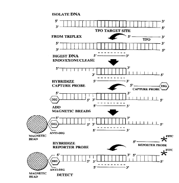
FIG. 1 shows the steps of a method of the present
invention. There are five individual steps in the TPA
procedure as shown in FIG. 1: DNA isolation; PNAS
formation, which in FIG. 1 is a triplex formation;
endo/exonuclease digestion; addition of the capture probe
with an affinity molecule, which in FIG. 1 is didoxigenein;
isolation of the PNAS/tails with capture probe by addition
of m~gnetic beads; addition of the reporter probe with its
label, which in this case, the label is FITC (fluorescein
2s isothiocyanate) and detection of the labeled PNAS/tail with
reporter and capture probes.
Detailed Description
The present invention includes methods for the
detection of a specific target nucleic acid sequence using a
protection molecule that forms a protected nucleic acid
sequence (PNAS) structure including the target nucleic acid
sequence. An assay using the methods of the present
invention may be referred to as TPA, target protection
assay. One embodiment of the present invention is a
CA 0223~804 l99X-04-24
W O 97/15691 PCT~US96/17191
method for the detection of specific DNA sequences. The
present invention also includes methods for the detection of
specific RNA sequences. In the disclosure herein, the
- nucleic acid DNA will be used but it is to be understood
that any nucleic acid, including RNA, can be used with the
methods of the present invention. Where specific nucleases
are referred to, any nuclease that can perform the specified
function can be substituted for the named nuclease.
The steps of a method of the present invention, the
lo Target Protection Assay (TPA), involves the combination
of several techniques to arrive at a unique nucleic acid
diagnostic tool that is specific for a target nucleic acid
seuqence. A preferred method of the present invention,
directed at a DNA target nucleic acid, involves the steps of
1) DNA isolation; 2) forrnation of the PNAS; 3) enzymatic
digestion of unprotected DNA; 4) capture; and 5) labeling
and 6) detection of the PNAS.
Many of the individual procedures s-lmm~rized
herein may use techniques known to those skilled in the art
of molecular biology, with several variations taught in the
literature, or commercially available in the form of kits. It
is to be understood that the present invention is not limited
by the specifically disclosed techniques, but any techniques
that are capable of performing the same function or result
2s can be substituted for the ones described. For the purpose
of example, a single technique will be described for each
step in the TPA procedure. Suitable alternative techniques
are noted where appropriate. However, the present
invention is not to be limited thereby as many other suitable
alternatives are intended to be included within the scope of
the invention.
The present invention includes within its scope such
nucleic acid targets as DNA (single and double stranded)
and RNA (single and double stranded). The methods of the
3s present invention are useful for specifically detecting the
CA 0223~804 1998-04-24
W O 97/lS691 PCT~US96/17191
16
presence of very low copy number nucleic acid targets in a
vast excess of non-target nucleic acids.
The methods of the present invention involve
protecting the target nucleic acid sequence from nuclease
attack with the protection molecule, a molecule such as a
single stranded DNA or RNA or a peptide nucleic acid
(PNA). The protection molecule is selected or designed to
bind specifically to the target nucleic acid sequence. The
protection molecule, in association with the target nucleic
acid sequence, forms a structure, the PNAS. For example,
the PNA~ includes, but is not limited to, triplex and duplex
nucleic acid structures, peptide nucleic acid and antibody
associated structures.
The methods of the present invention may include a
protection molecule associated with an af~mity molecule
that allows binding of the PNAS in solution to a fixed
substrate. The presence of the af~mity molecule permits
the removal of the excess extraneous nucleic acid. The
methods of the present invention further involve a reporter
molecule to perrnit visll~li7~tion of the presence of the
target nucleic acid.
An assay of the present invention that allows for the
lowest level of specificity involves the binding of the
protection molecule to the specific target nucleic acid
2s sequence to form the PNAS. Diagnostic technologies are
valuable only if they achieve high specificity (few false
positives) and high sensitivity (few false negatives). In
order to provide for a higher level of specificity, the
present invention comprises methods wherein the target
nucleic acid sequence is protected from nuclease attack
when bound by the protection molecule to form the PNAS,
and one or two enzymatically generated 5' DNA tails are
generated, on one or both sides of the PNAS for
hybridization with a capture probe containing an affinity
molecule. The capture probe is selected or designed to
CA 0223~804 1998-04-24
W O 97/15691 PCT~US96/17191
specifically bind to a tail region of a tail-contz~inin~ PNAS.
This assay results in two levels of specificity-binclin~ of the
protection molecule and binding of the affinity molecule to
- the tail of the target nucleic acid. The affinity molecule
allows 3~or t~e attac~rnent of the entire protec~ion structure
with the bound affinity molecule to be attached to a fixed
substrate. In this example of an assay with two levels of
specificity, either the affinity molecule or the protection
molecule are labelled with any type of label known to those
skilled in the art. The label would allow for detection of
the PNAS having an affinity molecule.
A third level of specificity can be added to the assays
contemplated by the present invention by generation of two
different tail regions, preferably one on each side of the
target nucleic acid, that extend beyond the target nucleic
acid sequence bound by the protection molecule. The tail
regions are included in the protected structure but are not
bound to the protection molecule and the structure is named
the PNAS/tails. One tail region could be bound to the
capture probe to anchor the target to a fixed substrate (via
the affinity molecule), and the other tail used to bind a
reporter probe with label to visualize the presence of the
nucleic acid target. The tail regions of the PNAS may or
may not be necessary for use depending on the level of
specificity desired. Preferrably, the capture probe and the
reporter probe are selected or designed to bind specifically
and exclusively to one tail or the other, thereby ensuring
that each of the two probes hybridizes to the PNAS/tail.
Increasing the number of levels of specificity
increases the specificity of the assay (no false positives),
however, excessive levels of specificity may decrease the
levels of sensitivity generated (high false negatives). The
present invention comprises assays that are dynamic
diagnostic technologies that can be customized to deal with
CA 0223~804 1998-04-24
W O 97/15691 PCTrUS96/17191
any specific nucleic acid target and yield any of a variety of
desired levels of specificity.
Nucleic ~ci~ Isolation
s
Nucleic acids may be isolated using any methods
known to those skilled in the art. Nucleic acids, as used
herein, means both DNA and RNA in all its forms found in
cells or constructed by molecular biological techniques.
l o The method for DNA isolation used will largely
depend on the amount and type of material to be extracted.
Virtually any DNA isolation procedure reported in the
Jiterature which produces genomic or mitochondrial DNA,
or any commercially available DNA isolation kit will
suffice. The method conternplates that the sample amount
of DNA to be used in each assay is concentrated in a
volume that can range from 0.1 to 1.0 mL depending on
the solubility of the DNA being tested. Larger DNA
samples may require use of greater sized volumes. The
methods of the present invention may test amounts of
sample nucleic acids between picogram amounts to
milligram amounts. The reactions components would have
to be adjusted, for example, to provide adequate amounts
for hybridization of the components. The reaction
components are proportionate to not only the size of the
sample tested but also to the relative number of target
sequences that are present. It is to be understood that the
a~nount of sample DNA will depend on the size and kind of
sample.
The DNA is placed in a buffer suitable for the
formation of PNAS, such as a duplex or triplex structure
using a duplex or triplex forming oligonucleotide (DFO or
TFO) or a peptide nucleic acid. Many procedures have
been reported for the isolation of high molecular weight
3s DNA from several sources including whole blood, isolated
CA 0223~804 1998-04-24
W O 97/15691 PCT~US96/17191
19
blood cells, serum and plasma, fresh, frozen or prepared
- tissues, and tissue culture cells.
RNA can also be isolated by any methods known to
those in the art. Published RNA isolation protocols lyse the
s cell in a chemical environment that denatures ribonucleases,
and fractionates the RNA type of interest from other RNAs
and other cellular macromolecules. The RNA isolation
method used is dependent upon the cell type from which the
RNA is isolated and the eventual use of the RNA.
There are published methods for preparing total
RNA from eukaryatic cells, and such methods are herein
incorporated by reference. In Favaloco, et al., 1979 and
Chomczynski and Sacchi, 1987, cells are lysed using
guanidinium isothiocyanate. This method has few
manipulations and yields clean RNA from many sources,
and is the method of choice for tissues that have high levels
of endogenous RNAse. In the third method of Palmiter,
1974, cells are lysed with phenol and SDS. This results in
clean, high molecular weight RNA from large quantities of
plant cells and also works well with some m~mm~ n cells
and tissues.
Published methods for preparing total RNA from
prokaryotic cells include: protocols for extracting RNA
from gram-negative and gram-positive bacteria, using
protease digestion and organic extraction to remove protein
and nuclease digestion to remove DNA (Reddy, et al.,
1990); and a simple protocol for rapidly isolating RNA
from E.coli without organic extractions, protease, or
nuclease treatment (Summers, 1970). Lastly a published
method (Aviv and Leder, 1972) can fractionate messenger
RNA from ribosomal and transfer RNA based upon the
exclusive presence of poly (A) tails on mRNA.
CA 0223~804 1998-04-24
W O 97/15691 PCTAUS96/17191
PNAS Formation
After isolation of the nucleic acid, the next step in the
methods of the present invention include forrnation of the
PNAS. This step introduces the first level of specificity to
the assay. This step involves the forrnation of the PNAS
using a target nucleic acid sequence-specific TFO or DFO
or PNA. Hereinafter, TFO will be used in the example of a
preferred embodiment, but it is to be understood that
triplex and duplex structures and PNA are contemplated by
the present invention.
The sequence of the TFO will depend on the speci~lc
target sequence to be detected. The most well characterized
triplex structure is the one formed between a double
stranded homopurine-homopyrimidine helix and a single
stranded homopyrimidine tract. Formation of such
structures are well known in the art. Specific details of the
formation of such structures are given in the following
references which are herein incorporated by reference. S.
W. Blume, J. E. Gee, K. Shrestha, and D. M. Miller. Triple
helix formation by purine-rich oligonucleotides targeted to
the human dihydrofolate reductase promoter. Nucl. Acids
Res. 20: 1777-1784 (1992).
In this first type of triple helix, the third
homopyrimidine strand binds to the major groove, parallel
to the hornopurine strand of the Watson-Crick double
helical DNA via Hoogstein hydrogen bonding. The third-
strand thymidine (T) recognizes adenine-thymine (A:T)
base pairs forIning T:A:T triplets, and the third strand
cytosine (C), protonated at the N-3 position, recognizes
guanidine-cytosine (G-C) base pairs forming C+: G: C
triplets. Homopyrirnidine oligonucleotide have been shown
to form local triplexes with corresponding homopurine
sites in larger double-stranded DNAs. An alternative
triplex structure is a double stranded hornopyrimidine-
homopurine helix and a single stranded homopurine tract
CA 0223~804 1998-04-24
W O 97/15691 PCTrUS96/17191
21
(TFO). Yet other alternative triplex structures comprise a
combination of the two described structures.
The design of the TFO will generally follow the
Pyrimidine-Purine-Pyrimidine binding rules described
s previousiy, or may be designed to form Purine-Purine-
Pyrimidine triplexes if necessary. Such structures are well-
known in the art. However, other binding motifs also
apply, examples: I. Rec A Mediated TFO binding in 4 base
regions (Rec A required to remain in solution); II. Triple
purine and triple pyrimidine triplexes. Rec A is a
recombinant enzyme that catalyzes the recombination
between two DNA strands with simil~r homology.
While not wishing to be bound by the following
theory, the premise of using TFOs to select specific regions
lS of DNA for diagnostic use requires one to have a conserved
sequence of DNA from the target sequence and to have a
long enough sequence to ensure hybridization and
selectivity. The human genome has approximately 5 x 10⁹
base pairs of DNA. In order to have a unique sequence this
would require an oligonucleotide of approximately between
16-20 nucleotides long. The actual number is probably
smaller due to the presence of intron sequences in the
DNA. Longer sequences increase the hybridization
between the TFO and DNA while decreasing the specificity.
The selection of the TFOs is based on an empirical
search for poly-purine/pyrimidine stretches in the target
region. In the methods of the present invention, several
confounding factors such as DNA/protein interactions
should not interfere with the binding of the TFO to its
target sequence. Also, secondary structure can be
influenced by temperature, which should allow for more
efficient TFO binding. Often the sequence is not entirely a
homopurine strand but contains intermixed pyrimidines.
Even though the introduction of pyrimidines could lower
the TFO's affinity for the duplex DNA, the entire sequence
CA 0223~804 1998-04-24
W O 97/15691 PCTAUS96/17191
still allows selective binding at the proposed hybridization
temperature. The conditions to form the triplex structure
may also vary depending on the target sequence, but must
be compatible with the nucleases used in the subsequent
step. For example, the conditions may need to be adjusted
for activity by Exonuclease III (Exo III) and the restriction
endonucleases chosen for the next step in the procedure (see
next section).
To aid in the forrnation of a triplex structure, a low
pH buffer (pH 6.8 - 7.4) would be optimal. This would
also serve to help stabilize the structure during the
enzymatic digestion step. Additional stabilization
procedures, known to those in the art, can also be
employed. For example, while TFOs may work well under
a variety of situations, there are two fundarnental problems
unique to triplex formation. One is that, for the CT motif,
acidic pH is required for triplex formation. The second is
that the recognition sequence is limited to oligopurines.
The ~lrst problem can be approached by altering the nucleic
acid with chemical modifications, such as those taught in
the art. See J. S. Lee, L. J. Woodsworth, P. Latimer, and
A. R. Morgan. Poly(pyrimidine).poly(purine) synthetic
DNA's containing S-methylcytosine forrn stable triplexes at
neutral pH. Nucleic Acids Res. 12: 6603-6614 (1984). This
is done by replacing dC with modified bases such as 5-
methyl-dC, C-5 propyne pyrimidine, 6-methyl-8-oxo-2'-
deoxyadenosine, or 2'-O-methylpseudocystein.
Another approach is to add a linker to increase and
stabilize the interaction with the target sequence. In an
additional approach the TFO can also be conjugated to
unique chemical groups to allow the formation of a triplex
structure when it normally would not. Not only can triple-
stranded DNA complexes be stabilized by a high ionic
strength or by the presence of cations like magnesium, but
also by triple-helix specific ligands called
CA 0223~804 1998-04-24
W O 97/15691 PCT~US96/17191
benzopyridoindole (BPI) derivatives, which intercalate in
triple helix complexes. The present invention contemplates
all of these methods that are well known in the art and
other binding schemes that function in the same m~nner.
Lower pH conditions are compatible, although not
necessarily optimal, with Exo III and most restriction
endonucleases. In addition, these conditions allow triplex
formation at the elevated temperatures (37~C) needed for
the subsequent digestion step.
An example of formation of a triplex structure is
given here. A >10-fold molar excess of the TFO is added
to the isolated DNA (10 pmoles TFO/ ,ug DNA) and the
triplex structure is allowed to form for 10 min. When the
DNA and TFO are mixed in equal amounts, the kinetics of
triplex formation has been characterized by half-decay
times (tl/2) of 150-390 seconds. By contrast, when the
TFO was in ten-fold excess over the DNA the kinetics were
faster and the tl/2 decreased to 19-28 seconds. The rate of
triplex appears to be about three orders of m~gnitude
slower than the rate of duplex recombination, which has a
rate constant in the order of 106. The apparent activation
energy associated with the rate constant of triplex
formation was small and negative (E1 = 26+15 kJ/mol).
The first order rate constant of triplex formation (k 1)
depends on temperature and was in the range of 10-7 to 10-
S s-1 (at 20~C and 33~C, respectively), with an apparent
activation energy that was large and positive (E l = 355+33
kJ/mol). The rate of triplex formation also showed a
dependence on ionic strength (I) of the buffer solution
(17,23,24). A decrease of I from 137 mM to 57 mM
resulted in a six-fold decrease in the association constant.
Enzymatic Digestion of DNA
This step in the methods of the present invention
3~ assures that S' tails of approximately at least 20 base pairs
CA 0223~804 1998-04-24
W O 97/15691 PCT~US96/17191
24
are generated upstream and downstream from the PNAS.
These tails are useful for the capture and detection steps.
This step also ensures that all non-specific nucleic acids are
digested as well as unbound TFO, DFO and PNA
s molecules, thus reducing potential false-positive .~i~n~l.s.
More specifically, once the PNAS is formed and
stabilized, a mixture of exo- and endonucleases are added to
the mixture. The endonucleases are sequence specific
restriction enzymes chosen to flank the target nucleic acid
site, leaving approximately 20 base pairs (usually more) of
nucleic on each side. Where the target nucleic acid
sequence is dsDNA, the exonuclease must be ds DNA
dependent which digests only one strand (either 3' to 5' or
5' to 3'), leaving large tracts of ss DNA available for
hybridization with specific probes. A preferred enzyme
(and the one used in all examples) is Exo III. Exo m is a
monomeric protein of 28,000 Daltons that catalyzes the
stepwise 3' to 5' removal of 5'-mononucleotides from ds
DNA with a free 3'-OH end. Exo III also contains an
2Q inherent 3' phosphatase activity and a RNAse H activity.
Thus, Exo III can also be used in methods of the present
invention that use RNA target sequences.
The enzymes shown in Table 1 may be used in the
present invention. The present invention is not limited to
the disclosed enzymes.
CA 02235804 1998-04-24
W O 97/15691 PCTfUS96/17191
Table 1
Properties of some m~mm~ n nucleases
s
Enzyme Substr~te Mode of pHZ Mgl ~ Rr~ ~ Mol. wt
nctionl product3
DNAsel ds/ssDNA Endo 7.1 + S' oli,s~os 31 Kdal
DNAse 11 ds/ss DNA Endo 4.1 - 3' oligos 38 Kdal
DNAse m ss Duplex DNA Exo 8 . 5 + 5'monodinu-~
DNAselV DuplexDNA Exo 3'5' 8.5 + 5' mono 42Kdal
DNAseV DuplexDNA Exo 3'5'15'3' 8.8 + 5' mono 12Kdal
DNAse Vl ssDNA Endo 9.5 + 5'oligo 45 Kdal
DNAseVII ss and nicked & Exo 3'5' 7.8 + S' mono 43 Kdal
ds DNA
DNA Vlll 5' ss and nicked Exo S'3' 9.5 + 5' oli,~os 31 Kdal
Correxo ss DNA nicked Exo 3'5'/5'3' 8.0 + 5' oligos 30-35
W'd ds DNA4 Kdal
Lysosomal or RNA or DNA Exo 5'3' S.S - 3' mono 70 Kdalspleen with 5' OH
exonuclease
I Endo = .~nrlnn~lrle~lytic. Exo = ~y~mlrl~r)iytic
2 optimum pH
3 O14;vllu~1c,vtide shown is the main reaction product.
oligos = olig(-ml~ oti~
mono = .. -r.. l~ tidr~
4 UV inadiated double stranded DNA
Exo III is commercially available from many sources
at a reasonable cost, and will create the desired single
stranded regions adjacent to the target DNA. Most
importantly, Exo III will not digest dsDNA that is in a
triplex structure, and thus the PNAS with the target
- 20 sequence will be protected from digestion. One unit of Exo
III will digest 50 ng of genomic DNA at 37~C in 10 min.
The main purposes of the endonucleases is to produce free
ds DNA ends close to the TFO target site to aid the Exo III
CA 0223~804 1998-04-24
W O 97/15691 PCT~US96/17191
26
Furthermore, endonuclease activity will increase the
solubility of the sample DNA and complete digestion would
elimin~te nontarget DNA as a source of non-specific
interactions. In some reactions, pretreatment with
noninterfering nucleases may be used to increase the
nucleic acid solubility and help minimi7e the solution
volume to be tested. This should allow the use of less Exo
III than would be required to digest full length genomic
DNA. In addition, complete endonuclease digestion is also
lo not necessarily required to obtain the desired product.
In its simplest form, methods of the present invention
can be fulfilled by this single protection step by
concomitantly introducing a molecule for the capture
system and a reporter molecule for target identification of
the PNAS, yielding an assay with a single level of
specificity. Additional nuclease steps may be necessary to
prevent interference from unbound TFO and non-specific
signals. In order to increase the level of the specificity,
additional steps involving additional oligonucleotide probes
can be added.
Qli~onucleotide Probe - Capture System
l~urther steps in the methods of the present invention
comprise the second level of specificity. These steps
involve the hybridization of a capture probe cont~ining an
affinity molecule (such as biotin or digoxigenin) to the
digested PNAS/tail and binding of the complex to a
derivatized solid support (such as magnetic beads,
microtiter plates, or membranes). This step allows greater
sample manipulation because it can be used for
concentration of the target sequences, buffer exchange, as
well as removal of non-target nucleic acids.
The sequence of the capture probe will be
complementary (Watson-Crick base pairing) to one of the
ss (single-stranded) DNA regions fl~nking the PNAS which
CA 0223~804 1998-04-24
W O 97/15691 PCT~US96/17191
was generated by the nuclease digestion step. For example,
a greater than l0-fold molar excess of the capture probe
can be added to the PNAS/tail under conditions favoring
specific hybridization. Such conditions are known to those
skilled in the art. For example, 2.0M NaCl, 0.2 M sodium
acetate, pH 4.5, 50~C, for l hour could be used. Following
hybridization, the complexes will be purified by co-
incubation with the derivatized solid support for an
additional l hour under the same conditions, followed by
adequate w~hing of unbound complexes (e.g. 8 times with
hybridization buffer).
At this point, the complexes may be dissociated from
the support, if desired, with a dissociation buffer. Such
conditions are known to those skilled in the art. For
example,l.0 M Tris-HCl, pH 9, 0.5 mM EDTA for 20 min.
could be used.
The options for affinity capture systems are
numerous and are well known in the art. Such capture
systems include, but are not limited to, the two most cited
systems, biotin (capture with streptavidin) and digoxigenin
(Dig, Boehringer-Mannheim, captured with anti-Dig
antibody). However, any other .~imi1~r system can be used.
In the case of solid supports, the situation is similar.
The use of derivatized membranes (such as nylon) have had
2s widespread application in the literature, and could be used
in the present invention where detection using film
exposure or phosphor im~ging (such as with radioactivity
or chemiluminescence) is desired. These supports also
work well with the available enzyme conjugate systems
(ic~1k~1ine phosphatase [AP] or horseradish peroxidase
[HRP]) with non-radioactive color producing substrates.
Another option for a solid support is a derivatized
microtiter plate. These plates are available with many
options from several sources. One advantage of microtiter
3s plates is the availability of many supporting systems for
CA 0223~804 1998-04-24
WO 97/15691 PCT~US96/17191
28
automated manipulation (i.e. w~hin~ steps) and detection
options (radioactivity, U.V. and visible light spectroscopy,
and fluoresence). This system has the disadvantage of
being limited to a relatively small volume (lO0 - 200
s ,ulJwell).
A system that is rapidly growing in popularity is the
use of derivatized magnetic beads (Dynal). Non-m~gnstic
beads (usually agarose or sepharose) have been used for
affinity capture and purification for many years. The
magnetic bead system is a preferred system for the
manipulations needed for the methods of the present
invention, and it will be the system used for the example
here. These beads are available derivatized with both
strepavidin and anti-Dig.
The assay could be completed at this point if this
level of specificity is acceptable. The capture probe or
protection molecule could be labeled so that the captured
PNAS could be detected.
2Q Oligonucleotide Probe Detection
The third level of specificity in the methods of the
present invention is achieved through the use of a reporter
probe. It is also at this step that the specific mechanism of
2s detection is introduced. The reporter probe consists of a
synthetic single stranded oligonucleotide complementary to
the opposite single stranded end (not being used for
attachment to the capture system) generated by the nuclease
digestion. The composition of this detection step will vary
depending on the method used. All methods of detection
will require the presence of a reporter probe that be
specifically detected as it binds to a specific sequence on the
captured PNAS. For this invention the method need only
be sufficiently sensitive to detect this specific probe-
CA 0223~804 1998-04-24
W O 97/1~691 PCT~US96/17191
29
complex interaction so that a positive results can be
defined.
The composition of the oligonucleotide probe will
depend on the method of detection used. For direct
s detection of the probe, the probe may simply be a specific
sequence of nucleotides complimentary to the specific
sequence on the PNAS/tail where the interaction is detected
by any physical method that can detect a specific interaction
of oligonucleotides. An example of such a detection
technique would be fluorescence anisotropy where the
relative amount of bound probe can be measured directly
without the removal of unbound probe.
In the case of fluoresence anisotropy, the relative
level of bound probe can be measured directly without the
removal of unbound probe. Methods based on separation
might perturb the equilibrium binding of the probe and
may led to erroneous results. In the use of anisotropy
spectrophotometric deterrnin~tion, the concentration of free
and bound material are measured by an observable change
in the chromophore (i.e. due to changes in the molecular
weight after hybridization). The fraction bound can be
expressed as fb = (robs - rin)/(rb - rin)~ where fb is the
fraction bound, rin is the initial anisotropy, rObs is the
observed anisotropy after hybridization, and rb is the total
binding (determined by titrating a small concentration of
the probe with an excess of binding agent). With this
information, the kinetics of binding can be seen for both
small molecules and rnacromolecules. This methodology
has been applied to observing oligonucleotide
3c hybridization in solution, and is used in the TPA assay.
Other physical methods may include evenescent wave
technology that detects changes in the physical properties of
a surface as proteins or nucleic acids specifically interact on
that surface. There are a number of related physical
methods that can be used where the specific interaction can
CA 0223~804 1998-04-24
W O 97/15691 PCT~US96/17191
be measured without separation of bound and free labeled
oligonucleotide .
Direct detection of the oligonucleotide probe can
involve a specific sequence of nucleotides complimentary to
the 5'tail on the PNAS/tail moiety where the
oligonucleotide is derivatized with a label that can emit a
signal when specifically bound to the target DNA Triplex.
For the detection to be specific, any unbound directly
labeled oligonucleotide would have to be separated from
lo the bound form prior to detection. Examples of labels that
can be directly incorporated into oligonucleotides include:
radioactive isotopes, such as 3H, 14C, 32p,125I that are
detected using scintillation or gamma counters, fluorescent
dyes that can be detected by fluorimeters, bioluminescent,
chemiluminescent or electrochemiluminescent labels that
can be detected using specific triggering reactions to
generate light that can be quantified in a luminometer.
The various types of labels and methods of labeling
nucleotide sequences are well known to those skilled in the
art. Many of these labeling formats can be used in the
above described assays with the first or second level of
specificity. Several specific labels or reporter groups are
set forth below.
For example, the label can be a radiolabel such as,
but not restricted to, 32p, 3H, 14C, 35S, 125I, or 131I. A
32p label can be incorporated into the sequence of the
probe by nick-tr~n~l~tion, end-labeling or incorporation of
labelled nucleotide. A 3H, 14C or 35S label can be
incorporated into the sequence of the probe by
incorporation of a labelled precursor or by chemical
modification. An 125I or 131I label can be incorporated
into the sequence of the probe by chemical modification.
Detection of a label can be by methods such as scintill~tion
counting, gamma ray spectrometry or autoradiography.
CA 0223~804 1998-04-24
W O 97/15691 PCT~US96/17191
The label can also be a Mass or Nuclear Magnetic
Resonance (NMR) label such as, for example, l3C, l5N, or
l 9 O. Detection of such a label can be by Mass
Spectrometry or NMR.
s Dyes and fluorogens can also be used to label the
probes. Examples of dyes include ethidium bromide,
acridines, propidium and other intercalating dyes, and
4',6'-diamidino-2-phenylindole (DAPI)(Sigma Chemical
Company, St. Louis, MO) or other proprietary nucleic
lo acid stains. Examples of fluorogens include fluorescein and
derivatives, phycoerythrin, allo-phycocyanin, phycocyanin,
rhodamine, Texas Red or other proprietary fluorogens.
The fluorogens are generally attached by chemical
modification. The dye labels can be detected by a
spectrophotometer and the fluorogens can be detected by a
fluorescence detector.
The probe can alternatively be labelled with a
chromogen to provide an enzyme or affinity label. For
example, the probe can be biotinylated so that it can be
utilized in a biotin-avidin reaction which may also be
coupled to a label such as an enzyme or fluorogen. The
probe can be labelled with peroxidase, ~lk~line phosphatase
or other enzymes giving a chromogenic or fluorogenic
reaction upon addition of substrate. For example, additives
such as 5-amino-2,3-dihydro- 1 ,4-phthalazinedione (also
known as LuminolTM) (Sigma Chemical Company, St.
Louis, MO) and rate enhancers such as p-hydroxybiphenyl
(also known as p-phenylphenol) (Sigma Chemical
Company, St. Louis, MO) can be used to amplify enzymes
such as horseradish peroxidase through a luminescent
reaction; and luminogeneic or fluorogenic dioxetane
derivatives of enzyme substrates can also be used.
Recognition sites for enzymes, such as restriction
enzyme sites, can also be incorporated into the probes to
3s provide a detectable label. A label can also be made by
CA 0223~804 1998-04-24
WO 97115691 PCTAUS96/17191
incorporating any modified base or precursor cont~ining
any label, incorporation of a modified base cont~ininp~ a
chernical group recognizable by specific antibodies, or by
detecting any bound antibody complex by various means
including immunofluorescence or imrnuno-enzymatic
reactions. Such labels can be detected using enzyme-linked
imm~lnoassays (ELISA) or by detecting a color change with
the aid of a spectrophotometer. It will be understood by
those skilled in the art that other reporter groups can also
be used.
Indirect detection of the oligonucleotide probe can
involve a specific sequence of nucleotides complimentary to
the specific sequence on the PNAS/tail where the
oligonucleotide is derivatized with a reagent or entity that
can be caused to produce a detectable signal in the presence
of another specific reagent or entity. An example of an
indirect detection system is the covalent derivatization of
the oligonucleotide probe with a unique chemical structure
that can be uniquely recognized by a binding partner; i.e., a
hapten label such as biotin or digoxigenin or a unique piece
of nucleic acid or nucleic acid related material where
avidin, anti-digoxigenin, or a complimentary strand of
nucleic acid itself is directly labeled and capable of
detection by a physical method after removing any free
label from specifically bound label. Another example of an
indirect label is an oligonucleotide that is covalently
derivatized with an enzyme that can convert a substrate into
a detectable compound or release energy that can be
detected by physical methods. Examples of enzyme-
substrate pairs that can be used for indirect detection
include:
l) Phosphatases such as ~lk~line phosphatase that can
be detected by addition of phosphorylated compounds
which when dephosphorylated by the result in compounds
that, a) absorb light at a wavelength different form the
CA 0223~804 1998-04-24
W O 97/15691 PCTAUS96/17191
substrate; b) can produce a specific fluorescence; c) become
luminescent; d) become a substrate for a second enzyme
that can be included with a second substrate to generate a
detectable signal.
2) Peroxidases, for example, horseradish
peroxidase, whose reaction products in the presence of
appropriate compounds can generate compounds that, a)
absorb light at a wavelength different form the substrate; b)
can produce a specific fluorescence; c) become luminescent;
d) become a substrate for a second enzyme that can be
included with a second substrate to generate a detectable
signal.
3) Luciferases that can be detected by addition of
appropriate substrates and cofactors which result in the
production of light. Alternatively, luciferases can be
included as the second enzyme in assays where the substrate
was a phosphorylated luciferin that is only acted upon by a
luciferase after removal of the phosphate. Other hydrolytic
enzymes other than the specific ones listed here can be used
as indirect enzyme labels.
The methods of the present invention can be used to
detect single copy or low copy number nucleic acid
sequences from any size sample, including large amounts of
nucleic acids. An unexpected benefit of the assays of the
present invention resides in the ability to process large
samples of nucleic acid and to detect and quantify specific
nucleotide sequences that make up only a minor component
of the complex mixtures of sequences in the large sample.
The sensitivity limits can be approxim~ted by
evaluation of available detection systems combined with the
amount of target that can be obtained from a specific
sample size. A very sensitive system for nucleic acid
detection is a bioluminescence technique based on the
photoprotein, AquaLite(~). This technique is described in
Actor et al.,(l996) J. NIH Res. 8 (10):62, herein
CA 0223~804 1998-04-24
WO 97/15691 PCT~US96/17191
34
incorporated by reference in it entirety. The system is
capable of detecting 3 x 10⁶ speci~lc sequences of DNA in a
hybridization immllnOaSSay technique with high signal to
background noise ratio.
s In the methods of the present invention, a
bioluminescent conjugate of AquaLite(~), coupled to an anti-
digoxigenin antibody, is used to detect a digoxigenenin
labeled reporter probe containing 2-3 digoxigenin
molecules used in the methods of the present invention. At
the present time, the lower limit of detection of the signal
produced by the bioluminescence protein requires that
there be 3 x 10⁶ signals produced. Amplification systems,
such as PCR would require amplifying a selected sequence
to reach this level of detection. In contrast, using the
methods of the present invention one could start with a
large original sample that contains at least 3 x 10⁶ specific
sequences and detect them directly from the large sample.
The limiting step is the signal detection system, not
the assay of the methods of the present invention. Other
techniques may be used to provide lower limits of
detection. With a signal amplification system in
combination with TPA, single copy genes could be detected
DNA samples from as little as 100~1 of a blood sample.
For example, a very early detection of infection with
2s HIV could be made with the methods of the present
invention. Without TPA, the earliest detection of HIV
could not occur until the infected person produced
antibodies to HIV, a period of 6 months after initial
infection. Using TPA, the blood could be tested
immediately after possible HIV infection by isolating all
white blood cells via leucophoresis, then extracting the
DNA, (approximately 5-8 mg DNA/500 mL of whole
blood), assaying with methods of the present invention
using a labeled reporter probe with 2-3 digoxigenin, and
detecting the HIV sequences with AquaLite~) coupled to an
CA 0223~804 1998-04-24
W O 97/15691 PCT~US96/17191
anti-digoxigenin antibody. Should there not be enough
sequences for the signal detection system in the initial
sample, subsequent blood samples could be taken and
pooled because TPA can be employed with such a large
s sample size of nucleic acid. This testing procedure could
provide very early detection of infection with HIV.
The present invention comprises a method for
detecting a target nucleic acid sequence, comprising
obtaining isolated nucleic acid sequences from a sample
o suspected of containing a target nucleic acid sequence;
contacting a protection molecule with the nucleic acid
sequences under hydridizing conditions sufficient to form a
PNAS; and detecting the PNAS. The methods may further
comprise the steps of digesting the isolated nucleic acids
cont~ining one or more PNAS with nucleolytic enzymes to
form a PNAS/tail; and hybridizing a capture molecule to
the PNAS/tail; prior to the step of detecting the PNAS.
Additionally, the methods may further comprise the step of
hybridizing of a reporter molecule to the PNAS/tail; prior
to the step of detecting the PNAS. A method for detecting
specific nucleic acid sequences, comprising obtaining
isolated nucleic acid sequences from a sample suspected of
containing a target nucleic acid sequence; contacting a
protection molecule with the nucleic acid sequences under
2s hydridizing conditions sufficient to form a PNAS; digesting
the isolated nucleic acids cont~ining one or more PNAS
with nucleolytic enzymes to form a PNAS/tail; hybridizing
a capture molecule to the PNAS/tail; hybridizing of a
reporter molecule to the PNAS/tail; and detecting the
PNAS.
The present invention comprises compositions for
detecting specific nucleic acid sequences, comprising a
protection molecule capable of binding with a specific
nucleic acid sequence. A composition of the present
3s invention may further comprise a capture molecule.
CA 0223~804 1998-04-24
W O 97/15691 PCTAUS96/17191
36
Additionally, a composition of the present invention may
further comprise a reporter molecule.
Procedure Variations in the Methods of the
s Present Invention
As discussed above, the methods of the present
invention include a wide variety of alternative methods
which can be substituted within each of the above described
steps. An entire method could be performed in situ (using
lo intact cells) and evaluated microscopically or in a flow
cytometer. In addition, the steps themselves may also be
modified to achieve the desired result. For example, Steps
2 (formation of the PNAS) and 3 (digestion of the
extraneous nucleic acids) may be combined into a single
procedure. This could be accomplished because the
conditions that are described for the formation of the
PNAS (Step 2) allow for rapid binding of the protection
probe to its target sequence (see above for theory of triplex
formation kinetics). As long as the formation of the
protection structure is significantly faster than the digestion
of the nucleic acid by the exonuclease (Step 3), there will
still be complete protection of the protection structure with
the target sequence. A lead time of at least approximately
10 minutes for the formation in Step 2 was included to
insure that the advantage went to the binding of the
protection probe over that of enzymatic DNA digestion,
however this may not be required in most cases.
In this respect, Steps 4 and 5 could also be combined
into a single hybridization/capture step with no purification
3G in between. Since each probe is unique to its own target
sequence, there should be no danger of cross hybridization
to produce false signals. This possibility is further reduced
by the fact that each probe carries a different label (i.e.
capture with Dig vs. reporter with FITC). Since the
3s hybridization and wash procedures are identical in each
CA 0223~804 1998-04-24
W O 97/15691 PCT~US96tl7191
step, combining the two would represent a significant
simplification of the steps of the methods of the present
invention. Ultimately the number of method steps is
dependent on the desired level of specificity. Excessive
s steps may have a negative effect on sensitivity. Those
skilled in the art would be well aware of the level of
specificity desired and the level at which the assay should
be performed.
The methods of the present invention are
lo especially useful for detecting specific genetic sequences.
The present invention comprises methods such as the
Target Protection Assay (TPA), which has the advantage of
allowing the processing of very large amounts (>1 mg) of
purified DNA, thus elimin~ting the need for artificial
amplification procedures such as PCR, while enabling the
detection of single target sequences. In addition, the three
levels of specificity - target protection, capture probe, and
reporter probe - drastically reduce technical problems such
as those associated with false positive DNA amplification
and/or hybridization signals.
The methods of the present invention can be
used for the detection of viruses and other microorganisms
such as pathogens of humans and ~nim~l~, as well as genetic
analysis of polymorphic gene sequences such as with HLA
typing. The methods of the present invention can be used
for taxonomical purposes for cells, microorganisms,
~nim~l~, plants or any other nucleic acid containing
org~ni.sm.~. The isolation of specific nucleic acid sequences
could be used for diagnosis of diseases found in humans,
~nim~l~, plants or other organisms. The methods of the
present invention can be used in forensics, paternity
determin~tions, or transplantation or organs or tissues, or
genetic disease analysis. Microbial nucleic acid sequences
are defined at the nucleic acid sequences from
microorganisms such as, but not limited to, viruses,
CA 0223~804 1998-04-24
W O 97/15691 PCT~US96/17191
38
bacteria, micoplasma, fungi, viroids, slow viruses, and
scrapie-like org~ni.~m.~.
The methods of the present invention can be
used for detection of nucleic acid sequences and thus are
s appIicable to many uses. The following is a list of uses of
the methods of the present invention:
Testing the blood supply to prevent the tr~n.cmi~sion of
infectious agents
Detection of infectious agents in blood, blood products, and
the organ-donor supply
Detection of HIV status early in the course of
infection
Con~ tion of diagnosis of pediatric AIDS
Diagnosis of hereditary disease
Early detection of infectious diseases from fluids or tissues
of infected hnm~ns, ~nim~l~ and plants.
Early detection of tumor cells in normal tissues
Detection of type I diabetes during fetal development
Determin~tion of drug resistance prior to ~(lmini~tration of
the drug
Forensic identity testing
For example, the methods of the present
invention can be used for the detection of nucleic acids in
samples taken from bodily fluids and from environmental
sources such as surfaces, air, or water. Because the
methods of the present invention can isolate specific nucleic
acid sequences from sarnples cont~ining large amounts of
nucleic acids, the source of the nucleic acid is not to be
limited by the examples herein taught. Any source of
nucleic acid can be employed with the methods of the
present invention.
This invention is further illustrated by the
following examples, which are not to be construed in any
way as imposing lirnitations upon the scope thereof. On the
contrary, it is to be clearly understood that resor~ may br~
CA 0223~804 1998-04-24
W O 97/15691 PCT~US96/17191
39
had to various other embodiments, modifications, and
equivalents thereof which, after reading the description
herein, ~nay suggest themselves to those skilled in the art
without departing from the spirit of the present invention
and/or the scope of the appended claims.
Example 1
General Format of Target Protection Assay Using
A ds DNA Target Sequence With PNAS Mediated
By Triplex Formation
Isolation of DNA
lS The following protocol is a representative procedure
for the rapid isolation of DNA from large amounts of
whole blood: 150 mL of blood collected in venipuncture
tubes (heparin, ACD or EDTA) is pooled together and
diluted with 150 ml Isoton II (Coulter Diagnostics) in a 500
ml centrifuge bottle. 30 ml of 10% Triton X-100 is added
and mixed vigorously for 3 seconds. Cell nuclei are
pelleted at maximum speed (12,000 x g) for 5 minutes.
After removal of the supernatant, the pellet is resuspended
in 10 ml PK mixture (10 mM Tris-HCl, pH 8.0, 1 mM
EDTA, 0.5% Tween 20, 0.5% NP-40, and 2.5 mg/ml
Protease K), incubated at 55~C for 15 min, 95~C for 10
min (to inactivate the Protease K), and then slowly cooled
to room temperature. The sample is then transferred to a
centrifuge tube and spun at 12,000 x g for 10 minutes. The
supernatant is recovered and the DNA is pelleted with the
addition of 0.2 volumes of lOM ammonium acetate and 2
volumes of ethanol. The precipitated DNA is pelleted at
5,000 x g for 10 minutes, washed twice with 70% ethanol,
and then resuspended in 0.5 ml sterile water. Mild
sonication or shearing may be required to obtain complete
CA 0223~804 1998-04-24
W O 97/15691 PCT~US96/17191
dissolution of the pellet. Approxim~tely 1 mg of total
genomic DNA should be recovered from 150 ml whole
blood (approx. 150 million nucleated cells). Any RNA
preparative technique can also be applied.
s
Formation of PNAS mediated by triplex formation
To the 0.5 ml DNA sarnple in water, add 50 ~l 10x TFO
buffer (0.25 M Tris-acetate, pH 7.0, 0.5 M NaCl, 100 mM
MgC12, 50 mM -mercaptoethanol, 0.10 mg/mI BSA, and
40 mM spermine-HCl), followed by 10 nmoles of the
specific TFO. Incubate for a period of time, and at a
temperature sufficient to permit the formation of stable
PNAS, for example at 37~C for 10 min, before proceeding
to the next step.
Enzymatic Digestion
Add 500 units of each restriction enzyme (50 ~ul in
most cases) and 4,000 units of Exo III (40 ,ul of a
100,000u/ml stock). Incubate the reaction at 37~C for an
additional 50 min, followed by inactivation of the enzymes
by a biochemical or biophysical method. The sample is
now ready for the next step in the procedure.
Capture Sys~em
To the digested DNA mixture, add 10 nmoles of Dig
labeled capture probe and 0.5 ml 2.5x hybridization buffer
(5.0 M NaCl, 0.5 M NaOAc, pH 4.5). Incubate the mixture
at optimal hybridization temperature for a period of time
sufficient to permit stable hydridization complexes to form,
for exa~nple 1 hour, followed by the addition of 100 ,ul of
anti-Dig coated magnetic beads, washed and resuspended in
hybridization buffer. After an additional 1 hour
incubation, isolate the beads using a m~netic particle
concentrator and wash eight times with 0.5 ml
CA 0223~804 1998-04-24
W O 97/15691 PCT~US96/17191
41
hybridization buffer. The sample is now ready for the
final step in the DNA triplex TPA procedure.
A FITC labeled reporter probe is used and detection
is accomplished using fluorescence anisotropy. After the
s initial anisotropy of a 1.0 mL solution containing 10
nmoles of reporter prove in hybridization buffer is
measured, it is added to the washed m~gnetiC beads. The
mixture is incubated for 1 hour at 50~C with gentle
rocking, followed by transfer of the entire contents
lo (including beads) to an Abbott TDM sample vial. The
anisotropy is then remeasured compared to the initial value
for analysis. The fraction bound can be expressed as fb =
(rObs - rin)/(rb - rin)~ where fb is the fraction bound, rin is
the initial anisotropy, robs is the observed anisotropy after
hybridization, and rb is the total binding (determined by
titrating a small concentration of the probe with an excess
of binding agent).
Example 2 HIV
Human immunodeficiency virus type 1 (HIV-l) is
one of the two etiologic agents of AIDS. Currently,
serologic assays which detect the presence of anti-HIV
antibodies are used to screen blood and blood products.
While generally reliable, these tests will occasionally
2s produce false positive results due to cross reactive
antibodies or false negative results if the infection is at an
early stage before the onset of a measurable immune
response. It is in the latter case that alternative methods
such as TPA (Target Protection Assay, a method of the
present invention) may be particularly useful, since large
amounts of sample DNA may be processed and tested in a
single assay tube. A direct assay for the virus using co-
cultivation with a susceptible cell line does exist, however
this method is labor intensive and requires several days to
3s complete. The following example will describe the
CA 02235804 1998-04-24
WO 97/15691 PCT~US96/17191
42
extraction of a large amount of blood for the worst case:
that of a recently infected individual with low levels of
infected CD4 positive cells.
1. Extract DNA from 150 ml whole blood (150 million
white cells) as described above in Example 1.
Resuspend puri~led DNA in 0.5 ml water.
2. Add 50 ,ul lOx TFO buffer and 10 nmoles TFO:
HIV-l TFO:
5' - TTT TCT TTT CCC CCC T - 3'
3. Incubate 10 min at 37~C.
4. Add 500 units Sau 3A and 4,000 units Exo III.
5. Incubate 50 min at 37~C, followed by 20 min at
60~C.
6. Add 0.5 ml 2.5x hybridization buffer and 10 nmoles
of Dig labeled capture probe:
HIV-l Capture probe:
5' - ACT GCC ATT TGT ACT GCT GT - Dig - 3'
7. Incubate 50~C for 1 hour.
8. Add 100 ,ul washed Dig coated magnetic beads.
9. Incubate 50~C for 1 hour with rocking.
10. Place tube in a m~gnetic concentrator and remove
liquid.
11. Wash 8x with 0.5 ml hybridization buffer.
12. Resuspend beads in 1.0 ml hybridization buffer
cont~inin~ 10 nmoles reporter probe previously
measured for fluorescence anisotropy:
HIV-l reporter probe:
5' - GAA TAG TAG ACA TAA TAG TA -
FITC - 3'
13. Incubate 50~C for 1 hour.
14. Remeasure anisotropy and analyze fraction of bound
probe (fb) by the formula given above.
CA 0223~804 1998-04-24
W O 97/15691 PCT~US96/17191
43
Alternatively, after step 13 the beads can be
repurified with the m~netic particle concentrator, washed
8x with hybridization buffer, and placed in a ~uorometer
s for direct fluoresence measurement (--eXC=490 nm,
--em=520 nm), or the beads can be placed on a slide for
viewing on a fluorescent microscope.
Example 3 - Borrelia bergdorferi
The spirochete B. bergdorferi is the c~ll.s~tive agent
of Lyme disease. This agent is transmitted primarily
through the bite of infected ticks, resulting in arthritic,
neurological, and rheumatoid symptoms, m~king clinical
diagnosis difficult. The primary tests for this agent are
serologic and bacterial culture, both of which are relatively
low in sensitivity, especially at the early stages of the
disease. Sources of test material include whole blood,
serum, j oint fluid, cerebrospinal fluid, and urine . The
following procedure is for 30 ml of whole blood:
1. Extract DNA from 30 ml whole blood (30 million
white cells) as described above in Example 1.
Resuspend purified DNA in 0.5 ml water.
2. Add S0 ,ul lOx TFO buffer and 2 nmoles TFO:
TFO: 5' - TCC GCC TTT TGT TGT TTT TC - 3'
3. Incubate 10 min at 37~C.
4. Add 100 units Ssp I, 100 units of Xho I, and 800
units Exo III.
5. Incubate 50 min at 37~C, followed be 20 min at
60~C.
6. Add 0.5 ml 2.5x hybridization buffer and 2 nmoles
of Dig labeled capture probe:
CA 0223~804 1998-04-24
= W O 97/15691 PCTAJS96/17191
44
Capture probe:
S' - CCA GGC AAA TCT ACT GAA ACG CTG -
Dig- 3'
7. Incubate 50~C for 1 hour.
s 8. Add 20 ~l washed Dig coated magnetic beads.
9. Incubate 50~C for 1 hour with rocking.
- 10. Place tube in a magnetic concentrator and remove
liquid.
11. Wash 8x with 0.5 ml hybridization buffer.
12. Resuspend beads in 1.0 ml hybridization buffer
containing 2 nmoles reporter probe previously
measured for fluorescence anisotropy:
reporter probe:
5' - TAG ACA AGC TTG AGC TTA AAG -
lS FITC - 3'
13. Incubate 50~C for 1 hour.
14. Remeasure anisotropy and analyze fraction of bound
probe (fb) by the formula given above.
Alternatively, after step 13 the beads can be
repurified with the magnetic particle concentrator, washed
8x with hybridization buffer, and placed in a fluorometer
for direct fluorescence measurement (-eXC=490 nm,
--em=520 nm), or the beads can be placed on a slide for
viewing on a fluorescent microscope.
~,xanlple 4 - B. dermatitidis
B. d e rmatitid is represents a family of fungal
pathogens who' s incidence of infection is increasing,
especially among immunocompromised patients (such as
organ transplant recipients). Current tests for fungal
pathogens include serology and cultures, which are
relatively slow and insensitive. A DNA-based test (non-
PCR) has also been reported, but requires initial culturing
3s of the pathogen before testing. The TPA procedure for
CA 0223~804 1998-04-24
W O 97/15691 PCT~US96/17191
this pathogen is identical to the previous examples with the
following modifications: Isolate the DNA from 0.3 g wet
yeasts or mycelia forms by the method of Lee and Taylor
(39), and resuspend the isolated DNA in O.S ml water. Use
s 1 nmole of a TFO of the sequence S' - TTC CTC CGT
CGT CCG CGC - 3' in the triplex formation step. Use 100
units of Rsa I and Msp I, and 800 units of Exo III in the
digestion step. Use 1 nmol of Dig labeled ca~,Lul~ probe of
the sequence S - GGT AGC CGT TTC TCA GGC TCC TC
lo - Dig - 3', and 50 ~1 Dig coated magnetic beads for
capture. Finally, use 1 nmol of a reporter probe of the
sequence 5'- GAG GTA GTG ACA ATA AAT ACT GAT
- FITC -3' for the detection step.
lS Example 5 - Babesia microti
B. microti is a tick transmitted protozoal pathogen
which infects humans and is found primarily in the U.S.
This is the primary etiologic agent associated with the
Nantucket fever outbreak off the coast of New Fn~l~nd.
Diagnosis is based mainly on serologic detection of anti-B.
microti antibodies or the vis~ tion of intraerythrocytic
inclusions. The TPA procedure for this agent is identical
to the above examples with the following modifications:
1. Extract DNA from 30 ml whole blood and resuspend
2s in O.S ml water.
2. Use 2 nmoles of each probe at the a~ro~liate step:
TFO:
S'-GGG GCG ACG ACG GGT GAC GGG G- 3'
Capture:
5'-TCT GAC CTA TCA GCT TTG GAC GGT-
Dig-S'
Reporter:
S'-TAG ATG TGG TAG CCG TTT CTC AGG-
3s FITC - 3'
= CA 0223~804 1998-04-24
W O 97/15691 PCTAUS96/17191
46
3. Use 100 units of Xho I and Mun I, plus 800 units of
Exo III for digestion.
li,x~mple 6 - Methicillin Resistant Staphylococcus
aureus
Methicillin resistant strains of S. aureus were initially
isolated soon after the drug was introduced for clinical use.
Resistant strains produce a penicillin-binding protein with
low affinity for -lactam antibiotics, thus rendering the
pathogen resistant. This protein is produced by an acquired
gene, mecA, which is the target for TPA detection (45).
DNA is isolated from bacterial colonies growing on
sensitivity disk agar (Nissui) by the method of Cassiday et
al (46), or directly from blood or serum as described
above. The following modifications will be used for the
TPA procedure listed above for the drug resistant form of
S. aureus:
1. Extract DNA from 30 ml whole blood and resuspend
in 0.5 ml water.
2. Use 2 nmoles of each probe at the appropriate step:
TFO:
2s 5' - CCA TTT TTC CCT GAG CTT TTT - 3'
Capture:
5' - TAA TTC TTC AGA GTT AAT GGG A -
Dig - 5'
Reporter:
5' - AAC ATG AAG ATG GCT ATC GTG TC -
FITC - 3'
3. Use 100 units of Sal I and Mnl I, plus 800 units of
Exo III for digestion.
CA 0223~804 1998-04-24
W O 97/15691 PCTAUS96/17191
47
li',x~mple 7
Assay combinations
The following is a listing of combinations of target nucleic
s acids and protection molecules. The present invention is
not limited to these examples and other combinations can be
used by those skilled in the art.
Target Nucleic Protection
Acid Molecule
ss DNA ss DNA
ss DNA with
crosslinkers
ss DNA that covers
restriction sites
with homologous
bases/ non-cross
linking
ss DNA with
crosslinkers and 5'
non-homologous
tail
ss RNA with
crosslinkers
ss RNA with non-
homologous RNA
tail on either side of
target
ss RNA with
crosslinkers
ss RNA
ss RNA +
Antibody to hybrid
ss RNA with non-
complc;ll.t;,.ku ,y
RNA tail (either
one or both sides of
target sequence) 1
ss RNA with non-
. complementary
RNA tail, 3' side
target
CA 02235804 1998-04-24
W O 97/15691 PCT~US96/17191
48
Target Nucleic Protection
Acid Molecule
ss DNA ss RNA with non-
RNA tail, 5' side
target 1
ss DNA extending
beyond flanking
regions and
covering restriction
sites3
ss DNA
homologous to
target site3
ss D N A
complementary to
target
ss RNA with non-
complementary
DNA tail (either
one or both sides of
target sequence) 1
ss RNA with non-
compl~
DNA tail, 3' side
target 1
ss RNA with non-
complelllen~,y
DNA tail, 5' side
target
ss DNA with non-
complelllelll~y
RNA tail (either
one or both sides of
target sequence)
ss DNA with non-
complementary
RNA tail, 3' side
target
ss DNA with non-
complelll~lll~l,y
RNA tail, 5' side
target
ss DNA with non-
complementary 5'
DNA tail
ssRNA +DNA
CA 02235804 1998-04-24
W O 97/15691 PCT~US96/17191
49
Target Nucleic Protection
Acid Molecule
ss DNA ss RNA with non-
complelllt;llkuy
RNA tail, 5' side
target 1
ss DNA target
specific only with
TFO
ss DNA with non-
complementary
5'tail2
ss RNA with non-
complementary
RNA tail on both
sides of target +
antibody to hydbrid
ss RNA with non-
compler.l~ t~f . y
RNA tail on both
sides of target
ss RNA with non-
compl~ "
5'tail2
ss RNA with non-
complementary 5'
DNA tail
ds DNA T . O DNA or RNA
PNA
PNA with non-
complementary 5'
DNA tail
Sequence specific
protein
ss RNA PNA
ss DNA +
Antibody to
DN~JRNA hybrid
ss ~-A3
ss D NA3
ss R\-A with cross
linkers
ss Dna with cross
linkers
CA 02235804 1998-04-24
WO 97/15691 PCT~US96/17191
Target Nucleic Protection
Acid Molecule
ss RNA Sequence specific
protein
ds RNA TFO (RNA)
TFO (DNA)
PNA
+ Antibody to DNA/RNA hybrid can also be used to aid in binding
2 binding aided by pH and ionic strength
3 PNA can also be added.
Location of Affinity Molecule
On the ss DNA probe
On ss DNA
On oligo homologous to upstrearn 5' DNA (target
nucleic acid) tail
On oligo homologous to 5' probe (protection probe)
1 5 tail
On ss RNA probe
On ss RNA
On oligo homologous to S' DNA (target nucleic acid)
tail generated
On oligo homologous to RNA (protection probe) tail
On oligo hornologous to RNA tail
On ss RNA cross-linked probe
On antibody
On antibody used in capture system
On antibody to hybrid protection structure
On PNA (peptide-nucleic acid)
On TFO (triplex fonning oligo)
On oligo complementary to 5' fl~nking region of
target
CA 0223~804 1998-04-24
W O 97/15691 PCTAUS96/17191
On TFO (triplex forming oligo)
On oligo complementary to 5' fl~nking region of
target
On oligo complementary to S' DNA probe tail
On oligo complementary to S' flAnkin~ region of
target
On oligo attached to PNA
On oligo attached to TFO (DNA/RNA)
On sequence specific protein
Location of Reporter Molecule
On ss DNA probe
On ss DNA
On oligo homologous to downstream S' DNA tail
On oligo homologous to upstream S' DNA tail
On oligo homologous to generated 5' DNA tail
On oligo homologous to 5' probe tail
On ss RNA
On ss RNA probe
On oligo homologous to RNA tail (probe)
On ss RNA crosslinked probe
On antibody
On antibody used in capture system
On antibody to hybrid protection structure
On PNA (protein-nucleic acid)
On TFO (triplex forming oligo)
On oligo complementary to 5' flanking region of
target
On oligo complementary to 5' DNA probe tail
On oligo complementary to 5' flanking region of
target
On oligo attached to PNA
On oligo attached to TFO
On sequence specific protein
3s
= CA 02235804 1998-04-24
= W O97/15691 PCT~US96/17191
52
Antibodies to the hybrid protection structure of
DNA/RNA, either DNA target with RNA protection probe,
or RNA target with DNA protection probe, can be used as
a capture system.
s
Peptide Nucleic Acid (PNA) can be used in the second
hybridization step.
Triplex Forming oligonucleotides (TFO) can be used in the
lo second hybridization step.
It should be understood, of course, that the
foregoing relates only to preferred embodiments of the
present invention and that numerous modifications or
alterations may be made therein without departing from the
spirit and the scope of the invention as set forth in the
appended claims.