Note: Descriptions are shown in the official language in which they were submitted.
CA 0223613~ 1998-04-29
DEVICE ANDI METHOD FOR OBTAINING
CLINICALLY SIGNIFICANT ANALYTE RATIOS
Backqround of the Invention
Immunochromatographic strip formats have become increas-
ingly popular for qualitative and semi-quantitative assays
which use visual detection schemes. This type of immunoassay
involves the application of a liquid test sample suspected of
containing an analyte to be detected to an application zone of
an immunochromatographic test strip. The strip is comprised
of a matrix material through which the test fluid and analyte
suspended or dissolved therein can flow by capillarity from
the application zone to a capture zone where a detectable sig-
nal, or the absence of such, reveals the presence of the ana-
lyte. Typically, the strip will include means for immuno-
specifically binding the analyte to be detected with its spe-
cific binding partner which bears the detectable label. In
one such scheme, as disc:losed in U.S. Patent 4,446,232; the
stIip contains an enzyme labeled, mobile binding partner for
the analyte which is in a zone downstream from the sample ap-
pli.cation zone. If analyte is present in the test sample, it
wi]Ll combine with its labeled binding partner to form a com-
plex which will flow along the strip to a detection zone which
contains a substrate for the enzyme label which is capable of
providing a colored response in the presence of the enzyme la-
beL. The strip may conta~in a zone in which analyte is immobi-
lized, so that labeled binding partner which does not combine
with analyte, due to the absence of analyte in the sample,
will be captured and thereby inhibited from reaching the de-
CA 0223613~ 1998-04-29
tection zone. There have been published various modifications
of this technique, all of which involve some competitive spe-
cific binding system in which the presence or absence of ana-
lyte in the test sample is determined by the detection or lack
thereof of labeled binding partner in the capture zone.
An alternative to the above described immunometric assay
which detects the free labeled antibody is the so called sand-
wich format in which the c:apture zone contains immobilized an-
tibodies against an epitope of the analyte which is different
than the epitope to which the labeled antibody is specific.
In this format, there is formed a sandwich of the analyte be-
tween the immobilized and labeled antibodies and it is there-
fore an immunometric assay which detects the bound labeled an-
tibody species.
Not all of the schemes for immunochromatography rely on
an enzyme labeled binding partner/enzyme substrate for provid-
ing the signal for detect;ion of the analyte. In U.S. Patent
4,806,311 there is disclosed a multizone test device for the
specific binding assay determination of an analyte and an im-
mok,ilized binding partne] therefore together with a capture
zon,e for receiving labeled reagent which migrates thereto from
the reagent zone. The capture zone contains an immobilized
form of a binding substance for the labeled reagent. The la-
beled reagent bears a chemical group having a detectable
physical property which is detectable on the basis of its own
physical properties, so that it does not require a chemical
reaction with another substance. Exemplary of such groups are
co].ored species of fluorescers, phosphorescent molecules, ra-
dioisotopes and electroactive moieties.
CA 0223613~ 1998-04-29
United States Patent 4,703,017 describes the use of visi-
ble particulate labels for the receptor. Various particulate
labels such as gold sol particles and visible dye containing
liposomes are mentioned.
In WO-96/34271 there is disclosed a device for determin-
ing a target analyte and creatinine in a fluid test sample
which device has an assay strip for the detection of cre-
atinine and a second assay strip for the detection of the tar-
get analyte. The creatinine concentration may be determined
colorimetrically or by the specific capture of labeled cre-
atinine binding partners. The concentration of the target
analyte is corrected based on the sample's creatinine concen-
tration which correction can either be done manually or by
means of a properly programmed reflectance analyzer.
EP 0 462 376 A2 disc].oses an immunochromatographic proce-
dure in which signal at the capture site and the conjugate re-
covery site of the strip are detected and the analyte concen-
tration is determined by the intensity of the signal at the
capture site relative to the signal at the conjugate recovery
site. Also of interest in this regard is U.S. Patent
5,569,608.
Immunochromatographic strip formats provide a viable sys-
tem for the determination of various analytes (whether they be
antigens or antibodies) but suffer from the limitation that
the!y yield results which are at best semi-quantitative when,
for some analytes, more precise, quantitative results are re-
quired. Accordingly, it would be desirable and it is an ob-
CA 0223613~ 1998-04-29
ject of the present invent;ion to provide a means for quantify-
ing the results of analyses carried out by the use of immuno-
chromatographic strip fornnats.
Summary of the Invention
The present invention involves a method for determination
of an analyte in a sample of body fluid which comprises the
steps of:
a) providing a tes;t strip comprising a matrix through
which the fluid sample can flow by capillarity, said
strip having a first region which contains mobile
specific binding partner for the analyte which bind-
ing partner beaLrs a detectable label and can react
with the analyte to form an analyte/labeled binding
partner complex:, at least one second region which
contains immobilized analyte or an immobilized bind-
ing partner which is specific for an epitope of the
analyte different than that to which the labeled
binding partner is specific, at least one third re-
gion which con.tains means for capturing the ana-
lyte/labeled specific binding partner complex which
is not bound in the second region and a fourth re-
gion which contains means for producing a detectable
signal the intensity of which corresponds to the
level of a second analyte whose concentration is
clinically related to that of the analyte whose con-
centration in t.he body fluid is being determined;
CA 0223613~ 1998-04-29
b) developing the matrix by applying a sample of a body
fluid suspected of containing the first and second
analytes thereto thereby allowing it to contact the
labeled specific binding partner so that the analyte
present in the fluid sample binds to the labeled
specific binding partner to form a complex while
leaving excess, unreacted labeled binding partner
free to further react whereby the fluid sample car-
ries the analyte/labeled partner complex and unre-
acted labeled binding partner along the matrix by
capillarity to the second region containing the im-
mobilized analy-~e in which region unreacted labeled
binding partner is bound to the immobilized analyte
in inverse relationship to the concentration of the
first analyte in the fluid test sample or is bound
to the immobilized specific binding partner in a di-
rect relationsh:ip to the concentration of analyte in
the fluid test sample; and the labeled specific
binding partner which did not bind to the second re-
gion is carried~ by capillarity to the third region
where it is captured by the capture means;
c) reading the sec:ond zone of the developed matrix on
an instrument having a detector capable of measuring
the signal from the detectable label to determine
the concentration of the labeled binding partner in
the second zone and reading the third zone of the
developed strip in a similar manner to determine the
signal from the labeled binding partner in the third
zone of the matrix;
CA 0223613~ 1998-04-29
d) determining a f.inal response signal by ratioing the
signals from the labeled binding partner immobilized
in the second region and the labeled binding partner
captured in the third region;
e) determining the concentration of the first analyte
in the fluid sa]mple by comparing the final response
signal determined in step (d) with final response
signals determined in a similar manner for fluid
samples contain:Lng known concentrations of the first
analyte; and
f) correcting the concentration of the first analyte
determined in slep (e) by determining the concentra-
tion of the second analyte in the fluid test sample
by measuring the intensity of the signal in the
fourth region o:E the matrix and converting this to a
concentration value of the second analyte and then
determining the ratio of the second analyte to the
first analyte whose quantitative concentration is
being sought.
Des;cription of the Invention
The present invention is practiced by first providing the
tes;t matrix through which the fluid test sample can flow by
capillarity. Typically, the matrix will be in the form of a
strip through which the t:est fluid flows horizontally. While
the matrix could be ass,embled in a layered format through
whi.ch the test fluid could flow vertically from top to bottom
CA 0223613~ 1998-04-29
or vice-versa, the follow:ing discussion is focused on the pre-
ferred strip format.
The strip can be prepared from any matrix material
through which the test fluid and an analyte contained therein
can flow by capillarity and can be of a material which is ca-
pa~le of supporting non-bibulous lateral flow. This type of
flow is described in U.S Patent 4,943,522 as liquid flow in
which all of the dissolved or dispersed components of the liq-
uid are carried through the matrix at substantially equal
rates and with relatively unimpaired flow, as contrasted to
pre!ferential retention of one or more components as would be
the! case if the matrix m,aterial were capable of absorbing or
imbibing one or more of t.he components. An example of such a
matrix material is the high density or ultra high molecular
weight polyethylene sheet material from Porex Technologies.
Ec~ually suitable for use as the matrix from which the chroma-
toqraphic strips can be fabricated are bibulous materials such
as paper, nitrocellulose and nylon.
Various immunochromatographic strip formats are suitable
for use in conjunction with the present invention. A particu-
larly suitable format is that which is disclosed in U.S. Pat-
ent; 4,446,232 in which there is described a device for the de-
termination of the presence of antigens, which device com-
prLses a strip of matrix material having a first zone in which
there are provided immobilized analyte and enzyme linked anti-
bodies specific to the analyte to be determined. The labeled
anlibodies can flow to a second zone when reacted with analyte
inlroduced into the first zone via the test sample but will
nol so flow in the absence of analyte in the test fluid due to
CA 0223613~ 1998-04-29
their being bound in the first region by interaction with the
immobilized analyte. The analyte is typically antigen, al-
though the format can be designed to detect the presence of
antibodies as analyte. Modifications to this format are dis-
closed in U.S. Patent 4,868,108. In another modification, the
enzyme substrate is disposed in the region of a second, immo-
bilized antibody to thereby capture the complex formed between
the enzyme labeled binding partner and the analyte. This sort
of format is particularly suitable for adapta- tion to the
present invention, although any physically detectable signal
generator may be used as lhe label since the present invention
need not be limited to the interaction of an enzyme and its
substrate to provide the detectable signal. Thus, by immobi-
lizing the conjugate in a discrete detection zone located
downstream on the strip from the zone in which the labeled
binding partner for the analyte is captured, there are pro-
vided two regions from w]hich the physically detectable prop-
erty of the label can be measured to determine its concentra-
tion. By measuring the signal from the detectable label in
the second region of the matrix (sometimes referred to as the
capture zone) and the signal from the physically detectable
property of the label in the third region (sometimes referred
to as the detection zone), in which an immobilized antibody
aga,inst the labeled binding partner (e.g. anti-mouse IgG when
the labeled binding partner is an antibody~ is the capture
mea~ns, and determining the ratio of these signals, the accu-
racy of the test for analyte concentration can be increased.
The accuracy is increasecl because this technique corrects for
inalccuracies in labeled conjugate deposition and/or non-
uniform flow through the matrix. More particularly, since the
aforementioned inaccurac:ies of labeled conjugate deposition
CA 0223613~ 1998-04-29
and non-uniform fluid flow are usually of small but signifi-
cant magnitude, they do not substantially disturb the binding
~ equilibrium. Therefore ~he ratio of the signals in the two
bin.ding regions is a more accurate measure of the analyte con-
cen.tration than is the signal from either region by itself.
This principle applies with equal force when the previously
described sandwich format is used.
The second and third zones of the matrix used in the pre-
sen.t invention may each be divided into two or more bands with
the second region prefera.bly containing 1 to 3 discrete bands
and~ the third region having 1 to 2 bands. By dividing these
regions into bands it is possible to increase the dynamic
range and/or precision o1E the assay due to the non-linearity
of reflectance to the number of detected labeled binding part-
ners. Dividing the regions into discrete bands can be desir-
abl.e because the measurement of small changes in detected la-
bel.ed binding partner is more robust at higher values of re-
flectance than at low values. The number of capture and/or
col.lection bands which a:re desirable will depend on the par-
tic-ular assay for which the strip is designed since dividing
the capture and collection zones into 2 or more discrete bands
wi].l increase the dynamic range of certain assays but not of
others. Dynamic range :refers to the fact that the overall
sic;nal can be increased if one focuses on more than a single
capture or detection ban(l within a zone. Thus, when the de-
tec:table label is one which is detectable by a reflectance me-
ter, there can be a large nonlinearity of reflectance and the
error associated with the reflectance meter used. For exam-
ple, the difference between 70%R and 75%R represents a fairly
small amount of detected label whereas the difference between
CA 0223613~ 1998-04-29
30%R and 35%R represents a high percentage of detected label.
With the use of certain :reflectance meters, the error in re-
flectance reading stays constant or increases as the reflec-
tance value decreases. Thus, it can be advantageous to use
one or more additional capture or detection bands if this
places the reflectance reading at a higher value where it is
more sensitive to particle concentration. In those assays in
which a wide dynamic range is not necessary, simply reading
and ratioing a single capture and a single detection region
can give good quantitative results. This is illustrated by
the following Example 1 in which deoxypyridinoline is the
first analyte, the capture zone is divided into 3 bands (P1, P2
and P3) while the detection zone is a single band (P4) and the
decode algorithm for the DPD assay is T/Pn where T is the sum-
mation of the signal from all four bands. Algorithms using
the!se reagent band reflectance values are constructed in such
a ~ay as to maximize the signal to noise ratio and thereby in-
crease the quantitation of the assay by reducing coefficients
of variation (CV). The particular algorithm chosen will de-
pend upon the number of reagent bands on the particular tes
strip being used and the sensitivity and/or precision of the
assay. Once the appropriate algorithm is chosen, the rela-
tionship between the algorithm value and the analyte concen-
tration is determined and fitted to a non-linear regression
function. The purpose of such fitting is to minimize the er-
ror relating the chosen algorithm value to that of the analyte
concentration. The regression function is used in order to
det:ermine a calibration c:urve which is used to relate the de-
termined algorithm value to that of the analyte concentration.
Onc:e this relationship has been established, the calibration
CA 0223613~ 1998-04-29
curve, which can be stored as an equation in the reflectance
instrument, is used to ca.Lculate the analyte concentration.
Since the capture and detection zones' reflectance
changes opposite to one another, i.e. the greater the signal
from the capture zone the less the signal from the detection
zone, the use of multiple capture and/or detection bands is
designed to alter this range of reflectance values. The
mechanism by which analyte concentration changes the band's
reflectance is a function of the chemistry of a particular as-
say. For sandwich assays, with increasing analyte concentra-
tion, the capture band reflectance increases and the detection
band reflectance band reflectance decreases. For competitive
assays, the capture band reflectance decreases and detection
band reflectance increases with increasing amounts of analyte
in the fluid test sample.
The need for additional capture and/or detection bands
depends on whether change!s in an additional band are signifi-
cantly larger in a given analyte region than in any of the
other bands. Depending upon the label (i.e. gold sol) concen-
trcltion, additional capture bands will also reduce the signal
changes at the detection zone. In certain assays, a second
capture band simply mirrors the first capture band but with
recluced signal changes, and, in such cases, its need can be
questioned. However, in those assays in which the detection
zone is too dark due to Low reflectance, the addition of cap-
ture bands can reduce the signal in this zone.
In general, the crux of the present invention involves
choosing a particular algorithm and additional capture and/or
CA 0223613~ 1998-04-29
detection bands so as to alter the signal in such a way that
it can be read with greater precision by a reflectance meter.
There are two steps :involved in developing an appropriate
algorithm. The first is 1o increase the signal to noise ratio
to as high a level as possible. The second is to define an
algorithm which easily fits an equation so that accurate val-
ues for any analyte conc~entration may be obtained. This is
demonstrated by the following study involving a strip contain-
ing three bands (2 capture bands and one detection band). The
two capture bands had different capture reagent concentrations
with the first capture band having a l0-fold lower capture
rea,gent concentration th,~n the second. This format demon-
strates that different combinations and concentrations of cap-
ture and collection bands can be used depending upon the
unique properties of each assay. Data were taken
(representing N=18 for each analyte level) using 3 different
CLINITEK~ 50 reflectance meters over a period of 2 days. Ta-
ble l shows the Figure oi Merit (FOM) differences between DPD
levels of the various band reflectance changes and the use of
various reflectance changes and the use of various algorithms.
The FOM is calculated as (Avgl-Avg2)/(SDl+SD2) where Avgl and
Avq2 are the mean measured values for analyte level l and ana-
lyl:e level 2 and SDl and SD2 are the standard deviations of
the mean values for analyte level l and analyte level 2.
CA 0223613~ 1998-04-29
TABLE 1
~/~R cnange FOM
- . Cap 1 Col 1 C,apture 1 ' Da~ct ~n 1 ' Cap1/ Algor 1 ~ ~otaUCap1
I ~e~ 1
~ o~ .9 . 1 1 . - ~
o ~ .~6 .
o 0 . ~.6 : 7 .~
~to_50 _. 7 .2 - -
In Table 1, Capture 1 is the IR corrected reflectance
data for the Capture 1 band, detection band 1 is the IR cor-
rected reflectance data for detection band 2, Capl/Detl is the
KtS transformed data of C'apture 1 divided by Detection 1 and
Algor 1 is:
C/{Capture Band 2/~ Capture Bands)*ABS(2-C)}
where C = 100*(1 + ~(Detection Bands/~)Reagent Bands))
and Total/Capl as:
~(All Bands)/Capture 1
where ABS represents the absolute value of the number. In
this illustration, the detection band performance decreases as
the DPD concentration increases, whereas larger signal changes
are noted for the capture band. For any algorithm, the goal
is to weight the reflect;ance values in such a way that the
signal to noise ratio in the region most critical to the assay
is maximized. This can be accomplished by use of FOM analysis
ancL Algor 1 is designed to weight the differences of the two
capture bands higher at low analyte concentrations where this
dif,ference is greatest with that weighting of the collection
CA 0223613~ 1998-04-29
band over the total at higher analyte concentrations. The
other goal of Algor 1 is to place this weighting in a way
which allows fitting to a common four parameter fit used in
many immunoassays.
The second step in algorithm development is to use an
equation which can be easily fitted and give accurate analyte
concentrations for in between values. While FOM is a good
met;hod for distinguishing between two discrete analyte levels,
it gives no information about the shape of the curve. The
best approach is often one that uses an analysis which mimics
the chemistry of the particular assay. For immunoassays this
is often a four parameter fit equation. The test for any fit-
ted equation and algorithm is the use of random samples with
various analyte concentrations and the calculation of the er-
ror (~ CV) and bias. The goal is to seek the lowest % CV with
minimal bias throughout the expected range of the assay. A
comparison of the ~PD results 0-250 nM/mM in urine for two
types of analyses is shown in Tables 2 and 3 for the three
band immunostrips used in this illustration.
TABLE 2
Algor1 Results
E~necte~l Value DPD ~ %Ct' ~ias : DPD/Cr SD %C~'
.. 8 ~. .: ~ I~ h
: .2 ~ 8.2 ... .. ~ ' 1.6
.~ 8 ,. ~I~n .. 6 16.1
n I ~ : u . -0.8 : '.f' ~ - -
, .' '~. 15 _.3 0.5 6. ~j. .. S
' 0 In4.2 _' 1' .2 14.2 9,, .~6 .9
! '"'~ 2_? ': .6 10 4 ~ -23 ' 41 (J.''9 :(1.6
CA 0223613~ 1998-04-29
TABLE 3
Total /P1 Results
Esnected Va~ue DPD cl;, %C~ Bias DPD/Cr ! SD %CV
C 0.7 _,,7 ' 0.7~3 1.~)3 ! I .
~ " ~ 2.0 0 nh3 ~.'~ 0,~ i~ ''5.5
~5 ~ 0.1 . 2.3 3 ,u3 1 ~ 0.0
. ~ u ~ .9 .~ 98 ' . 8 0.
u~ .2.~ .~3h, ~9 9~
O : ' 8.8 '.9 : 0 "."0~".' ' 1. ~ . .
~o :~5.6 : :.1 .5.1 ~4 .:'~ ~ 1. _ h,h
From the data of Tables 2 and 3, it can be determined
that for this particular analysis, the first algorithm has
less error, as measured by the % CV, at all DPD values. This
example illustrates the somewhat empirical method for finding
a correct algorithm. The chosen algorithm will be one which
has the lowest error associated with it for a given strip for-
mulation and format and a given clinical range for the ana-
lyt;e.
After the value for the target analyte has been obtained,
the instrument uses the reflectance value at one or more wave-
lengths of the reagent pad for the second analyte to determine
the concentration of this analyte in the fluid test sample.
In the case in which DPD is the target analyte and creatinine
is the second analyte, the precision (or the reduction of sig-
nal to noise) is critical to the assay for both analytes. Ab-
sent a high level of precision, the resulting error will pro-
duce a test which has little medical significance since as
little as a two fold increase in the DPD/creatinine ratio oc-
curs between the normal and osteoporosis states. The second
analyte is selected from those materials in the body fluid
wh:ich are clinically related to the first analyte. The most
CA 02236l3~ l998-04-29
16
notable example of a second analyte is creatinine, the end me-
tabolite when creatine becomes creatine phosphate which is
used as an energy source for muscle contraction. The cre-
atinine produced is filtered by the kidney glomeruli and then
excreted into the urine w.ithout reabsorption. In order to in-
crease the sensitivity of urinary assays and minimize the
problem of high urine flow rates which result in urine dilu-
tion, analyte/creatinine ratios are used in urine protein as-
says to normalize the uri.ne concentration. Common creatinine
assays include the alkal.ine Jaffe and Benedict-Behre methods
whi.ch are run at a high pH, typically in the range of from
11.5 to 12.5. More recently, there has been developed a cre-
ati.nine assay in which the urine sample is contacted with cu-
pri.c ions in the presence of citrate, a hydroperoxide and an
oxidizable dye which provides a colored response in the pres-
ence of oxygen free radicals and a pseudoperoxide. This
method is more fully described in U.S. Patent 5,374,561. Cre-
atinine quantitation may also be accomplished immunologically
as described in WO-96/34271. Those second analytes whose con-
centration in the body fluid sample is clinically related to
the concentration of the target analyte are not limited to
creatinine in urine nor is urine the only body fluid which can
be assayed by the method of the present invention. Thus, for
example, the body fluid tested can be whole blood, the first
(target) analyte can be HbA1C and the second analyte can be to-
ta:l hemoglobin since the apparent concentration of HbA1C can be
adjusted to the whole blood's total hemoglobin concentration
to factor out bias in the HbA1C assay. Inulin, administered
intravenously, is, like creatinine, an indicator of renal
flow. In serology based assays the first analyte can be total
prostate specific antigen and the second analyte free prostate
CA 0223613~ 1998-04-29
specific antigen. Another pair of analytes whose concentra-
tions are clinically related are alanine aminotransferase
(ALrr) and aspartate aminotransferase (AST) which are widely
distributed in human tissues. Both AST and ALT are normally
present in human plasma, bile and saliva. With viral hepati-
tis and other forms of liver disease, levels of AST and ALT
are elevated even before clinical signs of disease, such as
jaundice, appear. In toxic or viral hepatitis, ALT is charac-
teristically as high or higher than the ALT and the ALT/AST
ratio, which is normally <1, approaches or becomes greater
than unity. Furthermore, AST concentrations increase after
myocardial infarction thereby changing the ratio of these two
enzymes and their activity. Thus, clinically significant re-
sults can be obtained by determining the ratio of these two
analytes in serum.
Many clinically significant target analytes are present
in urine and are determinable by means of the present inven-
tion. Among these analytes are deoxypyridinoline (DPD), hu-
man serum albumin, drugs of abuse such as ampheta-
mines/barbiturates/cocaine, clinically important protein mark-
ers such as prostate specific antigen, kidney disease proteins
such as lactate dehydrogenase, N-acetyl-B-D-glucosamindase,
pregnancy or fertility associated hormones such as human
chorionic gonadotropin, follicle-stimulating hormone and
lutenizing hormone, markers of urinary tract infection such as
Tamm-Horsfall protein or lipopolysaccharide, beta-2-
microglobulin, amylase and chlamydial LPS. Determining the
rat.io of IgA/IgG to assess infection can be accomplished by
means of the present invention. Correcting their absolute
concentrations for variations in renal flow by ratioing these
CA 0223613~ 1998-04-29
18
concentrations to observeci creatinine concentrations increases
the precision and accuracy of the measurements.
While the means for detecting the signal from the devel-
oped strip will depend on the detectable label attached to the
labeled binding partner, the use of a reflectance meter is
typical when the label's detectable physical property is the
reflectance of light at a predetermined wavelength in the
visible or infrared region of the spectrum. In a preferred
embodiment, there is provided a reflectance meter with means
for moving the strip or the meter's detector element relative
to each other such as by use of a specimen table for the strip
which can be moved laterally under the readhead of the detec-
tor. This technique will assist in providing accurate quanti-
tation for regions of the strip which may not have been pre-
cisely located with respect to the detection means of the re-
flectance meter. More specifically, the location of the strip
relative to the detector can be under microprocessor control,
so that the reflectance from the second, third or fourth re-
gions of the strip, and individual bands within these regions,
can be individually determined.
The method of practicing the present invention is more
fully illustrated by the following examples:
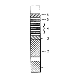
Example 1
A test strip for the determination of creatinine and deo-
xypyridinoline (DPD) containing six distinct areas assembled
together onto a polystyrene backing of 4 inches (101.6 mm) in
length and 0.2 inch (5.0 mm) in width is illustrated by Fig.
CA 0223613~ 1998-04-29
19
1. Referring to Fig. 1, area 1 is the creatinine pad with a
size of 0.2 x 0.2 inch. The creatinine pad was prepared as
follows to render it suit;able for the colorimetric determina-
tion of creatinine: Whatman 3 mm filter paper was first
treated by dipping it to a depth of 0.2 inch into a solution
containing 30 mM copper sulfate, 50 mM citrate, 750 mM glyc-
erol-2-phosphate, 0.2% hexane sulfonic acid, 50 mM phytic acid
and 0.2~ sodium dodecyl sulfonate (SDS) at pH 6.94. After
drying, the strip was dipped into a solution containing 33 mM
3,3',5,5'-tetramethylbenzidine, 73 mM diisopropylbenzene dihy-
droperoxide, 63 mM triisopropanolamine borate, 0.5% plasonde
and 0.032% ethyl orange. The intensity of the colored re-
sponse produced when the strip is contacted with an aqueous
medium containing creatinine is proportionate to the concen-
tration of creatinine. Area 2 is the buffer pad, prepared by
impregnation of Whatman F075-07 glass fiber with 0.5 to 1 M
glycine and 175 to 350 mM urea and having a size of 0.2 x 0.5
inch (12.7 mm). The buffer pad serves the purpose of buffer-
ing the pH of urine samples to the desired value. For exam-
ple, the pH of urine can range from 4.5 to 8 and a buffer pad
can be used to keep the samples at a pH >7 to favor the anti-
gen/antibody bonding reaction. There is a 0.1 inch (2.5 mm)
gap between creatinine pad 1 and buffer pad 2 for purposes of
isolating the creatinine reagent from the buffer pad reagent.
Area 3 is a gold sol-DPD antibody pad (first region containing
a labeled binding partner specific for the analyte). Areas 4
and 5 are the immunochromatography development area where the
capture and detection reagents are deposited onto one piece of
nitrocellulose having a size of 0.2 x 1.25 inch (31.75 mm).
Area 4 contains three capture bands (second region containing
immobilized analyte) with DPD immobilized to carboxyl termi-
CA 02236l3~ l998-04-29
nated polyethylene glycol with a band width of about 0.059
inch (1.5 mm) per band and a 0. 2 inch space between the bands
(from center to center). At 0. 2 inch from the center of the
third capture band is area 5 consisting of one anti-IgG col-
lection band (third region for immobilizing unreacted labeled
binding partner) with a band width of about 0.059 inch (1.5
mm). At 0. 2 inch above the collection band is the absorbant
pad 6 which serves to absorb the liquid which migrates from
the nitrocellulose area of the strip having a size of 0. 2 inch
x 0.5 inch (12.7 mm).
To perform the assay, the strip was dipped into the test
solution, i.e. a urine sample containing the DPD analyte to be
determined, for 3 seconds to a depth such that only the cre-
atinine zone and buffer pad were below the surface of the test
solution which allowed the test solution to flow up the strip
by capillarity through the capture bands of the capture re-
gion, the single band of the detection region and to the ab-
sorbant pad. At the end of the 3 second dip the strip was
placed on the read table of a CLINITEK~ 50 reflectance spec-
trometer and the device's start button pressed. The cre-
atinine pad reflectance was recorded at 3 minutes and the re-
flectance of the immuno DPD strip (all 4 bands) was measured
and recorded at 3 minutes. Reflectance signals for the DPD
assay were measured with IR and green filters whereas reflec-
tance for the creatinine assay was measured using red and
green filters. The device gives a response in decode values
which is derived by equations 1 to 5.
CA 0223613~ 1998-04-29
Equation 1
Decode for Creatinine = [R]qreen
[R]red
where [R]green is the reflectance measured with the green fil-
ter, [R]red is the reflectance measured with the red filter.
For the DPD assay, the band response signals were desig-
nated as indicated in Table 4.
TABLE 4
Band Signals Designation for DPD Assay
Band # ~y~ Designation
1 Capture band 1 Pl
2 Capture band 2 P2
3 Capture band 3 P3
4 Collection band 1 P4
The reflectance with the green filter is ratioed to the re-
flectance with the IR filter to reduce the error from the
variations between strips such as height and surface varia-
tions. The reflectance at the IR wavelength remains fairly
constant regardless of the gold sol intensity of the band.
The corrected reflectance, [Rn], is calculated according to
Equation 2.
Equation 2
[Rn] green X 65
[Rn] =
[Rn] IR
CA 0223613~ 1998-04-29
where n is the band number 1, 2, 3 or 4, [Rn] green is the re-
flectance of band n with green filter, [Rn]I~ is the reflec-
tance of band n with IR filter. The number 65 is the assigned
corrected reference value since the ~ reflectance with the IR
filter is about 65~.
The IR corrected reflectance value, [Rn], is then con-
verted to K/S according to Equation 3 to give the band re-
sponse signal for each band:
Equation 3
(1 - [Rn])
Band Signal = Pn =
2 x [Rn]
where band signal, Pn, is the K/S transformation reflectance
value, [Rn].
The response decode of each band is finally computed ac-
cording to Equation 4.
Equation 4
Decode for DPD assay =
Pl
where T is the summation of the band signal for all bands
(Equation 5).
CA 0223613~ 1998-04-29
23
Equation 5
T = , Pn
n = 1 to N
where N is the total number of capture bands and collection
bands which is 4 in the present example, Pn is the band signal
n and n is 1, 2, 3 or 4.
Standard curves for DPD and creatinine were generated us-
ing calibrators containing six levels of analyte concentra-
tions. Examples of standard curves are shown in Fig. 2 for
the DPD assay and Fig. 3 for the creatinine assay. The DPD
and creatinine concentrations for the urine test sample were
calculated from the DPD and creatinine standard curves respec-
tively. The DPD/creatinine ratio in nM/mM was then calculated
for urine sample A as per the following calculation:
DPD concentration calculated from the DPD standard curve
= 123 nM
Creatinine concentration calculated from the creatinine
standard curve = 10.2 mM
The DPD/creatinine ratio = 123 nM/10.2 mM = 12.1 nM/mm.
The cutoff for determination of a state of high bone re-
sorption is a DPD/creatinine ratio is 7.4 nM/mM. Lower than
7.4 is normal and larger than 7.4 is at the state of high bone
resorption. Therefore, in this example, the result indicates
a state of high bone resorption. A second urine sample was
analyzed in a similar manner and gave the following results:
CA 02236l35 l998-04-29
24
DPD concentration = 12 3 nM
Creatinine concentration = 20.5 mM
DPD/Creatinine ratio = 6.0 nM/mM.
Although the DPD concentration is the same, the ratio of DPD
to creatinine indicates a state of low bone resorption.
Five runs were made using the above procedure with urine
samples containing varying amounts of DPD and creatinine. The
expected and observed ratios as well as standard deviations, %
coefficient of variance and positive/negative biases are set
out in Table 5. From Table 5 it can be determined that a pre-
cision of less than 12% CV was obtained at three levels, 4.52,
7.54 and 12. 07 nM/mM of DPD to creatinine.
TABLE 5
DPDlCreatinine Assav Performance
4.52 4.29 . 1.~ -0.23
7.54 7.59 0.89 11.7% 0.05
12.07 t2.04 1.38 11.5% -0.03
A~en~c %CV = 11.9%
While the gold sol labeled antibodies are visually ob-
servable in the capture and collection zones of the strip,
clinically meaningful results are obtainable only through the
CA 0223613~ 1998-04-29
use of a reflectance meter. This ls the case because of the
use of multiple bands across the entire length of the strip.
In addition, the band signals require reflectance measurement
at different wavelengths (IR, green and red) using an instru-
ment with the capability to measure and record the reflectance
at these wavelengths. The reflectance measurements are ra-
tioed based on a predetermined algorithm using the instru-
ment's software. Furthermore, the analyte concentrations are
determined using standard curves stored in the instrument and
the DPD/creatinine ratio is computed using the software set up
in the instrument.
In the above example, the final response signal (decode)
for the DPD assay was determined using the algorithm decode =
[T/Pn] where T is the summation of the signal from all four
bands and Pn is the band signal of band 1. The use of this
algorithm enhances the accuracy of the assay because ratioing
the band signal minimizes the systematic error such as error
introduced by instrument to instrument variation. Other algo-
rithms may be used to determine the final response signal.
The response signal in this example was determined as:
Response Signal = [T/Capture Band 1]
or [T/P1]
where all band signals are K/S transformation reflectance val-
ues and T is the summation of capture bands and detection
bands.
The advantages of using band ratioing is demonstrated by
the data of Tables 6 and 7 from which it can be determined
CA 02236135 1998-04-29
26
that the precision with which the strip can determine the con-
centration of DPD is much greater than that which is obtain-
able when using only the signal from the capture zone.
TABLE 6
Response Signal = Band Ratioing Algorithm [T/P1]
E~D~-~ Rff~ve~
DPDJCR DPDICR SD %C~'
nMlmM nM~mM
4.15 4.23 0.53 12.5
8.09 8.10 0.28 3.5
10.37 10.44 1.16 11.1
TABLE 7
Response Signal = Capture Zone [P1] (no band ratioing)
r.,.,~ IL~l Re~:OVerOd
DPD/CR DPDICR SD %C~'
nMI~ nWmM
4.15 2.84 4.1 144
8.09 ?.74 0.46 6.0
10.37 1O.g9 2.0 18.5
Alternatively, the final response signal can be calcu-
lated as:
Response Signal = [Detection Band/Capture Band]
where all band signals are KtS transformation reflectance val-
ues. Alternatively, when the strip contains multiple capture
bands and detection bands, the final response signal can be
calculated as:
CA 0223613~ 1998-04-29
[Capture Band 1/Detection Band 1]
where all signals are K/S transformation reflectance values.
Another method of calculating the response signal involves us-
ing the algorithm:
Response Signal =
[Wcap * Capture Band 1/Wdet * Detection Band]
where all band signals are reflectance values and the Wc~p and
Wdet are weighting functions which weight the capture bands and
detection bands differently. Thus, a large number of algo-
rithms can be used to determine the final response signal.
The calculation of the response signal by ratioing the
signals from the labeled binding partner immobilized in the
second (capture) region of the strip and the labeled binding
partner immobilized in the third (detection) region is criti-
cal to increasing the precision of the assay by reducing the
signal to noise ratio. This enhanced precision is necessary
for the test to have clinical significance since only a two-
fold increase in the DPD/creatinine ratio occurs between the
normal and disease indicating osteoporosis states.
Further evidence of the improvement in analytical results
that can be achieved by the present invention is presented in
Tables 8-10. These table were prepared using the same data
set but 3 different algorithms ([T/P1] with band ratioing, %
reflectance of first capture band with no IR correction and no
CA 02236135 1998-04-29
28
band ratioing and % reflectance of first capture band with no
band ratioing but with IR correction) are compared.
TABLE 8
Performance Using [T/P1] Band Ratioing Algorithm
~o i ~ OPD. tttM I ~--t DYDI ~ tt~1
DP~ t~_rt ~ I 'h t~Y ~ I % ~ ~ o tNt I o ~ ~. cv ~o
1 29 1~. 1 4.2 1 .- I .71 t 08 1.97
2 . ~ t I ~10 i 7 1 4 1 .36 - .' -1- 7-
- 7 ~.~ I J. - . 6 1 -1 ' I a.l ~ 6~ -. -.1'
. ~ . 5 ~ Lt.- 23. 1 . _77 ~
.. ' . - . 4 ~ . ._ I I~A 12.~ .t7 - . ) - 3.
12 ~ . t I ._I .~ 10. 1 .. _ I ~.9~
tttt~tttD ~ t Dttt tttO ~2 _35
nc~ e s~rst ievei was e xàuatx ~n tne m an ~ n~t
TABLE 9
Performance Using %R of Capture Band 1
With No Band Ratioing and No IR Correction
I ~D n~ I ~Kttt~ DP .~M l~nttttyy~l DPl/oR ~htttM
. DM ~ Dtt_D ! ~t~ %C~rU--~ %B~tu ~ D~ tttD t %~ ' I Lt %u--
1.~ 10.'13t.26 ~ 2.9 1 ~ 143. 6 1 .~ .29
:' 3t.3 14. 3 .90 .301 . ~ I 9 ~ I t.
69 7 ~ l ''.60 - .~0 ~_~ 7 9 . I . 3
I l ~ 49 ~ 90~ 20. ' .~ 2
15 . ~ .6- .90 , ~ 1 ' - -. .- ~ 44 1 - .
26' .: _ .- .4-I 1.70 - .I .' . ' .0 -37 --
i DttetttD .6 ~ i nlt Ittlt I .9 I -_.t~Ct
no~e: ~r.e rirs~ ievel was exat~ed m tne mean r~ nnrt
CA 02236135 1998-04-29
29
TABLE 10
Performance Using %R of Capture Band
With No Band Ratioing But With IR Correction
I ~off#d Ro~vn~DP . ~ 1~l~1 PD~CRob~mM
I DPD.~M o~ Id %CV hu ','.81~ n~o lO~Oi o ' %cv ~ %-Uu
7.' 1 10.~ 13.49 -.60 ~ 2.8 ~ 6.~3 . .3~9
i : 3~ 1 13.4 3~ 4 : 17. .~ 20 1 ~ '3~ Q~5
6~._ 1.4 ~ 7~ ~ . 6~ 0
! 116. 1 71 7 .. 6. 2U.~ ! 5 ,,_ I .02
1~. 1 16.0 ,, , ~ 7 ~ 54 -37
. 2~3. ' 40.6 . 1..... .1 ~ ) _~ !18.18 ~.63 ~O~
i I I m~ol ~ IOI_O~ 9