Note: Descriptions are shown in the official language in which they were submitted.
CA 02238661 1998-06-23
SPECIFICATION
KILLER T CELL ACTIVATOR AND USE THEREOF
TECHNICAL FIELD OF THE INVENTION
The present invention relates to a pharmaceutical preparation
comprising a protective antigen, which is used for the activation of killer T
cells
having specificity for said protective antigen, and for the prophylaxis and
treatment of cancer and diseases caused by microbial infection. More
particularly, the present invention relates to said preparation in the dosage
form of a plaster. The present invention also relates to a method for
activating
protective antigen-specific killer T cells in a mammal, and a method for the
prophylaxis and treatment of cancer and diseases caused by microbial
infection in a mammal, comprising administering said protective antigen to
the mammal. The present invention further relates to use of a protective
antigen for the production of a protective antigen-specific lflller T cell
activator
and an agent for the prophylaxis and treatment of cancer and diseases caused
by microbial infection.
BACKGROUND OF THE INVENTION
As of date, there is no medication or treatment method for intractable
diseases causing serious conditions of patients, such as cancer and viral
infectious diseases (e.g., acquired immune deficiency syndrome (AIDS),
hepatitis B and hepatitis C and the like), which selectively attacks and
eradicates only the pathogenic cells, and an effective treatment method is
needed, which is free of side effects and adverse influence on the living
body.
Among potential methods for selectively eradicating malignant cells and
cells infected with virus is a method based on activation of the immune system
that a living body innately possesses, and one of such known methods for
inducing activation of manunalian immune system is vaccination. Vaccines
are divided into live vaccines containing live attenuated microorganisms and
inactivated vaccines containing inactivated microorganisms or infection-
protective antigens thereof. A component vaccine, which is an inactivated
vaccine, is prepared using, as an antigen, a microbial antigenic protein, a
surface protein of malignant cell or a peptide fraglnent thereof, so that it
can be
used as an effective means for activating an immune system with safety.
1
CA 02238661 1998-06-23
However, existing vaccination only activates humoral immunity and increases
production of antigen-specific antibodies. This method is effective for
removing viral particles released from cells and the like, but ineffective for
eliminating malignant cells and infected cells causing viral infections.
Of the immune systems in the living body, CD 8 molecule-positive killer T
cells (hereinafter also referred to as CTL) play an important role in the
selective
eradication of virus-infected cells and malignant cells. To be specific, virus-
infected cells process viral proteins, and malignant cells process mutated
proteins, respectively, within the cytoplasm into peptide frdgments, and
express the fragments on the cell surface on a"vesseP' called major
histocompatibility complex (hereinafter abbreviated as MHC) class I molecule,
thereby notifying the immune system in a living body of the abnormal nature
of the cells. CTL recognize said viral peptide or mutated peptide (carcinoma
antigen peptide)/MHC class I molecule complex and eradicate the cells that
expressed the complex.
In recent years, vaccinations aiming at activation and amplification of
CTL, which specifically recognize and eradicate virus-infected cells and
malignant cells, have been actively studied. Among others, dendritic cells
known to have antigen presenting capability to helper T cells (Th) have been
elucidated to have high antigen presenting capability to CTL as well via MHC
class I molecule, and the development of vaccination using the dendritic cells
has been drawing much attention P. I. Mayordomo et al. Nature Med., 1 (12),
1279-1302 (1995)]. This method, nevertheless, requires complicated
manipulations, such as isolation of progentors of dendritic cells and a long
term culture, since it involves isolation of progentors of dendritic cells
present
in peripheral blood in a slight amount, a long term in vitro culture thereof
for
proliferation, preparation of resultant activated and differentiated dendritic
cells with viral peptide or carcinoma antigen peptide to induce dendritic
cells,
said dendritic cells presenting antigenic peptide on the MHC class I molecule
on the cell surface, and transfer thereof into the living body. Such
complicated manipulation makes the possibility of its clinical application
scarce.
It is therefore an object of the present invention to provide a
2
CA 02238661 1998-06-23
pharmaceutical preparation which efficiently activates CTL that selectively
eradicate cells infected with virus or malignant cells, for the establishment
of
an effective method for the prophylaxis and treatment of cancer and diseases
caused by viral infection. It is another object of the present invention to
provide a method for activating CTL using this preparation, and a method for
the prophylaxis and treatment of cancer and diseases caused by viral
infection,
using this preparation.
SUMMARY OF THE INVENTION
In accord with the present invention, it has now been found that
Langerhans cells, which are dendritic cells constantly present in a great
number in cutaneous epidermis, can be activated by merely disrupting a
stratum corneum of the epidermis mechanically or chemically, the stratum
being a primary barrier in the living body against exogenous stimulus, thus
resulting in pronounced enhancement of antigen presenting capability to CTL,
and that the activated epidermal Langerhans cells show increased expression
of MHC class I molecule responsible for antigen presentation to CTL, as well
as
increased expression of ICAM-1, B 7-2, CD 40 and the like required for an
ensured adhesion to CTL, followed by transfer of the Langerhans cells into the
lymph node where a number of CTL precursor cells are present. It has been
also found that when an antigenic peptide is percutaneously inoculated by
coating a viral peptide or carcinoma antigen peptide to cutaneous epidermis
having disrupted stratum corneum barrier, or by applying a pressure sensitive
adhesive tape containing said antigenic peptide in an adhesive layer in a
releasable manner, within about 24 hours after barrier disruption, epidermal
Langerhans cells move into the lymph node while presenting said antigen on
MHC class I molecule on the cell surface showing an increased expression of
the MHC class I molecule. In the lymph node, the cells adhere to CTL
precursor cells to activate and ampli.fy CTL precursor cells. Consequently,
the precursor cells can be efficiently differentiated into said antigen-
specific
mature CTL. In addition, it has been confumed that the lymphocytes, taken
from mammals percutaneously inoculated with antigenic peptide by the
above-mentioned method, selectively lyse only cells pulsed with said antigen.
That is, the present invention provides the following.
3
CA 02238661 1998-06-23
(1) A protective antigen-specific CTL activator comprising the protective
antigen and a pharmaceutically acceptable carrier, the activator being used
for
an external application to an epidermal Langerhans cell surface activated by
mechanically or chemically disrupting a stratum corneum of the epidermis.
(2) The activator of (1) above, which is in the form of a plaster.
(3) The activator of (2) above, wherein said plaster comprises a support and
an
adhesive layer comprising the protective antigen in a releasable manner, said
adhesive layer being laminated on one side of the support.
(4) The activator of (3) above, wherein the surface of the adhesive layer is
divided into two areas and either area comprises the protective antigen.
(5) The activator of (2) above, further comprising a support and adhesive
layers
laminated on both sides of the support, wherein the adhesive layer on one side
comprises the protective antigen in a releasable manner.
(6) The activator of (1) above, wherein the protective antigen is selected
from
the group consisting of a pathogenic microorganism, a peptide to be an
infection-protective antigen thereof and a malignant cell-specific mutated
peptide.
(7) The activator of (6) above, which is an agent for the prophylaxis and
treatment of cancer or a disease caused by microbial infection.
(8) A method for activating protective antigen-specific CTL in a mammal,
comprising expressing MHC class I molecule on the surface of epidermal
Langerhans cell by disrupting the surface of a stratum corneum of the
epidermis, and percutaneously inoculating, to said epidermis, the protective
antigen in an amount effective for activating CTL, thereby to activate said
Langerhans cell in a protective antigen-specific manner and to move the cell
to
lymph node so that the CTL precursor cell reacts with said antigen presenting
Langerhans cell in the lymph node.
(9) The method of (8) above, wherein said stratum corneum is disrupted by
adhering the adhesive surface layer of a pressure sensitive adhesive tape to
the
surface of the stratum corneum of the epidermis and then stripping same in
not less than one cycle.
(10) The method of (9) above, wherein the surface of the pressure sensitive
adhesive tape is divided into two areas, an adhesive layer comprising a
4
CA 02238661 1998-06-23
protective antigen is laminated on one of the areas, the stratum corneum of
the epidermis is removed using an adhesive layer area without the protective
antigen, and the protective antigen is percutaneously inoculated by adhering,
to said epiderrnis, the surface of the area of the adhesive layer of the
pressure
sensitive adhesive tape, which contains the protective antigen.
(11) The method of (9) above, wherein adhesive layers are laminated on both
surfaces of the pressure sensitive adhesive tape, the adhesive layer on one
side
comprising a protective antigen in a releasable manner, the stratum corneum
of the epidermis is removed using the adhesive layer without the protective
antigen, and the protective antigen is percutaneously inoculated by adhering,
to said epidermis, the surface of the area of the adhesive layer of the
pressure
sensitive adhesive tape, which contains the protective antigen.
(12) The method of (8) above, wherein the protective antigen is selected from
the group consisting of a pathogenic microorganism, a peptide to be an
infection-protective antigen thereof and a malignant cell-specific mutated
peptide.
(13) The method of (12) above, which is for the prophylaxis and treatment of
cancer or a disease caused by microbial infection.
(14) Use of a protective antigen for activating protective antigen-specific
CTL in
a mammal, comprising externally applying said protective antigen to an
epidermal Langerhans cell surface activated by mechanically or chemically
disrupting a stratum comeum of the epidermis.
(15) The use of (14) above, wherein said stratum comeum is disrupted by
adhering the adhesive surface layer of a pressure sensitive adhesive tape to
the
surface of the stratum comeum of the epidermis and then stripping same in
not less than one cycle.
(16) The use of (15) above, wherein the surface of the pressure sensitive
adhesive tape is divided into two areas, an adhesive layer comprising a
protective antigen is laminated on one of the areas, the stratum comeum of
the epidermis is disrupted using an adhesive layer area without the protective
antigen, and the protective antigen is percutaneously inoculated by adhering,
to said epidermis, the surface of the area of the adhesive layer of the
pressure
sensitive adhesive tape, which contains the protective antigen.
CA 02238661 1998-06-23
(17) The use of (15) above, wherein adhesive layers are laminated on both
surfaces of the pressure sensitive adhesive tape, the adhesive layer on one
side
comprising a protective antigen in a releasable manner, the stratum corneum
of the epidermis is removed using the adhesive layer without the protective
antigen, and the protective antigen is percutaneously inoculated by adhering,
to said epidermis, the surface of the area of the adhesive layer of the
pressure
sensitive adhesive tape, which contains the protective antigen.
(18) The use of (14) above, wherein the protective antigen is at least one
member selected from the group consisting of a pathogenic microorganism, a
peptide to be an infection-protective antigen thereof and a malignant cell-
specific mutated peptide.
(19) The use of (18) above, wherein the activation of the protective antigen-
specific CTL in a mammal is used for the prophylaxis and treatment of cancer
or a disease caused by microbial infection.
(20) Use of a protective antigen for the production of a protective antigen-
specific CTL activator to be externally applied to the surface of epidermal
Langerhans cells activated by mechanically or chemically disrupting a
stratum corneum of the epidermis.
(21) The use of (20) above, wherein the activator is in the form of a plaster.
(22) The use of (21) above, wherein the plaster comprises a support and an
adhesive layer comprising the protective antigen in a releasable manner, said
adhesive layer being laminated on one side of the support.
(23) The use of (22) above, wherein the surface of the pressure sensitive
adhesive tape is divided into two areas and the protective antigen is
contained
in one of the areas.
(24) The use of (21) above, further comprising the use of a support and
adhesive layers laminated on both sides of the support, said adhesive layer on
one side comprising the protective antigen in a releasable manner, wherein the
activator is in the form of a pressure sensitive adhesive tape.
(25) The use of (20) above, wherein the protective antigen is at least one
member selected from the group consisting of a pathogenic microorganism, a
peptide to be an infection-protective antigen thereof and a malignant cell-
specific mutated peptide.
6
CA 02238661 1998-06-23
(26) The use of (25) above, wherein the activator is used for the prophylaxis
and treatment of cancer or a disease caused by microbial infection.
(27) A commercial package comprising the protective antigen-specific CTL
activator of the above (1) and a document stating that said activator can be
used for activating protective antigen-specific of CTL by externally applying
the
activator to the surface of epidermal Langerhans cells activated by
mechanically or chemically disrupting a stratum corneum of the epidermis.
BRIEF DESCRIPl1ON OF THE DRAWINGS
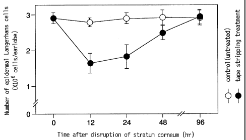
Fig. 1 shows time-course changes in the number of mouse earlobe
Langerhans cells after disrupting the stratum corneum by tape stripping.
Fig. 2 shows changes in the expression of accessory molecule in mouse
earlobe Langerhans cells after disrupting the stratum corneum by tape
stripping.
Fig. 3 shows changes in the expression of H-2Kb molecule on the surface
of mouse earlobe Langerhans cells after disrupting the stratum corneum by
tape stripping.
Fig. 4 shows expression of H-2Kb molecule in Lk'' cell.
Fig. 5 shows cytotoxic activity of cervical lymphocyte-derived effector cells
obtained from mice, to which three Iflnds of antigenic peptides were applied
separately at 12 hours after disrupting an earlobe stratum corneum by tape
stripping.
Fig. 6 shows cytotoxic activity of cervical lymphocyte-derived effector cells
obtained from mice, to which three kinds of antigenic peptides were applied
separately at 12, 24 and 48 hours after disrupting an earlobe stratum
corneum by tape stripping.
DETAILED DESCRIPTION OF THE INVENTiON
The inventive method for the activation of protective antigen-specific CTL
is based on the phenomenon that mechanical or chemical disrupting of
stratum corneum of the epidermis results in an increased expression of MHC
class I molecule on the surface of epidermal Langerhans cells, which is
accompanied by transfer of the activated epidermal Langerhans cells to lymph
node. Once a protective. antigen is inoculated percutaneously to said
epidermis during the transfer of said epidermal Langerhans cells to the lymph
7
CA 02238661 1998-06-23
node after an increased expression of MHC class I molecule, an auto peptide in
the MHC class I molecule is substituted by a peptide of the protective
antigen.
In the present invention, by the protective antigen is meant not only
microbial proteins which become infection protective antigens of pathogenic
microorganisms (e.g., bacteria, fungi, virus and the like), mutated proteins
specifically expressed in malignant cells, and immunogenic peptide fragments
thereof, but also live or dead attenuated microorganisms conventionally used
as live vaccines or inactivated vaccines. The microbial proteins are
exemplified by HIV-derived protein, surface antigens of hepatitis B and C
viruses, surface antigen of influenza virus and the like. The malignant cell-
specific mutated protein is exemplified by malignant transformation cell
differentiation antigen protein and the like. Said protective antigen may
consist of a single protective antigen or plural protective antigens to be
mixed
for use.
The activated epidermal Langerhans cells move into lymph node while
presenting antigen by an MHC class I molecule/protective antigen peptide
complex, and strictly bind to CTL precursor cell having MHC-restricted T cell
receptor which specifically recognizes said protective antigen in the lymph
node. The bond between the both cells may be an antigen-receptor bond,
MHC class I molecule-CD 8 bond, ICAM-I-LFA-1 bond or LFA-3-CD 2 bond.
The bond activates CTL precursor cells to cause amplification and
differentiation to antigen-specific mature CTL. The maturated and amplified
antigen-specific CTL patrol the entire body on lymph circulation. When they
recognize the target cell (e.g., virus-infected cell and malignant cell) which
shows said antigen, they bind thereto and release cytotoxic substance, such
as perforin and granzyme, to kill the target cell.
While the method for disrupting the stratum corneum is not particularly
limited, it is exemplified by a method comprising removing sebum, which
forms intercellular space of the stratum corneum, by the action of an organic
solvent such as acetone, and a method comprising mechanically disrupting
the stratum comeum. A preferable example of the latter is a method wherein
a pressure sensitive adhesive tape is adhered to the surface of a stratum
comeum of the epidermis and stripped therefrom (hereinafter the method is
8
CA 02238661 1998-06-23
referred to as pressure-sensitive adhesive tape stripping) in one or more
cycles.
[Bunya Shirai et al., Nitto Technical Report, 31 (2), 76 (1993)].
The pressure sensitive adhesive tape to be used for stripping preferably
has a peel adhesion of not less than 80 g/ 12 mm width relative to a Bakelite
board. Considering no difference in effects caused by peeling of keratin more
than necessary, the peel adhesion is preferably not more than 1600 g/ 12 mm
width. The adhesive may be any such as an acrylic polymer adhesive, a
rubber polymer adhesive and the like.
The percutaneous inoculation of the protective antigen is performed by
administering the inventive antigen-specific CTL activator to the epidermis
surface, from which a pressure sensitive adhesive tape was stripped,
simultaneously with the disruption of stratum corneum or within about 24
hours, preferably within about 12 hours, affter the disruption. The inventive
antigen-specific CTL activator is subject to no particular limitation in the
dosage form as long as it is an external agent, and typically contains a
protective antigen and a suitable base for external agent. Specific examples
include an ointment prepared by admixing a protective antigen with a suitable
base for an ointment and a pressure sensitive adhesive tape designed to
permit sustained release of the protective antigen, which contains an adhesive
layer as a base and the protective antigen inoculated to or embedded in said
adhesive layer (hereinafter to be referred to as a pressure sensitive adhesive
tape for antigen-specific CTL activation).
In the case of an ointment, the base for an ointment may be, for example,
fat and oil type base, such as petrolatum, para$'in, plastibase, silicone,
vegetable oil, wax and the like; emulsion base such as hydrophilic ointment,
hydrophilic petrolatum, purified lanolin and the like; water soluble base such
as macrogols; and the like. Where necessary, a preservative such as p-
hydroxybenzoate may be added.
The protective antigen can be percutaneously inoculated by applying said
ointment to the skin surface where the stratum corneum has been disrupted.
The protective antigen content of the ointment is preferably 0.1 mol/cm2 -1
mmol/cm2of one kind of antigen when the ointment is applied in a typical
amount.
9
CA 02238661 1998-06-23
A particularly preferable embodiment of the percutaneous inoculation of
the protective antigen is that using the inventive pressure sensitive adhesive
tape for activation of antigen-specific CTL, which is to be applied to the
skin
surface where the stratum corneum has been removed. The inventive
pressure sensitive adhesive tape for antigen-specific CTL activation is
designed
to permit appropriate release of the protective antigen and to stabilize said
protective antigen which has been inoculated to the surface of the adhesive
layer or embedded therein, to the degree that the function as the original
protective antigen is retained. The inventive pressure sensitive adhesive tape
for antigen-specific CTL activation preferably has an adhesive power which is
great enough to sufficiently adhere the tape to the sldn surface. The adhesive
to be used is essentially required not to lower or vary the antigenicity of
the
protective antigen. As long as such condition is met, the adhesive may be an
acrylic polymer adhesive or a rubber polymer adhesive. Particularly
preferable adhesive is that made from a hydrophilic polymer.
Examples of the hydrophilic polymer include water soluble natural
polymers such as gum arabic and carboxymethylcellulose,
polyvinylpyrrolidone, polyvinyl alcohol, polymethoxyethyl acrylate,
polyacrylic
acid and the like, which are obtained by polymerization of water soluble
polymer such as vinyl pyrrolidone, vinyl alcohol, 2-hydroxyethyl acrylate, 2-
methoxyethyl acrylate and acrylic acid, and copolymers of two or more of these
water soluble monomers. It may be an adhesive polymer obtained by
polymerization of hydrophobic polymer such as butyl acrylate and 2-
ethoxyhexyl acrylate to the extent that the hydrophilicity required in the
present invention is not impaired.
More preferable hydrophilic polymer is exemplified by an acrylic
copolymer comprising alkoxyalkyl acrylate and N-vinyl lactam. Alkoxyalkyl
acrylate, which is a monomer component of the acrylic copolymer, is
preferably an acrylate comprising an alkoxy having 1 to 4 carbon atoms and
alkyl or alkylene glycol having 2 to 4 carbon atoms. Specific examples
include alkoxyalkyl acrylate such as 2-methoxyethyl acrylate, 2-ethoxyethyl
acrylate, 2-butoxyethyl acrylate, 3-methoxypropyl aciylate, 3-ethoxypropyl
acrylate and the like, and alkoxyalkylene glycol acrylate such as
1 o
CA 02238661 1998-06-23
methoxytriethylene glycol acrylate, methoxydipropylene glycol acrylate and
the like, with preference given to 2-methoxyethyl acrylate and 2-ethoxyethyl
acrylate, in view of high hydrophilicity.
The other monomer component, N-vinyl lactam, may be N-vinyl lactam
which is a 5 to 7-membered ring. Examples thereof include N-vinyl-2-
pyrrolidone, N-vinyl-2-piperidone, N-vinyl-2-caprolactam and the like, with
preference given to N-vinyl pyrrolidone in view of safety and general
applicability.
While the alkoxyalkyl acrylate content is not particularly limited, it is
preferably 60-80 wt%, more preferably 65-75 wt%, of the entire acrylic
copolymer. While the N-vinyl lactam content is not particularly limited, it is
preferably 20-40 wtD/o, more preferably 25-35 wt9/o, of the entire acrylic
copolymer.
The hydrophilic polymer can be prepared by a method known per se.
For example, acrylic copolymer can be produced by any copolymerization
method such as solution polymerization, emulsion polymerization,
suspension polymerization bulk polymerization, optical polymerization and
the like.
The adhesive layer may further contain a hydrophilic or water soluble low
molecular substance to achieve a suitable degree of adhesion power. The
hydrophilic or water soluble low molecular substance may be a liquid
compound having a high boiling point of 100-400 C. Examples thereof
include polyhydric alcohol, sugar alcohol and the like, with preference given
to
a reducing sugar free of browning reaction (Maillard reaction) with a protein.
The polyhydric alcohol is exemplified by ethylene glycol, diethylene glycol,
triethylene glycol, liquid polyethylene glycol, propylene glycol, dipropylene
glycol, 1,1,1-trihydroxypropane, glycerol and the like, and the reducing sugar
is exemplified by sorbitol, sorbitan, erithyritol, xylitol, trehalose and the
like.
Alternatively, adducts with ether of glycerol and ethylene glycol,
propylene glycol and the like, such as polyoxyethyleneglycerol ether, adducts
with ether of sorbitol or sorbitan and ethylene glycol, propylene glycol and
the
like, such as polyoxypropylenesorbitol ether and polyoxyethylenesorbitan
ether, and the like may be used. These hydrophilic or water soluble low
1 1
CA 02238661 1998-06-23
molecular substances can be added in an amount within the range of from 5
wt% to 90 wt% of the adhesive constituting the adhesive layer.
In the above-mentioned hydrophilic adhesive layer, the entire adhesive
layer may be crosslinked, wherein 5-75% of the components constituting said
adhesive layer may be a gel-like insoluble matter which does not dissolve in
water. In this case, the adhesive layer may be crosslinked by the addition of
a
crosslinldng agent. The crosslinking agent may be a polyhydric epoxy
compound such as ethylene glycol diglycidyl ether, triglycidyl isocyanulate
and
the like, or an isocyanate compound such as CORONATE L and CORONATE
HL (Japan Polyurethane Corp.). These crosslinking agents can be added in
an amount of 0.01-5 parts by weight per 100 parts by weight of the adhesive
constituting the adhesive layer.
A different crosslinldng method of the adhesive layer may be
insolubilization by exposure to electron beam or y rays. The radiation in the
case of, for example, acrylic copolymer may be not less than 1 Kgy,
particularly
preferably not less than 25 Kgy and not more than 50 Kgy.
The adhesive layer is subject to no particular limitation as long as it has
sufficient adhesive power to stay on the skin surface of a living body. For
example, it has a peel adhesion of not less than 30 g/ 12 mm width, preferably
not less than 80 g/ 12 mm width, relative to a Bakelite board. When it
adheres unnecessarily strong to the skin of a living body, the skin tissue may
be disrupted. Thus, it is preferably designed to have a peel adhesion of not
more than 600 g/ 12 mm width. The thickness of the adhesive layer is such
as to afford necessary peel adhesion and to be free of cohesive failure of
adhesive layer upon peeling, which is typically 10-100 u m, preferably 20-80
u M.
The inventive pressure sensitive adhesive tape for antigen-specific CTL
activation can be in any dosage form depending on+ use, as long as it contains
a protective antigen in at least part of the adhesive layer in a releasable
manner. When said pressure sensitive adhesive tape is solely used for
percutaneous inoculation of a protective antigen, it may comprise an adhesive
layer laminated on one side of a support, wherein the adhesive layer contains
the protective antigen preferably uniformly in the entirety thereof. When the
1 2
CA 02238661 1998-06-23
pressure sensitive adhesive tape is used for both pressure sensitive adhesive
tape stripping aiming at disrupting the stratum corneum and percutaneous
inoculation of a protective antigen, the tape may comprise an adhesive layer
laminated on one side of a support, wherein the adhesive layer is divided into
two areas, one of which containing the protective antigen, or the tape may
comprise adhesive layers laminated on both sides of a support, wherein only
one of the adhesive layers contains the protective antigen. In either dosage
form, an adhesive layer area without a protective antigen is used for pressure
sensitive adhesive tape stripping and the adhesive layer area containing the
protective antigen is used for percutaneous inoculation of the protective
antigen. The both adhesive layer areas may be the same or different as long
as they satisfy the above-mentioned requirements of pressure sensitive
adhesive tape stripping and percutaneous inoculation of protective antigen.
The support to be used in the present invention is a flexible sheet and is
not limited to a specific material as long as it is strong enough to stand
damage
during handling. Examples thereof include plastic films made from
polyethylene, polypropylene, polyester, polyamide, polycarbonate, polysulfone,
polyvinyl chloride, polyether, polyurethane, ethylene-vinyl acetate copolymer,
acetate cellulose, nitrocellulose and the like.
The inventive pressure sensitive adhesive tape for antigen-specific CTL
activation is prepared by, for example, adding an aqueous dispersion or a
water/alcohol mixed dispersion adjusted to contain a protective antigen in a
desired concentration to an aqueous solution of the above-mentioned adhesive
hydrophilic polymer and a hydrophilic low molecular substance, or a
water/alcohol mixture, followed by thorough mixing and dispersing. Then,
the dispersion is applied on a flexible support sheet in a certain thickness
and
dried at a suitable temperature in the range of from 10 C to 2009C. The
drying temperature is preferably as low as possible, so that degeneration of
the
contents of the viscous dispersion can be prevented, which is typically about
30-100 C. The adhesive layer of the pressure sensitive adhesive tape
produced by this method now contains a protective antigen uniformly at a
desired concentration. In addition, since the entirety or part of the
protective
antigen is embedded in the adhesive layer thus formed, the protective antigen
1 3
CA 02238661 1998-06-23
is caused to be released in an appropriately sustained manner. A different
production method of the inventive pressure sensitive adhesive tape for
antigen-specific CTL activation includes applying the above-mentioned
aqueous dispersion or water/alcohol mixed dispersion containing an adhesive
hydrophilic polymer and a hydrophilic low molecular substance onto a flexible
support sheet in a certain thickness, and drying same at a suitable
temperature of 10-2009C, and where necessary, crosslinldng to give a pressure
sensitive adhesive tape without a protective antigen, which is followed by
application of an aqueous dispersion or a water/alcohol mixed dispersion
adjusted to contain a necessary amount of a protective antigen onto the
surface of the adhesive layer of said pressure sensitive adhesive tape in a
necessary inoculation amount of the protective antigen and evaporation of
water or alcohol. A pressure sensitive adhesive tape having an adhesive layer
area containing the protective antigen and an area that does not can be
prepared by suitably combining the above-mentioned two production
methods.
The inventive pressure sensitive adhesive tape for antigen-specific CTL
activation is preferably designed to contain a protective antigen in an amount
corresponding to 0.1 g mol/cm2-1 mmol/cm2 per antigen.
The inventive antigen-specific CTL activator sensitizes and amplifies the
antigen-specific CTL by the aforesaid stratum corneum disruption and the
subsequent percutaneous inoculation of the protective antigen. While
circulating through lymphoid, the resulting antigen-specific CTL specifically
recognize and bind to cells (e.g., virus-infected cell and malignant cell)
presenting said protective antigen and eradicate such cells. Thus, the
inventive antigen-specific CTL activator can be used for the prophylaxis and
treatment of cancer and diseases caused by microbial infection with, for
example, virus.
The inventive antigen-specific CTL activator is generally applicable to
animals having a T cell dependent cellular immune system. It is most
effectively used for mammals such as human, monkey, cow, horse, dog, cat,
sheep, goat, rabbit, mouse, rat, hamster, guinea pig and the like.
Inasmuch as the inventive antigen-specific CTL activator specifically
1 4
CA 02238661 1998-06-23
recognizes and binds to cells having a protective antigen and efficiently
sensitizes and amplifies CTL capable of eradication of such cells, by a simple
method involving disruption of stra.tum corneum of the epidermis and
subsequent percutaneous inoculation of the protective antigen by antigen-
specific CTL activator, it is extremely useful for the prophylaxis and
treatment
of intractable cancer and diseases caused by viral infection. The present
invention also provides a drastic method for the prophylaxis and treatment of
cancer and diseases caused by viral infection in mammals inclusive of human.
The present invention is described in more detail by the following
illustrative Examples.
Example 1
(1) Changes in epidermal Langerhans cell counts after removing the barrier of
stratum corneum of the epidermis by pressure sensitive adhesive tape
stripping.
The keratin of one side of earlobe of a C57BL/6 (B6) mouse was
disrupted by 10 repeats of stripping with a commercially available cellophane
tape, and epidermal cell suspensions were obtained by trypsinizing the earlobe
cut into a certain area at 12, 24 and 48 hours later. The suspensions were
stained with I-Ab antigen-specific antibody, which is one of the MHC class II
antigens, to count I-Ab antigen strong positive Langerhans cells by flow
cytometry. As a control, the other earlobe of the mouse free of tape stripping
was used. The results are shown in Fig. 1.
The mouse earlobe free of pressure sensitive adhesive tape stripping
showed normal Langerhans cell counts, whereas the cell count of the earlobe
which underwent stripping decreased to about half in 12 hours after the tape
stripping, thereby showing transit thereof to other tissues from epidermis.
The transit of the Langerhans cells reached maximum at about 12-24 hours
after tape stripping, and epidermal Langerhans cell count gradually restored
the normal level.
(2) Expression of activated accessory molecule of epidermal Langerhans cell
after disrupting stratum corneum.
One earlobe of a B6 mouse was disrupted by 10 repeats of stripping with
the same adhesive tape as used in Example 1-(1), and epidermal cell
1 5
CA 02238661 1998-06-23
suspensions were obtained by trypsinizing the epidermis cut into a certain
area at 0, 12, 24 and 48 hours later. The suspensions were stained with
antibodies specific to I-Ab, CD54 (ICAM-1), CD86 (B7-2) and aB TCR, and
analyzed by flow cytometry. As a control, the other earlobe of the same
mouse, which was free of tape stripping, was used. The results are shown in
Fig. 2.
The earlobe Langerhans cells which underwent pressure sensitive
adhesive tape stripping were activated 12 and 24 hours later, and showed an
increased expression of ICAM- 1, B7-2 and CD40 molecules responsible for
binding with CTL. The staining using anti- a0 TCR antibody as a control
antibody did not result in enhanced expression after barrier disruption. The
expression of ICAM- 1, B7-2 and CD40 molecules reached its peak at about
12-24 hours after barrier disruption, and returned to the level before
activation at 48 hours later. The mouse earlobe free of pressure sensitive
adhesive tape stripping showed no variation in the expression level of ICAM-
1,
B7-2 and CD40 molecules of Langerhans cell.
(3) Expression of MHC class I molecule on the epidermal Langerhans cell
surface after stratum corneum barrier disruption.
In the same manner as in Example 1-(2), the expression of H-2Kb
molecule, which is one of the MHC class I molecules, was examined. The
results are shown in Fig. 3. The epidermal Langerhans cell of the mouse
earlobe which underwent pressure sensitive adhesive tape stripping
apparently showed an increased expression of H-2Kb molecule in about 12-24
hours after barrier disruption, which molecule being responsible for
presentation of antigen to CTL (Fig. 3(A)). On the other hand, the Langerhans
cell of earlobe free of stratum corneum disruption showed no reinforcement of
expression of this molecule (Fig. 3(B)).
Reference Ebcample 1: Synthesis of antigenic peptide
In accord with a known literature, herpes simplex virus (HSV), vesicular
stomatitis virus (VSV)-derived antigenic peptide, and ovalbumin (OVA)-derived
peptide, all capable of being presented on H-2Kb molecule, which is one of the
MHC class I molecules, and activation of CTL in B6 mouse, were synthesized.
HSV *coprotein B 498-505
1 6
CA 02238661 1998-06-23
[reference: Bonneau et al., Virology, 195: 62-70 (1993)]
sequence: Ser Ser Ile Glu Phe Ala Arg Leu (SEQ ID NO: 1)
VSV NP 53-59
[reference: Van Bleek and Nathenson, Nature, 348: 213-215 (1990)]
sequence: Arg Gly Tyr Val Tyr Gln Gly Leu (SEQ ID NO: 2)
Ovalbnmin 257-264
[reference: Falo et al., Nature Med., 1: 649-653 (1995)]
sequence: Ser Ile Ile Asn Phe Glu Lys Leu (SEQ ID NO: 3)
Reference LbCample 2: Preparation of transformed L cell which expresses
H-2Kb molecule
A cell line was prepared, wherein H-2Kb molecule had been forcibly
expressed in L cell inherently having no H-2Kb molecule, for use in in vitm
confirmation of whether or not CTL, which are activated in a mouse when
antigenic peptide is applied to the tape-stripped skin, is specific to H-2Kb
molecule which presents antigenic peptide.
(i) Preparation of cell line transformed with H-2Kb molecule gene
A plasmid DNA, containing an H-2Kb gene inserted in the downstream of
a known promoter functional in L cell and having a tymidine kinase gene, was
prepared. The plasmid DNA was introduced into LP'- cell by a calcium
chloride method. The cells having the gene were screened using tymidine
kinase activity as an index to obtain a cell line having an Lkb gene.
(ii) Verified expression of H-2Kb molecule in Lkb cell
The prepared Lkb cell was stained with H-2Kb-specific antibody and
analyzed by flow cytometry. As a control of the cell, LP' cell before gene
introduction was used, and as a control of the antibody, an antibody different
from the H-2Kb-specific antibody but matching in antibody class alone was
used. As a result, L6 cell showed certain expression of H-2Kb molecule (Fig.
4).
Example 2
Sensitization of precursor CTL and activation of antigen-specific CTL by the
application of antigenic peptide to the tape-stripped skin.
In the following manner, it was confirmed that the activated epidermal
Langerhans cell sensitizes precursor CTL and activates antigen-specific CTL
17
CA 02238661 1998-06-23
by antigenic peptide pulse in the tape-stripped skin.
(i) One side of earlobe of a B6 mouse was disrupted by 10 repeats of stripping
with the same adhesive tape as used in Example 1-(1), and antigenic peptides
synthesized in Reference Example 1 were applied at 12, 24 and 48 hours later,
wherein the antigenic peptides were respectively dissolved in 70% ethanol and
applied in 10 u m per one side of the earlobe. At 1 week after the peptide
application, cervical lymph node was removed, and lymphocytes were cultured
for 2 days in vitro in an RPMI1640 medium containing a slight amount (5
U/ml)of IL-2. The Lkb cells labeled with 51Cr were pulsed with antigenic
peptide (5 mol) and LP'- cells were used as a control. Cytotoxicity assay
was
performed using cultured lymphocytes as effector cells, and 51Cr-labeled and
antigenic peptide-pulsed L~b cells and L~ cells as target cells.
(ii) Variation in ratio of CTL in cervical lymph node
Table 1 shows variation in the total number of lymphocytes and the
number of CD8-positive cells in cervical lymph node upon determination at
every antigenic peptide application. The antigenic peptides used were the
three kinds recited in Reference Example 1. The antigenic peptide-pulsed
epidennal Langerhans cell activated by tape stripping presented antigen in the
lymph node, and sensitized and amplified the cells of immune system. In
particular, the number of lymphocytes having a CD8 molecule, which is a CTL
marker, showed an increase. The increase in the number of CD8-positive
lymphocytes reached maximum at 12-24 hours after tape stripping, and
matched well with the changes in the Langerhans cell counts in epidermis.
Thus, it was strongly suggested that the activated epidermal Langerhans cells
moved to the lymph node and activated CTL in the lymph node.
1 8
CA 02238661 1998-06-23
Table I
Time after Total number Number of CD8-positive cells
Peptide stripping of cells (x107) (x107) (%)
HSV gp B untreated 0.82 0.25 (20.5)
HSV gp B 12 2.93 1.45 (49.5)
HSV gp B 24 1.86 0.86 (46.2)
HSV gp B 48 0.96 0.31 (32.3)
--------------------------------------------------------------------
VSV NP untreated 0.71 0.15 (21.1)
VSV NP 12 1.61 0.63 (39.1)
VSV NP 24 1.38 0.52 (37.7)
VSV NP 48 1.00 0.24 (24.0)
----------------------------------------------------------------------
Ovalbumin untreated 0.65 0.13 (20.0)
Ovalbumin 12 1.57 0.53 (33.8)
Ovalbumin 24 1.28 0.42 (32.8)
Ovalbumin 48 1.21 0.26 (21.5)
--------------------------------------------------------------------
(70% ethanol) untreated 0.73 0.19 (26.0)
- 12 0.72 0.19 (26.4)
- 24 0.77 0.20 (26.0)
- 48 0.80 0.23 (28.8)
(iii) Activation of antigen-specific CTL
Cytotoxicity assay was performed using the effector cells derived from
cervical lymphocytes obtained from the group that underwent separate
application of antigenic peptides of HSV gp B, VSV NP and OVA at 12 hours
after the tape stripping, the results of which are shown in Fig. 5. For
example,
CTL of cervical lymph node obtained from the group that underwent
application of antigenic HSV gp B peptide showed amplif cation specific to
HSV gp B, and recognized and certainly eradicated the cells presenting an H-
2Kb - HSV gp B peptide complex alone. In the groups to which VSV NP
1 9
CA 02238661 1998-06-23
peptide and OVA peptide were applied, cervical lymph node-derived CTL
selectively damaged the cells presenting respective inoculated antigenic
peptides. Naturally, greater effector cell/target cell ratios led to more
ensured
eradication of the target cells.
(iii) Effect on sensitization of CTL by time-course application of antigenic
peptide to the tape-shaped skin.
Cytotoxicity assay was performed using the effector cells derived from
cervical lymphocytes obtained from the group that underwent separate
application of antigenic peptides at 12, 24 and 48 hours after the tape
stripping, the results of which are shown in Fig. 6. In every group, the
peptide-pulsed epidermal Langerhans cells activated by tape stripping
sensitized CTL in an antigen-specific manner in the lymph node and
selectively damaged only the cells presenting respective antigenic peptides.
It
was clarified that, when antigenic peptide was applied at 12-24 hours after
the
tape stripping, the antigen-specific CTL were most strongly activated and
amplified in the lymph node.
Emmple 3
Production of pressure sensitive adhesive tape containing antigenic peptide.
In a closed type reactor equipped with a stirrer were charged 2-
methoxyethyl acrylate (70 parts by weight), N-vinyl-2-pyrrolidone (30 parts by
weight), azoisobutyronitrile (0.17 part by weight) as a polymerization
initiator,
and a mixed solvent of distilled water:methanol:isopropanol (250 parts by
weight, weight ratio 16:23:1). After displacement with nitrogen, the mixture
was stirred for 1.5 hours while maintaining the temperature in the reactor at
60-62'C. Then, stirring at 75 C for 2 hours completed the reaction. The
mixture was cooled to room temperature to give a viscous acrylic copolymer
solution. To this polymer solution were added polyoxypropyleneglycerol ether
(10 parts by weight, average molecular weight 400) and trehalose (10 parts by
weight, trademark TREHAOSE, available from HAYASHIBARA GROUP) and
completely dissolved. The solution containing this acrylic copolymer was
applied to a 6 m thick flexible and thin polyester film, and dried at 130 C
for
minutes to give a pressure sensitive adhesive tape having a 50 u m thick
adhesive layer. Separately, the antigenic peptide HSV gp B obtained in
CA 02238661 1998-06-23
Reference Example 1 was dissolved in 70% ethanol and applied to the surface
of the adhesive layer of the pressure sensitive adhesive tape in an amount of
50 mol per lcm2. Then, ethanol was evaporated to give a pressure
sensitive adhesive tape inoculated with HSV gp B peptide. A release paper
was adhered to this pressure sensitive adhesive tape to protect the adhesive
layer surface until use.
Esample 4
Sensitization of CTL and amplification of antigen-specific CTL by the
application of antigenic peptide-containing pressure sensitive adhesive tape
to
the taped-stripped sldn with disrupted stratum comeum barrier.
In the same manner as in Example 2 except that pressure sensitive
adhesive tape inoculated with HSV gp B antigenic peptide, which had been
obtained in Example 3, was applied immediately after tape stripping, instead
of antigenic peptide, it was confirmed that epidermal Langerhans cells
activated in an antigenic peptide specific manner by tape-stripping moved to
the lymph node and activated antigenic peptide-specific CTL. The results are
shown in Table 2.
Table 2
Time after Cytotoxic activity specific to
Peptide stripping HSV peptide-pulsed L cells (%)
HSV gp B untreated 0.2 0.8
HSV gp B 12 45.3 1.5
HSV gp B 24 37.0 1.1
HSVgpB 48 3.5 0.6
--------------------------------------------------------------------
- untreated 0.3 0.5
- 12 1.1 1.3
- 24 0.6 1.2
- 48 0.5 0.2
The epidermal Langerhans cells should have been activated after the
2 1
CA 02238661 2007-08-27
27103-182
passage of lead time necessary for the activation of epidermal Langerhans
cells
after tape stripping. Thus, when the antigenic peptide-containing pressure
sensitive adhesive tape is applied to the sldn surface upon tape stripping,
antigenic peptide is inoculated and antigenic peptide-pulsed epidermal
Langerhans cells move to the lymph node. As a result, antigenic peptide-
specific CTL are activated. When compared to the inoculation method of
Example 2, the antigenic peptide-specific C fL were amplified and activated
faster, and when measured by setting the stripping time as time 0, the total
amount of antigenic peptide-specific CTL amplified in a predetermined time
period from the application of the antigenic peptide-containing pressure
sensitive adhesive tape was found to have increased.
22
CA 02238661 1998-06-23
SEQUENCE LISTING
(1) GENERAL INFORMATION:
(i) APPLICANT: Nitto Denko Corporation
(ii) TITLE OF INVENTION: Killer T Cell Activator and Use Thereof
(iii) NUMBER OF SEQUENCE: 3
(iv) CORRESPONDENCE ADDRESS:
(A) ADDRESSEE: FETHERSTONHAUGH & Co.
(B) STREET: P.O. BOX 2999, STATION D
(C) CITY: OTTAWA
(D) STATE: ONTARIO
(E) COUNTRY: CANADA
(F) ZIP: K1P 5Y6
(v) COMPUTER READABLE FORM:
(A) MEDIUM TYPE: Diskette - 3.5 inch, 1.44 Mb storage
(B) COMPUTER: IBM-PC compatible
(C) OPERATING SYSTEM: PC-DOS/MS-DOS
(D) SOFTWARE:
(vi) CURRENT APPLICATION DATA:
(A) APPLICATION NUMBER:
(B) FILING DATE:
(C) CLASSIFICATION:
(vii) PRIOR APPLICATION DATA:
(A) APPLICATION NUMBER: JP 130031 / 1997
(B) FILING DATE: 20-MAY- 1997
(viii) ATTORNEY/AGENT INFORMATION:
(A) NAME:
(B) REGISTRATION NUMBER:
(C) REFFERENCE/DOCKET NUMBER:
(ix) TELECOMMUNICATION INFORMATION:
(A) TELEPHONE:
(B) TELEFAX:
(C) TELEX:
2 3
CA 02238661 1998-06-23
(2) INFORMATION FOR SEQ ID NO: 1:
(i) SEQUENCE CHARACTERISTICS:
(A) LENGTH: 8 amino acids
(B) TYPE: amino acid
(D) TOPOLOGY: linear
(ii) MOLECULE TYPE:
(A) DESCRIPTION: peptide
()d) SEQUENCE DESCRIPTION: SEQ ID NO: 1:
Ser Ser Ile Glu Phe Ala Arg Leu
(2) INFORMATION FOR SEQ ID NO: 2:
(i) SEQUENCE CHARACTERISTICS:
(A) LENGTH: 8 amino acids
(B) TYPE: amino acid
(D) TOPOLOGY: linear
(ii) MOLECULE TYPE:
(A) DESCRIPTION: peptide
(xi) SEQUENCE DESCRIPTION: SEQ ID NO: 2:
Arg Gly Tyr Val Tyr Gln Gly Leu
5
(2) INFORMATION FOR SEQ ID NO: 3:
(i) SEQUENCE CHARACTERISTICS:
(A) LENGTH: 8 amino acids
(B) TYPE: amino acid
(D) TOPOLOGY: linear
(ii) MOLECULE TYPE:
(A) DESCRIPTION: peptide
(xi) SEQUENCE DESCRIPTION: SEQ ID NO: 3:
Ser Ile Ile Asn Phe Glu Lys Leu
5
24