Note: Descriptions are shown in the official language in which they were submitted.
CA 02239815 1998-06-19
WO 98/20108 PCTIUS97/19834
A METHOD AND APPARATUS FOR HOLDING CELLS
FIELD OF THE INVENTION
The present invention is related to an apparatus
for holding cells. More specifically, the present invention
is related to an apparatus for incubating cells so that an
array of single cells, or a functional ensemble, can be grown
and individually analyzed in a dynamically controlled
environment.
BACKGROUND OF THE INVENTION
In adult humans, hematopoeitic stem cells are found
primarily in the bone marrow, although in newborns these
cells also are present in the blood of the umbilical cord.
Hematopoeitic stem cells are the progenitors (i.e.,
precursors) of mature blood cells in the body, and through a
process called hematopoiesis, stem cells continuously
regenerate the body's blood supply, including red blood cells
(which transport oxygen in the body), white blood cells
(which fight infections and comprise the body's immune
system), and platelets (which form clots to stop bleeding).
Hematopoiesis involves cell division (i.e., increase in cell
number) and differentiation (i.e., change in cell phenotype).
Chemotherapy and radiation therapy are important tools for
treating patients with cancer or requiring solid-organ
transplants, but these processes are (beneficially) toxic to
CA 02239815 1998-06-19
WO 98/20108 PCT/US97/19834
the hematopoeitic (i.e., blood) system because chemotherapy
and ionizing irradiation kill many of the stem cells in the
bone marrow. This immunosuppression and other blood toxicity
limit the effectiveness of many otherwise promising cancer
therapies because a critical low number of blood cells in the
body lead to life-threatening infection and bleeding.
Recovery from these therapies requires
replenishment of the patient's stem cells. Treatment with
growth factors currently is used to promote the recovery of
blood cells but is only partially effective following
immunosuppressive treatments. Alternatively, infusion of
human stem cells through a bone marrow transplant
increasingly is used by physicians to restore rapidly and
permanently a patient's ability to regenerate blood cells.
Transplants of bone marrow grew from 5,000 per year in 1990
to more than 40,000 per year by 1995 (Kline, Ronald, New
Marrow for Old, Technology Review, Nov./Dec. 1993, p. 43;
Anonymous Inside Surgery, Medical Data International Ed.,
Vol. 3, No. 8, Feb. 1996, p. 192). However, the large number
of reports in the media citing people who are looking for
appropriate bone marrow donors demonstrates that this process
can be extremely difficult because appropriate donors are
very rare in many cases. Although the best bone marrow
donors are siblings, only 25% of the time is a sibling a.
compatible transplant donor (Kline, Ronald, New Marrow for
Old, Technology Review, Nov./Dec. 1993, p. 43).
CA 02239815 1998-06-19
WO 98/20108 PCT/US97/19834
'-3-
The automated growth of stem cells through the use
of a unique bioreactor system would be a very important
advance for cancer research and therapy. For example, the
use of the bioreactor system could eliminate the need for
donors: some stem cells can be removed from a patient prior
to chemotherapy, stored during chemotherapy, and then large
numbers of stem cells generated in the bioreactor system can
be transplanted back into the patient. This strategy cannot
be implemented with current technologies for growing stem
cells because these approaches predominantly result in
hematopoeitic expansion to produce differentiated mature
blood cells at the expense of increasing the number of
pluripotent (most primitive) stem cells needed for long-term
replenishment of the bone marrow (Van Zant, Gary, Rummel, Sue
A., Koller, Manfred R., Larson, David B., Drubachevsky,
Ilana, Palsson, Mahshid and Emerson, Stephen G. Expansion in
Bioreactors of Human Progenitor Populations from Cord Blood
and Mobilized Peripheral Blood. Blood Cells (1994) 20:482-
491; Goff, Julie P., Shields, Donna S., Petersen, Bryon E.,
Zajac, Valerie F., Michalopoulos, George K. and Greenberger,
Joel S. Synergistic Effects of Hepatocyte Growth Factor on
Human Cord Blood CD34+ Progenitor Cells are the Result of
c-met Receptor Expression. Stem Cells (In Press) ; Moore,
MAS. Clinical Implications of Positive and Negative
= 25 Hematopoeitic Stem Cell Regulators. Blood 1991; 78:1-19;
Metcalfe, D. Hematopoeitic Regulators: Redundancy or
Subtlety? Blood 1993; 82:3515-3523; Bernstein, I.D.,
CA 02239815 1998-06-19
WO 98/20108 PCT/US97/19834
--4 - ,
Andrews, R.G., Zsebo, K.M. Recombinant Human Stem Cell
Factor Enhances the Formation of Colonies by CD34+ and
CD34+lin- Cells and the Generation of Colony-Forming Cell
Progeny From CD34+lin- Cells Cultured With Interleukin-3,
Granulocyte Colony-Stimulating Factor, or
Granulocyte-Macrophage Colony-Stimulating Factor. Blood
1991; 77:2316-2321; Musashi, M. Clark, S.C., Suodo, T. et al.
Synergistic Interactions Between Interleukin-11 and
Interleukin-4 in Support of Proliferation of Primitive
Hematopoeitic Progenitors of Mice. Blood 1991; 78:1448-1451;
Musashi, M., Yang, Y-C, Paul, S.R. et al. Direct and
Synergistic Effects of Interleukin-11 on Murine Hemopoiesis
in Culture. Proc Natl Acad Sci 1991; 88:765-769; Migliaccio,
G., Migliaccio, A.R., Druzin, M.L. et al. Long-Term
Generation of Colony-Forming Cells in Liquid Culture of CD34+
Cord Blood Cells in the Presence of Recombinant Human Stem
Cell Factor. Blood 1992; 79(10):2620-2627; Ikuta, K.,
Weissman, I.L. Evidence That Hematopoeitic Stem Cells Express
Mouse C-Kit but do not Depend on Steel Factor for Their
Generation. Proc Natl Acad Sci USA 1992; 89:1502-1506;
Miltenyi, S., Guth, S., Radbruch, A. et al. Isolation of
CD34+ Hematopoeitic Progenitor Cells by High-Gradient
Magnetic Sorting. In: Wunder E., ed Hematopoeitic Stem
Cells: Alpha Med Press 1994; 201-213; Traycoff, C.M., Kosak,
S.T., Grigsby, S., Srour, E.F. Evaluation of Ex Vivo
Expansion Potential of Cord Blood and Bone Marrow
Hematopoeitic Progenitor Cells Using Cell Tracking and
CA 02239815 1998-06-19
WO 98/20108 PCT/11S97/19834
'-5-
Limiting Dilution Analysis. Blood 85, No. 8:2059-2068 (April
15) 1995; Murray, L., Chen, B., Galy, A., Chen, S.,
Tushinski, R., Uchida, N., Negrin, R., Tricot, G., Jagannath,
S., Vesole, D., Barlogie, B., Hoffman, R., Tsukamoto, A.
Enrichment of Human Hematopoeitic Stem Cell Activity in the
CD34+Thy-l+Lin- Subpopulation from Mobilized Peripheral
Blood. Blood 85, No. 2:368-378 (January 15) 1995; Uchida,
N., Aguila, H.L., Fleming, W.H., Jerabek, L., Weissman, I.L.
Rapid and Sustained Hematopoeitic Recovery in Lethally
Irradiated Mice Transplanted with Purified Thy-1.1 Lin-Scal+
Hematopoeitic Stem Cells. Blood 83, No. 12:3758-3779 (June
15) 1995).
The underlying biological problem is that
differentiated daughter cells -- termed "committed
progenitors" -- produce and secrete molecules that appear to
inhibit the proliferation of nearby true stem cells (Ogata,
H., Bradley, W.G., Inaba, M., Ogata, N., Ikehara, S., Good,
R.A. Long-Term Repopulation of Hematolymphoid Cells With
Only a Few Hemopoietic Stem Cells in Mice. Proc. Natl. Acad.
Sci. USA. 92:5945-5949, June 1995; Li, C.L., Johnson, G.R.
Murine Hematopoeitic Stem and Progenitor Cells: I.
Enrichment and Biologic Characterization. Blood 85, No.
6:1472-1479 (March 15) 1995; Dunbar, C.E.,"Cottler-Fox, M.,
O'Shaughnessy, J.A., Doren, S., Charter, C., Berenson, R.,
Brown, S., Moen, R.C., Greenblatt, J., Stewart, F.M.,
Leitman, S.F., Wilson, W.H., Cowan, K., Young, N.S.,
CA 02239815 1998-06-19
WO 98/20108 PCT/US97/19834
-6-
Nienhuis, A.W. Retrovirally Marked CD34-Enriched Peripheral
Blood and Bone Marrow Cells Contribute to Long-Term
Engraftment After Autologous Transplantation. Blood 85, No.
11:3048-3057 (June 1) 1995; Traycoff, C.M., Kosak, S.T.,
Grigsby, S., Srour, E.F. Evaluation of Ex Vivo Expansion
Potential of Cord Blood and Bone Marrow Hematopoeitic
Progenitor Cells Using Cell Tracking and Limiting Dilution
Analysis. Blood 85, No. 8:2059-2068 (April 15) 1995; Murray,
L., Chen, B., Galy, A., Chen, S., Tushinski, R., Uchida, N.,
Negrin, R., Tricot, G., Jagannath, S., Vesole, D., Barlogie,
B., Hoffman, R., Tsukamoto, A. Enrichment of Human
Hematopoeitic Stem Cell Activity in the CD34+Thy-1+Lin-
Subpopulation from Mobilized Peripheral Blood. Blood 85, No.
2:368-378 (January 15) 1995; Uchida, N., Aguila, H.L.,
Fleming, W.H., Jerabek, L., Weissman, I.L. Rapid and
Sustained Hematopoeitic Recovery in Lethally Irradiated Mice
Transplanted with Purified Thy-1.1 Lin-Scal+ Hematopoeitic
Stem Cells. Blood 83, No. 12:3758-3779 (June 15) 1995);
Issaad, C., Croisille, L., Katz, A., Vainchenker, W.,
Coulombel, L. A Murine Stromal Cell Line Allows the
Proliferation of Very Primitive Human CD34+ +,/CD38-
Progenitor Cells in Long-Term Cultures and Semisolid Assays.
Blood 81, No. 11:2916-2924 (June 1) 1993; Pettengell, R.,
Luft, T., Henschler, R., Hows, J.M., Dexter, T.M., Ryder, D.,
Testa, N.G. Direct Comparison by Limiting Dilution Analysis
of Long-Term Culture-Initiating Cells in Human Bone Marrow,
Umbilical Cord Blood, and Blood Stem Cells. Blood 84, No.
CA 02239815 1998-06-19
WO 98/20108 PCT/US97/19834
-7-
11:3653-3659 (December 1) 1994; Greenberger, J.S. Long-Term
Hematopoeitic Cultures. In: Golde D, (ed). Methods in
Hematology. New York: Churchill Livingston, 11:203-243,
1984; Rothstein, L., Pierce, J.H., Aaronson, S.A.,
Greenberger, J.S. Amphotropic Retrovirus Vector Transfer of
the v-ras Oncogene Into Human Hematopoeitic and Stromal Cells
in Continuous Bone Marrow Culture. Blood. 65:744-752, 1985;
Greenberger, J.S. Recent Modifications and Technical
Improvements in Human Long-Term Bone Marrow Cultures.
Proceedings of the Symposium on Long-Term Bone Marrow
Culture, Kroc Foundation, September 1983, Alan R. Liss, New
York, pp. 119-133, 1984; Greenberger, J_S. The Hematopoeitic
Microenvironment. Critical Reviews in Hem/Onc, Elsevier
Science Publications B.V. 11:65-84, 1991; Goff, J.P.,
Shields, D.S., Michalopoulos, G.K., Greenberger, J.S.
Synergistic Effects of Hepatocyte Growth Factor on In Vitro
Generation of CFU-FM From Human Cord Blood CD34+ Progenitor
Cells. Thirty-Sixth Annual Meeting of the American Society
of Hematology, Nashville, TN, 12/1/94-12/6/94. Blood,
84(10) :Suppl. #280A, 1994; Pogue-Geile, K.L., Sakakeeny,
M.A., Panza, J.L., Sell, S.L., Greenberger, J.S. Cloning and
Expression of Unique Murine Macrophage Colony Stimulating
Factor Transcripts. Blood, 85:3478 3486, 1995; Goff, J.P.,
Shields, D.S., Michalopoulos, G.K., Greenberger, J.S.
Effects of Hepatocyte Growth Factor and IL-11 on Human Cord
Blood CD34+ Progenitor Cells. International Society for
Experimental Hematology Meeting, Duesseldorf, Germany,
CA 02239815 1998-06-19
WO 98/20108 PCT/US97/19834
-8-
8/25/95-9/1/95). Current technologies for the growth of stem
cells do not address this problem because these technologies
are designed to increase the total number of blood cells, not
the number of stem cells per se (Traycoff, C.M., Kosak, S.T.,
Grigsby, S., Srour, E.F. Evaluation of Ex Vivo Expansion
Potential of Cord Blood and Bone Marrow Hematopoeitic
Progenitor Cells Using Cell Tracking and Limiting Dilution
Analysis. Blood 85, No. 8:2059-2068 (April 15) 1995; Murray,
L., Chen, B., Galy, A., Chen, S., Tushinski, R., Uchida, N.,
Negrin, R., Tricot, G., Jagannath, S., Vesole, D., Barlogie,
B., Hoffman, R., Tsukamoto, A. Enrichment of Human
Hematopoeitic Stem Cell Activity in the CD34+Thy-l+Lin-
Subpopulation from Mobilized Peripheral Blood. Blood 85, No.
2:368-378 (January 15) 1995) Limiting the differentiation
of daughter cells is necessary to grow multiple exact
replicas of the original stem cells. By identifying in situ
the occurrence of cell division and the presence of
differentiated cells with microscope imaging, the bioreactor
system with z-robot pipette for medium exchange allows
solution of this problem: there will be automated exchange
of the primary growth medium in a well with a secondary
quiescence (i.e., "quieting") medium upon cell division. The
first medium promotes proliferation of the original stem cell
into exact replicas, and the second medium inhibits
differentiation of the resulting daughter cells into
committed progenitors.
CA 02239815 1998-06-19
WO 98/20108 PCT/US97/19834
'-9-
Understanding and continuing interest in culturing
human stem cells obtained from bone marrow and umbilical cord
blood has expanded greatly in the last five years. Human
stem cell candidates are identified as CD34+Thyl+Lin- (lin-) :
they express the cell surface antigens CD34 and Thyl but not
lineage specific antigens (lin-). Antigens are molecules on
cell surfaces recognized by specific monoclonal antibodies.
CD34+ cells in the bone marrow (approximately 1%) can be
isolated by immunomagnetic selection (incubating cells with
magnetic beads coated with monoclonal antibodies against CD34
and applying a magnetic field). The subpopulation of CD34+
cells (roughly 1 in 2 to 1 in 4) which do not express
antigens associated with differentiated or lineage committed
cells can also be removed using appropriate antibodies and
immunomagnetic selection or by labeling these antibodies with
fluorochromes and flow cytometry. The lin- cells obtained
after sorting represent around 1 in 50,000 cells from the
original population.
Previous work on developing technology for
culturing stem cells has focused on hematopoeitic expansion
(i.e., solely increasing the number of committed progeny and
mature blood cells) rather than increasing the number of
uncommitted lin- cells in the population. For example,
Stephen Emerson and Bernhard Palsson (University of Michigan,
in collaboration with Aastrom Biosciences, Inc.) developed a
batch-operated bioreactor for growing large numbers of CD34+
CA 02239815 1998-06-19
WO 98/20108 PCT/US97/19834
-10-
cells in which culture medium is recirculated over a series
of layered individual trays on which stem cells are
maintained (Van Zant, Gary, Rummel, Sue A., Koller, Manfred
R., Larson, David B., Drubachevsky, Ilana, Palsson, Mahshid
and Emerson, Stephen G. Expansion in Bioreactors of Human
Progenitor Populations from Cord Blood and Mobilized
Peripheral Blood. Blood Cells (1994) 20:482-491) . Waste and
catabolites are removed continuously from the reactor.
Modest increases in numbers of CD34+ cells were detected, but
the true lineage specificity of the amplified stem cell was
not demonstrated (Van Zant, Gary, Rummel, Sue A., Koller,
Manfred R., Larson, David B., Drubachevsky, Ilana, Palsson,
Mahshid and Emerson, Stephen G. Expansion in Bioreactors of
Human Progenitor Populations from Cord Blood and Mobilized
Peripheral Blood. Blood Cells (1994) 20:482-491).
Based on the results of previous studies in which
modest or no increases in the numbers of CD34+ cells were
detected (Van Zant, Gary, Rummel, Sue A., Koller, Manfred R.,
Larson, David B., Drubachevsky, Ilana, Palsson, Mahshid and
Emerson, Stephen G. Expansion in Bioreactors of Human
Progenitor Populations from Cord Blood and Mobilized
Peripheral Blood. Blood Cells (1994) 20:482-491; Verfaille,
C.M., Catanzarro, P.M. W. Li. Macrophage Inflammatory
Protein la, Interleukin 3 and Diffusible Marrow Stromal
Factors Maintain Human Hematopoetic Stem Cells for at Least
Eight Weeks In Vitro. J. Exp. Med 1994; 179:643-649), the
CA 02239815 1998-06-19
WO 98/20108 PCT/US97/19834
--11-
problem of stem cell differentiation during expansion through
a combination of biological and engineering technologies was
addressed. It was hypothesized that after one cell division
one daughter of the two resulting lin- cells might produce
inhibitors which limit proliferation and promote
differentiation. This hypothesis suggests that the stem
cells will be lost if growth conditions are not optimized --
i.e., if the medium is not controlled dynamically to shut
down differentiation. This model requires testing with an
assay in which individual cell phenotype is identified in
situ. By detecting the antigens for CD34, Thyl, and Lin
with monoclonal antibodies labeled with different
fluorochromes fluorescein isothiocyanate (FITC) and
phycoerythrein (PE), it was demonstrated that lineage
fidelity can be confirmed while maintaining cell viability.
These experiments were conducted in single wells of a 96-well
plate.
Achieving the goal of maximizing proliferation
(i.e., minimizing the time between cell divisions) and
minimizing differentiation of human stem cells clearly
requires an automated technology that can significantly
reduce the time needed to optimize growth conditions by
testing various combinations of the over 30 known
molecularly-cloned growth and inhibitory factors. With
current tissue culture techniques this task is essentially
CA 02239815 1998-06-19
WO 98/20108 PCT/US97/19834
impossible (Verfaille, C.M. Can Human Hematopoetic Stem
Cells Be Cultured Ex Vivo? Stem Cells 1994; 12:466-476).
From a broader perspective, the technology herein
will provide a revolutionary means for developing media for
tissue culture and protocols for growing cells through the
automated testing of a large number of biological variables
(e.g., medium composition, environmental conditions, and
presence of engineered genes). The opportunity extends into
cell biology, molecular biology, the rational development of
extracellular matrices for tissue culture and biomaterials,
and toxicology. The invention herein will be unique because
it enables academic researchers, applied clinicians, or
industrial scientists to focus their efforts on understanding
the processes of division and differentiation for individual
cells. Moreover, the invention herein will be superior to
any other available: bioreactors and systems for cell
culture which currently are commercially available only allow
identification of the properties of populations of large
numbers of cells while neglecting phenomena, such as
differentiation, which occur at the single-cell level and
control the properties of the population.
SUMMARY OF THE INVENTION
The present invention pertains to an apparatus for
holding cells. The apparatus comprises a mechanism for
CA 02239815 2006-06-06
-13-
incubating cells having a dynamically controlled environment in
which the cells are grown, which are maintained in a desired
condition and in which cells can be examined while the environment
is dynamically controlled and maintained in the desired condition.
The apparatus also comprises a mechanism for determining the state
of the cells. The determining mechanism is in communication with
the incubating mechanism.
The present invention pertains to a method for holding
cells. The method comprises the steps of incubating the cells in
a dynamically or controlled environment which is maintained in a
desired condition and in which the cells can be examined while the
environment is dynamically controlled and maintained in the
desired condition. Additionally, there is the step of determining
the state of the cells.
The present invention provides an apparatus for
incubating and determining the state of individual cells within a
plurality of cells comprising:
a mechanism for incubating the plurality of cells, the
incubating mechanism having a housing having a biochamber, said
biochamber being a dynamically controlled environment, which is
maintained in a desired condition and in which each individual
cell of the plurality of cells can be individually examined over
time while the environment is dynamically controlled and
maintained in the desired condition; and
a mechanism for automatically determining the state of said
individual cell of the plurality of cells over time while the
environment is dynamically controlled and maintained in the
desired condition, said determining mechanism in communication
with the incubating mechanism, said determining mechanism includes
a computer for automatically determining the state of said
individual cell of the plurality of cells over time.
BRIEF DESCRIPTION OF THE DRAWINGS
In the accompanying drawings, the preferred embodiment of
the invention and preferred methods of practicing the invention
are illustrated in which:
Figure la is a schematic representation of components of a
first embodiment of the present invention.
CA 02239815 1998-06-19
WO 98/20108 PCT/US97/19834
Figures lb, lc, ld and le are details of the
chamber of a first embodiment of the present invention.
Figure 2 is a demonstration of the recognition
patterns identified by the microscope software which can
detect a cell division.
Figure 3 is a representation of the paths of ten
human glioblastoma cells (superimposed to a common origin)
over a 12-hour period. Scale bars: 100 gm.
Figure 4a is an overhead view of a representation
of another embodiment of the present invention.
Figure 4b is a side view of a representation of a
z-robot pipette for media change operations.
Figure 4c is a schematic representation of a
z-robot pipette with diagnostic elements.
Figure 4d is a schematic representation of an
alternative embodiment of the housing with the chamber of the
system.
Figure 5 is a series of photographs showing a stem.
cell dividing.
CA 02239815 1998-06-19
WO 98/20108 PCT/US97/19834
15-
Figure 6 is a flow chart of the operational mode of
the system.
Figures 7a and 7b are immunofluorescently stained
human umbilical cord blood cells for the expression of CD34,
Thyl, and lineage specific markers, respectively.
DESCRIPTION OF THE PREFERRED EMBODIMENT
Referring now to the drawings wherein like
reference numerals refer to similar or identical parts
throughout the several views, and more specifically to
figures la-le, 4a and 4b thereof, there is shown an system
300 for holding cells. The system 300 comprises a mechanism
200 for incubating cells having a dynamically controlled
environment in which the cells are grown, which is maintained
in a desired condition and in which cells can be examined
while the environment is dynamically controlled and
maintained in the desired condition. The system 300 also
comprises a mechanism 202 for determining the state of the
cells. The determining mechanism 202 is in communication
with the incubating mechanism 200.
The incubating mechanism 200 preferably includes a
housing 204 having a Biochamber 10 in the housing 204. The
incubating mechanism 200 preferably includes a first well 206
and at least a second well 208 in which cells are grown. The
CA 02239815 1998-06-19
WO 98/20108 PCTIUS97/19834
-16-
first and second wells are disposed in the Biochamber 10 of
the housing 204. The incubating mechanism 200 preferably
comprises a transparent plate 207 in which the first and
second wells are disposed.
The housing 204 preferably has a first port
mechanism 210 through which the first and second wells in the
Biochamber 10 can be viewed. The first port mechanism 210
preferably includes a first window 209 disposed in the top of
the housing 204 and a second window 211 disposed in the
bottom of the housing 204 and in optical alignment with the
first window 209 to form an optical path for light entering
the first window 209 from outside the housing 204 and to exit
the housing 204 through the second window 211. The housing
204 preferably has a second port mechanism 214 in fluid
communication with the Biochamber 10.
The determining mechanism 202 preferably includes
an imaging mechanism 212 disposed adjacent the first port
mechanism 210 which engages the cells in the first and second
wells. The imaging mechanism 212 preferably comprises a
computer 42 for identifying whether a cell in the first well
206 or the second well 208 has multiplied. The computer 42
is connected to the imaging mechanism 212 to receive images
from the first and second wells from the imaging mechanism
212. The imaging mechanism 212 preferably comprises a
microscope mechanism 220 which view the first and the second
CA 02239815 1998-06-19
WO 98/20108 PCT/US97/19834
wells. The microscope mechanism 220 is disposed adjacent the
first port mechanism 210. The microscope mechanism 220 is in
communication with the computer 42. The determining
mechanism 202 preferably includes a moving mechanism 224 for
moving the first and second wells relative to the microscope
mechanism 220 so the microscope mechanism 220 can view the
cells in the first and second wells. The determining
mechanism 202 preferably includes a joystick 30 connected to
the microscope mechanism 220 to control the position of the
microscope mechanism 220 relative to the first and second
wells. The joystick function can also be controlled directly
through computer 42.
The imaging mechanism 212 preferably comprises a
camera mechanism 222 for imaging the cells in the first and
second wells. The camera mechanism 222 is connected to the
microscope mechanism 220 such that the camera mechanism 222
takes images of the cells in the first and second wells
through the microscope mechanism 220. The camera mechanism
222 is connected to the computer 42.
Preferably, the incubating mechanism 200 includes
a mechanism 216 for controlling the environment in the
Biochamber 10. The environment controlling mechanism 216 is
connected with the second port mechanism 214. The
environment controlling mechanism 216 preferably includes a
heating mechanism 218 in thermal communication with the
CA 02239815 1998-06-19
WO'98/20108 PCT/US97/19834
-18-
Biochamber 10 to maintain the cells in the first and second
wells at a desired temperature. The environment controlling
mechanism 216 preferably comprises a mechanism 226 for
controlling media pH in the first and second wells in
communication with the Biochamber 10, and the environment
controlling mechanism 216 preferably also comprises a
mechanism 228 for controlling pressure in the Biochamber 10
in communication with the Biochamber 10. The controlling
media pH mechanism 228 preferably includes a CO2 controller
14 with tank 16 and sensor 66. The CO. affects the pH of the
media in a well as is well known in the art. The controlling
pressure mechanism 228 preferably includes a pressure relief
fitting 70 and pressure relief valve 72.
The incubating mechanism 200 preferably includes a
robotic mechanism 230 for automatically dispensing and
aspirating media to and from the first or second wells. The
robotic mechanism 230 includes a reservoir mechanism 232 for
fresh and waste media regarding the first and second wells.
The determining mechanism 202 includes a diagnostic
mechanism 234 in communication with the robotic mechanism 230
for ascertaining an occurrence of a predetermined biological
event in the first or second wells.
The biological unit is any type of living organism
which divides for reproduction like a prokaryotic or
CA 02239815 1998-06-19
WO 98/20108 PCT/US97/19834
eukaryotic cell such as animal or plant cell including but
not limited.to:
a. Single invertebrate cell
b. Single vertebrate cell
C. Single parasite organism
d. Single micro-organism (protozoan,- bacterium,
trypanosome, amoeba, fungus)
e. A mammalian cell including but not limited
to:
1. Muscle cell
2. Fertilized ovum
3. Glandular cell
4. Endothelial cell
5. Immunoreactive cell (T-cell, B-cell, Nk-
cell, macrophage, neutrophil, basophil,
mast-cell, eosinophil)
6. Hematopoeitic stem cell
7. Keratinocyte
8. Neuron or neural cell including glial
cell
9. Mesenchymal cell or mesenchymal stem
cell
10. Skin cell
11. Embryonal stem cell
f. A plant cell including but not limited to:
CA 02239815 1998-06-19
WO 98/20108 PCTIUS97/19834
-20-
1. A cell from a member of the phylum
angiospermae (dicotyledoneae,
monocotyledmeael)
2. A cell from a member of the phylum
embryophyta (gymnospermae, filicineae,
hepaticae, lycopodmeae, equisetineae)
3. A cell from a member of the class
chlorophyta (green algae)
Also, the biological unit can be protozoa,
bacteria, single and multicellular organisms, as well as
embryonic life forms, including fish, amphibians, reptiles,
and all vertebrata. This can also apply to plant cells, as
mentioned above, whether they are single-celled such as
algae, slime, molds, yeasts, and other small single and
multicellular organisms. It is possible that some of these
organisms will be important for inserting transgenes to
produce recombinant molecules that will be of value in the
pharmaceutical or chemical industries, and the Biochamber 10
can be used in all of those kinds of experiments.
The present invention pertains to a method for
holding cells. The method comprises the steps of incubating
the cells in a dynamically controlled environment which is
maintained in a desired condition and in which the cells can
be examined while the environment is dynamically controlled
CA 02239815 1998-06-19
WO 98/20108 PCT/US97/19834
-21-
and maintained in the desired condition. Additionally, there
is the step of determining the state of the cells.
The operation of the preferred embodiment is now
described. Concerning biological research, a statistically
significant array of single cells can be observed at the
individual and descendent levels in real time to ascertain
how cellular growth and differentiation are altered by a
static or dynamically controlled environment. This capability
exceeds that provided by current technologies such as
suspension culture coupled with flow cytometry. In such
systems, information with good time resolution is
unattainable due to hazards or contamination risks associated
with breaching the cultivation system for sampling.
Additionally, flow cytometry provides population constituent
information, but the mother-daughter relationship information
is not preserved during analysis. Hence, vital information
is routinely lost.
For technologists, the system 300 will enable a
more rapid and complete assessment of the synergistic and/or
antagonistic effects of different combinations of factors
(e.g. hormones, cytokines, radiation, surface treatments,
environment, etc.) on cellular proliferation, function, and
other metrics. The system 300 allows this purpose to be
achieved.
CA 02239815 1998-06-19
WO 98/20108 PCT/US97/19834
-22-
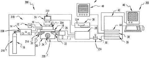
Figure la provides an overall schematic of one
embodiment of an automated single-cell culture system; Table
I provides a detailed description of the components in Figure
la. Figures lb-le provide a more detailed set of schematics
for the Chamber for one embodiment of the automated single-
cell culture system 300; Table II provides a detailed
description of the components of the Biochamber 10.
A preferable strategy used in the system 300
entails periodic monitoring and analysis of cells housed in
300 L wells of a disposable, plastic 96-well plate 207 under
a sterile, controlled environment using a robotic imaging
system (Fig. la, 20-46) . Cells are observed using an
Inverted Microscope 20 with extra-long working distance
(ELWD) condenser and phase-contrast objectives and
epifluorescence attachments. Digitized phase-contrast images
of cells are obtained using a video-Rate CCD Camera 32
connected to a PixelPipeline Imaging Board 38 installed in a
Macintosh Quadra 950 42 through a Time-Lapse VCR 36; the
Time-Lapse VCR records images for long-term archiving of
image data. Digitized fluorescence images of cells are
obtained using a Cooled CCD Camera 34 connected directly to
an interface board in the Quadra 950. Imaging operations on
the Quadra 950 are performed using Oncor-Image software.
Both phase-contrast and fluorescence images are displaced on
the Computer Monitor 46 using a Video Board 44 installed in
CA 02239815 1998-06-19
WO 98/20108 PC1:/US97/19834
-23-
the Quadra 950. Phase-contrast images also are displaced on
a High-Resolution Video Monitor 40.
The robotic components of the imaging system (Fig.
la, 18 and 22-30) are controlled by a Microscope Controller
28 which itself is controlled by commands from the Quadra 950
using Oncor-Image software through a RS-232 interface. The
Biochamber 10 is secured on a Motorized Stage 18 mounted on
the Inverted Microscope 20. The Motorized Stage 18 has a
resolution of 0.1 Am, an accuracy of 6 um, and a
repeatability of 2 urn. Preferably, the Biochamber 10 itself
with Motorized Stage 18 mounts directly on the Inverted
Microscope 20. Focus control is achieved for each well using
a Motorized Focus Drive Assembly and Controller 22 mounted on
the focusing knob of the Inverted Microscope 20.
Illumination is switched between transmitted light for phase-
contrast imaging and epillumination for fluorescence imaging
using a High-Speed Shutter for Transmitted Light 24 and a
High-Speed Dual Filter Wheel with Shutter for Fluorescence
26. The Motorized Focus Drive Assembly and Controller 22,
the motorized stage 18, the High-Speed Shutter for
Transmitted Light 24, and the High-Speed Dual Filter Wheel
with Shutter for Fluorescence 26 are connected electrically
to the Microscope Controller 28. Initial x-y positioning of
the Motorized Stage 18 stage and z-focal planes for each well
are chosen using a Joystick 30 connected to the Microscope
Controller 28 or by the computer 42.
CA 02239815 1998-06-19
WO 98/20108 PCT/US97/19834
=24-
Cells are maintained in individual wells of 96-well
plates under a sterile, controlled environment (i.e.,
physiological temperature, pH, P02! and humidity) inside a
anodized aluminum Biochamber 10 with glass windows on top and
bottom to provide an optical path for imaging. There are two
embodiments for the system 300: a Biochamber 10 (Fig. la and
Table I) and a Biochamber 10 also with z-robct for medium
exchange, as shown in figures 4a-4d. The Biochamber 10 for
the first embodiment (described in detail in Figs lb-e and
Table II) is approximately 6" by 5" by 2" high. Temperature
is regulated using a Thermocouple 58, Temperature Controller
12, and Heating Cartridges 62. Media pH is maintained using
standard bicarbonate-based buffers and a CO2 Controller 14
which sets atmospheric pCO2 at 5% by regulating the flow of
C02 from a CO2 Supply Tank with Regulator 16 through a
solenoid valve based on signals from a detachable CO2 Sensor
66 mounted on the side of the Biochamber 10. Pressure inside
the Biochamber 10 is fixed by a Pressure Relief Valve 72.
Control of pO2 in the Biochamber 10 can be maintained
similarly through a sensor and supply interfaced through two
additional chamber frontports. Fast response dynamics and
stable control are insured by rapidly mixing the Chamber's
atmosphere using a pinwheel turbine 78 driven externally by
house air.
Several parts of the Biochamber 10 are maintained
in an assembled state at all times. Glass Observation
CA 02239815 1998-06-19
WO 98/20108 PCT/US97/19834
-25-
Windows 54 are cemented into the base of the Chamber Body 50
and Chamber Cover 52; the Glass Observation Windows can be
removed for replacement but are not routinely because their
removal requires breakage. The Thermocouple Fitting 60 is
screwed into the right face of the Chamber Body; the Co,
Supply Fitting 68, Pressure Relief Fitting 70, and three
Unused Port Plugs 74 are screwed into the fron` face of the
Chamber Body. The turbine is assembled by securing one of
the Turbines 78 to the Turbine Shaft 80 with a Brass Bushing
82, screwing the two House Air Fittings 90 into opposing side
faces of the Turbine Housing 76, inserting the .urbine-shaft
assembly into the Turbine Housing such that a Turbine is
housed in the Turbine Housing, and securing the remaining
Turbine to the Turbine Shaft with the remaining Brass
Bushing. Next, the Turbine Housing O-Ring 86 is placed in a
groove on the front face of the Turbine Housing, the Turbine
Back Plate O-Ring 88 placed in a groove on the back face of
the Turbine Housing, the Turbine Housing Back Plate 84 placed
on the back face of the Turbine Housing by lining up the
groove on the Turbine Housing Back Plate with the Turbine
Back Plate O-Ring, and the assembly mounted onto the back
face of the. Chamber with two 1 1/4" x 3/16" hex-nut headed
screws. These screws are tightened to form gas-tight seals
between the Chamber Body and the Turbine Housing and between
the Turbine Housing and Turbine Housing Back Plate.
CA 02239815 1998-06-19
WO 98/20108 PCT/US97/19834
=26-
In operation, before use the disassembled
Biochamber 10 is autoclaved with 121 C steam for 15 minutes
for sterility. All components of the Chamber (50-90 in Table
II) are sterilized except for the Thermocouple 58, CO, Sensor
66 and the Pressure Relief Valve 72. The CO2 Sensor and
Thermocouple are sterilized by swabbing with a 70% aqueous
solution of ethanol in the sterile environment of a laminar
flow hood. The sterilized components are removed from the
autoclave and placed in the laminar flow hood along with a
disposable 96-well plate 207 containing cells. The 96-well
plate 207 has been maintained at 37 C in a humidified
atmosphere of 5% CO2 since cells were plated. The procedure
for plating cells is described subsequently in this
application. Spare wells in the plate in which cells were
not plated are previously filled with 100 E.cL of sterile
distilled water to maintain 95-100% humidity inside the
enclosed Chamber. The CO2 Sensor is mounted on the right
face of the Chamber Body 50 by tightening two 1 1/2" x 3/16"
hex-nut headed screws. The Pressure Relief Valve 72 is
connected to the Pressure Relief Fitting 70 with tygon
tubing. Next, the plate 207 is placed carefully into the
inset on the bottom of the Chamber Body 50 and secured with
a spring clip. The Thermocouple is inserted into the Chamber
through the Thermocouple Fitting 60 and tightened into place
with a Teflon fastener on the Thermocouple Fitting. The
Chamber is enclosed by placing the Chamber Cover Gasket 56 in
a groove on the top face of the Chamber Body and securing the
CA 02239815 1998-06-19
W008/20108 PCTIUS97/19834
=27-
Chamber Cover 52 in place on-top of the Chamber Body and
Chamber Cover Gasket by tightening eight 0.50" x 0.19" hex-
nut headed screws. Chamber assembly is completed by securing
the two Heating Cartridges 62_into channels in side walls of
the Chamber Body from ports in the front face of the Chamber
Body using one Heating Cartridge Retaining Screw 64 each.
Environmental control within the Biochamber 10 is
maintained by regulating temperature and the partial pressure
of C02 with two control systems. The Thermocouple 58 is
connected by insulated electrical wire to the input junction
of the Temperature Controller 12. The two Heating Cartridges
62 are connected by insulated electrical wire to the output
junctions of the Temperature Controller. The CO2 Sensor 66
is connected electrically to the input junction of the CO2
Controller 14. The output gas stream from the CO2 Sensor is
connected to the C02 Supply Fitting 68 on the front face of
the Chamber and the CO2 Supply Tank with Regulator 16
connected to the input gas stream to the CO2 Sensor. The
assembled Biochamber 10 with environmental controls is
allowed to thermally and atmospherically equilibrate for one
to two hours before placement on the Motorized Stage 18.
Temperature and pCO2are controllable to 37 0.5 C and 5 0.2%,
respectively, over the course of several days.
The Biochamber 10 with environmental controls next
is secured on the Motorized Stage 18 with a spring mount.
CA 02239815 1998-06-19
WO 98/20108 PCT/US97/19834
--28-
Cells for observation are chosen by scanning wells using the
Motorized Stage and Joystick 30 and phase-contrast and
fluorescence optics. Image fields of individual wells
containing cells for further investigation are selected based
on clarity of images. For each well, one or more fields are
selected. After selection of fields from up to preferably 96
wells for observation, the user initiates the automated Hart
of the imaging and analysis by selecting the appropriate
option. Each field selected then is scanned sequentially at
a user-defined interval (preferably between one and 60
minutes). It also is possible to scan at shorter or longer
intervals depending on the requirements of a particular
biological system. Each field is imaged under phase-contrast
optics with transmitted light illumination using the Video-
Rate CCD Camera 32 and under fluorescence optics with
epillumination using the Cooled CCD Camera 34.
The occurrence of cell division and differentiation
is detected by pattern recognition software. The number and
two-dimensional shape (e.g., area and perimeter) of "objects"
in each selected field are identified from phase-contrast
images after application of an optical gradient
transformation, thresholding, and dilation to detect "halos"
around each cell (see Fig. 2). Threshold values for shape
parameters which indicate whether each object is one or more
cells have been defined. The number of cells is then
determined in each well at that particular time point by
CA 02239815 1998-06-19
WO 98/20108 PCT/US97/19834
comparing the current values of the shape parameters with
values for previous time points. Cell division is detected
automatically as an increase in cell number between two time
points. Image analysis also provides information on (x-y)
positions which can be used to measure individual cell speed
and directional persistence time by application of a
persistent random walk model for migration, to determine the
fraction of a population which is motile, and to adjust the
position of the field to allow for cell movement while
centering cells in the field. The parameter cell speed and
directional persistence time for each individual cell and 5e-
motile for a population of individual cells are determined by
fitting a mathematical model for a persistent random walk in
an isotropic environment to observe data for the mean-squared
displacement of each individual cell based on a time sequence
of (xyl position at the control of the cell) . (DiMilla,
P.A., Albelda, S.M., Lauffenburger, D.A., and Quinn, J.A.
1992. Measurement of Individual Cell Migration Parameters
for Human Tissue Cells. AIChE J. 38(7): 1092-1104; DiMilla,
P.A., Stone, J.A., Albelda, S.M., Lauf<fenburger, D.A. and
Quinn, J.A. 1992. Measurement of Cell Adhesion and
Migration on Protein-Coated Surfaces. In Tissue-Inducing
Biomaterials, L.G. Cima and E. Ron, eds., Mater. Res. Soc.
Proc. Vol. 252, pp. 205-212; DiMilla, P.A., Stone, J.A.,
Quinn, J.A., Albelda, S.M. and Lauffenburger, D.A. 1993.
Maximal Migration of Human Smooth Muscle Cells on Type IV
Collagen and Fibronectin Occurs at an Intermediate Initial
CA 02239815 2004-06-08
-30-
Attachment Strength. J. Cell Biol. 122(3): 729-737; DiMilla,
P.A. Receptor-Mediated Adhesive Interactions at the
Cytoskeleton/Substratum Interface During Cell Migration. in
Cell Mechanics and Cellular Engineering, R.M. Hochmuth, V.C.
Mow, F. Guilak, and R. Tran-Son-Tay, eds., Springer-Verlag,
New York, pp. 490-514, 1994; Thomas, T.W. and DiMilla, P.A.
Effects of Substratum Compliance on the Motility, Morphology,
and Proliferation of Adherent Human Gliblastoma Cells. In
Proceedings of the 1995 Bioengineering Conference, BED-Vol.
29, R.M. Hochmuth, N.A. Langrana, and M.S. Hefzy, eds., ASME,
New York, pp. 153-154, 1995).
Data for the movement of human grade IV
SNB-19 glioblastoma cells is depicted in Fig. 3 and
demonstrates an application in neuroscience and cancer
research.
After completion of an experiment to identify
growth or attribute information about a given type of cell,
or after cells are grown as desired, the computer program is
stopped, the Biochamber 10 removed from the imaging system,
and environmental controls disconnected. The Chamber is
disassembled in a laminar flow hood. The 96-well plate is
saved. The Chamber components are now ready for
sterilization and use in a new experiment.
The second embodiment of the system 300 is designed
to augment the basic strategy. It implements a different
CA 02239815 1998-06-19
WO 98/20108 PCT/US97/19834
--31-
translation' strategy in the x-y plane and provides for
enhanced diagnostic and growth environment manipulation at
the level of a single well in the array.
The second embodiment of the system 300 adds to the
features of the first embodiment (continuous non-invasive
observation of single cells in multiple wells, sterility,
control of temperature to 0.5 C, control of pCO2 and p02to
0.1%, autoclavablity) with a z-robot pipette that can
automatically dispense and aspirate media to and from wells
in a 96-well plate. The z-robot thus endows the system with
the capability to alter the environment of each well and/or
add diagnostic reagents to ascertain the occurrence of
biological events or based upon the image recognition of a
biological event.
In the second embodiment, the generic motorized
stage has been replaced with a custom motorized stage which
allows the incorporation of the z-robot pipette. The
Biochamber 10 houses a 96-well plate 207 mounted on a movable
platter which is moved to specific (x-y) coordinates by a
pair of stepper motors. This design moves each well under
the microscope objective as well as move any selected well
for z-robot pipette servicing. An overview is shown in
Figure 4A.
CA 02239815 1998-06-19
WO 98/20108 PCT/US97/19834
-32-
The z-robot pipette dynamically controls the
composition of medium bathing cells to add growth and/or
quiescence factors automatically to individual wells based on
cell behavior. Software driving the operation of this z-
robot pipette is integrated with software for monitoring cell
behavior. It also is possible and preferred in some
applications of the system 300 to add, remove or change
medium based on external criteria, such as at particular time
intervals chosen by the user. The z-robot pipette also
transfers media from individual wells to supplemental
analysis systems.
The z-robot pipette for media exchange itself
consists of a modified micropipette tip, see Fig. 4b, mounted
on a support arm driven by a z-axis stepper motor to move up
and down and raise and lower the pipette tip for aspiration
and dispensing media in 1 to 95 iLL increments. Note that
although typically 100 L of medium is added to each 300 gL-
volume well, aspirating all of the medium from a well will
result in very large shears being applied to cells and likely
detach or otherwise disturb them. Preferably, the minimum
volume of medium which must remain in any well at any time is
5 L (corresponding to a depth of 125 Am).
Referring to figure 4b, the major components of the
pipetting system consists of a syringe pump 100 that can
deliver growth factors, quiescence factors, or any type of
CA 02239815 1998-06-19
WO'98/20108 PCT/US97/19834
liquid from multiple fluid reservoirs 101 through tubing to
a pipette tip 102. The syringe pump consists preferably of a
250 microliter syringe 103 (although other syringe sizes can
be used) that is driven by a stepper motor 104, which is in
turn controlled via a multi-port stepper motor driver card
105 and a computer 106. The stepper motor 104 drives the
plunger 107 of the syringe 103 up and down which results in
a dispensing action (if the plunger is being driven into the
syringe) or an aspiration action (if the plunger is being
driven out of the syringe). The syringe is connected to one
port of a distribution valve 108. The distribution valve can
be from 3 ports to 8 or more ports. One port is connected to
the syringe 103, one port is connected to the pipette probe
102, one port to an optional wash pump 111, and the remaining
ports to various fluid reservoirs 101. The distribution valve
108 is also stepper motor driven through stepper motor 109
which can be driven also from stepper motor drive board 105.
The syringe, stepper motor, stepper motor driver, and
distribution valve can be obtained from Advanced Liquid
Handling model MEP 2000 (Williams Bay, WI). A second
distribution valve can also be mounted in the system in
parallel with valve 108 to tie into more fluid reservoirs.
The reservoirs 101 are thermostat to 4 2 C by
thermostatting means 112, to allow good preservation of the
growth and quiescence medias and tied to the distribution
valve 108 through 1/16 inch Teflon tubing.
CA 02239815 1998-06-19
WO 98/20108 PCT/US97/19834
=34-
The distribution valve (and thus the syringe pump)
is plumbed via 1/16 inch Teflon or stainless steel tubing to
the pipette probe 102. The pipette consists of a stainless
steel probe with an ID of 1/32 inch (0.031 inch) that narrows
down to a tip ID of 0.013 inch. This pipette tip is used for
both dispensing growth and quiescence factors into the 96
well plate as well as aspirating media out of the plate. The
pipette probe has conductive coating on the outside of the
probe that provides a signal that can be read by the computer
106. This electrical signal provides feedback on how much
fluid there is in the well that the probe is in. This is
helpful in aspiration to know when no more fluid exists and
aspiration should stop. The pipette probe is driven in the
"Z" direction by a stepper motor 110 that is tied into the
stepper motor drive 105. This stepper motor drives the
pipette probe up and down to dispense into or aspirate out of
a selected well. The probe with conductive sensing can be
obtained from Diba Industries, Inc., (Danbury, CT). The
pipette stepper motor can be obtained from Advanced Liquid
Handling model MBD Crawler (Williams Bay, WI). The pipette
probe mounts into the biocontainment box by piercing through
a Teflon bulkhead. The Teflon bulkhead has a hole in it that
is sized to interference fit the OD of the pipette probe.
Thus a seal is made between the OD of the pipette and the ID
of the hole in the Teflon. This fit allows the pipette to
move up and down freely and yet provides a seal to keep the
environment within the Biochamber stable. The pipette moves
CA 02239815 1998-06-19
WO 98/20108 PCT/US97/19834
down into the well to a depth of 3 1 mm from the top of the
well for dispensing; the pipette moves down to the liquid
surface in the well for aspiration (as measured by the
conductive sensing mechanism on the probe tip); and the probe
moves up out of the well with a clearance of 10 to 13 mm to
clear the well as the well plate moves around on the x-y
stage.
An alternative embodiment is to have multiple
dispensing/aspiration tips so that dispenses to the 96 well
plate or aspirations can be done in parallel for higher
throughput. A wash is needed with the system to wash out
growth factors, quiescence factors or used media from the
plumbing lines. The preferred wash fluid is Phosphate Buffer
Saline (PBS). One approach is to use one of the reservoirs
101 for wash fluid to clean the system. Another approach is
to use a separate wash pump 111 with the system. The wash
pump 111 is a peristaltic pump with higher volumetric flow
capabilities that can be turned on by the computer 106 and
pump through higher flows of wash fluid. The wash fluid is
dispensed from the pipette tip 102 to a flush station within
the Biochamber 10, as shown by item 330 in figure 4d.
Referring to Figures 4a and 4c, various additional
analytical determination steps can be added to the system. A
second distribution valve 114 has been added to the system
and tied into distribution valve 108. This allows more ports
CA 02239815 1998-06-19
WO 98/20108 PCTIUS97/19834
-36-
to be added to the system. This allows more fluid reservoirs
101 to be added to the system or supplemental analysis
systems 116 and 118. These supplemental analysis systems
work in the following way: The pipette tip 102 is lowered
into a well of the 96 well plate; the syringe pump 100
aspirates out a specific amount of media or fluid from the
well through the pipette tip. This fluid is drawn all the
way into the syringe barrel 103. The distribution valve 108
and distribution valve 114 is switched so that the flow from
the syringe pump is directed out through these two valves to
supplemental analysis systems 1 (116) or 2 (118) or to any
port connected to the distribution valves. The syringe pump
would then pump out through the plumbing and valves to the
supplemental analysis systems. These supplemental analysis
systems could be any of the following examples, although not
limited thereto- Additional supplemental analysis systems
can be added based on user requirements.
Tissue culture medium or nutrients removed from
individual tissue culture wells by the robotic arm will (for
specific experimental uses) be deposited into a
protein/nutrient analysis system. Alternatively, all
material including cells will be removed for cell counting by
automated cell counters (Coulter, Co.). This tissue culture
medium can be analyzed by each of a variety of biochemical,
immunochemical, biological and chemical assays including but
not limited to the following:
CA 02239815 1998-06-19
WO 98/20108 PCT/US97/19834
=37-
1. Radioimmuno-assay for detection of produced hormones
such as insulin, growth hormone, prolactin, gastrin,
(other peptide hormones) or by radioimmuno-assay for
cellular production and release of cytokines including
but not limited to: IL-1 (interleukin 1, IL-2, IL-3,
IL-4, IL-5, IL-6, IL-7, IL-8, IL-9, IL-10, IL-11, IL-12,
IL-13, IL-14, IL-15, M-CSF, GM-CSF, C-CSF, HGF, NGF,
basic FGF, acidic FGF, PDGF).
2. Lentil lectin chromatography for detection of
glycosylated proteins using columns such as the
Sepharose 4 (3 (Pharmacia Corporation) column:
3. Di ethyl aminoethyl (DEAE) chromatography using a column
such as that produced by the Whatman Corporation.
4. Ionic exchange high pressure liquid chromatography
(HPLC) analysis using a column such as the Synchropak
AX300 column (Thompson Instrument Company).
5. Gel filtration (HPLC), using centriprep or centricon-30
(30,000 molecular weight cut-off) centrifugal
microcentrifuge (Amicon Corporation) samples using a
column such as the protein-PAK 300 SW (Millipore
Corporation).
CA 02239815 1998-06-19
WO 98/20108 PCT/US97/19834
38-
6. Reverse phase (HPLC) using an apparatus such as the
VydacC4 HPLC column (The Separations Group Corporation)
using the equilibration with Trifluoroactic acidic acid,
or acetonitrile (made by Pierce Corporation and Baxter
Corporation, respectively).
7. Sodium deodecylsulfate/polyacrylamide gel
electrophoresis (SDS/PAGE) analysis using commercially
available reagents from Integrated Separations Systems
Incorporated.
8. Protein analysis for glycosylation by tunicamycin or
N-glycosidase treatment (using reagents obtained from
Wurthington Biochemical Corporation and Genzyme
Corporation).
9. Proliferation stimulation assays (biological assays);
aliquots of tissue culture medium will be tested for
stimulation of tritiated thymidine incorporation (50-90
MMOL; Dupont Chemical Corporation) by target indicator
cell populations with known cell populations that
respond to each of a variety of cytokines in each growth
factor using published methods. (Pogue-Geile, K.L.,
Sakakeeny, M.A., Panza, J.L., Sell, S.L., Greenberger,
J.S. Cloning and Expression of Unique Murine Macrophage
Colony Stimulating Factor Transcripts. Blood, 85:3478
3486, 1995)
CA 02239815 1998-06-19
WO 98/20108 PCTIUS97/19834
'39-
10. Respiratory/oxidative physiologic functioning analysis
including analysis of pH, bicarbonate concentration,
chloride concentration, oxygen concentration.
11. Catabolic product production including assays for
ammonium urea, and consumption of glucose, fructose and
other sugar molecules contained within the particular
culture medium (including Dulbecco's modified Eagles
medium, McCoy's medium and other tissue culture media
prepared by commercial suppliers and available from
GIBCO Corporation or other suppliers).
12. Enzyme analysis including tests for proteases, sucrases,
and other sugar conjugating or degrading enzymes using
standard biochemical test kits available from SIGMA
Pharmaceutical Company, and available in standard
hospital clinical laboratories. Assays would include
those for amylase, acid phosphatase, alkaline
phosphatase, carbonic anhydrase, and others.
The purpose of the assays outlined above will be to
determine whether cells identified by the imaging mechanism
and Pattern Recognition software and computer analysis system
are in a specific physical, chemical or physiological state
and to correlate this state with a particular metabolic
CA 02239815 1998-06-19
WO 98/20108 PCT/US97/19834
-40-
process including those associated with either production or
consumption of the above factors.
In summary, the medium from tissue cultured cells
grown in the Biochamber wells would be tested for production
or degradation of proteins, simple or complex sugars,
individual amino acids, individual ions, and individual
molecules, both with respect to physical presence and/or
biological activity.
Another embodiment of the biocontainment box is
shown in Figure 4d, which shows a front view of parts of the
system. The* X-Y translation system is - shown as part 300,
which has an open space in the middle of it shown by the
dotted lines 302 and 304. This allows the objective lenses
306 to be moved into the X-Y translation table and be focused
onto the 96 well plate 308. The 96 well plate 308 is
positioned onto a mounting plate 310 which moves according to
the translation of the X-Y translation plate. Mounting plate
310 has an optical window 328 in it that is below the 96 well
plate. The mounting plate also contains a pipette probe
flushing station 330 which is used as a port to flush and
clean the probe. The biocontainment box 312 has double walls
314 and 316. Each of these walls is sealed to the mounting
plate 310 by a silicone seal 318. The biocontainment box is
stationary while the mounting plate 310 moves underneath it.
The biocontainment box 312 also has the following parts
CA 02239815 1998-06-19
WO 98/20108 PCT/US97/19834
=41-
mounted into it: pipette probe 320 is mounted in a Teflon
bulkhead 322 and driven in the Z-axis direction by stepper
motor 332; and an optical window 324 is mounted so that
light can pass through it into condenser 326 with is
positioned above the objective lenses 306, optical window
328, plate 308 and optical window 324. The biocontainment box
also has the controls for pH, C02, humidity, etc mounted into
it (not shown). The pipette probe is connected to the syringe
pump system via teflon line 334.
The X-Y translation plate moves any well of the 96
well plate over the objective so that the cells in the well
can be imaged; it also moves any well to the pipette probe
for dispensing or aspiration of media or cells, and it also
moves the flushing station to the probe tip so that the probe
can be flushed and/or cleaned.
Reservoirs for fresh and waste media, including
individual cocktails of growth factors, are located next to
the Chamber and maintained at 4 C. Small-volume syringe
pumps are used to deliver growth factors and base medium to
user-specified compositions, and waste media is aspirated
from wells using the same pipette. The pipette is cleaned
thoroughly between dispenses and aspirations by flushing with
a PBS solution.
CA 02239815 2004-06-08
-42-
The operation of the z-robot pipette has been
optimized such that the fluid forces applied to cells are
minimized while retaining a sufficient flow rate for rapid
medium exchange. The following parameters have been
examined: the dynamics and steady-state value of the flow
rate, the minimum volume of fluid which must be retained in
a well after aspiration, and the effects of locating the
nozzle off-center in the well. Medium is dispensed to wells
by drop-wise addition. Choosing the optimal parameters for
aspiration most quickly is supported by numerical simulations
of the fluid mechanics of this process using well-established
computational packages (e.g., Fluent). (DiMilla, P.A., Stone,
J.A., Albelda, S.M., Lauffenburger, D.A. and Quinn, J.A.
1992. Measurement of Cell Adhesion and Migration on
Protein-Coated Surfaces. In Tissue-Inducing Biomaterials,
L.G. Cima and E. Ron, eds., Mater. Res.'Soc. Proc. Vol. 252,
pp. 205-212; DiMilla, P.A., Stone, J.A., Quinn, J.A.,
Albelda, S.M. and Lauffenburger, D.A. 1993. Maximal
Migration of Human Smooth Muscle Cells on Type IV Collagen
and-Fibronectin Occurs at an Intermediate Initial Attachment
Strength. J. Cell Biol. 122(3):.729-737; DiMilla, P.A.
Receptor-Mediated Adhesive Interactions at the
Cytoskeleton/Substratum Interface During Cell Migration. In
Cell Mechanics and Cellular Engineering, R.M. Hochmuth, V.C.
Mow, F. Guilak, and R. Tran-Son-Tay, eds., Springer-Verlag,
New York, pp. 490-514, 1994; Goldstein A.S. and DiMilla,
P.A.
CA 02239815 1998-06-19
WO 98/20108 PCT/US97/19834
=43-
Overall, the detection of changes in cell phenotype
and operation of the z-robot pipette with the features of the
first embodiment are integrated. By applying the methodology
for phase-contrast imaging to fluorescent images (obtained
with a cooled CCD camera) for wells in which fluorescent
antibodies against specific antigens for the lin- phenotype
are added, the system 300 is able to, for example, identify
stem cells from other differentiated cells. As discussed in
more detail below, this approach allows one to determine
whether and when individual cells differentiate and change
phenotype. Kinetic data-for the rates of cell division and
differentiation can then be obtained. This data is then
analyzed using engineering models for probabilistic processes
to determine kinetic parameters for rationally optimizing and
scheduling changes of media.
A general description of an algorithm for image
analysis of a doubling event is now provided. When a cell
divides, there are characteristic morphological features that
are visible. The pinching of the middle and the swelling of
the size, for example. These are used to identify cells that
are dividing. In terms of the computer, these events are
recognized by changes in the x,y position, area, perimeter,
sphericity (a measurement of the closeness to a circle), and
eccentricity (a measurement of closeness to a square). Other
parameters can be added as new data queue is acquired.
Moreover, the trend of these parameters corresponds with the
CA 02239815 1998-06-19
WO 98/20108 PCT/US97/19834
=44-
time before the doubling. The parameters are stored in
computer memory in a queue for a certain length of time. As
new image data is taken, the least recent value is removed
from the queue. The trend of this data is compared to the
historically known trend that reflects a cell division. If
the match is within tolerance, then the computer is signaled
that the cell is about to or is in the process of dividing.
For example, with stem cells it is theorized that these cells
stop moving just before they undergo division. This would
signal a decrease in the change of the x,y positions. If the
change remains small enough for a significant period of time,
then a division may be occurring. However, this alone is not
enough to guarantee it, so the trends of the other variables
are compared also.
The mathematical nature of the trend comparison is
the following. The trend of each parameter up to the point
of cell division is curve fitted over a length of time. The
current parameters stored in a queue obtained from a cell for
a given time period are compared to this smooth curve and the
error between the two is calculated. If the error is within
a user-specified tolerance, the cell is considered to be
approaching a division.
A division could be missed if the cell is not
visualized frequently. This could result from having to
analyze, stain and/or view other wells or having too many
CA 02239815 1998-06-19
WO 98/20108 PCT/US97/19834
=45-
wells to successfully return to each well within a regional
period of time. If this is the case, the parameters from the
image are compared to the previous parameters. A set of
morphological and positional criteria established empirically
are used to determine if the two objects could have come from
a division or if one additional object all moved into the
view field.
Figure 5 shows the results from an analysis of a
cell dividing. The photographs show the phase contrast
images on the left and the bitplane pictures on the right.
The pi=ch_ng has already begun to occur in the top left
picture.
A protocol for purifying human hematopoeitic stem
cells using the system 300 is now provided as an example of
using cells generally with the system 300.
Human nucleated blood cells, obtained from either
umbilical cord blood, peripheral blood by leukophoresis or
bone marrow, are purified free of red cells by
centrifugation. Buffy coat leukocytes are then purified to
separate CD34+ cells (representing approximately 1 out of
10,000 nucleated cells in human adult bone marrow) using any
of several commercially available immunobead, or column
chromatography methods. A preferred method is the CellPro
Ceprate column, which is commercially available from CellPro
CA 02239815 1998-06-19
WO 98/20108 PCT/US97/19834
=46-
Corporation, Bothell, WA. Using the techniques described in
the information supplied by the manufacturer, nucleated cells
that are adherent to the column are then washed free from the
column by competition with a supply of reagent contained in
the package which separates the CD34+ human hematopoeitic
cells from the column. This is carried out by competition
displacement. The CD34+ cells are washed free through the
eluate and are collected. These cells are then sorted a
second time using a fluorescence-activated cell sorter and a
combination of monoclonal antibodies that are lineage-
specific. Those subsets of the CD34+ cells which bind FITC,
rhodamine, or other fluorescently labeled indicator of
lineage-specific antigens including CD38, are then collected
in the lineage-negative (fluorescent antibody-negative)
preparation volume. These cells are then prepared for a
second FACS (fluorescence activated cell sorting) step, and
now are separated into a final population of those reacting
positively with a fluorochrome dye for Thyl. This population
represents a final concentration of cells which represented
one out of 50,000 of the original nucleated cells from the
original specimen of peripheral blood, bone marrow or
umbilical cord blood. These cells are those known to be
highly enriched for multilineage hematopoeitic stem cells.
Assays confirming the homogeneity and purification of these
cells include the long-term culture initiating cell assay
(Sutherland, H.J., Landsdorp, P.M.,' Henkelman, D.H., Eaves,
A.C., Eaves, C.J. Functional Characterization of Individual
CA 02239815 2004-06-08
-47-
Human Hematopoeitic Stem Cells Cultured at Limiting Dilution
on Supportive Marrow Stromal Layers. Proc Natl Acad Sci USA
87:2584, 1990), or the
cobblestone island assay measuring those cells forming
cobblestone islands at day 14 or day 21 after coculture
(Ploemacher, R., van der Sluijs, J., van Beurden, C., Baert,
M., Chan, P. Use of Limiting-Dilution Type Long-Term Marrow
Cultures in Frequency Analysis of Marrow-Repopulating and
Spleen Colony-Forming Hematopoeitic Stem Cells in the Mouse.
Blood 10:2527-2533; 1991),
or the assay for CFU-blast (Ikebuchi, K., Wong, G., Clark,
S., Ihle, J., Hirai, Y., Ogawa, M. Interleukin 6 Enhancement
of Interleukin 3 Dependent Proliferation of Multi-Potential
Hemopoietic Progenitors. Proc. Natl. Acad. Sci. USA 84:9035;
1987), or the assay for
high proliferative potential colony-forming unit culture
[HPP-CFC1 (Pogue-Geile, K.L., Sakakeeny, M.A., Panza, J.L.,
Sell, S.L., Greenberger, J.S. Cloning and Expression of
Unique Murine Macrophage Colony Stimulating Factor
Transcripts. Blood, 85:3478 3486, 1995).
Each and any of these assays demonstrates
that the CD34+Lin-Thyl+ subpopulation of cells is enriched
for the presence of cells positive in these assays by a
factor of around 1000-10,000-fold. More importantly, these
enriched cells have been demonstrated to form multilineage
hematopoeitic cells in the peripheral blood in marrow of
SCID/Hu mice, or Nu/BIX in xenotransplant studies. These
CA 02239815 2004-06-08
-48-
cells have also been shown to reconstitute multilineage human
hematopoiesis in fetal sheep (Zanjani, E.D., Almeida-Porada,
G. and Flake, A.W. 1995. Engraftment and Multilineage
Expression of Human Hematopoeitic Stem Cells in Human-sheep
Chimeras. Stem Cells 13:101-111). Thus, by the two
xenotransplant models (SCID/Hu mouse, and fetal sheep) as
well as the in vitro assays described above, the CD34+Lin-
Thyl+ and Thyl- fraction of nucleated peripheral blood, bone
marrow, or cord blood cells is known to be highly enriched
for stem cells. The phenotype of CD34+Lin-Thyl+ is known to
be rapidly lost when cells are cultured in suspension culture
as other than single cells and as culture methods other than
an automated cell division linked bioreactor (Mayani, Hector,
Lansdorp, Peter M. Proliferation of Individual Hematopoeitic
Progenitors Purified From Umbilical Cord Blood. Experimental
Hematology 23:1453-1462 (1995); Van Zant, Gary, Rummel, Sue
A., Koller, Manfred R., Larson, David B., Drubachevsky,
Ilana, Palsson, Mahshid and Emerson, Stephen G. Expansion in
Bioreactors of Human Progenitor Populations from Cord Blood
and Mobilized Peripheral Blood. Blood Cells (1994) 20:482-
491; Sandstrom, C.E., Bender, J.G., Papoutsakis, E.T.,
Miller, W.M. Effects of CD34+ Cell Selection and Perfusion
on Ex Vivo Expansion of Peripheral Blood Mononuclear Cells.
Blood. 86, No. 3:958 970 (August 1) 1995)).
CA 02239815 1998-06-19
WO 98/20108 PCT/US97/19834
=49-
Using 96-well plates (LinBro Plastic Corporation),
the MB210 mouse cell line (preferred), or any of a variety of
human, murine or primate bone marrow stromal cell lines (or
no stromal cell line, or in place of the stromal cell line
extracellular matrix protein or proteoglycan such as
fibronectin, or heparin sulfate proteoglycan), is plated into
each of the 96 wells at 1x10⁵ cells/well in Dulbecco's
Modified Eagle's Medium supplemented with 10% fetal calf
serum, and the cultures are incubated at 37 C in a high
humidity incubator for 24 hours. The cultures are then
removed from the incubator and each well is surveyed to be
certain there is a lawn of confluent monolayer of the stromal
cell line. The stromal cells are then irradiated to 2000 cGy
preferred (1000-10,000 cGy) using a 250 KVP orthovoltage x-
ray unit (preferred) or any linear accelerator from 6 MeV -
MeV with focal plane at the tissue culture surface on the
monolayer of cells. The cells are irradiated in the same
medium and are returned to the incubator in the same medium.
The cultures are then washed free of medium with multiple
20 washes of Iscove's Modified, serum-free medium for each well,
and then Iscove's Modified, serum-free medium is added to
each well. The exact recipe for the Iscove's Medium is
described in the reference (Goff, Julie P., Shields, Donna
S., Petersen, Bryon E., Zajac, Valerie F., Michalopoulos,
George K. and Greenberger, Joel S. Synergistic Effects of
Hepatocyte Growth Factor on Human Cord Blood CD34+ Progenitor
Cells are the Result of c-met Receptor Expression. Stem
CA 02239815 2004-06-08
-5Q-
Cells 14(5):592-602 (1996). The
serum-free Iscove's Medium (preferred) can be substituted
with any commercially available serum-free medium
supplemented with vitamins, nutrients, lipid substitute,
bovine serum albumin or any of a variety of additives known
to support hematopoeitic progenitor cells in the absence of
serum, but supplemented with appropriate growth factors.
Each tissue culture well is then fed with 100 ,uL of Iscove's
complete medium, and with an additional volume of Medium-A
(this is the growth medium and contains optimal
concentrations of each of the following growth factors: IL-
11, IL-6, IL-1, HGF, G-CSF, basic FGF). This medium will be
referred to as growth medium or Medium-A. Each tissue
culture well in the 96-well plate is then supplemented with
a 10 ,uL drop containing one FAC-sorted single CD34+Lin-Thyl+
cell, hereafter referred to as "stem cell". The term "stem
cell" will be used in this application to refer to a cell
with a phenotype demonstrated by fluorescence antibody
binding which is positive for CD34+, negative for the lineage
markers (the combination of lineage antibody markers used in
any of a combination of 10-15 different lineage markers but
contains the preferred marker, CD38, and is either Thyl+ or
Thyl-). The term "stem cell" will be used to refer to that
cell which is either Thyl+ or Thyl- (varies between
experiments, preferred Thyl+) but which is definitely CD38-
and CD34+. Cells were stained with FITC and PE-conjugated
antibodies against hematopoietic cell surface differentiation
CA 02239815 1998-06-19
WO 98/20108 PCT/US97/19834
--5l-
markers. Figures 7A and 7B represent the same field of
cells. Examples of cells that are only positive for CD34 and
Thyl appear green (7A, asterisk). Cells that express lineage
markers appear red (7B). When the cells are positive for
S CD34 and Thy! as well as lineage markers, the wavelengths
combine and the resulting fluorescence is yellow (arrowheads
Figure 7A)
The plate of 96 wells containing one stem cell per
well is then transferred to the Biochamber 10.
The Biochamber 10 is then closed and the unit is
programmed to cycle according to medium change with each cell
division (linked to the pattern recognition of the CD34-E-Lin-
Thyl+ phenotype).
Details of the specific motions for each element of
the protocol will be described following a short summary.
Note that different operational modes are possible and
selection depends on, for example, whether the aim is
biologic production or more basic investigation. The
following summary is a representative production mode where
phenotype conservation and high proliferation are intended.
An example of operation for basic investigation is presented
later in the form of a flow chart.
CA 02239815 1998-06-19
WO 98/20108 PCT/US97/19834
-52-
Each individual well is tracked separately over a
cycle of the plate 207 underneath the viewing objective, and
this is programmed to be viewed every minute (preferred), can
be varied to every 20 seconds or as long as once per day or
once per week (or any longer desired length of time) or
anything in between. The viewing is accompanied by capture
of the image of the cell in the software of the computer
linked to the CCD camera. When a pattern associated with a
cell doubling is recognized by pattern recognition software,
the cell doubling event is recorded and operation of the
system 300 linked to the dye recognition phase. The dye
recognition phase is activated when the x-y stage moves the
well to underneath the z-robot where a combination of
fluorescent dyes are added for CD34 and CD38 (lineage marker)
as well as for Thyl (Thyl marker). After the well is moved by
the x-y stage back to he optical path/mechanism and waiting
for an appropriate interval (which depends on dye
properties), an image is then captured by the cooled CCD
camera of the cell doublet with three imaging purposes: 1)
detecting the color of the dye on CD34, 2) detecting the
color of the dye on the CD38, and 3) detecting the color of
the dye on Thyl. The pattern recognition software then
recognizes those wells in which a doublet was identified and
in which the pattern was noted to be conserved (both cells
CD34+ or both cells CD34- to substituting for the latter both
cells CD38-) and then the well is moved by the x-y stage to
the z-robot which carries out a media change, which will be
CA 02239815 1998-06-19
WO 98/20108 PCTIUS97/19834
53-
described in greater detail. The medium is then replaced
with Medium-B (quiescence medium) which will be described in
greater detail. The entire cycle then continues with
observation of that well at regular intervals (preferably
each minute, can vary from 20 seconds to more than one week),
and then when a second interval has passed (preferably six
hours or any time between 1 minute to more than a week), the
stage is instructed to translate that well to the z-robot
which changes the medium back to Medium-A or growth medium.
The entire cycle then repeats again with the pattern
recognition software analyzing that well and waiting and
scoring for the appearance of four cells. When the presence
of four cells have been detected/recorded, the colorimetric
fluorochrome staining mechanism of the z-robot adds
colorimetric dye, after an appropriate interval (preferably
five minutes) more images are taken with the cooled CCD
camera and image analysis to detect each of the three
fluorochromes, and those wells now which score positive for
all four cells with the phenotype of CD34+CD38-Thyl+ are
again translated to the z-robot which extracts the Medium-A
and replaces the medium with Medium-B. The entire cycle
repeats again surveying for the presence of 8 cells, then
again surveying for 16 cells, then again for 32 cells, then
again for 64 cells, and finally, 128 cells. Alternatively,
the system 300 can be operated such that at each cell
division (i.e. when 3, 4, 5, 6, ... up to more than 128 cells
are present) medium exchange from Medium-A to Medium-B
CA 02239815 1998-06-19
WO 98/20108 PCTIUS97/19834
54-
occurs. When a well contains 128 cells, the imaging protocol
is terminated and all the cells (in Medium-B (quiescence
medium)) removed from the well into a repository. When the
repository is replete with the contents of each well in the
plate (representing over 1000 cells), the contents at such
repository then are transferred to other parts of the
operation for use in gene transfer, bone marrow stem cell
transplantation to an awaiting patient, or cryopreservation
facility to .be used later for gene therapy or for an awaiting
patient.
Specifics of the protocol are now provided. The
96-well plate is placed in a sterile tissue culture hood
(Format or similar type) and using a Pasteur pipette, the
contents of individual 1.0 ml Eppendorf tubes, each
containing 10-20 /.cL of serum-free Iscove's medium which have
been supplemented with one sorted CD34+Lin-Thyl+ stem cell,
are transferred to each respective well of the 96-well plate.
Each well already contains 100 ML of the Iscove's medium, and
is now supplemented with 10-20 AtL of this medium containing
a single cell. The 96-well plate 207 is then transferred to
the Biochamber 10. The 100-120 /,2L in each well contains the
concentration of growth factors contained in Medium-.A (a
range for each of the factors will be included with a
preferred concentration indicated: hepatocyte growth factor
2S [HGF] , 10 ng/ml - 1 /. cg/ml, preferred 10 /2g/ml; IL-11, 1 ng/ml
100 . cg/ml, preferred 10 ,ug/ml; IL-1 10 ng/ml - 100 ug/ml,
CA 02239815 1998-06-19
WO 98/20108 PCTIUS97/19834
-55-
preferred 10 ug/ml; G-CSF, 1 ng/ml - 100 ,ug/ml, preferred 10
,ug/ml). Each of these growth factors are available from
commercial suppliers including Immunex and Amgen. It should
be noted these are but examples of Medium-A or growth medium.
There may be many different growth mediums, and not one
unique Medium-A, for a given type of cell. An acceptable
growth medium is one which allows the cell to reproduce in a
healthy manner. The cells are then placed in the Biochamber
10, which is set at a temperature of 37 C (can be varied from
31 C - 49 C), and 7% CO, (can be varied from 0.1% to 40% CO,),
and the humidity is kept at a high level (but can be low or
very high).
The Biochamber stage is moved so that each well is
placed over the microscope objective and in the optical path,
and the user moves the stage to identify the exact
coordinates of where the single stem cell is located. This
is done by hand and usually takes 15 minutes (this can take
between one minute and four hours to identify the location of
each cell). The coordinates of the individual cell are then
recorded and the cycling for observation of the cell is begun
with a magnification of 20 - 40x magnification, preferably
40x. The individual cell is located by image analysis and
its position and morphological parameters records by the
computer Y2. The computer is then set to cycle to capture
the image of the individual well during a six second viewing
of this cell (variation between one second and five minutes)
CA 02239815 1998-06-19
WO 98/20108 PCT/US97/19834
-56-
using the cooled CCD camera. The time during which the cooled
CCD Camera focuses on each well is set to be long enough to
capture the image within an acceptable signal-to-noise ratio
(preferably 20 seconds, can vary from one second - 20
minutes). The digitized image of the single cell in each well
is recorded and the stage then cycles over each of the 96
positions (corresponding to one per well) tracking the
cellular movement and recording the position of the cell
relative to the cross hairs which have been set to define the
position of. the cell in the initial image capture. The
survey of each well is carried out in cycle. The digitized
image is recorded sequentially for each well and stored in
the computer's memory. The images are compared to one
another serially over the time required for one cell doubling
to occur in each of the 96 single wells. With the growth
Medium-A being in each well, cell divisions are expected to
occur in each of the cells within six hours preferably (range
can be 10 minutes - two months).
At the time when a cell doubling is detected, as
captured by the image at the cycling over that particular
well and as reflected in the temporal trends in morphological
parameters for the computer's stored set of images and
registered as a cell doubling by pattern recognition
software, the stage is mechanically moved so that the well
corresponding to the cell doubling is underneath the z-robot
and fluorochrome dyes are added for determination- of the
CA 02239815 1998-06-19
WO 98/20108 PCT/US97/19834
-57-
phenotype of the two cells comprising the doublet. The
robotic pipette moves down and adds 10 /uL (range can be 1 :.4L
- 100 uL) of a combination of two fluorochrome dyes including
FITC-labeled anti-CD34 and phycoerythrin-labeled anti-CD3S.
The concentrations of dyes are those as supplied by t_e
manufacturer, and are commercially available. Fluorochromes
for detection of each monoclonal antibody (e.g., from
Pharmingen, Becton Dickinson, Dakocorp., Immunotech) will be
conjugated to each antibody, such that two separate non-
overlapping colors, each detected with the appropriate
fluorescence filters (available from but not limited to Sia:.a
and Chroma), will be distinguishable and registered by eat=
of two specific images. After the dye has been added to the
well with the cell doublet, two successive fluorescent images
are acquired with the imaging mechanism consisting of the
inverted microscope using epillumination with appropriate
excitation filter (placed in the optical path by the High
Speed Dual Filter Wheel with Shutter for Fluorescence) and
cooled CCD camera and stored as a digitized image on the
computer. Each image is exposed for a particular color,
either green or red, and for exposure times ranging from 0.1
seconds to 100 seconds. Optimal exposure time for each dye
is that determined by the fluorescence intensity of the dye
on the cell surface from detection prior to operation of the
system 300 and correlated to the positive control test
samples for the dye according to manufacturer's instructions.
Of the two successive images the first is acquired with a
CA 02239815 1998-06-19
WO 98/20108 PCT/US97/19834
-58- -
filter that allows imaging of emission in the green range of
the visible light spectrum (to detect the binding of
anti-CD34 antibody to any CD34 marker present on the surface
of the cells of the doublet) and the second acquired with a
filter that allows imaging of emission in the red range of
the visible light spectrum (to detect binding of anti-CD38
antibody to any CD38 marker present on the surface of the
cells of the doublet). This detection scheme is not limited
to successive examination for only two distinct colored
antibodies and can be extended to detect the presence of
additional markers on the cell surface reflecting finer
discrimination of cell phenotype. Two types of approaches
can be implemented for this extension of detection. In the
first, an additional antibody type labeled with a third
distinct colored dye other than red or green, such that the
emission bands of each of the dyes do not overlap, can be
included in the antibody cocktail to detect the presence of
a third cell-surface marker. This approach itself allows
further extension to fourth, fifth, and more than fifth
colors of dyes used to label distinct antibodies against
cell-surface markers provided that the emission of each of
the dyes can be distinguished unambiguously. In the second
approach for extending detection capability of cell
phenotype, after acquisition of the initial two successive
images using green and red excitation filters, the well is
washed sufficiently with PBS and another cocktail containing
an alternative set of antibodies labeled with green-emitting
CA 02239815 1998-06-19
WO 98/20108 PCT/US97/19834
and/or red-emitting dyes are added. Again images are
acquired successively with green and red excitation filters,
respectively. This second approach and the first described
approach for extending detection types are not mutually
exclusive and can be combined to allow for detection of any
desired number of cell-surface determinants by further
successive washes and dye-labeled antibody additions.
Preferably, the second addition of antibodies will contain a
green-labeled anti-Thyl antibody against Thyl. Each
fluorescent image acquired for each distinct dye and antibody
will be sorted in computer memory and images overlaid
digitally by computer for automated interpretation. Sets of
processed images which, with the above approach with
successive incubation with anti-CD34, anti-CD38, and
anti-Thyl antibodies, are green positive (i.e., CD34+ and
Thyl+ from positive detection of the green dye on cell
surfaces in successive green images) and red negative (i.e.,
CD38- from the absence of red dye on the cell surfaces in the
red image) will be recorded and the z-robot will carry out
media change. For images of cell in wells not meeting the
criteria of green positive/red negative but identified as a
cell doublet, a conserved phenotype has not been obtained,
and this well will be removed from the survey cycle of image
acquisition, pattern recognition, fluorochrome-labeled
antibody analysis, and media exchange for the remainder of
the process of expanding stem cells. The presence of a
non-conserved division indicates that the stem cell phenotype
CA 02239815 1998-06-19
WO 98/20108 PCT/U897/19834
-Go- .
has been lost and that no further increase in stem cell
number in this well is possible.
The pattern recognition software and stage movement
having registered that an individual well contains two cells
with the appropriate phenotype is described above. This well
is next moved by the x-y stage underneath the z-robot and 100
gL of media removed, and the waste media placed into the
receptacle vehicle in the Biochamber 10. (See figures
4a-4d.) The range of medium removed can be from 10 - 200 yL
(preferred 100 /.cL) . Standard conditions for media change
will be those associated with detection of the cell doublet
after fluorescence analysis. After withdrawing Medium-A,
Medium B containing the quiescence medium will be added.
This contains a 100 .uL volume of Iscove's Modified Medium,
serum-free, as described above but now containing MIP-1a,
TGFi3 each in the optimal concentration (preferred 10 ng/ml,
range 10 ng/ml - 100 ng/ml). This quiescence medium is but
one example of many possible quiescence mediums. The
quiescence medium can simply be a serum-free medium alone
added for twenty seconds to five minutes, or a medium with
TGF(3 or MIP-la, or TNF. There may be many possible
quiescence media that work; that is, which shut down the
maturation or differentiation process in the given cell but
maintain the cell in essentially a status quo state.
CA 02239815 1998-06-19
WO 98/20108 PCT/US97/19834
-61-
The bioreactor system 300 then continues to
cyclically examine all other wells and view this well to
which Medium-B has been added as a well in quiescence. A
timer within the computer program doing system operations is
set to leave the well in which Medium-B has not been added
undisturbed, except for imaging for pattern recognition of an
additional cell doubling event, for six hours (preferred,
range 10 minutes - two months). After the chosen time in
Medium-B has passed, this well is automatically returned by
x-y stage to underneath the z-robot and again imaged for
pattern. recognition to identify a cell doublet. Quiescence
medium, by definition, facilitates no cell division, and the
location of the cells in the doublet as well as their shape
will be similar compared to the time when the interval in
which the quiescence medium was present began (six hours
earlier).
The z-robot then aspirates Medium-B and deposit
this medium in the receptacle container within the
bioreactor. The z-robot then adds an equivalent volume of
Medium-A (the original growth medium). The optimal volume
removed of Medium-B is 100 uL, the optimal replacement volume
is 100 uL (range can be 10 ML - 200 12L).
After addition of Medium-A to the bioreactor well
containing two cells, the sequence of review of that well
consisting of imaging, pattern recognition, fluorescence
CA 02239815 1998-06-19
WO 98/20108 PCT/US97/19834
=62-
analysis and media exchange is resumed. After release of
that well from the "quiescence period," routine monitoring at
each previously set interval resumes. This interval is as
brief as ten seconds, and as long as two months. The well is
then assessed for the appearance of four cells
(quadruplicates). The pattern recognition software is reset
for that particular well to identify a four cell
quadruplicate. Upon recognition of the four cell
quadruplicate, the well is moved underneath the z-robot and
20 /[L of the dye combination added as described above. The
same dyes are added in the same concentrations as described
above. After an appropriate interval for incubation with the
fluorescent dyes (preferred 5 minutes, range one minute - 20
minutes), the filters are rotated to image the four cells
with the green and red excitation filters, respectively. The
exposure times are those as described above. The images are
then processed and those wells containing four cells with the
(CD34, green positive, CD38-, red negative) phenotype are
then registered as being maintained in the experiment. Those
wells containing one or more of the four cells with non-
conserved phenotype are eliminated from the experiment. The
z-robot then maintains this position over the well with the
four cells of conserved phenotype and withdraws 100 pL of
Medium-A, replacing it with 100 ,uL of the quiescence Medium-B
as described above. The amount of medium removed and
replaced can range from 10 /2L - 200- L.
CA 02239815 1998-06-19
WO'98/20108 PCT/US97/19834
--63-
The quiescence media interval is then reset for
that particular bioreactor well and the well is left
undisturbed, except for routine observation during each cycle
for the, quiescence interval (preferably six hours, variation
two minutes - two months).
The bioreactor system 300 is then reset to indicate
that the well will be surveyed for the appearance of eight
cells representing an octuplet. The bioreactor system 300
continues to cycle during its observation periods with review
of that well at each survey over the 96 wells, and continues
its pattern recognition program for 8, then 16, then 32, then
64, then 128 cells. The mechanical and computer algorithms
used for identifying increases in the number of cell doublet,
the presence of conserved phenotypes, and exchange of media,
including scheduling, are as described above, except the
threshold number of cells is increased.
When the pattern recognition software recognizes
the presence of eight cells in that well, the z-robot adds
the previously described concentrations of dyes in each two
colors, the imaging devices are used to acquire images of
green and red fluorescence, and those wells containing eight
cells with the conserved phenotype of green positive, red
negative are scored. The z-robot then withdraws 100 /2L of
growth medium (Medium-A) and replaces it with 100 /2L of
quiescence medium (Medium-B) and resets the clock for the
CA 02239815 1998-06-19
WO 98/20108 PCT/US97/19834
-64-
quiescence interval (six hours preferred, range one minute -
two months). Cycling is resumed as described above.
When the pattern recognition software detects the
presence of 16 cells in the well, recognition is accompanied
by movement of the z-robot to add dye, survey for the green
positive, red negative phenotype and then registers those
wells in which all 16 cells have the appropriate green
positive, red negative phenotype. The z-robot then removes
100 /2L of growth medium and replaces it with 100 uL of
quiescence medium as described above. That well is then
registered as being in quiescence. Cycling is resumed as
described above.
The pattern as described above is repeated for
those wells containing 32 cells with the green positive, red
negative phenotype. The robotic arm moves to this site and
withdraws 100 ML of Medium-B and replaces it with 100 gL of
the growth medium (Medium-A). Cycling is resumed as
described above.
Those wells recognized by the pattern recognition
software to contain 64 individual cells then are manipulated
as described above such that the z-robot adds the colored
dyes and images are acquired using each of the two filters.
Those wells containing 64 cells with the green positive, red
negative phenotype then have 100 /.iL of growth medium
CA 02239815 1998-06-19
WO 98/20108 PCTIUS97/19834
-65-
(Medium-A) removed and 100 gL of quiescence (Medium-B)
added. The time interval in quiescence (preferred six hours)
will again be re-initiated. Cycling is resumed as described
above before replacement of quiescence medium with growth
medium.
Pattern recognition software resumes surveying that
well in its growth medium until such time as 128 cells are
detected in a well. The z-robot then delivers the
fluorophore-labeled antibodies and images (with respective
excitation filters) for labeled cells in the well. A well
containing 128 objects of green positive, red negative is
then scored as having achieved the optimum cell division
number for that experiment while maintaining the conserved
stem cell phenotype. This is the preferred experiment in
practice. The number can go up to 256 or higher numbers if
this is preferred for-the individual experiment in question.
Additionally, the plate can have many more than 96 wells and
is not limited to such number of wells.
The bioreactor system 300 then removes the growth
medium and replace with 100 ML of quiescence medium in this
well. The z-robot then harvests all the contents of the
well, including the 128 stem cells, and place these in a
specific receptacle marked "finished". This receptacle (which
may actually be one or more receptacles) is kept in
quiescence medium at refrigerator temperature (3 C preferred,
CA 02239815 1998-06-19
WO 98/20108 PCT/US97/19834
-66-
temperature from -270 - 37 C). When 10 "unfinished" wells
are obtained containing over 1,200 stem cells, these are
taken from the bioreactor system 300 and placed in the
"completed" designation receptacle for use in gene therapy,
5- cryopreservation for later use in a patient transplant, or
delivered to the physician/clinical group requesting
amplified stem cells for a stem cell transplantation.
Another example of operational mode is depicted in
the attached flow charge, shown in figure 6, to illustrate
event sequencing and overall system operational integration.
The mode shown would allow one to fulfill the goal of
recording cellular events in time while manipulating the
cellular environment to study response that could, among
other things, provide information relevant to directing
growth to an optimal target (e.g. prior example). With
respect to the enabling technological application,
conservation of stem cell phenotype during division, the mode
allows for a switch to quiescence medium and a diagnosis of
the phenotypic outcome of the first division event. As noted
before, other modes are possible.
(1) First the array is initialized; cell
coordinates are recorded along with image parameters which
have been previously described (e.g. sphericity). (2)
Thereafter, the optimal scanning cycle is begun which is in
the phase contrast or fluorescence modes. (3,4) The cells
CA 02239815 1998-06-19
WO 98/20108 PCT/US97/19834
-67-
in each well are viewed and image parameters acquired and
compared to prior values. (5) When the threshold criteria
are attained for an event such as cell division, the well is
(6) flagged for a medium change. (7) Otherwise, if the scan
S cycle is not complete (cells in wells remain to be examined),
the next cells in the array are viewed, analyzed, and
parameters recorded.
When all the cells in the well have been imaged,
the (8) medium manipulation and diagnostic reagent addition
cycle commences. The stage translates to the z-robot
station, and (9) each well in a row that has been flagged as
exhibiting a cell starting division is directed under the
pipette. (10) The pipette apparatus aspirates old medium
and adds cruiescence medium; the well is then flagged for
subsequent antigen analysis which will be performed after
division is complete. (11) If a well is not flagged for a
medium change, the (12) need to add diagnostics to a well to
enable the determination of cell phenotype is determined
before moving on to other wells. The determination is based
on being flagged following a prior medium change and being
scored as having undergone a division based on the most
recently stored image parameters. (13) When flagged for the
addition of diagnostic reagents, the z-robot makes the needed
additions. The well is then flagged for fluorescence or
alternate analysis which will be conducted once the well scan
cycle is restarted. (The flagging will automatically engage
CA 02239815 1998-06-19
WO 98/20108 PCT/US97/19834
=68-
the needed filters, illumination sources, and relevant
sections of. the imaging code within the restarted image
cycle). (14,15) This servicing continues until all wells
flagged for either medium change or diagnostic addition have
been serviced by the z-robot. (Note: the system allows
alternate operation where all medium changes are done first,
the scan cycle is restarted, and thereafter diagnostic
addition is done. This mode is more suited when events are
spread in time and parallelizing medium change and reagent
addition are not needed). (14) When the z-robot service
cycle is complete (YES), the scan cycle is restarted to
detect the onset of division or to score the outcome of a
division completed in quiescence medium as indicated by the
added diagnostic reagents.
The use of the quiescence media is designed as
follows:
The transcriptional regulators for cell division
can be separated from those associated with adherence and
cell differentiation by time. In other words, the addition
of specific growth factors sends "three trains down three
tracks", by replacing the growth media by quiescence media
one train will go to the end of a very small track
independently of the effects of more added growth factors
(cell division), however, the adherence and cell
differentiation trains will stop. Thus, at some point, there
CA 02239815 1998-06-19
WO 98/20108 PCT/US97/19834
-:69-
are two cells both stopped at the undifferentiated state. By
re-addition of growth factors in the growth medium, the three
trains are started down three tracks again, by addition of
the quiescence media stop trains two and three again. This
occurs over and over. Time is the quiescence media will
determine whether the cells can recover and be able to start
three trains down three tracks. The bioreactor can also be
used to determine the length of time in quiescence media to
optimize the ideal situation (train 1 goes to completion,
trains 2 and 3 stop).
The bioreactor can be used to study cells where
there is no a priori information about the cell as follows.
Basic tissue culture media such as Dulbecco's modified Eagles
medium, RPMI 1640 medium, or other tissue culture liquids
available from commercial suppliers is used without any
additional serum or growth factors. The subject cells of
interest are put in the reactor and the cell shape monitored.
If cells are alive their size does not change and they
double; if they are dying or experiencing toxicity, their
size shrinks and they do not divide. Therefore, the
bioreactor can be used first to screen different commercially
available medias for what keeps the cell alive (and/or
dividing) and then to add different types of serum including
fetal calf, horse, goat, or other commercially serum to see
what would supplement growth. These parameters would be the
CA 02239815 1998-06-19
WO 98/20108 PCT/US97/19834
70-
same looking at cell size survivability, and division. Then
recombinant growth factors such as those listed herein.
Although the invention has been described in detail
in the foregoing embodiments for the purpose- of illustration,
it is to be understood that such detail is solely for that
purpose and that variations can be made therein by those
skilled in the art without departing from the spirit and
scope of the invention except as it may be described by the
following claims.
CA 02239815 1998-06-19
WO 98/20108 PCT/US97/19834
71-
APPENDIX
CA 02239815 1998-06-19
WO 98/20108 PCT/US97/19834
-72-
Vr
cd -cy
N N N CO
00 ~O O p m
v~ ra O N^ o O p
W
o w o ~' H co o
Q" dq C `' o c~ O
cis N O
00 > sr!
c%s M
U co -0
V Q - N
O U Nw U cn o o
-c H o a~i UO e v C~ v
F w > L,, s- in a- O ~ O
o .o a. v U 0
o o - c cn
v o _a Cj O o c!:~ on 00 o o
v N .C O ~? N N
O C',
~' U Z O Z O U y tY L1.
rn U o
En
N t1. Co
CZ
C:) O
L)
o ~F oF" EO~ rr'"ae u
v
U
v
U U
v ~a+l c~ w co C%~ W U sr '~
cd
O CZ W
0 U RCi
a
U
o
U) 0
0 0 co
s~. v F"'
E v o >,
U ; cc 0 0
z a U c~ 4-
N '' N N ~O
O
cc U f ~' U
2 00
o E
O
U
CA 02239815 1998-06-19
WO 98/20108 PCTIUS97/19834
-73-
0
bq a~ -i7 -d
ro O ^ 00 cu C
tUi~ N N O w w b O N cOd 4- O cd
cd > U O s. N O +i^ O 'fir ~~"
= O "fl 00
p w O' O b N 00
00 w C .D Q O cow w (U
ACA t7 N G1 .b h UO > O Q cd O 0
yr y a~ v, w ~
- 0 3' Q o
a) co -v dA o- Q d U in.
X cn c/~ C cd a) O O b t 0 0 -!z
o rs rn O U o o 110
U
v W ~' v Q > 0 o ~ 0 0 0 ~- tn 0
rn rs 3
O y y U
U.
En
M. ~Ok U U A 0 U 0 O
N ~" -d -v N U cn p
cz 4t p" l(j co
In. uj N "a0 sL.0 o 0, . O 00 O N "d O O r 4r 0
M N U O ,~ =C M d1 O U O~ O U O O
O > "T yU Q w o ON
y 00 U! ]. p COO -= co Utd U y N O 'h 'C '~ in O
O O M 0 0 y O O p M O "' Oq O M
Q U o~ :awE 3~dU yc- 3:cp.~r~
0 14
/=~ L
0 y
0
w :z
ri .-1 Q rig a)
v vD E a
¾ xH xw
O N ~i
N N N N
CA 02239815 1998-06-19
WO 98/20108 PCT/US97/19834
-74-
00
4 rn
a 3 x c-1 s
CU (D cz co C> 4 cz
ca
00 En
> 00
co CD := 6 w E en
0 >1 N x o a c, I.. 72
C) 0
A 2 O 0000 O O
U O a v co
O O U Q= t'1 sO~ >
N a
M
O bUA O i~ U U W O
c J.-
N O " ai N ^d c0 U cd
r~ En CID
> "1 N N a) U- Q
C/D
rn O crs sa, ,~ x o C7 Urn
o w .c
00 = = U f o CZ cn = CL) C: ) '~
C)
00 d O C I U O p "7 M O co -t 'Cl c:,
cis s
00
t-
cz
w o vpOw Wd H _a.
C _O O 'C sU D\ M 0 N O >
C.) O p 0 C's L" Q U a 0 -O
^C 'C1
0 N c..O N O > M O vOi NNy ~Or M ~f-
a vs
to U
a a O 0
0
cz
i.. ~
cd
cd
U
E P4
o A ca U
U U Q
rn
T
U
O U N Q)
00 0
= U E=,
00 M
O M M
CA 02239815 1998-06-19
WO 98/20108 PCT/US97/19834
-75-
~
72
co 0 ; ^ v ten' o
Ra a) Q rr
cts
bA cOi~ .-w Q) .~ N 't7 N
O 0 N O O
ca oon 0 3 o U oA cis ai w
a-- cz
ccs 00
c~ U E co 00 o
V a ~: CZ w. E N N -
N bA .- "r O r7 C) .-r ^ T.
Z as
cri
bA N N N C N p O
N O O v 'JM I- Q. p 0
N _
> -O w a C, 0 0
Q) U .1.., O N IZ "G7 V O ue..
O M 'O U
i E0
to cz
co
- O ccs
o 0
o rs.
x o N 0
x ern
> U
00
CA 02239815 1998-06-19
WO 98/20108 PCT/US97/19834
-76-
^~
NN
000
tll F'-' N X co to
O^ O
O N '~ M -fl fl ~, Ste,
M U taq 0p ~, cu
CLO
v a~ o .ti an 3 .0 do
pip p N to t c, N ccu ~y-
L~4 'Ci N O co 0 w -0 00
w 03
En CIS
co N +-' N vim-) = . ai Q., N - `,,,
bUD C 0 ray
0 3 V s~ O "d 'L7 bA
j3 i N O ta0 c~i~ . to xt w U' u N cz
U a a aoatFoa.~' ,~ 3 p a cz
..~ O x M p U U U
L) a co w
co 4.) N p., 't - 7 iu a~ kr)
-~- U O 0 0 1- a") ct2 th
p 3 -fl pp C v'
U "C F" N 3 co oO o O
cu 0
o m C A U " o C)
On w co Q
Q.) co q
-0 CZ to
0 cu
U-0 CVJS U v to ch C -0 L1 U o U U C C U
E
0
U)
0
n
(D
o
U -o > v
~.. z o
o U m
En
0 10
.fl 0
0 cz ma
_
L) U
0
U
av
.a o kdr)
E~ E
0
9
CA 02239815 1998-06-19
WO 98/20108 PCT/US97/19834
-77-
4-4
o o sm. c o O ai
U U "C7 O U. al 'U U- U
000
CD 'r. E C's to 0 w
co En -M rn
bA ?C ~.i U U yG N U
00
co C) 00
a p .b co H
`~ Oõ o
to E
Q.) kn
N
U =b Q+ U w U O ct
O C O s UO W fl U O E ?,
y O N U `.~ cz N U L~
Q) cz co N c0 p
kn "i7 bA
U N cz U Ti ¾ O "d U
w cis - an o E U
N O U- ' L ' U U
.54 L) Q, N O O
Gq pA cz
O
p^ c~i oo U cl, O .c U -b L)
I C) 4-q a.) 0
O ... O ~-o cUe = O o = b
w10 ~ 5.o T'-~ U
w a) a)
b
C,3 E cu
co E CZ := -0
o L) 3 o u o 3 N
v~UovOH, fz. E-- o L)cnL) O~~s U U a~
a,o
.C bA N C)
cUd ly
o bbD bA
N U
U +-
0 0 U U c-q
vUi
.o 0 0 OD bA U) C)
vD
U) o C U U ai
U H H x x OU
iv 00 C14 \.c
w
CA 02239815 1998-06-19
WO 98/20108 PCT/US97/19834
-78-
~. bA o . . E
O O ;~
0 v 3 v 0. 00
co dq r-
Q, z c,_, v p , p .C cv
(L) 0 in. C-4
O O ~~., "O 00 cC
oA CL1 H t , cci
F., cz
C) U u, v
v N v U a) w
L) cz 21
C) o O o o Q~H-d' ~x rb
U
ro 0 o H cct U co H
cl, tn -0 CZ
X ^ w C N .~
>+ v N v f1~ G- Q O cad u N v 00
M to u) >
v~ ,, vs , rn qp G GA
`
00 'm
:It pH U a4 .o = coa V = co m ai
00 00 Q
'o 00
HUH E~U~ C7w cis Ux = =p;xOE~
00
O M
co vs
bA
= ~ w c~.., O ~ _
LL v O Q. rn
0
fs v
ry U) U
0 r ; cis ~ H '
00
CA 02239815 1998-06-19
WO 98/20108 PCT/US97/19834
-79-
cu 'm ~o
cz
H H a H , o 0
pA
O
L > .O U a) ~h (D Lo
cd "a
rn s.
y O y N aJ rn- a) ,.- U O
00 C,3 V
o 0 - - ~..~ v wry 0
In to
00
.+ vUi
OA .N .C N U ~, 00
co a
cl,
U co O bA ap OA N
to .N-+ cn .
U) a~ = - O ' U) U y O co
N W O' N U T O ?f O _= cio Ck>
- E ~aa)) .-= 3 O ~-. cUCt O O v H
0 C14 t--
r- CC 14~ 0
t7 O ccU O O p 3 h O 'C N U b U
U cn
rl (D
=C' v N
x a) C qj
45 E CO -; 5 '5 0
C14 0 co C) 0 L)
CZ CU
co co (D w
> U o w o o aA ew .c
C's
00
m co
80 cz
-o bO i U) W o 4H a? w as bA x v
ccs .~ 'N co
N O U t. _
(n CL>
O cFC U) U cU~ U p N O U O~ E N
U H C~ O G O V~ - C/] x -- 'G H E
a)
O
_ O ao
N 0!} OA
bA E E
U U) ~L
cd U w
O O cci
C
H H 0.'! H E--~ H 'x
00 00
00 00 00 00 00