Note: Descriptions are shown in the official language in which they were submitted.
CA 02240471 1998-06-25
WO 97/24598 PCT/GB96/03256
1
MEm~r~n Fc»t TESTING A CELL SAMPhE
Technical Field
~ 5 The present invention relates to a method of measuring
cell membrane permeability and is applicable to all types
of cells, including red cells, white cells, platelets,
fibroblasts, tissue cells, amoebae, fungi, bacteria, all
eucaryotic and procaryotic cells as well as synthesized
cells or particles.
ackq_round Art
Permeability is the passage of matter in a fluid or
gaseous state through another material, usually in a solid
state, measured as a rate or total volume transferred
across a membrane per unit time per unit surface area at
standard temperature and pressure. Biologically, many
membranes, especially cell membranes, are selectively
permeable enabling cells to transfer nutrients, hormones,
gases, sugars, proteins or water across their membranes.
This transport may be passive, depending solely upon the
partial pressures or concentrations of the substances on
either side of the membrane or it may be active, requiring
energy to counter existing concentrations. Different cells
have different molecule specific rates of permeability
which are closely related to the cell's function.
Current tests of red cell permeability produce a
single value for permeability, typically by measuring the
change in concentration of a radio labelled molecule (often
water) in or around a cell (or a population of cells).
Disclosure of Invention
~ According to the present invention there is provided
a new method in which a sample of cells suspended in a
liquid medium, wherein the cells have at least one
measurable property distinct from that of the liquid
CA 02240471 2003-11-27
2
medium, is subjected to analysis to determine a measure
of cell permeability of the sample of cells by a method
including the steps:
(a) passing a first aliquot of the sample cell
suspension through a sensor,
(b) measuring said at least one property of the
cell suspension,
(c) recording the measurement of said property for
the first aliquot of cells,
(d) subjecting a second aliquot of the sample cell
suspension to an alteration in at least one parameter of
the cell environment which has the potential to induce a
flow of fluid across the cell membranes and thereby alter
the said at least one property of the cells,
(e) passing said second aliquot through a sensor,
(f) measuring said at least one property of the
cell suspension under the altered environment,
(g) recording the measurement of said at least one
property for the second aliquot of cells,
(h) comparing the data from steps (c) and (g) as a
function of the extent of said alteration of said
parameter of the cell environment and change in the
recorded measurements of said at least one property to
determine a measure of cell permeability of the sample.
In one embodiment of the invention, there is
provided a method of testing a sample of cells suspended
in a liquid medium to determine a measure of cell
permeability of cells in the sample comprising:
(a) feeding the sample cell suspension into another
liquid medium having a continuously changing osmolality
gradient to produce an altered sample cell suspension by
attempting to induce a flow of fluid across cell
membranes and thereby change the shape of cells in the
sample suspension;
(b) passing the altered cell suspension through a
sensor;
CA 02240471 2003-11-27
2a
(c) measuring a property of the altered sample cell
suspension which is related to volume of the cells;
(d) recording a sensor measurement for the cells;
(e) subjecting the recorded sensor measurements to
analysis to identify a value of the recorded measurement
at which cells in the altered sample cell suspension
achieve a spherical shape;
(f) determining the volume of the cells in the
altered sample cell suspension based on analysis of the
recorded measurements of step (e) and calculating the
surface area of the cells from the volume determination;
and
(g) calculating cell permeability as a measure of
the volume of fluid which crosses cell membranes as the
cells undergo a change in shape in response to
continuously changing osmolality.
Blood cells travel through the entire body once a minute
continually transporting gases and metabolites. Blood cells
also act as messengers or surrogate hormones, transmitting
information around the body. It has been discovered that this
peripatetic existence allows the blood cells to signal distant
pathology. For example, when the brain dies, when a limb has
an occluded blood supply or the kidney fails to remove
essential toxins, the blood cell's membrane permeability
changes. Cell membrane permeability
CA 02240471 2003-11-27
3
has never been measured routinely and only rarely measured
experimentally. Until now, there have been no rapid or
reliable methods of performing such measurements. It has
also been discovered that red cell permeability is complex,
dynamically changing as molecules cross the cell's membrane
depending on, for example, the shape and structure of the
cell and membrane pump activity. The method of the present
invention produces existing measures of permeability, but
more usefully it produces more sensitive, accurate and
ZO descriptive measures of cell permeability within sixty
seconds with no sample preparation.
Preferably, the property of the cells which differs
from the liquid medium is one which is directly related to
the volume of the cell. Such a property is electrical
resistance or impedance which may be measured using
conventional particle counters such as the commercially
available instrument sold under the trade name Coulter
Counter by coulter Instruments Inc.. Preferably, the
sensor used to detect cells and measure a change in the
cells' property is that described in our co-pending
International application WO 97/24600. In this apparatus the.
cell suspension is caused to flow through an aperture where it
distorts an electrical field. The response of the electrical
field to the passage of the cells is recorded as a series of
voltage pulses, the amplitude of each pulse being proportional
to cell size.
In the preferred method of the present invention, a
measurement of cell permeability is determined by obtaining
a measure of the volume of fluid which crosses a sample
cell membrane in response to an altered environment. The
environmental parameter which is changed in the method may
be any change which results in a measurable property of the
cells being altered. Preferably, a lytic agent is used to
drive fluid across the cell membranes and thereby cause a
change in cell volume. Preferably therefore, the
~
CA 02240471 2003-11-27
4
environmental parameter change is an alteration in
osmolality, most preferably a reduction in osmolality.
Typically, the environment of the first aliquot is isotonic
and thus the environment of the second aliquot is rendered
hypotonic. Other suitable lytic agents include soap,
alcohols, poisons, salts, and an applied shear stress.
It is possible to subject only a single aliquot of
sample suspension to one or more alterations in osmolality
to achieve this effect, although is preferred to use two
or more different aliquots of the same sample suspension.
Most preferably, the sample suspension is subjected to a
continuous osmotic gradient, and in particular an osmotic
gradient generated in accordance with the method of our co-
pending International application wo 97/24797.
In the preferred method of our co-pending
International application WO 97/24598. A number of measurements
of particular cell parameters are made over a continuous series
of osmolalities, including cell volume and cell surface area,
which takes account of the deviation of the cells from
spherical shape particles commonly used to calibrate the
instruments. An estimate of in vivo cell shape made so that an
accurate measurement of cell volume and cell surface area at
all shapes is obtained. A sample suspension is fed continuously
into a solution the osmolality of which is changed continuously
to produce a continuous concentration gradient. Reducing the
osmolality of the solution surrounding a red blood cell below
a critical level causes the cell first to swell, then rupture,
forming a ghost cell which slowly releases its contents, almost
entirely haemoglobin, into the surrounding medium. The surface
area of the each cell remains virtually unchanged on an
increase in cell volume due to a reduction in osmolality of the
cell's environment as the cell membrane is substantially
inelastic. The time between
CA 02240471 2003-11-27
initiation of the alteration of the environment in each
aliquot to the passage of the cells through the sensing
zone is kept constant so that time is not a factor in any
calculation in cell permeability. An effect of feeding the
5 sample under test into a continuously changing osmolality
gradient, is to obtain measurements which are equivalent to
treating one particular cell sample with that continuously
changing gradient.
l0 Preferably, the measurements are recorded on a cell-
by-cell basis in accordance with the method ~f our co-
pending International application WO 97/24598. The number of
blood cells within each aliquot which are counted is typically
at least 1000 and the cell-by-cell data is then used to produce
an exact frequency distribution of cell permeability. Suitably
this density can be displayed more visibly by using different
colours to give a three dimensional effect, similar to that
seen in radar rainfall pictures used in weather forecasting.
Alternatively, for a single solution of any tonicity, the
measured parameter change could be displayed against a number
of individual cells showing the same change. In this way a
distribution of cell permeability in a tonicity of given
osmolality can be obtained.
As discussed above, the methods in our co-pending
applications can provide an accurate estimate of cell
volume, or other cell parameter related to cell volume, and
cell surface area over a continuous osmotic gradient for
individual cells in a sample. A plot of change in cell
volume against osmolality reveals a characteristic curve
showing how the cell volume changes with decreasing
osmolality and indicates maximum and minimum rates of flow
across the membrane and the flow rates attributed to a
particular or series of osmotic pressures.
CA 02240471 1998-06-25
WO 97/24598 PCT/GB96/03256
6
Having obtained measures of osmotic pressure (Posm)
cell volume, surface area (SA) and other relevant
environmental factors, it is possible to obtain a number of .
measures of cell permeability:
1 ) Cp rate
This coefficient of permeability measures the rate of
fluid flow across a square meter of membrane in response to
a specified pressure. All positive rates represent a net
flow into the cell, while all negative rates are the
equivalent of a net flow out of the cell. The rate is
determined by:
Cp rate = O cell volume J D Pay / SA at S.T.P.
2) Permeability Constant pkn
This set of permeability measures describe each
pressure where the net permeability rate is zero, and are
2 0 numbered pko , pk~ . . . pk~ .
(i) pko coincides with the minimum absolute pressure
(hypotanic) to which a cell can be subjected without loss
of integrity. A pressure change of one tenth of a
milliosmole per kg (0.0001 atms) at pkfl produces a change
in permeability of between one and two orders of magnitude
making pko a distinct, highly reproducible measure.
(ii) pk~ is a measure of the cells' ability to
volumetrically regulate in slightly hypotonic pressures.
After a certain pressure, the cell can no longer defeat the
osmotic force, resulting in a change in the cell's volume. '
pk~ provides a measure of the cells ability to perform this
regulation, thereby measuring a cell's maximum pump
transfer capability.
CA 02240471 1998-06-25
WO 97/24598 PCT/GB96/03256
7
(iii) pkZ, a corollary of pk~, is a measure of the cells
ability to volumetrically regulate in ertanic pressures,
and occurs at low differential pressures, when compared to
the cell's typical in vivo hydrostatic pressure.
. 5
The permeability constant pkn is described by the
following equation:
pk~ = O Palm / SA at S . T . P .
When calculating pkfl, D PQSm = (isotonic pressure) -
(pressure where net flow is zero).
When calculating pk~, O Posm = (isotonic pressure) -
(first hypotonic pressure where net positive flow begins).
The calculation of pk2 is identical to pk~ , except d Poi
measures the first hypertonic pressure where net positive
f low is not zero.
3) CPD
This dimensioniess value is the comparison of any two
Cp rates, and is expressed as the net amount of fluid to
cross the cell membrane between any two lytic
concentrations. it provides a volume independent and
pressure dependent comparison of permeability rates. This
measure may be used to compare permeability changes in the
. same individual over a period ranging from minutes to
34 months.
4 ? CP,~,~
' This is the maximum rate of flow across the cell's
membrane. For almost all cells, there are two maxima, one
positive (net flow into the cell) and one negative (net
flow out of the cell) situated either side of pko. Cp~X is
CA 02240471 1998-06-25
WO 97/24598 PCT/GB96/0325G
8
determined by detecting the maximum positive and negative
gradients of the continuous curve of change in cell volume
against osmolality. ,
5j Membrane Structural Resistance (MSRj .
This is a measure of the structural farces inside a
cell which resist the in-flow or out-flow of water. It is
determined by the ratio of Cp~X to all other non-zero flow
rates into the cell. As the membrane is theoretically
equally permeable at all pressures, change from the maximum
flow rate outside the pressure range of pk~ to pk2 are due
to mechanical forces. It is clear that pko is an entirely
mechanical limit on the cell because as Cp~.ate aPProaches
zero, MSR approaches ~, thereby producing more strain than
the membrane can tolerate.
MSR = Cp~x / Cpr8te x I00%
2 0 6 j Cpml
This is a measure of the physiological permeability
available to an individual per unit volume of tissue or
blood, or for the whole organ or total body, and is
calculated by:
CPml --~ O cell volume / D Poi / m~ per ml of whole
blood.
The method of the present invention has a wide range
of uses, in particular:
1. A means of measuring permeability and permeability
rates on any type of cell. '
2. A means for detecting and differentiating normal and
abnormal membrane permeabilities and their causes.
CA 02240471 1998-06-25
WO 97/24598 PCT/GB96/03256
9
3. An in vitro substitute for in vivo animal tests or
human experimentation on new drugs, or toxicology
experiments, and in particular the effect from unknown
substances upon membrane permeability, such as nerve
agents, anaesthetics, drugs, radiation and chemical warfare
agents.
4. Membrane research.
5. Taxonomy. Different species have different membrane
permeabilities which has been known but never used as a
basis for taxonomy.
6. A model for other cells, particularly nerve cells,
which are dependent upon membrane pumps for nerve impulse
propagation.
7. In medicine for blood banking. Currently donated
blood units have their shelf life limited to three weeks
because some donated blood units do not survive in storage
longer than this. However, the majority of units are viable
for many more weeks but hospitals do not risk using a non-
viable unit for transfusion. The permeability measurements
of the present invention provide a means of determining the
viability of blood, enabling a quick and cheap method of
determining if a unit has expired. It can also be used as
a basis for deciding when to discard a unit before the
three week limit, thereby reducing the risk of a bad
transfusion and potentially saving millions of units each
year.
- 8. As a means for the detection of disease, diagnosis of
disease, confirmation of diagnosis, monitoring prognosis
of disease, monitoring treatment efficacy and monitoring
remission in humans and all other species.
CA 02240471 1998-06-25
WO 97/24598 PCT/GB96/03256
9. As a means of investigating pathophysioiogy in all
species. There are many diseases that have been found to
have altered cell membrane permeability that were ,
previously unknown. For example it is altered when insulin
5 binding to the red cell is increased as in anorexia ,
nervosa, when anoxia induced by respiratory failure or
congenital diaphragmatic hernia, or in thalassaemia
intermedia, due to an undetermined mechanism. Hitherto
cell permeability has never been used to monitor blood flow
10 to a limb. One new and unexpected discovery is that
occlusion of the blood flow to the lower limb sufficient to
require femoral artery bypass, invariably and profoundly
changes the red cell membrane permeability.
15 10. As a means for detecting and confirming death. At
death, there is an alteration of cell membrane permeability
that is quicker and cheaper to measure than an EEC.
11. Screening of routine samples for abnormality as an
20 indication of disease.
brief Description of Drawings
The present invention will now be described in detail
with reference to the accompanying drawings, in which:
Figure 1 shows schematically an instrument used to
sample and test blood cells;
Figure 2 shows velocity profiles for the discharge of
fluids from fluid delivery syringes of a gradient generator
section of the instrument of Figure 1;
Figure 3 shows a block diagram illustrating the data
processing steps used in the instrument of Figure 1;
Figure 4 shows an example of a three-dimensional plot
of osmolality against measured voltage for cells of a blood
sample analyzed in accordance with the present invention;
Figure 5 shows another example of a three-dimensional
plot of osmolality against measured voltage ,which
CA 02240471 1998-06-25
WO 97124598 PCT/GB96/03256
11
illustrates the frequency distribution of blood cells at
intervals;
Figure 5 shows a series of three-dimensional plots for
a sample tested at hourly intervals;
Figures 7 and 8 show results for spherical latex
particles as part of an instrument calibration routine;
Figure 9 shows superimposed plots of osmolality
(x-axis) against measured voltage and true volume,
respectively;
Figures l0a to lOd show the results from the test of
a healthy individual;
Figure 11 shows Price-Jones curves of the results
shown in Figures l0a to 10d;
Figure 12 shows a graph of osmolality against cell
volume and indicates a number of different measures of cell
permeability;
Figure 13 shows a graph of osmolality against net
fluid flow; and
Figure 14 shows a three-dimensional frequency
distribution plot and cell parameters for an abnormal
individual.
Detailed Description
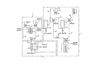
Figure 1 shows schematically the arrangement of a
blood sampler for use in the method of the present
invention. The blood sampler comprises a sample
preparation section 1, a gradient generator section 2 and
a sensor section 3.
A whole blood sample 4 contained in a sample container
5 acts as a sample reservoir for a sample probe 6. The
sample probe 6 is connected along PTFE fluid line 26 to a
diluter pump 7 via multi-position distribution valve 8 and
multi-position distribution valve 9. The diluter pump 7
draws saline solution from a reservoir (not shown) via port
#1 of the multi-position distribution valve 9. As will be
explained in detail below, the diluter pump 7 is controlled
CA 02240471 1998-06-25
WO 97!24598 PCT/GB96103256
12
to discharge a sample of blood together with a volume of
saline into a first well l0 as part of a first dilution
step in the sampling process.
In a second dilution step, the diluter pump 7 draws a
dilute sample of blood from the first well l0 via muiti-
position distribution valve 11 into PTFE fluid line 12 and
discharges this sample together with an additional volume
of saline into a second well 13. The second well 13
1Q provides the dilute sample source for the gradient
generator section 2 described in detail below.
Instead of using whole blood, a pre-diluted sample of
blood 14 in a sample container 15 may be used. In this
case, a sample probe 16 is connected along PTFE fluid line
30, multi-position distribution valve 11, PTFE fluid line
12 and multi-position distribution value 9 to the diluter
pump 7. In a second dilution step, the diluter pump 7
draws a volume of the pre-diluted sample 14 from the sample
container 15 via fluid line 30 and mufti-position
distribution value 11 into fluid line 12 and discharges the
sample together with an additional volume of saline into
the second well 13 to provide the dilute sample source for
the gradient generator section 2.
The gradient generator section 2 comprises a first
fluid delivery syringe 17 which draws water from a supply
via mufti-position distribution valve 18 and discharges
water to a mixing chamber 29 along PTFE fluid line 20. The
gradient generator section 2 also comprises a second fluid
delivery syringe 21 which draws the diluted sample of blood
from the second well 13 in the sample preparation section
1 via mufti-position distribution valve 22 and discharges
this to the mixing chamber 19 along PTFE fluid line 23 '
where it is mixed with the water from the first fluid
delivery syringe 17. As will be explained in detail below,
the rate of discharge of water from the first fluid
' CA 02240471 2003-11-27
13
delivery syringe 17 and the rate of discharge of dilute
blood sample from the second fluid delivery syringe 21 to
the mixing chamber is controlled to produce a predetermined
concentration profile of the sample suspension which exits
the mixing chamber 19 along PTFE fluid line 24. Fluid line
24 is typically up to 3 metres long. A suitable gradient
generator is described in detail in the Applicant's co-
pending International application wo 97/24529.
As will also be explained in detail below, the sample
suspension exits the mixing chamber 19 along fluid line 24
and enters the sensor section 3 where it passes a sensing
zone 25 which detects individual cells of the sample
suspension before the sample is disposed of via a number of
waste outlets.
In a routine test, the entire system is first flushed
and primed with saline, as appropriate, to clean the
instrument, remove pockets of air and debris, and reduce
carry-over.
The diluter pump 7 comprises a fluid delivery syringe
driven by a stepper motor (not shown) and is typically
arranged initially to draw 5 to lOml of saline from a
saline reservoir (not shown) via port #1 of multi-position
distribution valve 9 into the syringe body. A suitable
fluid delivery syringe and stepper motor arrangement is
described in detail in the Applicant's co-pending
International application WO 97/24797. Port #1 of the multi-
position distribution valve 9 is then closed and port #0 of
both multi-position distribution valve 9 and multi-position
distribution valve 8 are opened. Typically 1001 of whole blood
is then drawn from the sample container 5 to take up the dead
space in the fluid line 26. Port #0 of multi-position
distribution valve 8 is then closed and any blood
CA 02240471 1998-06-25
WO 97/24598 PCT/GB96/03256
14
from the whole blood sample 4 which has been drawn into a
fluid line 27 is discharged by the diluter pump 7 to waste
via port #1 of mufti-position distribution valve 8.
In a first dilution step, port #0 of mufti-position
distribution value 8 is opened and the diluter pump 7 draws
a known volume of whole blood, typically 1 to 20 ~C1, into
PTFE fluid line 27. Port #0 is then closed, port #2 opened
and the diluter pump 7 discharges the blood sample in fluid
line 27 together with a known volume of saline in fluid
line 27, typically 0.1 to 2m1, into the first well 10.
Port #2 of mufti-position distribution value 8 and port #0
of mufti-position distribution value 9 are then closed.
Following this, port #0 of mufti-position distribution
valve 11 and port #3 of mufti-position distribution valve
9 are opened to allow the diluter pump 7 to draw the first
sample dilution held in the first well 10 to take up the
dead space in PTFE fluid line 28. Port #0 of multi-
position distribution valve 11 is then closed and port #1
opened to allow the diluter pump 7 to discharge any of the
first sample dilution which has been drawn into fluid line
12 to waste via port #l.
In a second dilution step, port #0 of iriulti-position
distribution valve 11 is re-opened and the diluter pump 7
draws a known volume, typically 1 to 20 ~,1, of the first
sample dilution into fluid line 12. Fluid line 12 includes
a delay coil 29 which provides a reservoir to prevent the
sample contaminating the diluter pump 7. Port #0 of multi-
position distribution valve 11 is then closed, port #3
opened, and the diluter pump 7 then discharges the first
sample dilution in fluid line 12, together with a known
volume of saline, typically 0.1 to 20m1, into the second
well 13. Port #3 of mufti-position distribution valve 11
is then closed. At this stage, the whole blood sample has
been diluted by a ratio of typically 10000:1. As will be
CA 02240471 2003-11-27
explained below, the instrument is arranged automatically
to control the second dilution step to vary the dilution of
the sample suspension to achieve a predetermined cell count
to within a predetermined tolerance at the start of a test
5 routine.
In the gradient generator section 2, the first fluid
delivery syringe 17 is primed with water from a water
reservoir. Port #3 of multi-position distribution valve 22
10 is opened and the second fluid delivery syringe draws a
volume of the dilute blood sample from the second well 13
into the syringe body. Port #3 of multi-position
distribution valve 22 is then closed and port #2 of both
multi-position distribution valve 18 and mufti-position
15 distribution valve 22 are opened prior to the controlled
discharge of water and dilute blood sample simultaneously
into the mixing chamber 19.
Figure 2 shows how the velocity of the fluid
discharged from each of the first and second fluid delivery
syringes is varied with time to achieve ,a predetermined
continuous gradient of osmolality of the sample suspension
exiting the mixing chamber 19 along fluid line 24. The
flow rate of the sample suspension is typically in the
region of 200u1 s ~ which is maintained constant whilst
measurements are being made. This feature is described in
detail in the Applicant's co-pending application WO 97/24529.
As shown in Figure 2, a cam profile associated with a cam which
drives fluid delivery syringe 21 accelerates the syringe
plunger to discharge the sample at a velocity V1, whilst a cam
profile associated with a cam which drives fluid delivery
syringe 17 accelerates the associated syringe plunger to
discharge fluid at a lower velocity V2. Once a constant flow
rate from each delivery syringe has been established at time
To, at time T1 the cam profile associated with fluid delivery
syringe 21 causes the rate of sample discharge to decelerate
linearly over
CA 02240471 2003-11-27
16
the period T2-T~ to a velocity V2, while simultaneously, the
cam profile associated with fluid delivery syringe 17
causes the rate of fluid discharge to accelerate linearly
to velocity V~ . During this period, the combined flow rate
of the two syringes remains substantially constant at
around 200~1s~~ Finally, the two syringes are flushed over
the period T3-T2.
Once both the first fluid delivery syringe 17 and the
second fluid delivery syringe 21 have discharged their
contents, the first delivery syringe is refilled with water
in preparation for the next test. If a blood sample from
a different subject is to be used, the second fluid
delivery syringe 21 is flushed with saline from a saline
supply via port #1 of multi-position distribution valve 22
to clean the contaminated body of the syringe.
The sample suspension which exits the mixing chamber
19 passes along fluid line 24 to the sensor section 3. A
suitable sensor section is described in detail in the
Applicant's co-pending International application WO 97/24600.
The sample suspension passes to a sensing zone 25 comprising
an electrical field generated adjacent an aperture through
which the individual cells of the sample suspension must pass.
As individual blood cells of the sample suspension pass through
the aperture the response of the electrical field to the
electrical resistance of each individual cell is recorded as
a voltage pulse. The amplitude of each voltage pulse together
with the total number of voltage pulses for a particular
interrupt period, typically 0.2 seconds, is also recorded and
stored for subsequent analysis including a comparison with the
osmolality of the sample suspension at that instant which is
measured simultaneously. The osmolality of the sample
suspension may also be determined without measurement from a
knowledge of the predetermined continuous osmotic gradient
generated
CA 02240471 1998-06-25
WO 97/24598 PCTlGB96103256
17
by the gradient generator section 2. As described below,
the osmolality (pressure) is not required to determine the
cell parameters.
Figure 3 shows how data is collected and processed.
Inside each instrument is a main microprocessor which is
responsible for supervising and controlling the instrument,
with dedicated hardware or low-cost embedded controllers
responsible for specific jobs within the instrument, such
l0 as operating diluters, valves, and stepper motors or
digitizing and transferring a pulse to buffer memory. The
software which runs the instrument is written in C and
assembly code and is slightly less than 32 K long.
When a sample is being tested, the amplitude and
length of each voltage pulse produced by the sensor is
digitized to 12-bit precision and stored in one of two 16K
buffers, along with the sum of the amplitudes, the sum of
the lengths, and the number of pulses tested. Whilst the
instrument is collecting data for the sensors, one buffer
is filled with the digitized values while the main
microprocessor empties and processes the full buffer. This
processing consists of filtering out unwanted pulses,
analysing the data to alter the control of the instrument
and finally compressing the data before it is sent to the
personal computer for complex analysis.
Optional processing performed by the instrument
includes digital signal processing of each sensor pulse so
as to improve filtering, improve the accuracy of the peak
detection and to provide more information about the shape
and size of the pulses. Such digital signal processing
produces about 25 16-bit values per cell, generating about
25 megabytes of data per test.
Data processing in the personal computer consists of
a custom 400K program written in C and Pascal. The PC
CA 02240471 1998-06-25
WO 97/24598 PCT/GB96/03256
18
displays and analyses the data in real time, controls the
user interface (windows, menus, etc.) and stores and prints
each sample. ,
The software also maintains a database of every sample
tested enabling rapid comparison of any sample which has
been previously tested. Additionally, the software
monitors the instrument s operation to detect malfunctions
and errors, such as low fluid levels, system crashes or the
l0 user forgetting to turn the instrument on.
The voltage pulse generated by each cell of the sample
suspension as it passes through the aperture of sensing
zone 25 is displayed in graphical form on a VDU of a PC as
a plot of osmolality against measured voltage. The sample
suspension passes through the sensor section at a rate of
200~c1s j . The second dilution step is controlled to achieve
an initial cell count of around 5000 cells per second,
measured at the start of any test, so that in an interrupt
period of 0.20 seconds, around 1000 cells are detected and
measured. This is achieved by varying automatically the
volume of saline discharged by the diluter pump 7 from the
fluid line 12 in the second dilution step. Over a test
period of 40 seconds, a total of 200 interrupt periods
occur and this can be displayed as a continuous curve in a
three-dimensional form to illustrate the frequency
distribution of measured voltage at any particular
osmolality, an example of which is shown in Figures 4 and
5.
The measured cell voltage, stored and retrieved on an
individual cell basis is shown displayed on a plot of
voltage against the osmolality of the solution causing that
voltage change. Using individual dots to display the '
measured parameter change for each individual cell results
in a display whereby the distribution of cells by voltage,
and thereby by volume, in the population is shown for the
CA 02240471 1998-06-25
CVO 97/24598 PCT/GB96/03256
19
whole range of solutions covered by the osmoiality
gradient. The total effect is a three-dimensional display
. shown as a measured property change in terms of the
amplitude of the measured voltage pulses against altered
parameter, in this case the osmolality of the solution, to
which the cells have been subj ected and the distribution or
density of the cells of particular sizes within the
population subjected to the particular osmolality. The
effect is to produce a display analogous to a contour map,
which can be intensified by using colour to indicate the
areas of greatest intensity.
When full data is available on the distribution of
cell size in a particular population of cells subjected to
haemolytic shock in a wide range of hypotonic solutions, at
osmolaiities just below a critical osmolality causing lysis
a gap in the populations is visible. As shown in Figure 4,
ghost cells are fully visible or identifiable in the three-
dimensional plot and the unruptured cells are clearly
identifiable, but between them is a region defined by
osmolality and cell volume where relatively few individuals
appear. The existence of this phenomenon, which we have
termed the "ghost gap", has not previously been recognised.
If the entire series of steps are repeated at timed
intervals on further aliquots of the original sample and
the resulting measured voltage is plotted against
osmolality, time and frequency distribution, a four-
dimensionai display, is obtained which may be likened to a
change in weather map. This moving three-dimensional
display, its motion in time being the fourth dimension,
provides an additional pattern characteristic of a
particular blood sample. This is shown in the series of
images in Figure 6. The images shown in Figure 6 are the
results of tests carried out at hourly intervals at a
temperature of 37°C. As the measurements are so exact, the
CA 02240471 1998-06-25
WO 97/24598 PCT/GS96/03256
repeat values are superimposable using computer sequencing
techniques.
As shown, cells slowly lose their ability to function
5 over time, but they also change in unexpected ways. The
size and shape of the cells in a blood sample change in a
complex, non-linear but repeatable way, repeating some of
the characteristic patterns over the course of days and on
successive testing. The patterns, emerging over time, show
10 similarity among like samples and often show a
characteristic wave motion. The pattern of change may vary
between individuals reflecting the health of the
individual, or the pattern may vary within a sample. Thus
a sample that is homogeneous when first tested may split
15 into two or several sub-populations which change with time
and their existence can be detected by subjecting the
sample to a wide range of different tonicities and
recording the voltage pulse in the way described. As shown
in Figure 6 , after the f first f ew hours the cell becomes
20 increasingly spherical in the original sample, it then
becomes flatter for several hours, then more spherical
again, reaches a limit, and then becomes thinner and
finally may swell again. It has been determined that the
rate at which observed changes take place are inf luenced by
pH, temperature, available energy and other factors.
The three-dimensional pattern provides data which
enables identification of the precise osmolality at which
particular cells reach their maximum volume, when they
become spheres. With appropriate calibration, which is
described in detail below, and using the magnitude of the
voltage pulse, it is possible to define precisely and
accurately the actual volume of such cells and thereafter
derive a number of other cell parameters of clinical
interest.
CA 02240471 1998-06-25
WO 97/24598 PCT/GB96J03256
21
The amplitude of the voltage pulses produced by the
sensor 25 as individual cells pass through the electrical
. field are proportional to the volume of each cell.
However, before a conversion can be performed to provide a
measure of cell volume, the instrument requires
calibration. This is performed using spherical latex
particles of known volume and by comparison with cell
volumes determined using conventional techniques.
Experimental results have shown that the mapping of
measured voltage to spherical volume of commercially
available latex particles is a linear function.
Accordingly, only a single size of spherical latex
particles needs to be used to determine the correct
conversion factor. In a first calibration step, a sample
containing latex particles manufactured by Bangs
Laboratories Inc. having a diameter of 5.06~Cm i.e. a volume
of 67.834m3, was sampled by the instrument. The three-
dimensional plot for the latex particles is shown in Figure
7 with a plot of osmolality against mean voltage shown in
Figure 8. In this particular test, the instrument produced
a mean voltage of 691.97mV. The spherical volume is given
by the equation:
Spherical volume = measured voltage x K"o~t$
where K,~o~ts is the voltage conversion factor.
Re-arranging this equation gives:
spherical volume
~o~cs- measured voltage
which in this case gives,
CA 02240471 1998-06-25
WO 97/24598 PCT/G~96/03256
22
67.834
Kvotts- =0 ~ 0980
691.9?
This value of K"otts is only valid for the particular
instrument tested and is stored in a memory within the
instrument.
In a second calibration step, a shape correction
factor is determined to take account of the fact that the
average blood cell in the average individual has a bi-
concave shape. Applying the above voltage conversion
factor K"otts assumes that, like the latex particles, blood
cells are spherical and would therefore give an incorrect
cell volume for cell shapes other than spherical. In the
present invention, a variable shape correction function is
determined so that the mean volume of the blood cells at
any osmolality up to the critical osmolality causing lysis
can be calculated extremely accurately.
To illustrate this, a sample was tested at a number of
accurately known osmolalities and the volume of the blood
cells measured using a standard reference method, packed
cell volume. A portion of the same sample was also tested
by the method of the present invention using the instrument
of Figure 1 to measure the voltage pulses from individual
cells at the corresponding osmolalities. The results of
these procedures are shown in Table 1 and plotted as two
superimposed graphs of osmolality (x-axis) against measured
voltage and true volume, respectively, in Figure 9.
At an isotonic osmolality of 290mosm, the true volume,
as determined by the packed cell volume technique, was
92.Of1, whilst the measured mean voltage was 670mV.
The true isotonic volume of the cells is given by
equation:
CA 02240471 1998-06-25
WO 97/24598 PCT/GB96/03256
23
VOlume~so = VOltagejso X Kyolts x Kshape
where Voltage~so is the measured voltage and KSha~ is a
shape correction factor.
Re-arranging:
Volumei$o
Kshape Voltage~so X K~olts
IO
which in this example gives,
_ 92.0 _
Kshape' 670 x 0. 0980 rl' 4
Table 1 shows the shape correction factor Ksnape for
each of the other aliquots and demonstrates that the factor
to be applied to each sample is different with the maximum
shape correction being applied at isotonic osmolaiities
where the blood cells are bi-concave rather than spherical.
To automate the calculation of Ksha~e at any osmolality of
interest a shape correction function is required. The
following general function describes a shape correction
factor based on any two sensor readings i.e. measured
voltages:
f ( Ksna~ ) - f ( SRl , SR2 )
where SRl is a sensor reading (measured voltage) at a
known shape, typically spherical, and SR2 is a sensor
reading (measured voltage) at an osmolality of interest,
typically isotonic.
Analysis has shown that this is a linear function and
that:
CA 02240471 1998-06-25
WO 97/24598 PCT/GB96/03256
24
f(K ~ = 1+ (SR1-SR2) x K
shape ( SRl ) a
where Ka is an apparatus dependent constant, which is
determined as follows:
Kshape at an osmolality of 290 mosm is known (see
above), applying the values SRl = 1432mV, SR2 = 670mV and
Ksha~ = 1.4 to the above equation gives:
1.4 = 1+ ~ (1432-670) ~ x K$
1432
re-arranging:
Ka = 0.7518
This value of Ka is constant for this instrument.
The true isotonic volume of a blood sample is
determined by comparing the measured voltage at an isotonic
volume of interest with the measured voltage of cells of
the same blood sample at some known or identifiable shape,
most conveniently cells which have adopted a spherical
shape, whereby:
VOlume~so = VOltage~so X Kyolts X f (Kshape)
(SRl-SR2)
= SR2 x 0.0980 x + x 0.7518 '
SRl
In the present invention, the point at which the blood
cells become spherical when subjected to a predetermined
CA 02240471 1998-06-25
WO 97/24598 PCT/GB96/03256
continuous osmotic gradient can be determined very
accurately. Figures 10a-lOd show the results for a normal
blood sample from a healthy individual. Figure l0a shows
a three-dimensional plot of measured voltage against
5 osmolality, Figure lob shows a graph of osmolality against
percentage change in measured voltage for a series of tests
of a sample, Figure lOc shows the results in a tabulated
form, and Figure lOd shows superimposed graphs of mean
voltage and cell count for the test, respectively, against
10 osmolality. As shown, the cell count, which is initially
5000 cells per second at the beginning of a test, reduces
throughout the test due to the dilution of the sample in
the gradient generator section 2. The mean voltage rises
to a maximum at a critical osmolality where the blood cells
15 achieve a spherical shape and then reduces. Using standard
statistical techniques, the maxima of the curve in Figure
lOb, and therefore the mean voltage at the maxima, can be
determined. The mean voltage at this point gives the value
SRl for the above equation. It is then possible to select
20 any osmolality of interest, and the associated measured
voltage SR2, and calculate the true volume of the cell at
that osmolality. Typically, the isotonic osmolality is
chosen, corresponding to approximately 29omosm.
25 For the above test, at 290 mosm, SRl = 1432mV and SR2
- 670mV. Accordingly:
~ -1 1432-6?0 x 0.7518
f Kshape 290 + 14 3 2
Kshape 290 - 1. 4 0
and therefore:
Volumeiso - SR2 X Kyolts x Kshape
= 670 x 0.0980 x 1.40
- 91.92 fl,
CA 02240471 1998-06-25
WO 97/24598 PCT/GB96/03256
26
and:
Volumes - SR1 x K~o;ts x Kshape
- 1432 x 0.098 x 1.0
S = 140.34 fl.
Knowledge of the mean volume of the sphered cells
allows calculation of spherical radius as:
Volume$~= 4~3
3
from which the spherical radius
3 x Volumes s
r=
4n
1
3x140.34 3
ra
4 7I
=3 . 2 2E.am
Having determined volumeigo, volumes and the spherical
cell radius, it is possible to calculate a number of other
parameters. In particular:
1. Surface Area (SA)
Since the surface area SA is virtually unchanged at
all osmolalities, the cell membrane being virtually
inelastic, and in particular between spherical and
isotonic, the surface area SA may be calculated by
substituting r into the expression:
CA 02240471 1998-06-25
WO 97/24598 PCT/GB96/03256
27
SA = 4~rrZ
- 47tX(3.22)z
- 130.29~m2
2. Surface Area to Volume Ratio (SAVR)
Given that the walls of a red cell can be deformed
without altering their area, once the surface area SA is
known for a cell or set of cells of any particular shape,
the surface area is known for any other shape, thus the
surface area to volume ratio SAVR can be calculated for any
volume. SAVR is given by the expression:
4 rrrz SA
SAVR= -
Volumeiso Voiume~so
130.29
91.99
- 1.42
3. ~phericity Index (SI)
The present invention can easily measure the SAVR, a
widely quoted but hitherto, rarely measured indication of
cell shape. For a spherical cell, it has the value of 3/r,
but since cells of the same shape but of different sizes
may have different SAVR values, it is desirable to use the
sphericity index SI which is a dimensivnless unit
independent of cell size, given by the expression:
r
SI=SAVR x -
3
= 1.52
CA 02240471 1998-06-25
WO 97/24598 PCT/GB96/03256
28
3.22
=1.42 x
3
4. Gell Diameter {D)
When the normal cell is in the form of a bi-concave
disc at isotonic osmolality, it is known that the ratio of
the radius of a sphere to that of the bi-concave disc is
0.8155. On this basis, therefore, the diameter D of a cell
in the form of a bi-concave disc is given by:
2r
D=
0.8155
2x3.22
0.8155
- 8 . 19/im
The same parameter can be determined for all other
osmolalities. The frequency distribution of the cell
diameters is given both as dispersion statistics as well as
a frequency distribution plot. The present invention
provides an automated version of the known manual procedure
of plotting a frequency distribution of isotonic cell
diameters known as a Price-Jones curve. The present
invention is capable of producing a Price-,Tones curve of
cell diameters for any shape of cell and, in particular,
isotonic, spherical and ghost cells (at any osmoiality) and
is typically based on 250,000 cells. This is shown in
Figure 10.
5. Cell Thickness {CT)
CA 02240471 1998-06-25
WO 97/2x598 PCT/GB96103256
29
When the cell is in the form of a bi-concave disc, an
approximate measure of the cell thickness can be derived
from the cross-sectional area and the volume. The area is
of course derivable from the radius of the cell in
spherical form. The cell thickness can therefore be
calculated as follows:
VO lame i so
CT =
~rrZ
91.92
~tx3.22Z
- 2 . 8 2 ~Cm
6. Surface Area per millilitre {SAmI)
The product of the surface area (SA) and the cell
count (RBC) is the surface area per millilitre (SAml)
available for physiological exchange. The total surface
area of the proximal renal tubes that are responsible for
acid-base regulation of the body fluids is 5 m2. The total
surface area of the red blood cells that also play an
important part in the regulation of the acid-base balance
is 4572mZ, almost 3 orders of magnitude larger. RBC is
calculated internally from a knowledge of the flow rate of
the diluted blood sample, a cell count for each sample and
the dilution of the original whole blood sample.
Typically, RBC is approximately 4.29 x 10⁹ red cells per
ml.
SAml = SA x RBC {per ml)
- 13 0 . 2 9 ~mz x 4 . 2 9 109
~ 0.56 m2 ml ~
CA 02240471 1998-06-25
WO 97/24598 ~'CT/GB96/03256
7. Cell Permeabilit~~ tC~
The plot of cell volume against osmolality in Figure ,
I2 reveals a characteristic curve showing how the cell
5 volume changes with decreasing osmolality and indicates
maximum and minimum rates of flow across the membrane and
the flow rates attributed to a particular or series of
osmotic pressures. Many of the cell permeability
measurements are primarily dependent upon the change in
10 volume of the cells at different pressures. Table 2 shows
the volume measurements produced by the method of the
invention and the change in volume at each mosm. Such a
table is calculated automatically from continuous functions
and is not usually seen by the user. The results are shown
15 plotted as a graph of net fluid exchange against osmotic
pressure in Figure 13.
Having obtained measures of osmotic pressure (Poi),
cell volume, surface area {SA) and other relevant
20 environmental factors, it is possible to obtain a number of
measures of cell permeability:
a) ep rate
25 This coefficient of permeability measures the rate of
fluid flow across a square meter of membrane in response to
a specified pressure. All positive rates represent a net
flow into the cell, while all negative rates are the
equivalent of a net flow out of the cell. The rate is
30 determined by:
Cp rate = D cell volume / O Poi / SA at S.T.P.
Using the discrete values from Table 2 and Figure 13,
the Cp rate between 200 and 141 mOsm is given as:
Cp ratez9o.v4~ - {139.94-93.98) / (141-290) /130.29 ~m2
45.96 fl/149 mOsm/130.29
2.36 x 10 3 fl/mOsm/~Cmz
CA 02240471 1998-06-25
WO 97/24598 PCT/GB96/03256
31
- 2.36 x 10⁹ fl/mOsm/m2
- 2.36 ml/mOsm/mz
b) Permeability Constant pk~
This set of permeability measures describe each
pressure where the net permeability rate is zero, and are
numbered pk~ , pki . . . pk~ .
(i) pko coincides with the minimum absolute pressure
(hypotonic) to which a cell can be subjected without loss
of integrity and is shown in Figure 12. A pressure change
of one tenth of a milliosmole per kg (0.0001 atms) at pko
produces a change in permeability of between one and two
orders of magnitude making pko a distinct, highly
reproducible measure.
(ii) pk~ is a measure of the ceiis' ability to
volumetrically regulate in slightly hypotonic pressures and
is also shown in Figure 12. After a certain pressure, the
cell can no longer defeat the osmotic force, resulting in
a change in the cell's volume. pk~ provides a measure of
the cells ability to perform this regulation, thereby
measuring a cell's maximum pump transfer capability.
( iii) pk2, a corollary of pk~ , is a measure of the cells
ability to volumetrically regulate in ertonic pressures,
and occurs at low differential pressures, when compared to
the cell's typical in vivo hydrostatic pressure (not
shown).
- The permeability constant pk~ is described by the
following equation:
3 5 pk~ = O Pay / SA at S . T . P .
CA 02240471 1998-06-25
WO 97/24598 PCT/GB96I03256
32
When calculating pko, O Pay _ (isotonic pressure} -
{pressure where net flow is zero).
When calculating pk~, D Posm = (isotonic pressure) -
(first hypotonic pressure where net positive flow begins).
The calculation of pk2 (not shown) is identical to pk~,
except d Poi measures the first hypertonic pressure where
net positive flow is not zero.
Using the discrete values from Table 2 and Figure 13,
pKo = 141. 5 mOsm Kg ~ / 13 0-2 9 ~Cm2
1.086
C) CPO
This dimensionless value is the comparison of any two
Cp rates, and is expressed as the net amount of fluid to
cross the cell membrane between any two pressures. It
provides a volume independent and pressure dependent
comparison of permeability rates. This measure may be used
to compare permeability changes in the same individual over
a period ranging from minutes to months.
From CP rate above, CP rate29o.~4~ was determined to be
2.41 ml/mOsm/m2.
CPD - (CP rats 1 - CP rate 2/CP rate 2) x 100
CPS - (2.41-2.36}/2.36 x 100
= 2.07% change
Cprx
This is the maximum rate of flow across the cell's
membrane. For almost all cells, there are two maxima, one
positive (net flow into the cell} and one negative (net
flow out of the cell} situated either side of pko. Cp~x is
determined by detecting the maximum positive and negative
gradients of the continuous curve of change in cell volume
against osmolaiity. From the results, Cp~X into the cell
CA 02240471 1998-06-25
WO 97124598 PCT/GB96/03256
33
is +0.670 fl/mOsm and Cp~X out of tie cell is -0.722
f 1/mOsm.
ey Membrane Structural Resistance (MSR)
This is a measure of the structural forces inside a
cell which resist the in-flow or out-flow of water. It is
determined by the ratio of Cp~x to all other non-zero flow
rates into the cell. As the membrane is theoretically
equally permeable at all pressures, change from the maximum
flow rate outside the pressure range of pk~ to pk2 are due
to mechanical forces. It is clear that pko is an entirely
mechanical limit on the cell because as Cp~ate aPProaches
zero, MSR approaches ~, thereby producing more strain than
the membrane can tolerate.
MSR = Cp~X / Cp~et~ x 100%
f) Cpml
This is a measure of the physiological permeability
available to an individual per unit volume of tissue or
blood, or for the whole organ or total body, and is
calculated by:
CPmi - O cell volume / O Poi / m3 per ml of whole
blood.
From the above calculations, in 1 ml there are 4.29 x
109 red cells each with a surface area of 130.29 ~,mz
and SAmI = 0 . 5 6 m2 ml ~
At Cp~X (for instance) the flow rate into the cell was
0.677 fl/130.29~m2
- 5.20 x 10 3 fl/~m2
CA 02240471 1998-06-25
WO 97/24598 PCT/GB96/03256
34
Thus in 1 ml of whole blood the net volume of fluid
crossing the membrane was
- 5.20 x 10 3 fl/~mz x 0.559 x 10¹² ~,mz/ml ,
- 2.91 ml/ml of whole blood
Cpnec
CP~et is def fined as the rate at which f luid can be
forced across a unit area of membrane at standard
temperature and pressure over unit time and is a pressure
independent measure of the coefficient of permeability,
given by the equation:
( Vo lumes~-Vo lumen $o )
Cp~et'
SA
140.34-91.92
130.29
0.372 -z
3.72 ml m
Figure 14 illustrates the three-dimensional frequency
distribution of a sample from a patient having an HbCC
disease. As shown, the plot is grossly abnormal.