Note: Descriptions are shown in the official language in which they were submitted.
CA 02241817 1998-06-29
WO 97/25913 PCTIUS96/03319
SPECTROSCOPIC SYSTEM WITH DISPOSABLE CALIBRATION DEVICE
BACKGROUND OF THE INVENTION
1. Field of the Invention
This invention relates to spectroscopic instruments
requiring calibration to make measurements on animal tissues or
other materials, and in particular, to spectroscopic instruments
incorporating a disposable calibration target that ensures proper
calibration of the spectroscopic instrument, and prevents
scratching of optically sensitive windows through which
measurements are taken. Once used, the calibration target cannot
be reused, thereby helping to control the spread of infection in
tissues or helping to control contamination of materials.
More generally, this invention relates to a method and
device for calibrating many different types of measurement
instruments, and in particular, to a disposable calibration
device and method which uses that device for calibrating
measurement instruments that perform measurements on a material
or tissue. The calibration device includes a calibration target
that ensures proper calibration of the measurement instrument,
prevents scratching of windows through which measurements are
taken, and also prevents reuse of the disposable calibration
target, thereby helping to control the spread of infection if the
measurements are made on tissues, and helping to prevent
contamination if the measurements are made on materials.
2. Background of the Related Art
Spectroscopy is currently used for a wide variety of
purposes including to evaluate in-vivo or in-vitro tissue
samples. One type of spectroscopy, reflectance
1
CA 02241817 1998-06-29
WO 97/25913 PCT/US96/03319
spectroscopy, involves diffusely reflecting light from tissue
non-invasively. Such spectroscopic measures must be calibrated
prior to use, especially when made for medical or other critical
applications. Instrument calibration can be affected by
variations in light source intensity, spectral characteristics,
lens-aging, lens cleanliness, temperature, detector sensitivity
changes, and electronic drifting.
Many current instruments provide for a calibration to be
performed on a routine basis in order to compensate for these
changes in the instrument performance and response. Those
calibration methods typically involve measuring the response of
a test target with characteristics that remain stable with time
and over a range of temperatures. Those methods can also be used
to compensate for instrument to instrument variations and any
changes that an individual instrument may experience over its
working lifetime.
Typically, spectral transmittance, fluorescence (normal and
time resolved) and Raman spectroscopy are used to evaluate
biological tissues and other materials in order to determine the
materials present and measure their concentrations. These
methods are also affected by the scattering, reflecting absorbing
and transmitting properties of the instrument optics, detectors,
sources and the media under examination. This is due to the fact
that the amount of light reaching the tissue to be measured is
a function of those parameters, and in the case of fluorescence
and Raman emissions, reabsorption of emission spectra.
Although others have proposed calibration fixtures that
compensate for these variations in instrument performance, none
2
CA 02241817 1998-06-29
WO 97/25913 PCT/US96/03319
have provided a simultaneous solution to both the calibration
issue and the problems associated with the spread of infection
in a medical setting. Furthermore, calibration standards that
are designed to be reused can become damaged by sunlight,
temperature, humidity and other effects which could lead to
errors in calibration.
Bilirubin
The above spectroscopic instruments can perform a variety
of biological measurements. One such application of
spectroscopic systems involves detection of bilirubin. Bilirubin
i3 produced from the breakdown of hemoglobin in red blood cells.
Under normal conditions the bilirubin is conjugated by glucoronyl
transferase, an enzyme present in the liver, and then excreted
through the biliary system.
Newborn infants and prematurely born infants are
particularly susceptible to hyperbilirubinemia.
Hyperbilirubinemia describes the state where there is excessive
bilirubin in the body. Often this is due to the lack of
functioning glucoronyl transferase enzyme in their liver, or
excessive red blood cell breakdown associated with
erythroblastosis fetalis.
One method for bilirubin testing include blood based lab
assay testing. The "heel stick" blood lab assay is currently the
only accepted methodology for quantitative bilirubin testing
results in the United States. Of course, this invasive approach
requires that the drawing of blood to perform the test.
Non-invasive measurements of the bilirubin concentration in
the skin would eliminate the need to draw blood samples from
3
CA 02241817 1998-06-29
WO 97/25913 PCT/US96/03319
patients for bilirubin analysis. It also provides easy patient
interface. Bilirubin can be measured in the aqueous of the eye
based on the fluorescent signature. Bilirubin can also be
directly measured in the scelera (white) of the eye based on the
fluorescent signature. Reflectance measurements can also be made
on the tympanic membrane of the ear. Finally,
reflectance/scattering based measurements can be made on the
skin.
Many attempts have been made to measure cutaneous bilirubin
non-invasively. This is because bilirubin from the blood stains
the skin as well as other tissues of the body--Jaundice refers
to the condition when the bilirubin is visible in the skin and
sclera. These attempts include the development of visual
reference standards, and transcutaneous reflectance spectroscopy.
The absorption spectra of bilirubin, oxidized blood, and melanin,
the dominant absorbers in the skin. The concentration of these
pigments have distinct absorption spectra. Reflectance
bilirubinometers have obtained reasonable correlations between
bilirubin levels determined transcutaneously and serum bilirubin
concentrations in homogeneous patient populations, but have
failed to give satisfactory correlations when used over a
heterogeneous population. Since patient populations are rarely
homogeneous, transcutaneous bilirubin have not been widely
accepted clinically.
one system which implements a non-invasive cutaneous testing
approach for bilirubin and is in wide use in Japan, is the
Minolta Jaundice Meter. That approach, however, has not been
approved for use in the United States, but is nevertheless, used
4
CA 02241817 1998-06-29
WO 97/25913 PCTIUS96/03319
for screening purposes in some U.S. institutions. In addition,
that approach does not account for variations in skin color and
thickness.
Another approach to testing for bilirubin that does not
require the drawing of blood is a breath analysis approach
introduced by a group from Stanford. This approach does not have
quantitative accuracy required to have a high correlation to
serum bilirubin. Hence, it appears to only have potential use
as a screening technique.
General Measurement Systems
More generally, there has been an increase in the use of
light as a diagnostic tool in many areas of medicine. This
development has become more pervasive with the development of
appropriate and inexpensive light sources, detection devices and
optical fibers that allow for minimal invasiveness.
Moreover, there are many types of measurement systems that
require calibrations to be performed on a routine basis in order
to compensate for changes in instrument performance and response.
This is true for both radiation based measurement systems, i.e.,
systems that send electro-magnetic radiation to the tissue or
material to be measured and then detect the return radiation, and
acoustic based measurement systems, i.e., systems that send
acoustic waves or energy to the tissue or material to be measured
and then detect the return acoustic signal. The calibration
techniques in both cases typically involve measuring the response
of a test target with characteristics that remain stable with
time and over a range of temperatures. Those techniques can also
be used to compensate for instrument to instrument variations and
CA 02241817 1998-06-29
WO 97/25913 PCTlUS96/03319
any changes that an individual instrument may experience over its
working lifetime. Often such measurement systems must be
periodically calibrated and sometimes must be calibrated prior
to each and every use. This calibration becomes especially
important when measurements are made for medical or other
critical applications.
Radiation measuring systems such as the spectrometer system
discussed above, are currently used for a wide variety of
purposes including to evaluate tissue or materials. These
measuring systems require calibration for a variety of reasons
including variations in the radiation source intensity, changes
in spectral characteristics of the tissue or material, component
aging and cleanliness, changes in temperature, radiation detector
sensitivity changes, and electronic drifting.
Examples of radiation type measurement systems that often
require some type of calibration include in addition to
spectrometers, instruments such as laser radar, radar or any
other radiation measuring instrument that outputs radiation to
a tissue or material and then measures some aspect of the return
signal.
Acoustic type measuring systems are also used for a wide
variety of purposes including to evaluate tissue or materials.
Often these measurement systems must also be periodically
calibrated and sometimes must be calibrated prior to each use.
Acoustic measurement systems also require calibration for a
variety of reasons including variations in the output energy of
the acoustic wave source, changes in spectral characteristics of
the tissue or material, changes in temperature, detector
6
CA 02241817 1998-06-29
WO 97/25913 PCT/US96/03319
sensitivity changes, and electronic drifting.
Examples of acoustic type measurement systems that often
require some type of calibration include acoustic spectrometers,
and interferometers or any other system which uses an acoustic
wave measuring instrument that outputs acoustic energy a material
and then measures some portion of the return signal.
Various types of calibration techniques and devices have
been attempted. For example, U.S. Patent 5,365,925 describes a
calibration boot which includes a plurality of materials, which
is placed over an optical catheter for the purpose of making a
multi-point calibration of reflected or backscattered light.
U.S. Patent 5,311,273 describes a method of using four black body
radiators to provide calibration of an infrared spectrometer.
However, neither of these approaches involves an inexpensive
calibration target that can be easily discarded after each use,
and thus does not prevent a user from taking a measurement
without going through a calibration step.
U.S. Patent 4,981,355 describes a calibration device for the
in vitro calibration of a light guide, whereby a polyethylene
material has a plurality of light scattering particles and a
plurality of light absorbing particles which yields a neutral
density filtering type of effect, uniformly distributing light
in the plastic parts of the calibrator. The calibrator can be
positioned into a sterile tray which is protected by a tear off
plastic. Once the calibration is complete, the surgeon removes
the catheter from the calibrator and the tray in which it is held
and then presumably disposes of the calibration device and its
tray. This approach, however, is neither simple nor inexpensive.
7
CA 02241817 1998-06-29
WO 97/25913 PCTIUS96/03319
U.S. Patent 4,796,633 describes a calibration reference
apparatus that fits over a light guide. A stop limits the extent
to which the light guide can be advanced into the cavity whereby
an endface of the light guide is spaced from a region of the
surface to define a gap. The end wall and the gap are adapted
to return a known ratio of the light directed into the gap from
the end face of the light guide. Again, however, this approach
does not involve an inexpensive, disposable calibration device.
U.S. patent 4,744,656 discloses a calibration boot that
snaps into place over an optical catheter allowing calibration
of the catheter before use. Once the calibration is complete,
the boot is removed and the optical catheter is ready for use.
Each new catheter comes with a new boot. However, the boot is
not present during the measurement and there is no provision to
prevent reuse of the boot.
SUHIIKARY OF THE INVENTION
An object, therefore, of the invention is to provide an
optical system which utilizes an optical instrument with a
calibration device.
Another object of the invention is to provide an
spectroscopic system which utilizes a spectrometer as the optical
instrument and a disposable calibration device.
Another object of the invention is to provide a
spectroscopic system that utilizes a calibration device which can
be inexpensively mass produced.
Another object of the invention is to provide a
spectroscopic system which utilizes a disposable calibration
8
CA 02241817 1998-06-29
WO 97/25913 PCTIUS96/03319
device that helps prevent infection of tissue to be measured.
Another object of the invention is to provide a
spectroscopic system which uses a calibration device which
provides an optically clear, scratch-free window between the
optical instrument and the tissue or material to be measured.
Another object of the invention is to provide a spectrometer
system with a calibration device that serves to compensate for
the effects of variations from one spectrometer system to the
next.
Another object of the invention is to provide a spectrometer
system with a calibration device that serves to compensate for
changes over time in properties of the spectrometer instrument
in the spectrometer system.
Another object of the invention is to provide a spectrometer
system with a calibration device that serves to compensate for
changes over a wide range of temperatures in properties of an
individual optical instrument.
one advantage of the spectrometer system is that once used,
the calibration device cannot be re-used, thereby ensuring
against infection from one person to another person in that the
calibration device is discarded after a measurement is performed.
An advantage of the calibration device in general is that
it can be used in radiation type measurement systems.
Another advantage of the calibration device in general is
that it can be used in acoustic type measurement systems.
Another general advantage of the calibration device is that
it helps reduce the possibility of contamination from one
material to another material.
9
CA 02241817 1998-06-29
WO 97/25913 PCT/US96103319
One feature of the invention is that it can utilize an
optical instrument operating in the ultra-violet, visible and/or
the infrared regimes.
Another feature of the invention is that it can utilize a
spectrometer as the optical instrument according to one
embodiment of the invention.
Another feature of the invention is that it utilizes a
disposable calibration device that can include material that has
a stable or predictable spectroscopic signature.
Another feature of the invention is that it utilizes a
disposable calibration device with a window through which
radiation can be transmitted to tissue or material to be
measured.
Another feature of the invention is that it utilizes a
calibration target that can be peeled away from the window.
Another feature of the invention is that the calibration
target can have a tear tab which allows the calibration target
to be easily handled without disturbing the window or calibration
target in contact with the window.
Another feature of the invention is that the calibration
target can be attached to the window by a static cling brought
about by a proper selection of materials for the window and the
calibration target.
Another feature of the invention is that the calibration
device can include a structure which can be cone-shaped.
Another feature of the invention is that the cone-shaped
structure has a proximal end that attaches to the optical
instrument with which it is used.
CA 02241817 1998-06-29
WO 97/25913 PCTIUS96/03319
Another feature of the invention is that the calibration
device can include an outer annulus which comes into contact with
the tissue or material to be measured.
Another feature of the invention is that the calibration
device can include a landing annulus which aids in arranging the
window on the tissue or material for taking a measurement.
These and other objects, advantages and features are
accomplished by the provision of a spectrometer system,
comprising:
a spectrometer instrument which transmits radiation to a material
or tissue in order to effect measurements; a calibration device
holder; a calibration device which can be arranged in said
calibration device holder, said calibration device, comprising:
a structure including a window through which the radiation can
be transmitted; and a removable calibration target arranged on
said window and capable of returning a portion of said radiation
for calibrating the spectrometer instrument, whereby the
removable calibration target can be removed from said window to
allow a measurement to be made on the material or tissue.
In one approach, the window in the spectrometer system can
include material through which said radiation can pass, and the
removable calibration target includes a tear tab which can be
gripped to remove said removable calibration target from said
window.
The structure and window can comprise a barrier or infection
shield between the material or tissue and the spectrometer
system.
The spectrometer instrument in the spectrometer system can
11
CA 02241817 1998-06-29
WO 97/25913 PCT/US96103319
include: an optical unit for outputting output radiation and for
receiving received radiation and detecting said received
radiation as spectral return information; and a processor coupled
to said optical unit for receiving and processing said spectral
return information.
These and other objects, advantages and features are
accomplished by the provision of a spectrometer system,
comprising: a spectrometer instrument which transmits radiation
to a material or tissue in order to effect measurements; a
calibration device holder; a calibration device which can be
arranged on said calibration device holder, said calibration
device, comprising: a structure through which the radiation can
be transmitted; and a removable calibration target arranged about
said structure and capable of returning a portion of said
radiation for calibrating the spectrometer instrument, whereby
the removable calibration target can be removed from said
structure to allow a measurement to be made on the material or
tissue.
These and other objects, advantages and features are
accomplished by the provision of a method for transcutaneous
determination of bilirubin concentration in tissue, including the
steps of: performing a calibration measurement on a calibration
target and storing resulting calibration data; illuminating said
tissue with light; detecting a frequency spectrum of light
reflected from said tissue; calculating, from a first portion of
said spectrum, a first parameter indicative of a maturity of said
tissue; calculating, from a second portion of said spectrum, a
second parameter indicative of an amount of melanin in said
12
CA 02241817 1998-06-29
WO 97/25913 PCT/US96/03319
tissue; calculating, from a third portion of said spectrum, a
third parameter indicative of a blood content of said tissue;
calculating, from a fourth portion of said spectrum, a fourth
parameter indicative of an uncorrected bilirubin concentration
in said tissue; calculating a corrected bilirubin concentration
in said tissue as a function of said first, second, third, and
fourth parameter; adjusting said corrected bilirubin
concentration using said resulting calibration data to yield a
calibrated and corrected bilirubin concentration, whereby said
calibrated and corrected bilirubin concentration compensates for
unit to unit and time varying changes in source luminosity,
delivery optics, collection optics, detection sensitivity,
electronic drift, and environmental conditions such as
temperature and humidity.
These and other objects, advantages and features are
accomplished by the provision of a spectrometer system,
comprising: a spectrometer system, comprising: a housing
including a calibration device holder; a spectrometer instrument
arranged in said housing, said spectrometer instrument
transmitting radiation through said calibration device holder to
a material or tissue in order to effect measurements; and a
calibration device which can be attached to said calibration
device holder, said calibration device, comprising: a structure
including a window through which the radiation can be
transmitted; and a removable calibration target arranged on said
window and capable of returning a portion of said radiation for
calibrating the spectrometer instrument, whereby the removable
calibration target can be removed from said window to allow a
13
CA 02241817 1998-06-29
WO 97/25913 PCT/US96/03319
measurement to be made on the material or tissue.
The optical unit of the spectrometer system can further
comprises a grating for diffracting said return radiation
according to wavelengths therein toward said detector array.
These and other objects, advantages and features are
accomplished by the provision of a method for calibrating a
spectrometer system that outputs radiation from an output end,
comprising: placing a calibrating device over the output end of
the spectrometer system, wherein the calibration device has a
removable calibration target; activating the spectrometer system
to perform a calibration measurement; and removing the removable
calibration target from the calibration device.
The removing step can include removing the removable
calibration target from the calibration device while leaving a
window attached to the spectrometer system, and said radiation
is output through that window.
These and other objects, advantages and features will become
more apparent from the following description of embodiments
thereof taken in conjunction with the accompanying drawings.
BRIEF DESCRIPTION OF THE DRAWINGS
Figure 1A shows a schematic view of a measurement system in
a calibration mode, and Figure 1B shows the same system in a
measurement mode wherein the calibration target has been removed
and radiation is now reaching the tissue or material to be
measured.
Figure 2A shows a schematic representation of a preferred
embodiment of the calibration device used in the calibration
14
CA 02241817 1998-06-29
WO 97/25913 PCT/US96/03319
mode, and Figure 2B shows the calibration device after the
calibration target is removed (peeled) from the window.
Figures 3A and 3B correspond to Figures 2A and 2B, but with
the radiation entering from the right hand side and the
calibration target is attached to the window within the
structure. Figure 3C shows a measurement system which utilizes
a disposable calibration device as in Figures 3A and 3B, and
Figure 3D shows the same measurement system with the calibration
device removed. Figure 3E summarizes the steps involved for
calibrating the above measurement system and then taking a
measurement on material or tissue.
Figures 4A and 4B show a top view and a side view,
respectively, of a calibration device similar to the calibration
device in Figure 3A. Figures 4C and 4D show the same views as
Figures 4A and 4B, respectively, with the calibration target
removed. Figure 4E shows the calibration target with two pull
tabs at its sides and a perforation down the middle designed to
prevent reuse.
Figures 5A, 5B, and 5C show three more perspective views of
the calibration device, where Figures 5B and 5C show the
calibration target removed.
Figure 6 shows a calibration device according to another
embodiment of the invention.
Figure 7A shows a side view of the calibration device
according to yet another embodiment of the invention, and Figure
7B shows the calibration device as viewed from above.
Figures 8A, 8B, and 8C show a front, side and back view,
respectively, of a spectrometer system, and Figure 8D shows a
CA 02241817 1998-06-29
WO 97/25913 PCT/US96103319
spectrometer system in a charging stand, according to one
embodiment of the invention.
Figure 9A is a schematic diagram of certain elements of a
spectrometer system 803 including a spectrometer instrument, and
Figure 9B shows a cut away view of an optical unit in that
spectrometer instrument.
Figure 10 shows how spectroscopic system performs bilirubin
measurements on a patient.
Figure 11 shows the results of data taken using the method
of Figure 10 versus a standard serum bilirubin (heel stick)
method.
DETAILED DESCRIPTION OF PREFERRED EMBODIMENTS
A spectrometer system according to one embodiment of the
invention will be presented that uses a disposable calibration
device for calibration. First, however, Figures 1A through 7B
discuss a general calibration device which can be used in any
type of measurement system--be it an acoustic type measurement
system or a radiation type measurement system.
Figure 1A is a schematic view of a system 3 in a calibration
mode. System 3 includes an instrument 10 which transmits
electro-magnetic radiation 39. Alternatively, instrument 10 can
be used which transmits acoustic waves. Reference number 39 will
be used to represent electro-magnetic radiation or acoustic
radiation just as reference number 10 will be used to represent
an instrument that outputs either electro-magnetic radiation 39
or acoustic waves 39. If instrument 10 outputs electromagnetic
radiation 39, that radiation can lie within the visible,
16
CA 02241817 1998-06-29
WO 97/25913 PCT/US96/03319
infrared, ultra-violet regimes, and/or within the rf, microwave
and millimeter wave regimes. With regard to electromagnetic
radiation 39, instrument 10 can be a spectrometer, laser radar,
radar or any other radiation measuring instrument that outputs
radiation to a material 40 and then measures some portion of the
return signal. With regard to acousto-optic waves 39, instrument
can be a acoustic measuring/imaging device that outputs
acoustic waves and measures the return acoustic wave signal. The
discussion that follows is drawn to electro-magnetic radiation
39, it being understood that an analogous discussion applies for
the case in which acoustic waves are output from instrument 10.
Radiation 39 is transmitted toward and through shield 20 toward
a calibration target 30. Shield 20 serves as a barrier between
instrument 10 and material or tissue 40 to be measured and hence
functions to reduce contamination of material or tissue 40. One
major (but not the only) purpose of shield 20 is to guard against
possible infection when living tissue 40 is measured. Hence,
shield 20 might also be referred to as an infection shield.
Shield 20 must be at least partially transmissive to
radiation 39 such that a portion thereof appears as radiation
39'. Radiation 39' passes through region 35 and reaches surface
41 of calibration target 30. Surface 41 can be the same material
as calibration target 30 or a specially applied layer. Surface
41 reflects or scatters radiation back. Note that throughout
this specification, reflection and scattering are used
interchangeably and are meant to indicate that radiation travels
back toward instrument 10. Also, region 35 can be a variety
adhesives, gels, pastes, or other materials. The combination of
17
CA 02241817 1998-06-29
WO 97/25913 PCT/US96/03319
shield 20, region 35 and calibration target 30 comprise
calibration device 45. Once system 3 with instrument 10 is
calibrated, calibration target 30 is removed, and system 3 is now
ready to take measurements on material 40 through shield 20.
Figure 1B shows system 3 in measurement mode in that
calibration target 30 has been removed and radiation 39' is now
reaching tissue or material 40 to be measured through shield 20.
With regard to electromagnetic radiation 39, instrument 10
can be a spectrometer, laser radar, radar or any other radiation
measuring instrument that outputs radiation to a material 40 and
then measures some portion of the return signal. With regard to
acousto-optic waves 39, instrument 10 can be a acoustic
measuring/imaging device that outputs acoustic waves and measures
the return acoustic wave signal.
Figure 2A shows a schematic representation of a preferred
embodiment of device 45 used in the calibration mode for an
instrument 10 (not shown). Device 45 includes shield supporting
structure 250 with window 260 (structure 250 and window 260
comprising shield 20 from Figure 1A). In an alternative
embodiment, window 260 can simply be an opening and the
discussion regarding window 260 should be read to encompass
either an opening or a structure where appropriate. Also, in
this embodiment, supporting structure 250 has a cone-type shape
cut off at top 265 and window 260 is circular shaped and is
arranged to cover top 265. It should be understood, however,
that the shape of shield structure 250 need not be limited to
this cone-type shape and window 260 need not be limited to a
18
CA 02241817 1998-06-29
WO 97/25913 PCTIUS96/03319
circular shape. Finally, device 45 includes calibration target
270 (corresponding to target 30 from Figure 1A) with tab 280.
Device 45 receives radiation 39 (which will be considered
from here on out to be essentially the same as radiation 39' in
accordance with a preferred embodiment) from instrument 10 which
passes through window 260 and region 35 and then reaches surface
41 of calibration target 270. Window 260 must be at least
partially and preferably nearly completely transparent to
radiation 39. Region 35 can be an adhesive, gel, liquid and/or
free space. A preferred embodiment, however, has window 260
statically charged with respect to surface 41 of calibration
target 270, thereby holding calibration target 270 in place.
Radiation 39 is then incident on surface 41 of calibration target
270.
Calibration target 270 should be selected to have a known
reflection spectrum for calibration purposes (note that radiation
is scattered or reflected from 270). For instruments 10 which
perform measurements of intensity independent of wavelength, a
high reflection surface 41 of calibration target 270 may be
advantageous. This might include radar, laser radar and
interferometric type instruments. Note however, that such
instruments might also benefit from other lower reflecting
calibrating surfaces 41 of calibration target 270 as well.
Instruments 10 such as spectrometers should use calibration
targets that have a well defined or known spectral
characteristic. Once system 3 with instrument 10 is
calibrated, calibration target 270 is removed (peeled) from
window 260 by pulling on a tear tab 280 as shown in Figure 2B.
19
CA 02241817 1998-06-29
WO 97/25913 PCTIUS96/03319
Tear tab 280 allows the user to remove the calibration target 270
from shield window 260 of shield 20. System 3 is now ready to
take measurements on material 40 through window 260.
Figures 3A and 3B correspond to Figures 2A and 2B, but with
radiation 39 entering from the right hand side and calibration
target 270 attached to window 260 within structure 250. In this
case, an outer annular ring 306 comes into contact with tissue
or material 40 to be measured. Structure 250 includes an annular
ring or ridge 312 which secures device 45 to instrument 10 (not
shown).
Referring to Figures 3A and 3B, device 45 receives radiation
39 from instrument 10 which passes through window 260 and reaches
surface 41 of calibration target 270. Again region 35 can be an
adhesive, gel, liquid and/or free space, but a preferred
embodiment, has window 260 statically charged with respect to
surface 41 of calibration target 270, thereby holding calibration
target 270 in place. Radiation 39 passes though window 260 to
yield radiation 39' which is preferably identical to radiation
39. Radiation 39' then is incident on surface 41 of calibration
target 270.
Once calibration has been completed, calibration target 270
is removed from window 260 using tear tab 280 as shown in Figure
3B. Outer annular ring 306 is then arranged to contact tissue
or material 40 for a measurement.
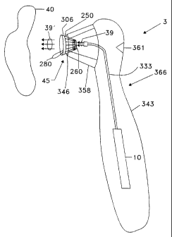
Figure 3C shows a measurement system 3 which utilizes a
disposable calibration device 45 for instrument 10. Here,
instrument 10 is an optical instrument such as a spectrometer and
radiation 39 is optical radiation which can be in the visible,
CA 02241817 1998-06-29
WO 97/25913 PCTIUS96/03319
uv and/or infrared regions. System 3 includes a housing 343
which is approximately the size of a human hand. Instrument 10
is coupled to calibration device 45 via optical fiber 333.
Calibration device 45 is inserted into an opening end 346 of a
cone-shaped holder 358 of housing 343. Cone shaped holder 358
can have any shape depending among other things on the shape of
calibration device 270 and hence will alternatively be referred
to as a calibration device receiving element. Holder 358 can be
a separate piece or part of housing 343. It is preferable that
holder 358 be capable of receiving calibration device 45, to
allow a calibration measurement to be made, but then allowing
calibration target 270 to be readily removed for the actual
measurement on material or tissue 40, and then allowing
calibration device 45 to be removed so that system 3 is again
ready to receive a new calibration device 45.
Curved portion 366 of housing 343 allows the hand to
comfortably hold system 3. A person can initiate a calibration
or measurement as the case may be, by pressing a push button 361
with his or her thumb. Once a calibration measurement has been
performed, tear tab 280 used to peel calibration target 270 away
from window 260 (not shown in this view), and system 3 is now
ready to make a measurement on material or tissue 40.
Figure 3D shows the same measurement system with calibration
device 45 removed. A new calibration device 45 must be inserted
into end 346 of system 3 and the above discussed process of
calibration must be repeated and calibration target 270 peeled
away before system 3 is ready to perform an new measurement.
Alternatively, a cap 375 can be placed over end 346 between
21
CA 02241817 1998-06-29
WO 97/25913 PCT/US96/03319
measurements.
In all of the above embodiments, calibration target 270 can
have calibration information fitted directly on surface 41 of
calibration target 270, and which can be read by instrument 10.
This calibration information can include a message read by
instrument 10 which initiates a system shut down after one or a
predetermined number measurements are performed. For the case
of shut down upon a single measurement, contamination is avoided,
because that system 3 cannot be reused on a new or different
material or tissue until a new calibration device 45 replaces the
used calibration device. In an alternative approach, this
calibration information can be directly input into system 3 by
a user using input 311.
Figure 3E summarizes the steps involved for system 3 to take
a measurement on material or tissue 40. In particular, step 382
involves placing calibration device 45 on end 346 of system 3.
At this point, calibration 45 device still has calibration target
270 covering window 260. A calibration measurement is performed
by system 3 at step 384 by pressing push button 361 which
activates instrument 10. Step 388 involves removing calibration
target 270 from window 260 using tear tab 280. Step 392 then
involves performing a measurement on tissue or material 40 to be
measured. This might involve a single measurement or multiple
measurements (if cross contamination is not an issue) on the same
or a similar tissue or material. That is, if measurements are
being performed on a human's tissue, several measurements might
be repeated in the same vicinity of that person's tissue.
Similarly, if measurements are being made on some type of
22
CA 02241817 1998-06-29
WO 97/25913 PCT/US96/03319
material, multiple measurements can be made in the vicinity of
that measurement provided that cross contamination is not an
issue. Finally, once the measurement or measurements have been
completed, calibration device 45 is removed, discarded, and
replaced with a new calibration device 45 at step 396.
Alternatively, used calibration device 45 can be removed,
discarded, and cap 375 can be placed over end 346 until a new
measurement is to be made.
Figures 4A and 4B show a top view and a side view,
respectively, of calibration device 45 similar, but not identical
to device 45. Figures 4C and 4D show the same views as Figures
4A and 4B, respectively, with calibration target 270 removed.
Device 45 can include cross-hatched lines 404, 406, and 408.
Lines 404, 406, and 408 can be placed on the backside 414 of
calibration target 270 as well as along inner-sides 424 of
structure 250 and outer annular ring 306 of calibration target
270 which can aid in the placement of window 260 on material
issue 40. Cross-hatched lines 404, 406, and 408 are designed to
be aligned prior to calibration. Once the calibration
measurement is made, calibration target 270 is removed, thereby
making system 3 ready to make a calibrated measurement. If a
user then tries to reattach calibration target 270, they will
note that lines 404, 406 and 408 are no longer properly aligned.
Also, surface 41 can be made so that once a calibration
measurement is made, calibration target 270 no longer attaches
or sticks to window 260. Cross-hatched lines 404, 406 and 408
define six zones (here each zone is shown as a wedge, but the
shape can be of any form). Also, note that an additional cross-
23
CA 02241817 1998-06-29
WO 97/25913 PCTIUS96/03319
hatched line is shown which further divides two of the wedges and
hence that the number of zones need not be limited to six. Each
of the cross-hatched lines are made to appear on both calibration
target 270 and window 260. The different zones on calibration
target 270 have different reflectivities or different reflectance
signatures. The different zones on calibration target 270 are
matched up with corresponding zones on windows 260 at the
manufacturing stage. The different zones on calibration target
270 thereby create a rotary reflectance signature. In this
manner, calibration is only valid if the rotary reflectance
signature is duplicated with each measurement. If calibration
target 270 is not properly oriented, the calibration would not
be valid. This helps to avoid the reuse of device 45.
Calibration target 270 can be manufactured with two pull
tabs at its sides as shown in Figure 4E. Here, two pull tabs 531
and 533 are attached to two halves 535 and 537 of target 270.
The two halves 535 and 537 have a mechanical perforation 539.
When target 270 is pulled away from window 260 (see Figures 2A
or 2B), it breaks along perforation 539, thereby making it
difficult to reuse. The remaining half of target 270 can be
pulled away using the remain tab. Perforation 539 need not be
a straight line, but can be curved or spiral shaped. If
perforation 539 is a spiral, a single tab (e.g., tab 531) can be
used, in which case target 270 is unraveled and peeled away from
window 260 either from its perimeter to its center (if the tab
is on the perimeter of target 270), or from its center to its
perimeter (if the tab is on the center of target 270). The
number of revolutions of the perforation spiral can vary from
24
CA 02241817 1998-06-29
WO 97/25913 PCT/US96/03319
less than one to three or more.
Device 45 in Figures 4B and 4D has annular ring 301 which
contacts the material or tissue 40 to be measured. Device 45
also has a collar section 405 that attaches to the optical outlet
(not shown) of instrument 10. Diameter Dl is defined to be the
diameter of annular ring 306 and diameter D2 is defined to be the
diameter of window 260, and height H is defined to be the
distance from window 260 to annular ring 306.
Figures 5A, 5B, and 5C show three more perspective views of
device 45 (Figures 5B and 5C have calibration target 270
removed).
Figure 6 shows a calibration device 45 according to another
embodiment of the invention. Here, a landing annulus 690 is
affixed to structure 250. Landing annulus 690 serves to fix the
angle radiation is incident on surface 680. Landing annulus 690
is preferably transparent to radiation 39. Calibration occurs
as before with the presence of calibration target 270. A
measurement is taken and then calibration target 270 is removed
and annulus 690 remains in place. Device 45 is then placed on
surface 680 such that annulus 690 lies flat on surface 680,
thereby ensuring that radiation 39 is incident approximately
normal to surface 680 as it was to surface 41 of calibration
target 270. On the other hand, depending on the type of
measurement, it may be preferable due to unwanted spectral
reflections, to have radiation 39 incident at an angle off normal
to surface 680. Landing annulus 690 can be a separate piece
affixed to structure 250 and comprised of any type of rigid
material such as various plastics. If infection to surface 680
CA 02241817 2006-10-27
WO 97/25913 PCT/US96/03319
of tissue 40 is an issue, then landing annulus 690 should be
removable from structure 250. Alternatively, annulus 690 can
simply be an extension of window 260 itself.
Structure 250 is preferably fabricated from molded plastic
with a smooth window zone defined for window 260. Using plastic
molding allows structure 250 to be fabricated at low cost and in
a wide variety of shapes and sizes. Calibration target 270 can
also be fabricated from plastic and may also have a dye or other
material added as surface 41 to provide sufficient spectral
detail to effect the necessary calibration. Calibration target
270 can be attached to window section 260 in such a way that once
removed, it cannot be readily re-attached. One implementation
is to fabricate calibration target 270 using a statically
clinging type plastic, and to fabricate structure 250 using an
appropriate material such as an acrylic called polymethyl
methacrylate (PMMA) both of which are available from 3M
Corporation.
Figure 7A shows a side view of calibration device 45
according to yet another embodiment of the invention. Here,
calibration target 270 is held in place by ridge 700 alone or
together with static cling between target 270 and window 260.
Ridge 700' can be part of window 260 or a separate piece. Figure
7B shows calibration device 45 as viewed from above.
Snectroscopic Measurements
United States Patent 5,353,790 presents a method and apparatus
for determining bilirubin concentration in human tissue such as
26
CA 02241817 1998-06-29
WO 97/25913 PCTIUS96/03319
skin. In particular, the patent discusses reflecting light from
skin to be tested to determine bilirubin concentration. The
approach corrects for maturity-dependent optical properties of
the skin including the amount of melanin in the skin and the
amount of blood in the skin. Reflected red to infrared light is
used to determine the maturity-dependent optical properties,
reflected red light is used to determine melanin content, and
reflected yellow-orange light is used to determine the amount of
blood in the skin. These quantities are used, in combination
with reflected blue light, to calculate cutaneous bilirubin
concentration.
Spectroscopy System
Figures 8A, 8B, and 8C show a front, side and back view,
respectively, of a spectrometer system 803, and Figure 8D shows
a spectrometer system 803 in a charging stand 871 according to
one embodiment of the invention. Figure 8A shows a front portion
809 of spectroscopic system 803 which utilizes a disposable
calibration device 845 (corresponding to the previously discussed
disposable calibration device 45) for a spectrometer 810. As
will be discussed with reference to Figure 9, spectrometer 810
can include a microspectrometer such as that offered by American
Laubscher Corporation of Farmingdale, LI, NY called the VIS/NIR
microspectrometer.
The elements in spectrometer system 803 which have similar
counterparts in the previously discussed system 3, will also have
the earlier reference numbers indicated in parenthesis.
Spectrometer system 803 can operate in the visible, uv and/or
27
CA 02241817 1998-06-29
WO 97/25913 PCT/US96/03319
infrared regions. Spectrometer system 803 includes a housing 843
which is approximately the size of a human hand. Spectrometer
810 is coupled to calibration device 845 via optical fiber 833
(see Figure 8B). Calibration device 845 is inserted into an
opening end 846 of cone-shaped holder 858 of housing 843. Curved
portion 866 of housing 843 allows the hand to comfortably hold
spectrometer system 803.
Figure 8B shows a side view of spectrometer system 803
including spectrometer 810 and push button 861. Spectrometer 810
is mounted on a printed circuit (pc) board 818 which is powered
by batteries 822. Batteries 822 can be recharged when placed in
a power adapter stand at charger connection 826. A liquid
crystal display (LCD) device 832 is also coupled to pc board 818
through window 841 displays measurement results, instructions,
warnings, etc... . Spectrometer 810 is controlled by a processor
(see Figures 9A and 9B) also mounted on pc board 818.
Figure 8C shows a back view of system 803 which includes
back portion 891 and a full view of LCD device 832. A person can
initiate a calibration and then a measurement by pressing push
button 861 with his or her thumb. In particular, once a
calibration measurement has been performed, tear tab 280 (see
previous figures) is used to peel calibration target 270 away
from window 260, and system 803 is now ready to make a
measurement on a patient. LCD device 832 indicates when
spectrometer system 803 is ready to make a calibration
measurement. LCD device 832 further indicates when the
calibration measurement has been completed and system 803 is
ready to make an actual measurement, and when system 803 has
28
CA 02241817 1998-06-29
WO 97/25913 PCT/US96/03319
completed the measurement. LCD device 832 also displays the
results of those measurements. LCD device 832 can also display
a message or other indicator showing that the particular
calibration target 270 has already been used and that no
additional measurements can be made until a new calibration
measurement is made. This can be achieved by the presence of a
limit switch (not shown) at the end of tip 858 which detects the
presence device 45. Once the limit switch is engaged, the
calibration is enabled and a measurement counter is initialized
to zero. Calibration is then performed. System software
increments the counter each time a measurement is made to a
predetermined maximum. Once the maximum number of measurements
is reached, system software indicates that a calibration is again
required, and the measurement counter is again initialized to
zero. Should the limit switch be disengaged at any time in the
measurement sequence, indicating the removal of the disposable
tip, the display indicates that a new calibration sequence must
be begun immediately. This prevents an operator from using one
calibration target more than once.
Figure 8D shows spectroscopic system 803 with a charging
stand 871 for storing and charging system 803. Charging stand
871 includes a center portion 873 for receiving system 803.
Center portion 871 serves as both a stand and a recharging unit.
Stand 871 has an electrical cord (not shown) which can be plugged
into an outlet. Stand 871 includes an electrical receiving unit
which receives charger connection 826 (see Figure 8B). An
indicator light 876 indicates when spectroscopic system 803 is
properly placed in center portion 873 so that recharging is
29
CA 02241817 1998-06-29
WO 97/25913 PCT/US96/03319
taking place. Stand 871 further includes a side receiving
portion 875 which can be used to place a supply 877 of
calibration devices 845.
Figure 9A is a schematic diagram of certain elements of
system 803, and in particular, of spectrometer instrument 810
which includes an optical unit 914, a central processor unit
(cpu) 905, and memory 909. Figure 9B shows a cut away view of
optical unit 914 including an optical source 918, a detector
array 923, an optical grating 951 and output 955 which couples
optical unit 914 to cpu 905 via bus 961_
Referring to Figure 9A, spectroscopic instrument 810
includes central processor unit (cpu) 905 and memory unit 909
which controls optical unit 914. Optical unit 914 may include
an optical source 918 which may be a tungsten halogen bulb, a
noble gas filled tungsten bulb or several LED's covering the
desired regions of the optical spectrum. The optical source 918
may also be placed at location 858 in the device housing to
illuminate the subject directly, without coupling into a fiber.
Output 955 is connected to cpu 905 via bus 961, thereby allowing
optical unit 914 to be controlled by cpu 905.
Figure 9B shows more detailed view of one embodiment of the
invention which utilizes a microspectrometer offered by American
Laubscher Corporation of Farmingdale, LI, NY called the VIS/NIR
microspectrometer. The cut away view of optical unit 914 shows
optical source 918 and with detector unit 933 which includes a
detector array 923 and a reflection grating 951. Optical
radiation 940 is output from optical source 918 and is
transmitted via fiber 833 to the target (not shown) to be
CA 02241817 1998-06-29
WO 97/25913 PCT/US96/03319
measured. The return signal 941 travels back down optical fiber
833 and is output from fiber end 958 into a type of waveguide 962
(cut away) and is incident on reflection grating 951. Reflection
grating 951 achieves self-focussing of radiation 941 to different
points or detectors on diode array 923 depending on the intensity
of wavelengths in radiation 941.
System 803 operates as follows. The following discussion
will include reference to Figure 4A (showing calibration device
45 with calibration target 270), Figure 8B (showing spectroscopy
system 803 and device 45), and Figures 9A and 9B (showing
spectrometer instrument 810 with optical unit 914). First,
calibration target 270 starts out being arranged on window 41 of
device 45 and a user pushes button 361 which indicates that
radiation 940 is output to calibration target 270. Calibration
target 270 has a known spectral characteristic. The actual
return radiation 941 results in a detected intensity at
individual detectors on detector array 923, thereby yielding a
measured calibration characteristic. This measured calibration
characteristic is compared to the expected or known spectral
characteristic of calibration target 270 and a resulting
adjustment value (which could be an array of values) is
determined. Calibration target 270 is then removed and a
measurement of tissue or material 40 is made by outputting
radiation 940 as above. A resulting spectral characteristic is
then output from detector array 923 which in turn is adjusted by
cpu 905 using the adjustment value or characteristic to yield a
calibrated spectral characteristic. The calibrated spectral
characteristic can then be used to determine some measurable
31
CA 02241817 1998-06-29
WO 97/25913 PCT/US96/03319
characteristic of material 40. One such measurement is a non-
intrusive bilirubin measurement according to one embodiment of
the invention as will now be discussed.
Bilirubin Measurement Process
Bilirubin can be measured in the aqueous of the eye based
on the fluorescent signature. Bilirubin can also be directly
measured in the sclera (white) of the eye based on the
fluorescent signature. Reflectance measurements can also be made
on the tympanic membrane of the ear. Finally,
reflectance/scattering based measurements can be made on the
skin.
Current literature has indicted that the aqueous levels are
likely to yield the same results as serum levels of albumin bound
bilirubin. However, measurements on five jaundiced adults showed
very low signal levels. Direct measurements in the aqueous are
also difficult due to low signal levels. This is probably due
to the photoconversion taking place in that location, i.e., too
much light is allowed into the aqueous in a typical person.
There are also difficulties in the evaluation of human factors
(such as the fact that infants may not stare in a particular
direction for an extended period of time) for an infant
measurement. Consequently, direct measurement in the aqueous is
not preferred due to the low signal-to-noise ratio and poor human
factors.
Direct measurements in the sclera is advantageous in that
the yellow color is clearly visible and hence the presence of
bilirubin is obvious. Also, this approach is advantageous over
32
CA 02241817 1998-06-29
WO 97/25913 PCTIUS96/03319
a skin based measurement, because it avoids the issue of
variations in skin color of thickness. The approach was tested
on five jaundiced adults. The approach yielded good signal
levels unlike the measurements in the aqueous. However,
repeatability was not very good. Also, data indicated a type of
photobleaching affect from the excitation light, even during the
data collection interval, spatial distribution was also not
constant due among other things to eyelid shading. Finally,
measurements on subjects shifted dramatically after those
subjects spent some time outside compared to the measurements
before those subjects went outside. Consequently, direct
measurement in the sclera although yielding a high signal-to-
noise, is not very repeatable and encounters poor human factors.
Direct measurements on the tympanic membrane suffers from
several shortcomings including poor vascularization, difficulty
in determining levels of bilirubin in the membrane, poor human
factors, particularly on premature babies.
Reflectance/scattering cutaneous measurements seem to be the
most promising non-invasive approach to measuring bilirubin.
Also, cutaneous measurements provide a simple interface with
which to work.
U.S. Patent 5,353,790 shows a technique makes it possible
to separate different constituents. That patent discusses the
absorption spectrum of melanin and shows that the melanin
absorption spectra essentially decreases linearly with wavelength
in the visible region. Moreover, since the melanin absorption
varies orders of magnitudes over the visible regime, variations
in the pigmentation will cause large absolute changes in the
33
CA 02241817 1998-06-29
WO 97/25913 PCTIUS96/03319
absorption at the shorter wavelengths, but the same magnitude
changes will cause relatively minuscule absolute changes in the
very long wavelengths (> 800 nm). The melanin pigmentation
measured in the far red wavelength range (650-750 nm) was found
to have a pivot point at around 837 nm.
Spectroscopic system 803 takes advantage of the above
phenomena and uses spectral reflectance to determine a serum
bilirubin level in mg/dL (milligrams of bilirubin per deciliters
of blood) as will now be discussed.
Figure 10 shows how spectroscopic system 803 performs
bilirubin measurements on a patient. The steps performed are an
improved approach discussed in U.S. Patent 5,353,790 by Jacques
et al., the contents of which are incorporated herein by
reference. Step 702 involves performing a calibration
measurement in a manner similar to that described in Figure 3E.
This involves simply outputting radiation to the calibration
target, measuring the return signal (due to reflection where
reflection is meant to include any type of scattering) to yield
a measured calibration spectrum or calibration data which is
compared to an expected calibration spectrum which is known a
priori depending on the material on surface 41 (see, for example,
Figures 2A or 3A). Also, the difference between the expected or
known spectrum and the measured spectrum can serve as the
calibration data which can be used to modify actual measured
data, thereby compensating for unit to unit and time varying
changes in source luminosity, delivery optics, collection optics,
detection sensitivity, electronic drift, and environmental
conditions such as temperature and humidity. The processor on
34
CA 02241817 1998-06-29
WO 97/25913 PCT/US96/03319
PC board 818 (see Figures 8A-8C) can perform the above
comparison. Alternatively, part or all of the comparison can be
performed with specifically designed digital and/or analog
hardware.
Step 704 involves making a reflection measurement (which
includes scattering) off of the tissue by illuminating the tissue
with light and detecting a frequency spectrum of light reflected
from said tissue. Step 708 involves converting the reflection
(scattering) measurement from step 702 into optical density.
Step 712 then involves calculating from a first portion of the
spectrum, a first parameter indicative of a maturity of the
tissue. Step 716 involves calculating from a second portion of
the spectrum, a second parameter indicative of an amount of
melanin in the tissue. Step 720 involves calculating from a
third portion of the spectrum, a third parameter indicative of
a blood content of the tissue. Step 724 involves calculating
from a fourth portion of the spectrum, a fourth parameter
indicative of an uncorrected bilirubin concentration in the
tissue. Step 728 involves calculating a corrected bilirubin
concentration in the tissue as a function of the first, second,
third and fourth parameters.
Figure 11 shows the results of data taken using the method
of Figure 10 versus a standard serum bilirubin (heel stick)
method. The subjects were 72 full term babies of varied ethnic
background, with 20 African Americans, 2 Hispanic Americans, 48
white Americans, and 2 Asian Americans. "R'I represents the
correlation coefficient between the measurement method described
in Figure 10, versus the standard method of serum bilirubin. The
CA 02241817 1998-06-29
WO 97/25913 PCT/US96/03319
correlation coefficient shown is 0.9165 with a perfect
correlation given as 1.0000. The tests represent a purely
prospective application of the process of Figure 10.
Numerous and additional modifications and variations of the
present invention are possible in light of the above teachings.
It is therefore to be understood that within the scope of the
appended claims, the invention may be practiced otherwise than
specifically claimed.
36