Note: Descriptions are shown in the official language in which they were submitted.
CA 02241984 1998-07-02
W O 97125914 PCTrUS97/00461
MEDIC~L ~UIDEWIRE
Background of the Invention
This invention relates to medical guidewires which
are used by physicians to access body lumens or other
remote areas within the body.
Guidewires are widely used in conjunction with
therapeutic devices, such as catheters, which are
10 threaded over the guidewire to gain access to an area
within the body requiring diagnosis or treatment.
Typically, a guidewire has a significantly smaller outer
diameter than therapeutic devices and can therefore be
inserted into the body to access remote areas more
15 easily. When the guidewire is positioned at the desired
location within the body, the therapeutic device is
passed over and guided along the guidewire to the
location. The therapeutic device is then utilized to
diagnose and/or treat the area at the location.
Summarv of the Invention
This invention features a medical guidewire in the
form of an elongated hollow tube, i.e., a tube with a
continuous wall such as is formed by extrusion and
drawing, having a sufficiently stiff proximal end, a
25 flexib~e, atraumatic distal end, and a thin wall
thickness to ~i~;ze the open area within the tubular
structure of the medical guidewire. In one aspect, small
lumens of the body can be accessed with a tubular
guidewire according to the invention, by conventional
30 guidewire t~chn; ques, and then an even smaller,
subselective guidewire can pass through the open area of
the initial guidewire to access even more restricted
regions of the bo~y. Furthermore, the relatively large
open area within the medical guidewire, as compared to
35 its small outer diameter, enables therapeutic means, such
CA 02241984 1998-07-02
W O 97/2~gl4 PCT~US97/00461
as liquid medicants, surgical instruments, or diagnostic
tools, to pass through the medical guidewire to access a
desired location within the body. Thus, small lumens of
the body can be accessed through the tubular structure of
5 the medical guidewire, using through the guidewire
procedures, to provide certain types of diagnosis and/or
treatment at the desired location within the body while
also enabling access of conventional over-the-wire
instruments. The small outer diameter of the medical
10 guidewire of the invention enables conventional over-the-
wire instruments to be used in conjunction with the
guidewire.
In one aspect, the invention features a medical
guidewire constructed for insertion into the body for
15 providing access to a body passage, the guidewire having
a main proximal portion in the form of an elongated tube
ext~n~;ng the majority of the length of the guidewire,
and a distal tubular portion integral with the main
proximal portion. The distal portion includes a distal
20 end and a flexible section having a slot cut in the wall
of the distal tubular portion. The slot crosses the
right sections of the distal tubular portion at an
oblique angle and extends continuously about the distal
tubular portion, in excess of at least one rotation about
25 the distal tubular portion, to increase the flexibility
of the distal tubular portion with respect to the main
proximal portion.
In another aspect, the invention includes an
elongated member having an outer diameter sized to pass
3Q through the medical guidewire such that the elongated
member may be inserted through the main proximal portion
and the distal tubular portion and extend beyond the
distal end of the distal tubular ~ection further into the
body. In preferred embodiments, the elongated member is
35 a subselective guidewire or an obtuxator.
CA 02241984 1998-07-02
W O 97125914 PCT~US97/00461
In another particular aspect, the slot in the
flexible section of the medical guidewire crosses the
right sections of the distal tubular portion at an
oblique angle and extends continuously through a
5 plurality of rotations about the distal tubular portion
to increase the flexibility of the distal tubular portion
with respect to the main proximal portion.
Preferred embodiments include the following
features.
The main proximal portion and the distal tubular
portion are formed from nitinol and at least the distal
tubular portion is annealed progressively to cause distal
portions of the distal tubular portion to be more
~lexible than proximal portions thereof.
1~ A subselective guidewire is used in combination
with the medical guidewire and is sized to pass through
the medical guidewire, through the main proximal portion
and the distal tubular portion, to extend beyond the
distal end of the distal tubular portion further into the
20 body. The outer diameter of the main proximal portion is
pre~erably of the order of 0.020 inches or smaller and
the outer diameter of the subselective guidewire is
preferably of the order of 0.014 inches or smaller.
The slot in the wall of the distal tubular portion
25 extends continuously through a plurality of rotations
about the tube, preferably through at least five
rotations. The width of the slot may vary along the
length of the slot such that the width is less in
pro~ l portions of the slot, e.g., from about 0.001
30 ~n~he~ to about 0.002 inches, relative to distal portions
thereof, e.g., from about 0.004 inches to about 0.005
~ nch~, Alternatively, the width of the slot may be
constant, e.g., between about 0.002 inches to about 0.004
inches.
CA 0224l984 l998-07-02
W O 97/25914 PCT~US97/00461
- 4 -
The oblique angle is greater at proximal portions
of the slot than distal portions thereof and may decrease
progressively, proximally to distally, along the slot,
from about 45~ to about 10~.
The distal tubular portion may further include a
transition section, located proximally of the flexible
section, that has portions of the wall of the distal
tubular portion removed to impart an intermediate range
of flexibility such that the transition section is more
10 flexible than the main proximal portion and less flexible
than the flexible section. The transition section
includes at least one slot shorter than the slot in the
flexible section. The transition section slot has a
discontinuous slot section which includes a series of
15 relatively short slots, generally helically aligned,
separated by unslotted portions of the wall of the distal
tubular portion. In aggregate, the discontinuous slot
section extends more than one rotation about the distal
tubular portion. The length of the discontinuous slot
20 section and the length of the flexible section each
extend about 2 to 3 cm of the length of the distal
tubular body.
The transition section may further include a
pattern of perforations through the wall of the distal
25 tubular portion. The pattern of perforations is located
pro~; ~lly of the relatively short slot and extends about
3 to 5 cm of the length of the distal tubular portion.
The pattern size and shape of the perforations are
selected to cause the corresponding region of the distal
30 tubular portion to be more flexible than proximal
portions thereof and less flexible than distal portions
thereof.
A sealing element extends along at least a portion
of the distal tubular portion and is joined to the wall
35 of the tubular member on both sides of the slot. The
CA 0224l984 l998-07-02
W O97l25914 PCTrUS97/00461
sealing element has at least one fluid-delivery
perforation aligned with the slot to deliver fluids to a
desired location within the body. The ~1 ing element
may be a polymeric tube, in shrunken state, that
5 surrounds at least a portion of said distal tubular
portion or may be a polymeric tube, in a rA~;~lly
~rAn~ed state, that contacts the inside of at least a
portion of said distal tubular portion. Alternatively,
an ela~tomer may fill at least a portion of the slot in
10 said distal tubular portion.
A distal tip portion, located distally of the
distal tubular portion, has a rounded distal extremity
for atraumatic insertion into the body and is fabricated
from a polymeric material chosen from the group
15 comprising PET, polyimide, or polyethylene. The distal
tip portion includes a polymeric core disposed within a
polymeric sleeve which is attached to the polymeric core.
The polymeric core and the polymeric sleeve are made from
a nylon material and the polymeric core may further
20 contain a radiopaque material. A nitinol core, having a
tapered distal portion is disposed within the polymeric
core and extends proximally through at least a portion of
the distal tubular portion. The diameter of the nitinol
core that e~tends into at least a portion of the distal
25 tubular portion is smaller than the internal diameter of
the distal tubular portion to provide a clearance between
the nitinol core and the inner surface of said distal
tubular portion.
Alternatively, the distal tip portion is hollow
30 and includes a longitudinal slit in opposing walls of the
distal tip portion. The longitudinal slit is biased
closed and upon application of an internal force, i.e., a
longitll~; nA 1 thrusting force of an elongated member or
fluid pressure, is capable of opening to provide a
35 passageway through the distal tip portion.
CA 02241984 1998-07-02
W O 97125914 PCTAJS97/00461
Another aspeot of the invention features a method
of treating a patient using a medical guidewire that
implements the structural features of the invention. The t
medical guidewire is inserted into a body lumen and the
5 elongated member is inserted into the medical guidewire.
The elongated member is sized to pass through the mediaal
guidewire so that the elongated member may be inserted
through the main proximal portion and the distal tubular
portion of the medical guidewire. The medical guidewire
10 and the elongated member are guided to a desired location
in the body lumen requiring treatment and the elongated
member is extended beyond the distal end of the medical
guidewire, further into the body, to access remote, small
passages at the desired location.
Preferred embodiments include the following
features.
While the medical guidewire and the elongated
mem~er are guided to the desired location in the body
lumen, the position of the elongated member within the
20 medical guidewire is adjusted to alter the stiffness of
the distal tubular portion of the medical guidewire.
F~rther, a surgical inY~ll ~nt may be slid, in
guided contact, over the medical guidewire to access the
desired location such that at least one surgical
25 operation using the surgical insLr~ ~nt may be performed
at the desired location.
In another aspect, the invention features a method
of treating a patient using the medical guidewire of the
invention. The medical guidewire is inserted into a body
30 lumen and guided to a desired location in the lumen
requiring treatment. Liquid is infused through the
medical guidewire and exits the medical guidewire though
the slot in the distal tubular portion of the medical
guidewire to enter the body lumen at the desired
CA 02241984 1998-07-02
W O 97/2~914 PCTrUS97/00461
-
- 7
location. The medical guidewire is removed from the body
lumen.
Other features and advantages of the invention
will become apparent from the following detailed
5 description, and from the claims.
~rief Description of the Drawinqs
Fig. 1 shows a medical guidewire according to the
invention, while Fig. la is an enlarged cross-sectional
view of the medical guidewire taken along line a-a in
10 Fig. 1;
Fig. 2-4 shows side views of embodiments of a
portion of the medical guidewire of the invention;
Fig. 5 shows a side view of an embodiment of the
medical guidewire, having a sealing element, while Fig.
15 ~a shows a side view of an alternative embodiment of the
sealing element;
Figs. 6 and 7 are side views, partially in cross-
sectional, of embodiments of the medical guidewire of the
invention;
2~ Figs. 8 and 8a are side views, partially in cross-
sectional, of further embodiments of the medical
guidewire of the invention, while Fig. 8b shows a cross-
sectional view of a further embodiment of the medical
guidewire of the invention.
~escriPtion of the Preferred Embodiments
Referring to Figure 1, a medical guidewire 10 is
shown for use as a primary access device for entering a
body lumen, particularly relatively small body lumens,
such as, for example, the vasculature of the brain or
30 distal coronary arteries. The overall length of the
medical guidewire is about 180 to 220 cm.
The medical guidewire comprises a hollow tube
defined by a continuous wall, such as can be formed by
extrusion and drawing. The inner and outer diameters of
CA 02241984 1998-07-02
WO 97125914 PCT~US97/~0461
-- 8 --
the medical guidewire are sized to reduce the wall
thickness of the guidewire to optimize the open area of
the lumen within the guidewire. This construction
facilitates the delivery of therapeutic means, such as,
5 liquids, surgical instruments, or diagnostic tools,
through the medical guidewire 10 while also providing the
means for secondary access, i.e., by guiding a catheter
over the medical guidewire to access the desired
location. To maintain adequate strength, stiffness, and
10 tor~ue characteristics, the medical guidewire 10 is
formed from a flexible and resilient metal, preferably
nitinol.
In particular, the medical guidewire 10 has an
outer diameter 14 of the order of 0.020 inches (0.51 mm)
15 and a wall thickness 16 of the order of 0.002 inches
~0.051 mm) to ~; ;ze the expanse of open area 18.
Medical guidewire 10 is comprised of at least two
integral portions, main proximal portion 20 and distal
tubular portion 24. Main proximal portion 20 is about
20 1~5 cm to about 205 cm in length and is in the form of an
elongated tube, i.e., an elongated hollow cylinder, and
although ~lexible, has a sti~fness o~ about 50-100 N-mm2
to impart sufficient lateral stiffness and torque
transmission capabilities along its length.
Re~erring as well to Figure 2, distal tubular
portion 24 i5 located distally of main proximal portion
2Q and is from about 15 to about 25 cm in length. Distal
tubular section is also in the form of a tube and is
integral with main proximal portion 20. For purposes of
30 this invention, the term "integral" means that the distal
tubular portion is attached to main proximal portion by a
~uitable method (e.g., by welding, brazing, heat
shr;nk;ng, or gluing) in an end to end, abutting
relationship or in an overlapping relationship.
35 Alternatively, "integral" means that distal tubular
CA 02241984 1998-07-02
W O 97125914 PCTrUS97/00461
-
_ g
portion and main proximal portion are fabricated from the
same continuous tube, but comprise distinct portions in
that tube. Distal tubular portion 24 has a stiffness of
~bout 25-50 N-mm2 or less, to impart flexibility to
5 medical guidewire 10. Additionally, the flexibility of
medical guidewire 10 may be varied by progressively
annealing either a portion, e.g., distal tubular portion
24, or the entire length of medical guidewire 10.
Distal tubular portion 24 is comprised of flexible
lQ section 30 having slot 32. The length of flexible
section 30 is about 2 to 3 cm. Slot 32 is preferably cut
completely through wall 33 of distal tubular portion.
A~ternatively, to vary the stiffness of distal tubular
portion 24, a portion of slot 32 may be replaced with a
15 grooved section (not shown), which is only partially cut
through the wall of distal tubular portion 24. In either
embodiment, slot 32 crosses right sections, e.g., section
34, of the distal tubular portion at an oblique angle,
e.g., angle e , and extends continuously for at least one
20 rotation, preferably through a plurality of rotations,
e.g., from about 3 to 15 rotations. It should be
understood that right section 34 is perpendicular to the
longitll~; n~ 1 axis 12 of medical guidewire 10 and that
oblique angle e is an angle formed between right section
25 34 and the center axis of slot 32 (represented by line
35~.
To vary the stiffness within flexible section 30,
such that distal portions of flexible section 30 are more
flexible than proximal portions thereof, oblique angle e
30 is greater at proximal portions of slot 32 than distal
portions thereof, e.g., obli~ue angle e1 is greater than
ob~ique angle e2. The decrease of angle e proximally to
distally is progressive, i.e., with each rotation about
distal tubular portion 24, angle e decreases uniformly
35 between 1 to 8~ or more per rotation. Alternatively,
CA 0224l984 l998-07-02
W O 97i25914 PCTAJS97tOO461
-- 10 --
angle e decreases variably, i.e., with each rotation,
angle e decreases variably. Preferably, angle e varies
from about 45~ to about 10~ proximally to distally.
Additionally, the stiffness of flexible section 30
5 may be varied by increasing the width of slot 32
proximally to distally. For example, the width of slot
32 at proximal portions of flexible section 30 is from
about 0.~01 inches to about 0.002 inches (O.OOZ5 cm to
about 0.051 mm) and the width of slot 32 at distal
10 portions thereof is from about 0.004 inches to about
0.005 inches (0.102 mm to about 0.127 mm).
Alternatively, the width of slot 32 may be constant,
e.g., from 0.002 inches to 0.004 inches (0.051 mm to
0.102 mm).
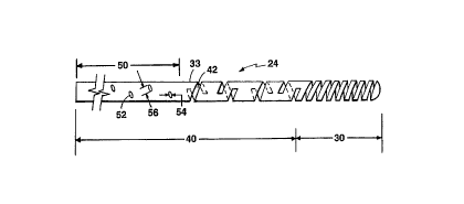
Referring now to Figure 3, distal tubular section
24 may further comprise transition section 40, located
proximally of flexible section 30. Portions of wall 33
in transition section 40 are removed to impart an
intermediate range of flexibility to transition section
20 40, i.e., transition section 40 is more flexible than
main proximal portion 20 and less flexible than flexible
section 30. Wall 33 may be removed only partially, such
a~, for example, having a groove or a notch cut only
partially through wall 33, or wholly, such as, for
25 example, a slot cut completely through wall 33, or a
combination of both. Transition section 40 includes at
least one discontinuous slot 42, and preferably a
plurality of discontinuous slots 42, separated by
unslotted portions 43 of wall 33. Discontinuous slot 42
30 is preferably generally helically aligned about wall 33
of distal tubular portion 24 and proceeds through more
than one rotation about distal tubular portion 24. In
particular, if a reference line "X" is drawn on wall 33
parallel to longitt~; n~ l axis 12, discontinuous slot 42,
35 ha~ing one end 44 beginning on or near reference line
-
CA 02241984 1998-07-02
W O 97i25914 PCTrUS97/00461
"X", proceeds generally helically about distal tubular
portion 24 such that second end 46 is located past
reference line "X", i.e., discontinuous slot 42 proceeds
through more than one rotation about distal tubular
5 portion 24.
The portion of transition section 40 having
discontinuous slots 42 extends about 2 to 3 cm of the
length of distal tubular section 24. Each discontinuous
51ot 42 has a length of about 0.03 inches to about 0.05
10 inches (0.076 cm to about 1.27 mm), preferably about 0.04
inches (1.02 mm). The distance ~etween discontinuous
slots 42, or the length o~ unslotted portion 42, is about
0.01 inches to about 0.1 inches ~0.025 cm to about 0.25
cm). The pitch of discontinuous slot 42 is preferably
15 about 45~ and width 48 is preferably constant, between
about 0.001 inches to about 0.002 inches (0.025 mm to
about 0.051 mm). Alternatively, width 48 may vary as
described above.
Referring to Figure 4, transition section 40 may
20 further comprise a pattern of perforations 50 through
wall 33 of distal tubular section 24. The pattern of
perforations is located proximally of discontinuous slots
42 and extends through a length of about 3 to 5 cm of the
length of distal tubular section 24. The pattern size
25 and shape of the perforations is selected such that the
pattern of perforations 50 is less flexible than
discontinuous slots 42 and more flexible than main
proximal portion 20.
Pattern of perforations 50 consists of a plurality
30 of angled slots 52, that are cut through wall 33.
Alternatively, angled slots 52 may be only partially cut
through wall 33 and may have various depths to alter the
stiffness of distal tubular section 24 at pattern of
perforations 50.
CA 0224l984 l998-07-02
W 097i25914 PCTrUS97/00461
- 12 -
Angled slots 52 are oriented at a particular
pitch, preferably 45~, and are disposed at 90~ or 120~
intervals along wall 33 of distal tubular portion 24.
Width 54 of angled slots 52 is about 0.001 inches to
5 about 0.005 inches (0.025 mm to about 0.127 mm). ~ength
56 of each angled slot 52 is about 0.020 inches (0.51 mm~
and each slot is separated by a length of about 1.0 to
about 1.5 mm. The desired degree of flexibility in the
pattern of perforations 50 may be varied by varying slot
10 width 54, slot length 56, and the distance between slots.
Additionally, the shape of angled slots 52 may be varied
to vary the flexibility.
Distal tubular portion 24 is preferably
manufactured from a nitinol tube. To impart the desired
15 flexibility characteristics to flexible section 30 and
transition section 40, slots 32, 42, and 52 are formed
into distal tubular portion 24 by, for example, EDM
ma~h; n; ng, chemical masking, electro-chemical etching, or
laser etching.
Referring to Figure 5, a sealing element 60 seals
and provides additional strength to medical guidewire 10.
In the preferred embodiment, sealing element 60 is a
~leeve that is heat shrunk to ~UL loul.d at least a portion
of distal tubular portion 24. The thickness of sealing
25 element 60 is about 0.0005 inches (0.013 mm) and is
fabricated from either PET, polyimide, polyethylene,
polyurethane, teflon, EVA, silicon, or hydrophilic gel.
The outer diameter of distal tllhlll ~r portion 24 is
preferably reduced at portions including sealing element
30 60 such that the outer diameter of medical guidewire 10
remains constant. For example, distal tubular portion
outer diameter 64 is less than distal tubular portion
outer diameter 66 such that the outer diameter of medical
guidewire 10 is not increased at portions including
35 sealing element 60.
CA 0224l984 l998-07-02
W O 97/25914 PCTrUS97/00461
- 13 -
Alternatively, sealing element 60 may be a sleeve
that is radially expanded from inside medical guidewire
10 (not shown), to contact the inside wall 26 of at least
a portion of distal tubular portion 24, or may be an
5 elastomer 61 which fills at least a portion of the slots
located in distal tubular portion 24 (Fig. 5a).
Sealing element 60 may include at least one fluid-
delivery aperture 62, preferably a plurality of fluid-
delivery apertures 62, aligned along slot 32.
10 Additionally, if sealing element 60 covers a portion of
slots 42 and/or 52 in transition section 40, fluid
delivery perforations 62 are aligned along portions of
these slots. Fluid-delivery apertures 62 enable a fluid
to be infused through medical guidewire 10 and out
15 through the fluid-delivery apertures 62 into a body lumen
to the site of interest and are preferably about 1 to 5
mils in diameter. For example, the infused fluid may be
a saline solution which can be used to flush the site of
interest or to clean surgical instruments used in
20 combination with the medical guidewire ~0 during a
surgical procedure or diagnosis. Alternatively, the
fluid may be a liquid medicant to treat the site of
interest or a radiographic or ultrasonic contrast agent
to monitor fluid flow through the site of interest using,
25 for example, x--ray or ultrasound techrliques. Fluid--
~elivery apertures 62 are fabricated using a laser or a
heated needle to perforate sealing element 60.
Referring to Figure 6, in one embodiment, a distal
tip portion 70 is attached to distal tubular portion 24.
30 To enable atraumatic insertion of medical guidewire 10
into the body, distal tip portion 70 is flexible, is
about 0.5 inches to about 3 inches (1.27 cm to about 7.62
cm~ long, and has a rounded distal extremity 72, the
outer diameter of which is about 0.014 inches (0.356 mm).
35 ~istal tip portion 70 includes sleeve 74 which abuts
CA 02241984 1998-07-02
W O 97125914 PCT~US97/00461
- 14 -
distal end 36 of distal tip portion 24. The inner and
outer diameter o~ distal tubular portion 24 at distal end
36 is substantially similar to the inner and outer
diameter of sleeve 74. Sleeve 74 further surrounds core
5 76. Core 76 encases tapered core 78. Sleeve 74 and core
76 are made from a polymeric material, preferably nylon.
Further, the availability of a range of stiffness for
sleeve 74 and core 76 permits the overall stiffness of
distal tip portion 70 to be tailored to the desired end
10 use of medical guidewire 10. Additionally, core 76 may
be filled with a radiopaque material so that a physician
can view its position within the body using an x-ray.
To attach distal tip portion 70 to distal tubular
portion 24, sealing element 60 surrounds a portion 79 of
15 the proximal end of sleeve 74. Sleeve 74 is bonded to
core 76, and core 76 is bonded to tapered core 78,
preferably with flexible adhesives, such as, for example,
urethane.
Tapered core 78 is approximately 6-~0 cm in length
20 and preferably has three sections, distal section 80,
tapered section 82 and proximal section 84. Distal
section 80 is approximately 1 to 2 cm in length,
preferably 1 cm, and has a constant diameter, which is
pre~erably about 3 to 4 mils (about 0.076 mm to 0.101
25 mm~. Tapered section 82 is located proximally of distal
section 80 and is also preferably about 1 to 2 cm in
length. The proximal end 83 of tapered section 82 has
the largest diameter of tapered core 78, e.g., about 6
mils (0.152 mm), and tapers uniformly to distal end 85 of
30 tapered section 82, where the diameter is about 3 mils
~o.076 mm). Alternatively, tapered core 78 may have a
constant diaméter. Proximal section 84 is located
proximal~y of tapered section 82 and is about 4 to 6 cm
~n length and extends proY;~lly through a portion of
3 distal tubular portion 24. The diameter of proximal
CA 02241984 1998-07-02
W O 97/2~914 PCTrUS97/00461
section 84 is substantially the same as the diameter of
the proximal end 83 of tapered section 84, i.e., about 6
mils (0.152 mm). Therefore, there is clearance between
the inner diameter of distal tubular portion 24 and the
5 proximal section of tapered core 78 to enable the passage
of fluids. Preferably, tapered core 78 is fabricated
from a solid nitinol wire.
Referring to Fig. 7, in an alternative embodiment,
distal tip portion 90 comprises duck bill 96 bonded to
10 sealing element 60 by, for example, a flexible adhesive,
such as urethane. Duck bill 96 is fabricated from a
polymeric material (e.g., PET, polyimide, or
polyethylene) and includes a longitll~;n~l slit 98 in
opposite walls of distal tip portion 90. Longit~;n~l
15 slit 98 is ~iased closed, however, upon an application of
an internal ~orce, such as, for example, fluid pressure
or a longitll~;n~l thrusting force, longitll~in~l slit 98
opens to provide a passageway through distal tip portion
90 .
Referring to Fig. 8 et seq., medical guidewire 10
includes elongated member 100, which is inserted through
main proximal portion 20 and distal tubular portion 24
and may, in certain applications, be ext~n~e~ beyond
distal end 36 of distal tubular portion 24 further into
25 the body. To enable the insertion of elongated member
100 through medical guidewire 10, for coronary,
neurology, or urology applications, elongated - he~ lOO
has an outer diameter of the order of 0.014 inches (0.356
mm~ or smaller and the outer diameter of the medical
30 guidewire is of the order of 0.020 inches (0.051 cm) or
smaller. Alternatively, for cardiac, gastro-intestinal,
or peripheral vascular applications, elongated member has
an outer diameter of the order of 0.03 inches (0.076 cm)
or smaller and the outer diameter of medical guidewire is
35 of the order of 0.035 inches (0.089 cm) or smaller. The
CA 02241984 1998-07-02
W O 97125914 PCTrUS97/00461
- 16 -
length of elongated member 100 is typically about 180 to
220 cm, or more. This length is sufficiently long to
enable a user to manipulate elongated member 100 at
proximal end 102 and still provide sufficient length at
5 distal portion 104 such that elongated member can extend
beyond distal end 36 o~ distal tubular portion 24 further
into the body.
In a preferred embodiment, elongated member 100 is
a subselective guidewire, as shown in Fig. 8, preferably
10 ~ormed ~rom a solid nitinol wire, but alternatively,
formed from a nitinol coil. Alternatively, elongated
member 100 is an obturator 112 (Fig. 8a) or an ultrasound
imaging transducer 114 (Fig. 8b--To enable the passage o~
ultrasonic sound waves through medical guidewire 10, a
15 sonolucent window 115 may be located in distal tubular
portion 24 or distal tip portion 70 may be fabricated
~rom a sonolucent material). Additionally, elongated
member 100 may be in the form of other surgical or
diagnostic instruments, such as, for example, a basket
20 retrieval system, a surgical instrument (e.g., a cutter),
a laser fiber, an optical fiber, a balloon, a radioactive
element, an antenna, or an electrode or series o~
electrodes.
~ethod of Use
A user inserts medical guidewire 10 into a body
lumen using, for example, the Seldinger techn;que.
Pre~erably, to increase the stiffness of medical
guidewire 10, elongated h~r lOO is inserted through
medical guidewire lO prior to inserting medical guidewire
30 10 into the body lumen. The medical guidewire, including
elongated member 100, is guided within a body lumen to a
location within the body lumen requiring treatment. As
medical guidewire 10 is guided within a body lumen, a
user manipulates elongated member 100 along longitudinal
35 axis 12 within distal tubular portion 24 to vary the
CA 0224l984 l998-07-02
W O97i25gl4 PCTnUS97/00461
stiffness of medical guidewire 10 at distal tubular
portion 24. When medical guidewire 10 is positioned at
or near the location within the body lumen requiring
treatment, the user, by manipulating the proximal end o~
5 elongated member 100, extends elongated ~ h~ lOO beyond
distal end 36 of distal tubular section 24 further into
the body. The elongated member 100 thus accesses remote,
small passages at the desired location for diagnosis
and/or treatment of the area. For example, in the
10 illustrated embodiment, elongated member 100 is a
subselective guidewire, which enables a user to access
more distal or tortuous regions of the body. Other
~urgical instruments or diagnostic or therapeutic means
used within small lumens of the body may be employed as
15 elongated member 100 to per~orm their respective
procedures at the desired location.
Alternatively, after medical guidewire 10 is
guided to a desired location within the body, medical
guidewire 10 is used to infuse liquids to that location.
20 For example, liquids, such as saline solution, medicants,
or x-ray contrast liquids, are infused through slot 32,
through slots 32, 42 and 52~ through fluid-delivery
apertures 62 (when distal tubular portion includes
sealing element 60), through open distal end 36, and/or
25 through longitll~;n~l slit 98 in distal tip portion 90.
Medical guidewire 10 may further be used to guide
surgical or diagnostic instruments over medical guidewire
10 to access a desired location in a body lumen. When
the instrument is positioned at the desired location
30 within the body lumen, at least one surgical or
diagnostic procedure using the in~LL~ -nt is performed.
The instrument may be removed and replaced with a
different instrument as required by the treatment,
CA 02241984 1998-07-02
W O 97f25914 PCT~US97/00461
- 18 -
diagnosis, or surgical procedure being per~ormed by the
user.
What i5 claimed is: