Note: Descriptions are shown in the official language in which they were submitted.
CA 02242033 1998-06-30
MEDICAL DEVICE FOR IMPROVING SKIN FIXATION OF INDWELLING
CA~ Ks AND OTHER TRANSCUTANEOUS
IMPLANTS WITH A REDUCED RISK OF INFECTION
The present invention relates to transcutaneous
medical devices and to a process for their production.
Transcutaneous medical devices are implants which
pass through the skin and remain in the body for a lengthy
period, such as, for example, indwelling catheters.
Examples of transcutaneous implants are catheters for
peritoneal dialysis and catheters for long-term perfusion
therapies. With long-term use of these and other implants,
there is a risk of infection through bacteria or other micro-
organisms entering the body. Various movements of the body
can exert transient tensile and compressive forces on the
passages for the implants through the skin, whereby fissures
periodically form at the interface between the passage through
the skin and the skin tissue, through which microorganisms can
enter and infect the body.
Several proposals for fixation of transcutaneous
implants and prevention of infections with transcutaneous
devices such as, for example, catheters for peritoneal dialysis
using cuffs are disclosed in the literature.
Cuffs are hollow cylinders which are a few milli-
meters to a few centimeters long around the catheter. They
are placed on the catheter, singly or multiply, by pulling on
or by sticking on an appropriate tape. The task of the cuff
is to enter with its outer surface into close contact with the
o.z. 5221
23443-644
CA 02242033 1998-06-30
body tissue and thus fix the catheter and prevent microorganisms
migrating in at the catheter/body tissue interface. To achieve
this aim, the outside of the cuff consists according to U. S.
Patent 5,057,075 of Dacron, of a material regarded as
compatible with the body or of a porous material (U~ S. Patent
5,308,338; U. S. Patent 5,141,499), into which body cells can
grow.
As an additional measure to prevent microbial
infections entering the body through the passage through the
skin, cuffs are occasionally employed in combination with
antiseptic substances. U. S. Patent 5,308,338 discloses a tube
which passes inside the catheter and through which antiseptic
liquids can be delivered to the cuff material.
U. S. Patent 5,049,140 describes the use of anti-
microbial substances in the cuff material.
Further patents disclosing a fixation and/or
prevention of infection in connection with the use of
transcutaneous catheters are, for example: U. S. Patent
5,098,413; U. S. Patent 5,057,075; U. S. Patent 4,772,269;
U. S. Patent 4,687,471 and U. S. Patent 4,623,329. The
priority descriptions are of particular geometric embodiments
of catheters for individual types of use.
Although the use of cuffs in the prior art can extend
the period after which the catheter must be changed owing to
signs of infection, the problems of fixation and prevention of
infection of transcutaneous devices for long-term use have not
yet been satisfactorily solved. In particular, the growing-in
o.z. 5221
23443-644
CA 02242033 1998-06-30
of body tissue into porous materials does not result in a
durable connection reliably preventing the penetration of
infectious organisms. The use of antimicrobial and/or
antiseptic substances is to be regarded as a temporary measure
which is susceptible to failure and difficult to implement.
Collagen-containing composite materials are known
from a different technical area to be materials which readily
form adhesions to the human body (German Patent 36 327 316).
A primary object of the present invention is to make
it possible to fix medical devices in or on the body and thus
to prevent an infection entering the body through the passage
into the body.
It has been found, surprisingly, that transcutaneous
medical devices which have on their surface a fibrous material
with free collagen fibers which have a natural structure prevent
to a high degree a microbial infection entering the body through
the point where the transcutaneous medical device passes through
the skin.
The body tissue forms adhesion, without infection or
rejection, with the collagen fibers of the transcutaneous
medical device and thus forms a durable unit with the
transcutaneous medical device. This blocks entry of infectious
organisms into the body.
The present invention therefore provides a
transcutaneous medical device which has on its surface a
fibrous material having free collagen fibers which have a
natural structure. The device is preferably a catheter. The
o.z. 5221
23443-644
CA 02242033 1998-06-30
fibrous material may be in the form of a felt having on its
surface free (or exposed or uncovered) fibers of the collagen.
The present invention furthermore provides a process
for producing the transcutaneous medical device, which comprises
applying to its surface a fibrous material having free collagen
fibers which have a natural structure.
According to a preferred embodiment of the present
invention, in the transcutaneous medical device defined above,
the fibrous material is prepared by:
production of a collagen felt from collagen fibers
having a natural structure;
infiltration of this felt with a polymerizable
monomer;
carrying out a free-radical polymerization in the
presence of a polymerization inhibitor which acts from a
surface of the infiltrated felt; and
detachment of a not completely polymerized surface
layer of the resulting polymer to expose the collagen fibers
by using a suitable solvent.
The present invention furthermore describes a process
for applying a fibrous material to a transcutaneous medical
device, which process comprises:
prcduction of a collagen felt from collagen fibers
having a natural structure;
infiltration of this felt with a polymerizable
monomer;
carrying out a free-radical polymerization in the
o.z. 5221
23443-644
CA 02242033 1998-06-30
presence of a polymerization inhibitor which acts from a
surface of the infiltrated felt;
detachment of a not completely polymerized surface
layer of the resulting polymer to expose the collagen fibers
by using a suitable solvent to produce a fibrous material; and
subsequent application of the fibrous material to a
surface of the transcutaneous medical device.
The fibrous material comprises a collagen-containing
composite material whose surface forms a felt of free collagen
fibers having a natural structureO This collagen/polymer
composite material may be produced by first obtaining, for
example, as disclosed in German Patent Publication
"Biocompatible composite material and process for its
production" (German Application 195 29 036.4), a felt from free
collagen fibers having a natural structure, and subsequently
infiltrating with a polymerizable monomer preparation. If the
monomer preparation contains no polymerization initiator, the
polymerization may be started for example by radiation
induction. The polymerization must in all cases be carried out
with surface quenching. This results in a collagen-containing
composite material with an incompletely polymerized layer on
the surface. This layer is detached with a suitable solvent
to expose the collagen fibers. It is possible to influence
the thickness and characteristics of the surface layer which
is not cured during the polymerization by the choice of quench-
ing parameters.
According to another embodiment of the present
invention, in the transcutaneous medical device defined above,
O.Z. 5221
23443-644
CA 02242033 1998-06-30
the fibrous material is prepared by:
production of a collagen felt from collagen fibers
having a natural structure;
infiltration of this felt with a polymerizable
monomer;
application of the felt infiltrated in this way to
a surface of the transcutaneous medical device;
carrying out an incomplete free-radical polymeriza-
tion in the presence of a polymerization inhibitor which acts
from a surface of the infiltrated felt; and
detachment of the incompletely polymerized surface
layer of the resulting polymer to expose the collagen fibers
by using a suitable solvent.
Besides application of the fibrous material to the
transcutaneous medical device, this material may also be
produced directly on a surface of the transcutaneous medical
device.
Another embodiment of the invention is a process for
applying a fibrous material to the surface of a transcutaneous
medical device, which process comprises:
production of a collagen felt from collagen fibers
having a natural structure;
infiltration of this felt with a polymerizable
monomer;
application of the felt infiltrated in this way to
a surface of the transcutaneous medical device;
carrying out an incomplete free-radical polymeriza-
tion in the presence of a polymerization inhibitor which acts
o.z. 5221
23443-644
CA 02242033 1998-06-30
from a surface of the infiltrated felt; and
detachment of the incompletely polymerized surface
layer of the resulting polymer to expose the collagen fibers
by using suitable solvents.
Brief Description of the Drawings
Fig~ 1 is a section through the skin (from Faller, A:
der Korper des Menschen, 5th edition, Thieme, Stuttgart 1972),
and diagrammatic representation of a catheter with a cuff-like
device for fixation in the skin.
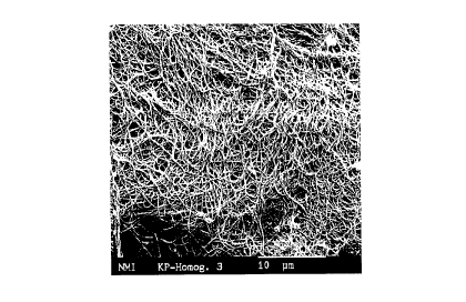
Fig. 2 is a microphotograph of the fibrous material
used in the subcutaneous medical device according to one
preferred embodiment of the present invention.
Detailed Description of the Invention
A very preferred feature of the present invention is
to use special materials in the transcutaneous medical devices.
These collagen/polymer composite materials have on their surface
free collagen fibers having a natural structure. By "free" is
meant that the collagen fibers are exposed outwards without
being covered by the polymer. These composite materials are
presumably responsible for the surprisingly rapid and complete
incorporation and adhesion of these materials to body tissue
such as, for example, cutaneous connective tissue.
Transcutaneous medical devices which comprise these collagen/-
polymer composite materials at specific points, for example in
the form of cuffs, form adhesions via these materials to the
body tissue. This results in fixation of the transcutaneous
device on or in the body and, if these collagen/polymer
o.z. 5221
23443-644
CA 02242033 1998-06-30
composite materials are located where the transcutaneous device
passes through the body, impedes migration of microbial
infectious organisms into the body (see Fig. 1).
In Fig. 1, the reference symbols and the reference
numbers have the following meanings.
a. Epithelial layer (epidermis). b. True skin
(corium), layer with connective tissue papillae (stratum
papillare)~ c. Reticular layer of the true skin (stratum
reticulare). d. Subcutaneous fatty tissue. 1. Meissner's
corpuscle. 2. Opening of a sweat gland on a ridge. 3. Free
nerve fiber. 4~ Convolution of the sweat gland. 5. Lamellated
corpuscle (Vater-Pacini) in longitudinal section. 6. Cornified
layer (stratum corneum). 7. Cornifying layer (stratum
granulosum and stratum lucidum). 8. Layer of living epithelial
cells (stratum germinativum). 9. Capillary loops in the
connective tissue papillae. 10. Cut surface of a small nerve.
11. Interlaced bundles of connective tissue in the true skin.
12. Efferent duct of a sweat gland. 13. Cross-section through
a lamellated corpuscle. 14. Fatty tissue lobules. 15.
Catheter in section. 16. Cuff made of the collagen/plastic
composite material. 17. Region of adhesion to the true skin.
In Fig. 1, the top indicates outside of the body
(i. e. skin) and the bottom indicates inside of the body.
Use of a Fine-fiber Collagen
The property which causes the implant material
according to the invention to form adhesions with the skin
results from the fine collagen fibers which are anchored in
o.z. 5221
23443-644
CA 02242033 1998-06-30
the implant material and project out of the implant surface.
These fibers have a diameter of up to 500, preferably of up to
about 200, nm, and correspond in their structure to natural
collagen (see Fig. 2). The collagen fibers formed by human
cells are therefore able readily to unite with the collagen
fibers of the composite material and ensure favorable incorpora-
tion of the transcutaneous medical devices with the body tissue.
Usual commercially obtainable collagen products are
more or less denatured with loss of their native structure.
The following process can be used to produce fine-
fiber collagen having a natural structure. Collagen, for
example from bovine or rat tail tendons, is dissolved in a
weak acid such as dilute acetic acid and then purified, prefer-
ably by dialysis and centrifugation. The centrifuge supernatent
containing the collagen molecules is then removed and
transferred into sterile vessels. Alteration of the pH and of
the salt concentration results in the collagen molecules
becoming organized in felt-like mats of fine-fiber collagen.
Production of the Collagen/Polymer Composite Materials
Crucial for successful application of a collagen-
containing composite material is exposure of the native
collagen fibers on the surface during production.
This may be achieved with the material described
herein by impregnation of the collagen mats (or felts) with
curable monomers for example methyl methacrylate (MMA),
subsequent free-radical polymerization (curing) in the presence
of polymerization inhibitors which act out from the surface of
g _
o.z. 5221
23443-644
CA 02242033 1998-06-30
the impregnated collagen mats, and then removal of the top most
layer of the collagen/polymer composite material for partial
exposure of the collagen fibers (exposed felt).
A process which is based on the principle of
inhibition of the polymerization reaction at the surface by
oxygen is preferably used. In this process, the collagen mats
impregnated with a monomer are polymerized not in closed molds
but with access for oxygen or for an oxygen-containing gas
mixture. Since polymerization reactions can be inhibited by
oxygen, an uncured outer layer remains on the surface of the
samples and is subsequently removed by treatment with a suitable
solvent, for example acetone. It is possible in this way to
expose the collagen fibers on the surface. The thickness of
the layer can be controlled by a choice of the appropriate
parameters (oxygen concentration, duration of the curing
process, light intensity, temperature, solvent).
It is possible in principle to use as curable
monomers all substances, singly (homopolymers) or in combina-
tion with other monomers (copolymers), which polymerize by a
free-radical reaction, such as, for example, styrene, vinyl
compounds, maleic anhydride or alkyl acrylates and meth-
acrylates, where the alkyl group may contain 1 to 12 C atoms.
The structure can be linear, branched, cycloaliphatic, aromatic
or substituted aromatic. It is furthermore possible to use
heterocyclic monomers which have either nitrogen, sulfur or
oxygen in the side chain. The monomers can be used as single
components or in the form of monomer mixtures or monomer/-
polymer mixtures with or without fillers.
-- 10 --
O,z. 5221
23443-644
CA 02242033 1998-06-30
The polymerizable monomer mixture may contain monomers capable of free-
radical polymerization, preferably (meth)acrylates, particularly
preferably methacrylates.
The polymerizable monomer mixture may furthermore contain one or more
compounds from the following group: methyl methacrylate, ethyl
methacrylate, n-butyl methacrylate, isobutyl methacrylate, 2-ethylhexyl
methacrylate, cyclohexyl methacrylate, isobornyl methacrylate, tetrahydro-
furfuryl methacrylate, benzyl methacrylate, morpholinoethyl methacrylate,
diethylene glycol dimethacrylate, triethylene glycol dimethacrylate,
diurethane dimethacrylate (product of the reaction of trimethylhexamethylene
diisocyanate with two moles of 2-hydroxyethyl methacrylate),
isopropylidenebis(2(3)-hydroxy-3(2)-(4-phenoxy)propyl methacrylate) and/or
methacrylic acid.
In addition, the polymerizable monomer mixture may contain one or more
compounds from the following group: styrene, a-methylstyrene,
styrenesulfonic acid, vinyl compounds and/or maleic anhydride.
Vinyl compounds may be ethylene, propylene or butylenes, but also vinyl
chloride or butadiene. Other components of the monomer mixture may be
solvents and/or fillers, and polymerization initiators.
Polymerization initiators which can be used include azo nitriles, alkyl
peroxides, acyl peroxides, hydroperoxides, peroxo ketones, peresters and
peroxocarbonates, peroxodisulfate, persulfate and all usual photoinitiators.
The polymerization can likewise be initiated thermally or by electromagnetic
-- 11 --
O.Z. 522
23443-644
CA 02242033 1998-06-30
radiation such as, for example, UV light or ~ radiation.
To produce an incompletely polymerized layer on the
surface of the collagen composite material, the polymerization
is carried out in the presence of oxygen or of an oxygen-
containing gas mixture. The incompletely polymerized layer
produced in this way is detached with a suitable solvent such
as, for example, acetone, methyl ethyl ketone, acetonitrile or
THF.
There are several possibilities for applying, accord-
ing to the invention, materials which have been obtained inthis way and have free collagen fibers to a surface of bodies
of transcutaneous medical devices:
application of the still plastic material, that is
to say a mixture of monomers and collagen, to a body of a
medical device with subsequent surface-quenched curing and
detachment of the incompletely polymerized surface;
production of hollow cylinders, for example by
infiltration of the collagen felt in the wall space of a mold.
The highly viscous monomer/collagen material is then removed
from the mold, impaled on a spike and polymerized with surface
quenching in oxygen or an oxygen-containing gas mixture.
After detachment of the incompletely polymerized outer layer
with a solvent, this results in hollow cylinders of the
polymer/collagen composite material with a smooth inside and
an outside with free collagen fibers. Hollow cylinders with
various dimensions can be produced by choice of different
molds. The molds preferably used have:
- 12 -
o.z. 5221
23443-644
CA 02242033 1998-06-30
a length of from 0.2 to 7 cmi
an external diameter of from 0.5 to 2 cm;
an internal diameter of from 0.2 to 1.5 cm.
Hollow cylinders which have free collagen fiber ends
on the outside and which have been produced in this or another
way can be employed in various embodiments for the fixation of
transcutaneous implants, for example as:
a cuff which is drawn over the catheter and fixed at
the required point by annular tension forces, use of a
biocompatible adhesive such as, for example, histoacrylic
adhesive or in other ways;
a sheath which is fitted via socket connectors into
the catheter; and
a sheath which is fixed vertically in the skin and
is left for some weeks to incorporate into the cutaneous tissue~
Then a catheter is pushed through the sheath, and the annular
gap between the catheter and the inner wall of the sheath is
sealed with silicone oil or another suitable material~
To assist the incorporation process in the use accord-
ing to the invention of the collagen/polymer composite materialsfor transcutaneous medical devices, the surface of the
transcutaneous device can be coated, shortly before its use,
with a gel which may contain antibiotics, substances promoting
tissue growth and/or other active substances. To prepare this
gel, an aqueous solution or suspension of the active substances
is prepared and is then solidified by adding gel-forming
substances.
o-z. 5221
23443-644
CA 02242033 1998-06-30
Constituents of the gel
Antibiotics
Antibiotics are employed in the gel formation in an amount which, taking
account of diffusion losses, prevents sepsis for several days. Particularly
suitable antibiotics are penicillin, streptomycin and other, mainly lipophilic
antibiotics.
Growth factors
Adequate amounts of a commercially available epidermal growth factor or
other factors such as, for example, fibroblast growth factor or platelet derived10 growth factor are added. It may be advantageous to add dextran or calcium
phosphate together with the growth factors in order to achieve a release-
slowing effect (European Patent No. 530,458, Japanese Patent
Publication No. 6 3-105765).
Formation of the gel
The aqueous solution or suspension containing the active substances can be
converted into a gel in several ways:
- addition of fibrinogen, thrombin and aprotinin;
- addition of collagen as disclosed by Parson-Wingerter and Saltzman
(Biotechnol. Prog. 9, pages 600 - 607/1993);
- addition of a mixture of collagen and calcium phosphate; and
20 - addition of sodium alginate and subsequent initiation of gel formation by
addition of CaCI2.
The following example is intended to illustrate the invention in detail:
18 cylindrical specimens (diameter: 4 mm, length: 15 mm) were impianted
dorsally in two rows subcutaneous from cranial to caudal in 3 young male
rats. These 18 specimens consisted of:
3 x poly(methyl methacrylate) (PMMA);
- 6 x PMMA/collagen composite material, collagen fibers not crosslinked;
- 6 x PMMA/collagen composite material, collagen fibers crosslinked with
- 14 -
O.Z. 522
23443-644
CA 02242033 1998-06-30
glutaraldehyde; and
3 x polytetrafluoroethylene .
The collagen-containing specimens were produced as follows: collagen
having a natural structure was infiltrated with a methyl methacrylate
monomer mixture (see German Application 195 29 036.4) and then cured in
a bowl-shaped curing mold under a halogen lamp with a radiation maximum
at 340 nm for 10 min. This curing was carried out in a desiccator which had
previously been flushed with oxygen. The samples were then immersed with
agitation in acetone for 10 min to detach the uncured surface layer. To
10 produce the specimens with crosslinked collagen, the collagen having a
natural structure was fixed with glutaraldehyde before infiltration with the
monomer mixture.
Since the oxygen had access only to that part of the sample surface which
was on top in the bowl-shaped curing mold during the curing, in the
subsequent acetone treatment free collagen fibers were exposed only on this
part of the surface (almost 50% of the total surface).
All the specimens were incorporated without rejection and inflammation into
the cutaneous tissue. However, the determination of the tensile forces
necessary to extract the implants from the cutaneous tissue of the sacrificed
20 rats, which was carried out after 28 days, showed marked differences:
-- 15 --
O.Z. 522
23443-644
CA 02242033 1998-06-30
MateriaiAverage tensile forces used to
extract the specimens [N]
Polytetrafluoroethylene < 1 ~ O
PMMA 3.0
PMMA/collagen composite 6.5
material, not crosslinked
PMMA/collagen composite 5.7
material, crosslinked
The forces to be used to extract the specimens in the case of the collagen-
containing composite materials show the good union of the specimens with
the body tissue, especially since the free collagen fibers are exposed only on
almost 50% of the total surface of the specimens.
-- 16 --
O.Z~ 5221
23443-644