Note: Descriptions are shown in the official language in which they were submitted.
CA 02242328 l998-07-06
- W O 98/19615 PCT~US97/20698
D~VICES AND METHODS USEABLE FQR FO~MING SM~LL ~NlN~S I~
T~ T.RNS CAPSIJLE:S OF Mi~MMAT.TAN EYE:S
Field o~ the Invention
The present invention relates generally to medica-
devices~ and more particularly to an electrical surgical
device which is usable to form relatively small (e.g., 1
2mrn) openings in the lens capsule o~ the eye.
Bac~.oulld of the Invention
1~ Cataracts have become one o~ the most common causes o~
visual imp~i rment and blindness in our ageing population.
Surgery is currently the only method o~ restoring vision t~
patients who have become visuall~ impaired or blinded by
cataracts.
Traditional cataract removal surgery requires the
intact cataract-a~fected lens to be removed through a 7-
lOmm incision formed in the anterior aspect of the lens
capsule of the eye. ~fter the in-tact cataract affected
lens has been removed through the 7-lOmm incision, a
prosthetic lens (e.g. r a transparent lens formed of a
biocompatable polymer) is then inserted through such 7-lOmm
incision and implanted within the lens capsule to serve as
a replacement for the previously-removed cataract-affected
lens. ~hese classic cataract removal procedures have
proven to be successful in restoring vision, but often are
associated with post-operative complications due to the
large 7-lOmm incision fonmed in the lens capsule.
More recently r another surgical procedure r known as
phacoemulsificationr has been developed for removing
3~ cataract-affect lenses. In these phacoemulsification
-
CA 02242328 l998-07-06
W O 98/19615 PCT~US97/20698
--2--
procedures, an ultrasonically vibrating probe is inserted
into the lens capsule through an incision formed therein.
The ultrasonically ~ibrating emulsification probe is then
manipulated about to ef~ect complete emulsification of the
cataract-a~fected lens. The emulsi~ied lens matter is then
aspirated out of the lens capsule. Thereafter, the desired
prosthetic lens replacement is inserted into the lens
capsule. Although the diameter of the phacoemulsification
probe may be relatively small, it is still typi~ally
necessary to utilize a caps~lar incision of 2.~-6.0mm in
length to allow the probe to be manipulated about
su~iciently to accomplish complete emulsi~ication and
removal of the lens matter. Furthermore, it is often
necessar~ to further enlarge the capsular incision through
which the phacoemulsification probe was inserted to the
usual 7-lOmm size to permit subse~uent insertion of a
pre~ormed prosthetic lens implant into the lens capsule.
The need to form a relatively large incision in the
anterior lens capsule to permit passage of a prosthetic
lens replacement into the lens capsule may soon be obviated
due to certain deve~opments in the lens removal/replacement
technology. For example, certain iniectable lens
replacement materials, known in the art and/or presently
being developed, may be passed into the interior of the
lens capsule through a needle or tubular cannula. Because
these injectable lens replacement materials may be passed
into the lens capsule through a relatively smal~ opening,
the advent of these injectable lens replacement materials
may el;~;n~te any need for the formation of large incisions
in the lens capsule to allow a prosthetic lens to be
inserted into the lens capsule.
Also, certain rotatable lens removing devices, such as
those described in United -States Patent No. 5,437,678
(Sorensen) as well as in United States Patent Applications
Serial No. 08/421,421, and 08/658,846 may be inserted into
. CA 02242328 1998-07-06
W O 98/19615 PCTrUS97/20698
the lens capsule through small (e.g., less than 3mm)
openings and may be held substantially stationary during
lens reduction and removal, thereby avoiding any need for
~orming any large (e.g., greatex than 3mm) capsular
incision.
In view of the de~elopment of moder~ i~jectable le~s
replacement materials and rotatable ~ens removing devices
which may be inserted and operated through relatively small
openings in the lens capsule, it is now possible to per~orm
an entire cataract removal and prosthetic lens replacement,
through a small (e.g., less than 3mm) opening in the
anterior lens capsule, while allowing the remainder of the
anterior lens capsule to remain in-tact and unincised.
However, the process of creating a small (e.g., less
1~ than 3mm~ opening in the anterior lens capsule is
problematic due to the tendency of the lens capsule
material to tear or "run" when punctured.
Accordingly, there presently exists a need for a new
device capable of consistently making smal-l (e.g., less
than 3mm) openings in the anterior lens capsu~e of the eye.
SI ry of the Invention
The present invention provides electrosurgical devices
and methods for forming small (e.g., less than 3~m)
openings in the anterior lens capsule of the m~mm~l ;An eye.
In accordance with the invention, there is provided an
electrosurgical probe device generally comprising: a) an
elongate probe which has a proximal end, a distal end and
a longitudinal axis pro~ectable therethrough, and, b) an
annular electrode tip located on the distal end of said
elongate probe, said annular electrode tip having a lens
capsule contacting surface which lies substantially in, or
is substantially disposable in, a plane that is non-
parallel to the longitudinal axis of the probe, said probe
being thereby insertable into the eye ~uch that the len~
capsule contacting surface of the annular electrode tip is
CA 02242328 1998-07-06
- WO 98/19615 PCTAUS97/20698
in con~act with the lens capsule~ whereby passage of
electrLcal current through annular electrode tip will
create an opening in the lens capsule.
Further in accordance with the invention, the above-
summarized probe device may be of a monopolar or bipolartype. If the device is of a monopolar type, a second
electrode (e.g., a plate electrode) will necessarily be
attached to, or placed in sufficient proximity to ~e
electrically coupled to, the body of the patient to
complete an elec~rical circuit or between the electrode tip
of the device and the second electrode. On the other hand,
i~ the device is of the bipolar type, the second electrode
will be formed or mounted upon the body of the probe, and
no externally-attached second electrode will be re~uired.
Still further in accordance with the invention, there
is provided an electrosurgical system for forming small
(e.g., less than 3mm) openings in the anterior lens capsule
of the ~-m~ n eye, said system comprising an
electrosurgical pro~e device of the above-described
2~ character, further in combination with: a handpiece sized
and con~igured to be grasped by the human hand, said
handpiece having a distal end and a proximal end; an
electrical signal generating apparatus which is connectable
to said handpiece, an on/off switch for ~tarting and
stopping a flow of current from the electrical signal
generating apparatus to the handpiece; connector apparatus
to connect the distal end of the handpiece to the prox; m~ 1
- end of said probe device and to pass said electrical
current from the hand piece to the annular electrode tip of
the probe.
Still fur~her in accordance with the invention, there
is provided a method for forming an opening in the lens
capsule of the eye, said method generally comprising the
steps of: a) providing an electrosurgical probe device
comprising an elongate probe having a prox; m~l end, a
CA 02242328 1998-07-06
W O 98/19615 PCTrUS97/20698
distal end, and an electrode tip formed on the distal end
thereof; b) inserting the probe into the eye such that the
electrode tip is in contact with the lens capsule of the
eye; c) passing electrical current through the probe and to
the electrode tip, ~aid electrical current being of
sufficient magnitude to form an opening in said lens
capsule at the location where said electrode tip is i~
contact with the ~ens capsule.
Further objects and advantages of the present
invention will become apparent to those skilled in the art
upon reading and understanding of the following detailed
description and the accompanying drawings.
Brief DescriPtion of the Drawinq~
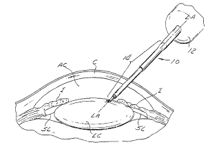
Figure 1 is a perspective view showing an
electrosurgical probe device of the present invention
operatively inserted into a human eye.
Figure 2 is a perspective view of a system of the
present invention comprising a) an electrical signal
generating apparatus, b) a handpiece, and c) an
electrosurgical probe device of the present invention
mounted on the distal end of the handpiece.
Figure 3a is a partial perspective view of a connector
assembly, ~ormed on the distal end o~ the handpiece shown
in Figure 2.
Figure 3 is a rear perspective view of a monopolar
embodiment of the electrosurgical probe device of the
present invention.
Figure 4 is a longitudinal sectional view of the
monopolar electrosurgical probe device shown in Figure 3.
Figure 5 is a perspective view of a segment of
hypotubing from which a notch has been removed to form an
annular electrode tip usable in the electrosurgical probe
devices of the present invention.
Figure 5a is a perspective view of an ann~lar
electrode tip formed from the notched segment of hypotubing
CA 02242328 1998-07-06
W O 98/1961S PCTAUS97/20698
shown in Figure 5.
Figure 6 is a perspective cut-away view of the
monopolar embodiment shown in Figures 3-4.
Figure 7 is a perspective cut-away view of a first
~ipolar embodiment of an electrosurgical probe device of
the present invention.
Figure 8 is a cross sectional view through line 8-8 of
Figure 7.
Figure 9 is a longitudinal sectional view of a second
(alternative) bipolar embodiment of an electrosurgical
probe device of the present invention.
Figure 10 is a cross sectional view through line 10-10
of Figure 9.
Figures lla-llc are graphic representations of power
vs. time, illustrating variation in the electrical wa~e
forms and power levels which may be utilized in conjunction
with the device and system of the present invention.
Detailed DescriPt:ion oE the Prei~e~red Embc--lim~nts
The following detailed description and the
accompanying drawings are provided for the purpose of
illustrating and describing presently preferred embodiments
of the invention only, and are not intended to limit $he
scope of the invention in any way.
With reference to the drawings, there is provided an
electrosurgical probe device 10 which is usable to ~orm
small openings (e.g., less than-3mm) in the anterior lens
capsule LC of the eye. In general, the electrosurgical
probe device 10 comprise6 a proxlmal attachment/contact hub
12 having an elongate probe member 14 extending distally
therefrom, and an annular electrode tip 16 mounted on the
distal end of the elongate pro~e portion 14. The annular
electrode tip 16 is disposed at an angle relative to the
longitl-~; nal axis LA of the elongate probe portion 14.
Figure 1 shows a preferre~ method by which the
electrosurgical probe device 10 of the present invention
CA 02242328 1998-07-06
W O 98119615 PCTrUS97/20698
may be inserted into the eye E for th~ purpose o~ forming
a small te.g., le6s than 3mm) opening in the anterior lens
capsule LC of the eye E. It will be noted from Figure 1
that the lens capsule LC is ~uspended within the eye E,
behind the anterior chamber AC and cornea C. The lens
capsule LC is maintained in its normal anatomical location
by various anchoring structure~ o~ the eye, including
suspensory ligaments S~ which are located posterior to the
iris I of the eye E. In operation, a small incision is
formed in the cornea C and the elongate probe porti~n 14 of
the device lQ is inserted through such incision, and is
advanced through the anterior chamber AC of the eye E until
the annular electrode tip 1~ comes into contact with the
anterior aspect o~ lens capsule ~C. Thereafter, electrical
current is passed through the annular electrode tip 16 so
as to cause the tip 14 to penetrate the lens capsule LC t
thereby forming an annular opening which is approximately
of the same size and configuration as the annular electrode
tip 16.
As the annular electrode tip ~6 elec~rosurgically
penetrates the lens capsule LC, heat will cauterize or melt
the portion of the lens capsule ~C which surrounds the
opening formed by the electrode tip 16, thereby
strengthening or reinforcing the edges of such opening.
Such strengthening or reinforcement of the periphery of the
opening will prevent or deter subsequent tearing, undesired
enlargement or extension of the opening as the cataract
removal and/or lens replacement instruments are passed
~hrough the opening.
With reference to Figure 2-2a, the electrosurgical
probe device 10 of the present invention may be constructed
and configured so as to be usable as an attachment to an
electrocautery system of a type typically used in
ophthalmological surgery. Such electrocau~ery systems
typically comprise a handpiece 18, an electrical signal
CA 02242328 1998-07-06
W O98/19615 PCT~US97/20698
generator 20 and an on/off switch such as a ~oot pedal ~2.
One example of a commercially available electrocautery
system o~ the type shown in Figures 2-2a is the Surgitron~
Model FFPF available from Ellman International, Inc., 113
5 Railroad Avenue, Hewlett, New York 11557.
In the embodiment shown in Figures 2-2a, a connector
assembly 24 is formed on the distal end of the handpiece 18
to facilitate connection with an electrical contact to the
electrosurgical probe device 10 of the present invention.
In the embodiment shown, the connector assembly 24
comprises a generally cylindrical, distally extending
contact post 26 surrounded by an internally threaded,
rotatable, ~uer-lock sleeve 24. The contact post is
insertable into a receiving contact bore ~ormed on the
proximal end of the probe device 10, and the Luer-lock
sleeve is then usable to engage and lock the probe device
10 to the handpiece 18 such that the contact post 26 of
handpiece 18 is held in firm electrical contact with the
probe device 10.
It will be appreciated that the electrosurgical probe
device 10 generally described hereabove, may be
specifically constructed in either monopolar or bipolar
embodiments, as more fully described herebelow:
i. Monopolar Embo~ L~
Figures 3-6 show a monopolar electrosurgical probe
device 10a of the present inventionr which is usable in
conjunction with an electrocautery system of the type shown
in Figures 2-2a.
As shown in Figures 3-6, the monopolar electrosurgical
probe device 10a is constructed such that the annular
electrode tip 16a is the only electrode present on the body
of the probe device 10a. A second electrode, such as a
plate electrode (not shown) r must be separately attached to
or brought into proximity with the body of the patient in
3~ order that an electrical circuit or capacitive coupling be
CA 02242328 1998-07-06
W O 98/19615 PCT~US97/20698
completed ~etween the annular electrode tip }6a and such
externally-placed second electrode (not shown).
As shown, the proximal hub 12a of the monopolar probe
device lOa comprises an electrically conductive, generally
cylindrical, hub 30 having a Leur-lock flange 32 ~ormed
therearound~ An insulative covering 34, formed o~
polyvinyl chloride ~PVC~ or other suitable plastic, may be
formed about the outer surface of the cylindrical hub 30,
but should not interfere with the engagement of the Luer-
lock ~lange 32 to the internal threads of the rotatablesleeve 28 of the handpiece connector assembly 24.
The distally-extending probe portion 14a of the
monopolar probe device lOa may comprise a rigid base tube
36 which is continuous with and protrudes distally from the
~rusto-conical pro~;mal hub 30 as shown. A first hypotube
member 38 is inserted into a distal portion of the base
tube 36, and extends axially therefrom, as shown. A second
hypotu~e member 40 is inserted into a distal portion o~ the
~irst hypotu~e memher 38, and extends distally therefrom,
as shown. The annular electrode tip 16a is formed on the
distal end of this second hypotube member 40.
One means by which the annular electrode tip may be
~ormed on the distal end of the second hypotube member 40
is illustrated in Figures 5 and ~a. With reference to
Figures 5~5a the second hypotube member 40 has a notch 42
cut away therefrom. Such notch 42 is configured such that
its distal edge is perpendicular to the longitudinal axis
LA of the second hypotube member 40, and its proximal edge
is curved or acutely angled relative to such longitudinal
axis LA. This results in the formation of a substantially
cylindrical ring at the distal end of the second hypotube
member 40, such ring being connected by a remnant portion
44 of the second hypotube 40 to that pro~;~l portion of
the second hypotube 40 located proximal to the notch 42.
The distal surface DS of the cylindrical ring formed
CA 02242328 1998-07-06
W O 98/19615 PCT~US97/20698
--10--
at the distal end of the second ~ypotube member 40 is
initially disposed in a plane which is perpendicular to the
longitudinal axis LA. ~owever, the remnant portion 44 is
bent in a direction away ~rom the longitudinal axis ~A such
5 that the plane P of the distal surface of the ring member
at the distal end of the second hypotube member 40 forms an
internal angel A relative to the longit~l~;n~l axis L~.
Also, electrically conductive wire 54 is soldered or
otherwise affixed to the interior of the second hypotube
member 40.
Upon assembly, the pro~;m~l portion of the second
hypotu~e member prepared as shown in Figure 5a, is inserted
into the distal portion o~ the bore of the first hypotube
member 38, and is affixed thereto. Similarly, the first
hypotube member 38 is affixed to the base tube 36 which in
turn is affixed to the frusto-conical proximal hub 30.
Such direct affixation of the hypotube member 38, base tube
36 and frusto-conical proximal hub 30 ma~ be sufficient to
provide reliable electrical contact and conduction
therebetween. However, if reliable electrical contact and
conduction between such components is not accomplished by
their direct a~fixation to one another, an added electrical
conductive wire 54 may optionally be soldered or otherwise
electrically connected to any or all of the hypotube member
2~ 38, base tube 36 and/or prox;~-l hub 30, to facilitate
electrical conduction therebetween.
The angle A of the distal surface DS of the annular
electrode tip 16a of the probe device lOa may vary,
depending on the intended positioning of the
electrosurgical probe device lOa within the eye E.
Typically r it will be desirable to position the
electrosurgical probe device lOa in a manner similar to
that shown in Figure 1. Thus, in most cases, it will be
desirable ~or the distal surface DS of the annular
electrode tip 16a to form an internal angle A of no less
CA 02242328 l998-07-06
W O 98/19615 PCTAUS97/20698
~han 90~ and typically in the range of 90~-1~0~. In the
embodiment shown, a fi:rst insulative sheath 50 is heat-
shrunk or otherwise secured about the proximal portion of
the second hypotube member 40 and the distal portion o~ the
first hypotube member 38. This insulative ~heath 50 helps
to securely join the first 38 and second 40 hypotube
members together and also provides an insulative outer
covering thereon. A second insulative sheath 52 is then
~ormed about the proximal portion of the first insulative
sheath 50 and about the base tube 36.
The monopolar electrosurgical probe lOa is attachable
to the connector assembly 24 of the electrocautery system
shown in Figures 2-2a by inserting the contact post 26 of
the connector assembly 24 into the proximal hub 30a such
1~ that the outer surface of the contact post 26 is in direct
abutting contact with the inner surface of the frusto-
conical hub 30a. Thereafter, the rotatable sleeve 24 is
rotata~ly advanced such that the internal threads of the
rotatable sleeve 34 will engage the Luer-lock flange 32a of
the proximal hub 30. In this manner, electrical current
~rom the electrical signal generator 20 will pass through
the hand piece 18, from the contact post 26 and to the hub
30a of the probe device lOa. Such current will then pass
from the hub 30 through the electrically conductive walls
o~ the first 38 and second 40 hypotube members and/or
through the electrically conductive wire 54. The current
will then pass from the annular electrode tip 16a to a
second elec~rode (not shown) which has been attached to or
brought into proximity with the patient's body to complete
the electrical circuit or establish the required capacitive
coupling.
ii. Bipolar EmboAi ~s
Figure 7-8 show a first embodiment o~ a bipolar
electrosurgical probe lOb of the present invention, while
Figures 9-10 show an alternative bipolar electrosurgical
CA 02242328 1998-07-06
W O 98/1961~ PCTrUS97/20698
-12-
probe lOc which has a structure substantially similar to
(and which shares many common structural attributes with)
that of the monopolar embodiment lOa described hereabove
and shown in Figures 3-6.
With reference to Figur~ 7-8, this bipolar probe
device lQb of the present invention comprises an elongate
probe portion 14b formed of an inner tubular electrode
member 60, an outer tu~ular electrode member 62 and an
insulating tubular she~th 64 positioned therebetween. The
inner tubular electrode 60 member, outer tu~ular electrode
member 62 and insulative tubular sheath 64 are disposed
coaxially about a common longitudinal axis LA. The distal
end o~ the inner tubular electrode 60 forms the annular
electrode tip 16b. In the embodiment shown, this annular
electrode tip 16b is ~ormed ~y cutting the distal end o~
the inner tubular electrode 6Q such that the it's distal
sur~ace is perpendicular (i.e., at a 90~ angle) relative to
the longitudinal axis LA. It will be appreciated, however,
that the dïstal end of the inner tubular electrode 60, the
outer tubular electrode 62 and/or the interposed sheath 64,
may be cut at various angles relative to the longitudinal
axis ~A, so as to provide di~ferent angular dispositions of
the annular electrode tip 16b. Similarlyl the distal end
of the outer electrode member 62 as well as the insulation
sheath 64 may be axially spaced and fastened to one another
such that the respective distal ends of the inner tubular
electrode 60, insulation sheath 64 and outer electrode
members 62 will form such angle.
The elongate probe portion 15b of this first bipolar
probe lOb shown in Figure 7-8 extends distally from and is
connected to a proximal hub (not shown) which may be
substantially the same as the proximal hub 12a described
hereabove with respect to the monopolar probe device lOa.
However, in this bipolar embodiment, only the inner tubular
electrode 60 is electrically connected to the pro~;~l hub,
CA 02242328 1998-07-06
W O 98/19615 PCTrUS97/20698
-13-
a~d the outer tubular electrode 62 is connected separately
by a separate electrical connection to the signal
generating apparatus 20, thereby completing the desired
bipolar circuit of this embodiment
As shown in Figure 7~ the distal end o~ the outer
tubular electrode 62 may terminate a spaced distance
proximal to the distal end of the inner tubular electrode
60. Also, the distal portion of the tubular insulating
sheath 64 which protrudes beyond the distal end of t~e
~uter tubular electrode may be tapered, in the manner sho~n
in Figure 7. In this manner, when the ~ipolar probe device
lOb is inserted into the eye, the distal end of the inner
tubular electrode 60 is positioned in contact with the lens
capsule ~C. ~hereafter, when energized, electrical current
will flow between the distal end of the inner tubular
electrode 60 (which forms the annular electrode tip 16b)
the adjacent distal portion of the outer tubular electrode
62. Thus, in this first bipolar embodiment, there is no
need for a separate external electrode to be attached to or
brought into proximity with the patient's body, as is
required of the above-described monolar polar probe device
lOa.
Figures 9 and ~0 shows an alternative or second
embodiment of a bipolar probe device lOc which is similar
in construction to the monopolar probe lOa described
hereabove. This second embodiment of the bipolar probe lOc
comprises a proximal hub 12c having an elongate probe
portion 14c extending ~ lly therefrom, in a distal
direction. An annular electrode tip 16c is formed on the
distal end of the probe portion 14c. The cylindrical hub
30c, first hypotube member 38c, second hypotu~e mem~er 40c,
remnant portion 44c, annular electrode 16c and electrically
conductive wire 54c are constructed, con~igured and
assembled in the same manner as described hereabove with
respect to the monopolar embodiment.
CA 02242328 l998-07-06
W O 98/19615 PCT~US97/20698
-14-
However, in this second bipolar embodiment, an outer
electrode tube 70, formed of electrically conductive
material, surrounds the insulative sheet 50c. Such outer
electrode tube 70 is connected to an electrically
conductive wire 5~ which e~tends through the insulative
casing 34c of the proximal hub 12c and i8 connectable to
the electrical signal genera~ing device 20. The outer
electrode tube 70 is distally coterminous with the
insulative sheath ~Oc, such that only the remnant portion
44c of the second hypotube member 40c and the annular
electrode tip 16c protrude distally beyond the distal end
of the outer_electrotube 70.
In operation, this second bipolar em~odiment of the
device lOc is inserted into ~he eye such that a distal
portion o~ the probe portion 14c extends thro~gh the
anterior chamber AC, and the distal surface DS of the
annular electrode tip 16c is in contact with the anterior
lens capsule. ~hereafter, electrical current from the
electrical signal generator 20 may pass through the
electrically conductive wire members 54c, 55 and/or other
electrically conductive portions of the pro~e de~ice lOc as
described hereabove, such that current will ~low ~rom the
annular electrode tip 16c to the distal portion of the
outer tubular electrode 40 through the electrically-
conductive fluid environment within the anterior chamber ofthe eye.
It will be appreciated that the annular electrode tip
16, 16a, 16b, 16c described hereabove may comprise any
appropriate geometrical configuration, and may have an open
center (e.g., a ring or hoop) or alternatively may have a
solid center (e.g., a disc having a generally annular outer
edge). Furthermore, it will be appreciated that the lens
capsule contacting surface, such as the distal surface, of
the annular electrode tip 16, 16a, 16~, 16c need not be
substantially flat or planar, and may be slightly concave
CA 02242328 1998-07-06
- W O 98/19615 PCT~US97/20698
or of any other suitable configuration. In this manner,
when reference is made in thi~ patent application to the
plane" in which the lens capsule contacting sur~ace of the
distal electrode tip lie, it will be appreciated that such
plane may ~e projected throu~h a concaved or wavy surface
of an average variant thereo~. ~lternatively, in
embodiments wherein the lens capsule contacting surface of
the annular electrode tip 16, 16a, 16b, 16c is flat, such
entire ~lat edge may lie within the referenced plane.
iii. Preferred Methods of OPeratin~ the Devices
Any and all of the above-described embodiments of the
present invention are preferably operated in accordance
with a general method wherein at least a distal portion of
the elongate probe porti~n 14, 14a, 14b, 14c is inserted
1~ through an incision in the cornea C and is advanced through
the anterior chamber AC until the lens-capsule-contacting
distal surface DS of the annular electrode tip 16, 16a,
16b, 16c is in contact with the lens capsule LC. If a
monopolar embodiment of the probe device 10a is used a
secondary electrode will be attached to or brought into
close proximity with the body of the patient at a location
which is suitable to complete an electrical circuit between
the annular electrode tip 16a of the probe 10a and such
second electrode. On the other hand, i~ one of the bipolar
embodiments of the probe device 10b, 10c are used, there
will be no need to provide a separate second electrode wit~
is attached to or placed in proximity with, the body of the
patient.
Therea~ter, the electrical signal generating device 20
is actuated so as to cause current to flow between the
annular electrode tip 16, 16a, 16b, 16c and either the
separately attached secondary electrode (monopolar
embodiment) or the on-board outer electrode tube 62, 70.
Any suitable electrical wave ~orm and power level may
be used. In this regard, in at least some applications it
CA 02242328 1998-07-06
W O 98/19615 PCTrUS97/20698
-16-
will be desirable to use a continuous, pulsed or
superpulsed wave form which pro~ides an average power level
of approximately 10 watts to form the desired opening in
the anterior lens capsule.
In embodiments of the system wherein the wave form is
intended to be pulsed or superpulsed, the signal-generating
device 20 will preferably include a mechanism for setting
~ the desired pulse duration, pulse train duration, pulse
bunch duration and/or duty cycle, examples of which are
shown graphically in Figures lla, llb and llc.
With re~erence to lla, there is shown the average
power generated by single pulse, of known pulse duration
PD.
Figure llb shows the average power generated by a
train of individual pulses, each of said individual pulses
having a pulse duration PDr and the overall train of pulses
having a pulse train duration PTD.
Figure llc shows a superpulsed embodiment of the
invention wherein bunches of small individual pulses, each
of ~aid individual pulses having a pulse duration PD of 10
milliseconds, are generated periodically on a given repeat
period RP.
It will be appreciated that the electrical signal
generating device 20 may be preprogrammed to deliver
2~ desired energy levelsr and/or wave ~orm(s) in response to
each triggering of a fixed on-off switch. Alternatively,
the signal generating device 20 may be rheostatically
controlled by way of a ~oot pedal or other type of
rheostatic control device r and the amount and duration of
energy delivered through the annular electrode tip 16, 16a,
16b, 16c will be determined by the current position of the
foot pedal or other rheostatic control mechanism.
In the above-described manner, the electrosurgical
probe device lO of the present invention is ~sa~le to form
an opening in the lens capsule LC of a size which is only
CA 02242328 1998-07-06
W O98/1961~ PCT~US97120698
slightly larger than the outer diameter of the annular
electrode tip 16, 16a, 16b, 16c. Furthermore, when the
preferred wave form and power setting are used, the
resultant electrosurgical opening of the lens capsule will
additionally form a heat-fused region around such openiny,
thereby preventing the anterior aspect of the lens capsule
~rom being torn, enlarged or extended during the subsequent
insertion and manipulation of the cataract removal
device(s) and/or prosthetic lens implant introduction
cannula.
The present invention has been described hereabove
with reference to certain presently prefèrred em~odiments
only. No attempt has been made to exhaustively describe
all possible embodiments in which the invention may be
practiced. Indeed, various additions, deletions,
modifications and alterations may be made to the above-
described pre~erred embodiments without departing from the
intended spirit and scope of the invention. Accordingly,
it is intended that all such reasonable additions,
deletions, modifications and alterations be included within
the scope of the following claims.