Note: Descriptions are shown in the official language in which they were submitted.
CA 02243649 1998-07-20
WO 97/29778 PCT/CA97/00089
BIORESPONSIVE PHARMACOLOGICALLY-ACTIVE
POLYMERS AND ARTICLES MADE THEREFROM
FIELD OF THE INVENTION
This invention relates to pharrrlacologically-active polymeric compounds; to
substrates, such as implantable medical devices formed thereof or coated
therewith; and
to methods of using said compounds or said substrates for providing
pharmacological
agents at a desired location in a mammal.
BACKGROUND TO THE INVENTION
Medical devices, may be classified in two major groups: 1) total implantable
e.g.
artificial joints, heart valves, and the like; and 2) access devices
associated with an exit
site, hereinafter termed "AD", which may be further divided into a) those
which exit
throu(yh an orifice, e.g. urinary catheters, endotracheal tubes, and the like,
and b) those
which cross exit transcutaneously, e.g. venous access devices, peritoneal
dialysis
catheters, and the like.
Devices associated with an exit site are colonized by bacteria in a time-
dependent
fashion, which effectively limits their long-term use. Colonization of these
devices may
occur through two routes; 1. the intraluminal route, acquired by improper
aseptic
technique; 2. the extraluminal route, following colonization of the exit site
and the
subcutaneous tunnel, bacteria are transported along the sinus which forms
between the
catheter and the host tissue. Intraluminal contamination has been
significantly reduced
for most applications through improved connectors and aseptic handling
techniques (1,
2). ln contrast, limited progress has been achieved in protecting the exit
site from
bacterial colonization and subsequent infection (3).
SUBSTITUTE SHEET (RULE 26)
CA 02243649 1998-07-20
WO 97/29778 PCT/CA97/00089
2
Approximately 40% of all hospital-acquired infections are related to the
urinary
tract (4), and urinary tract infections (UTI) are among the most common
factors leading to
life-threatening Gram negative sepsis (5). Currently, the commonest route of
colonization
for indwelling urinary catheters is via the periurethral space. Bacteria
colonizing the
perineum and permimeatal region adhere to the catheter extraluminal surfaces,
then
ascend along the catheter in the periurethral space. Even with the use of
closed sterile
urinary drainage systems, the risk of urinary catheter associated UTI remains
in excess of
5% per day of catheterization (6).
Infections associated with percutaneous access devices (PAD) account for the
second greatest source of nosocomial bacterial infections. Among the devices
in this
category are peritoneal dialysis catheters, central venous catheter lines for
total parenteral
nutrition, and chemotherapy venous access catheters. All of these devices are
employed
over extended periods of time, ranging from several weeks to two or more
years.
Endogenous organisms (e.g., Staphylococeus epidermidis) are the most common
bacteria
recovered during episodes of peritonitis (7, 8). The addition of a prosthetic
device to the
host increases the risk of infection four-fold (9). In the case of
peritonitis, patients may
require hospital admission for treatment and in approximately 20% of the
patients surgery
is required to remove the dialysis catheter if the infection is resistant to
antibiotic therapy
or is rapidly recurrent after cessation of therapy (10).
Classically, PAD catheters are fabricated from smooth latex, silicone rubbers,
or
polyurethanes. These materials do not allow the skin epithelial cells to
permanently
adhere to them either mechanically or chemically. Following implantation of a
PAD
catheter, the epidermal cells begin to migrate, each seeking to surround
themselves
completely with other epidermal cells. Under normal wound healing
circumstances, the
granulation tissue, which forms near the skin surface, provides an ideal bed
over which
these cells can migrate. The presence of a PAD catheter across the skin at the
exit site
prevents contact of the epidermal cells with sister cells This results in the
inward
migration of the epidermal cells towards the subcutaneous tissues and the
development of
a wet sinus tract between the surface of the skin and the tubing. Necrotic
epidermal cells
and keratin line the sinus tract creating an ideal environment for microbial
colonization.
Organisms colonizing the exit site sinus spread along the external surface of
the catheter
SUBSTITUTE SHEET (RULE 26)
CA 02243649 2008-02-29
3
forming an adherent biofilm (11). DacronT"' cuffs are usually employed to
prevent
dislodgement of the catheter, and act as a "biological barrier" to the ingress
of bacterial
cells along the exit site sinus. It is the failure of host tissue integration
with the DacronTM
cuff prior to bacterial colonization which leads to exit site-tunnel
infections. Gristina
(12) has described this situation as a "race to the surface". That is, if host
tissue is
allowed to integraate with the implanted device before bacteria are able to
adhere,
infection does not usually occur. This integration process can require several
weeks
following device implantation.
Current treatment modalities (local and systemic) are frequently associated
with a
high rate of re-infection. For reasons that are poorly understood, bacteria
associated with
foreign bodies can be 100 times or more resistant to systemically applied
antimicrobials
than are their planktonic (free-floating) counterparts (13, 14).
Silver coated catheters have been used to prevent exit site infections
associated
with chronic venous access (15) and peritoneal dialysis (16) catheters.
However, long-
term studies have failed to demonstrate a significant reduction in the number
or severity
of exit site infections. In addition, bacterial resistance to silver can
develop over time and
carries with it the risk of multiple antibiotic resistance (17).
Various antibiotics have also been used to coat the surfaces of catheters
through
non-covalent bonding. Trooskin et al. (18, 19) describes a method by which
catheter
surfaces were soaked in antibiotic solutions prior to their implantation.
Duran et al. (20)
covalently bonded a photoactivated hydrogel onto the surface of silicone
materials. The
antibiotic vancomycin was then immobilized within the hydrogel matrix. In both
of the
above studies, most of the antibiotic was essentially released over several
days rather than
the requisite efficacy of several weeks. The antibiotic ciprofloxacin has been
impregnated into DacronTM fibres using a textile pad/heat technique which
binds the drug
to the fibre by non-covalent interactions. After 24 hours of exposure to a
phosphate
buffer washing solution, more than 80% of the drug was released from the
fibres (21).
Exit site infections can only be controlled when bacterial colonization is
prevented for an
extended period of weeks to enable complete host tissue integration.
In addition to the traditional diffusion-controlled delivery systems described
by
Duran (20), there exist several more sophisticated in situ drug delivery
polymers which
CA 02243649 1998-07-20
WO 97/29778 PCT/CA97/00089
4
can alter the efficacy of drugs by improving target delivery and changing
controlling
parameters of the delivery rate. These include polymeric liposomes (22,23),
bioadhesives
(24,25), bioerodible polymers (26,27), chemical and physical stimuli
responsive polymers
(28,29) and polymer drugs (30,31). Applications of these materials have
included the
delivery of antitumor drugs in cancer therapy (30), insulin for diabetics (29)
and
antimicrobial drugs (gentamicin) for vascular grafts (32). In studies on
vascular grafts,
Karch et al. (32) combined gentamicin non-covalently with a fibrin sealant,
which was
then coated onto Dacron surfaces. This bioerodable system takes advantage of
the
degradative features of the biopolymer, fibrin, to release the gentamicin. The
degradation
process depends on the hydrolysis of amide bonds and dissolution of the fibrin
network to
accelerate physical release of the diffusion drug. Following implantation in a
porcine
model, gentamicin release was elevated for the first few days, but decreased
significantly
shortly thereafter. In addition, over 50% of the specimens containing the
fibrin-
gentamicin matrix were found to be infected upon retrieval. To-date, polymer-
based
antimicrobial delivery systems have failed to demonstrate in vitro or in vivo
efficacy over
extended periods of time.
Many sessile and sedentary plants and animals have hard, non-desquamanating
surfaces and are therefore subject to the same biofouling pressures as are
engineering
surfaces. The "natural" antifouling properties associated with some of these
non-fouling
surfaces have been the subject of several research programs sponsored by the
U.S. Navy
and other organizations affected by biofouling activities. Extracts from
Gorgonian coral
(33,34), eel grass (35), and marine sponges (36) have all been employed as so-
called
natural antifoulants in marine coating formulations.
Although one or more of these compounds may hold promise as antifouling
agents, perhaps with applications as biomaterial additives, natural compounds
suffer from
three major disadvantages in this regard, viz, 1) they are often available
only in limited
quantities; 2) the compounds are frequently difficult to synthesize de novo,
and possess
multiple chiral centers; and 3) their range of application is often limited
both in terms of
species selectivity and environmental conditions. These same considerations
should be
applied to emerging antimicrobial coatings applied to biomaterial surfaces.
Although the
concept of a "natural products" antimicrobial may hold an aesthetic appeal,
there is no
SUBSTITUTE SHEET (RULE 26)
CA 02243649 2008-02-29
evidence that these compounds are either safer or more effective than those
synthesized
de novo.
Short-term studies with silver-sulphadiazine- and chiorhexidine-coated
polyurethane vascular-access catheters showed a reduction in the incidence of
exit site
5 infections in a rat model (37). Fewer IV injected S. epidermidis cells
adhered to
rifampicin-treated than to untreatedDacronT" vascular grafts in sheep (38).
Although the
results of these in vivo animal studies were promising, they were all
conducted over
relatively short periods of time following the introduction of the
biomaterial.
Biomaterial-related infections can only be controUed when bacterial
colonization is
prevented for an extended period of time (weeks), enabling complete host
tissue
integration.
The bisbiguanide, chlorhexidine has shown good efficacy against surface-
associated bacteria in oral environments. Chlorhexidine has a broad spectrum
of activity
against Gram-positive and Gram-negative bacteria as well as a variety of
fungi, including
Candida spp. In addition to its bacteriostatic and bactericidal properties,
chlorhexidine
tends to bind very avidly to mucosal tissues, tooth surfaces, and dental
implant materials
(39). Chlorhexidine coatings on dental implant surfaces have demonstrated
excellent
short-term in vivo activity against S. mutans, Porahvromonas gingivalis, and
other dental
pathogens (37). In addition, local tissue integration with the implants is not
adversely
affected by the presence of this compound (40).
Macromolecules containing nalidixic acid and its structural analogue
quinolones
as pendent moieties are known (56,57).
A treatment strategy which significantly reduces the rate of urinary catheter
bacterial colonization would reduce the incidence of urinary tract infections
and
associated sequellae. In the case of an AD, infections can only be controlled
when
bacterial colonization is prevented for an extended period of time (weeks),
enabling
complete host tissue integration as was described above.
However, there remains a serious need to prevent and control exit site
bacterial
infections over extended periods of time.
CA 02243649 2006-04-05
WO 97/29778 PCTlC.A.97/00089
6
REFERENCE LIST
The present specification refers to the following publications,
1_ Buxkart J.M. 1988. ASAIQ-Trans. 34:433-436.
2. Churchill D.N., Taylor D.W., Vas S.I. & Oreopoulos D.G. 1989.. Perit.Dial
lnt.
9:159.
3_ Piraino G_, Berzzaz-dini J. & Sorkira. M. 1987_ Arrx.J.Kidney Dis_ 10:281-
286.
4_ Kunizz C.M. 1987 iua Detection, prevention and management of uutiaiary
tract
infections. Lea & Febiger, Philadelphia, pp.245-297.
5_ Waren J.W_, Platt R., Thomas R.J., Rosner B. and Kass E.H. 1978.
N.Engl.J.Med. 299: 570-573.
6. Khoury A.E., Lam K., Ellis B. and Costerton J.W., 1992. ASAJO J. 38:174-
I78_
7_ S.C.H. Division 1989. Canadian Peritonitis Registry. Statistics Canada,
Ottawa.
8_ Williams P.S., HendyM.S. and Ackrill P. 1987. Perit.Dial. Bull. 7:183-186.
9. Christensen G.D., Baddour L.M., Hasty D.L., Lowrance G.H_ & Simpson W.A.
1989. In: Bisno A.L. & F.A. Waldvogel ed. Infections associated Nvith
indwelling
medical devices.American Society for Microbiology, Washin,gton,D.C_pp.59-70.
10_ Holmes C.J. & Evans R. 1986. Perit. Dialy. Bul16:168-I77.
11. Read R.R., Eberwein P., Dasgupta M.K., Grant S.K., Lam K_, Nickel J.C_ &
Costerton J.W., 1989. Kid. Interzxat. 35:614-621.
12_ Gristina A., 1987, Science, 237: 1588-1597
13. Hoyle B,.D., et.al., 1990. J.Antimicrob. chemother. 26:1-5.
14. Khoury A.E., et.al.,1993. Proceedings of 18'h Izatemational Congress of
Chemotherapy, Stockholm.
15. Groeger J.S., et.al., 1993. Ann. Surg. 218:206-210.
16. Mittelman M.W., et.al., 1994. Ann. Corx Peritoneal Dialysis, Orlando. Fl_
17. Silver S. et al., 1988. Ann. Rev. Microbiol. 42:717-743.
18. Trooskin S.Z., Donetz A.P., Baxter J., Harvey R.A. & Greco R.S. 1987.
Nephron.46:263-267.
19_ Trooskin S.Z., Donetz A.P., Harvey R.A. & Greco R.S. 1985. Surgery, 97=547-
551.
SuBSTiT[ fTE SHEET (RULE 26)
CA 02243649 1998-07-20
WO 97/29778 PCT/CA97/00089
7
20. Duran L.W., Marcy J.A. & Josephson M.W. 1992. Surfaces in Biomaterials,
Minneapolis, 37-41.
21. Phanluf, M.D. 1993. J. Biomed.Mater.Res., 27, 233-237.
22. Stefely J.S., Markowitz M. A. & Regen S.L. 1988. J.Am.Chem.Soc. 110:7463.
23. Szoka F.C. & Papahadjopoulos D. 1981. In: Knight C.G., ed. Elsevier,
Amsterdam, pp. 51.
24. Hui H.W. & Robinson J.R. 1985. Int.J. Engi. Ed. August: 196.
25. Longer M.A., Ch'ng H.S. & Robinson J.R. 1985. J.Pharm.Sci. 74:406.
26. Heller J. 1988. J. Controlled Release, 8:111.
27. Mathiowitz E., Ron E., Mathiowitz G. & Langer R. 1989. Polym.Prepr.30:460.
28. Hsieh D.S. & Langer R. 1983. In; Roseman T.J. & Mansdorf S.Z. ed. Marcel
Dekker, New York.
29. Makino K.E., Okano Mark T. & Kim S.W. 1990. J. Controlled Release, 12:235.
30. Ouchi T., Kobayshi H. & Banda T. 1990. Brit. Polym. J. 23:22 1.
31. Takemoto K. & lnaki Y. 1988. Acta. Polym. 39:33.
32. Karch M., Forgione L. & Haverich A. 1993. Clin. Mat. 13:149-154.
33. Keifer, P.A., Rhinehart K.L. & Hooper, I.R. 1986. J.Org.Chem. 5:4450-4454.
34. Vrolijk, N.H., Targett N.M., Baier R.E. & Meyer A.E. 1990. Biofouling.
2:39-54
35. Harrison, P.G. & Chan A.T. 1980. Mar.Biol. 61:21-26
36. Sears M.A., Gearhart D.J. & Tittschof D. 1990. J.Chem.Ecol. 16:791-799
37. Bach A., Bohrer H., Motsch J., Martin E., Geiss H.K. & Sonntag H.G. 1994.
J.Antimicrob.Chemother. 33:969-978
38. D'Addato M., Curti T., Freyrie A., Agus G.B., Bertini D. 1994.
Cardiovasc.Surg.
2:254-258.
39. Sodhi R.N.S., Grad H.A. & Smith D.C. 1992. J.Dent.Res. 71:1493-1497
40. Burchard W.B., Cobb C.M., Drisko C.L. & Killoy W.J. 1991. 6:418-426.
41. Remes A. & Williams D.f. 1992. Biomater. 13:731.
42. Labow R.S.. et ai., 1994. J. Biomater. Sci. Polym. Edn., 6, 169-179.
43. Santerre J.P., et al., 1993. J.Biomed.Mater.Res. 27, 97-109.
44. Labow R.S., Erfle D.J., Santerre J.P. 1996. Biomaterials, 17, 2381-2388
45. Santerre J.P. et al.. 1994. J.Biomed.Mater.Res., 28:1187-1199.
SUBSTITUTE SHEET (RULE 26)
CA 02243649 2008-02-29
8
46. Santerre J.P., Labow R.S. 1997, J.Biomed.Mater.Res. 33, in press.
47. Wang F.G.B., Santerre J.P., Labow R.S. 1997, J.Biomed.Mater.Res. 33, in
press.
48. Khoury A.E., Nicholov R., Soltes S., Bruce A.W., Reid G. & DiCosmo F.
1992.
Int.Biodeterior. Biodegrad. 30:187-199.
49. Mittelman M.W., et al. 1992. Biofouling 6:39-5 1.
50. Mittelman M.W., Nivens D.E., Low C. & White d.c. 1990. Microb.Ecol. 19:269-
278.
51. Rodriguez G.G., Phippps D., Ishiguro K. & Ridgway H.F. 1992. Appl.
Environ.
Microbiol. 58:1801-1808.
52. Vestal J.R. & White D.C. 1989. Bioscience. 39:535-541.
53. Olsen M.W., Nickel J.C., Khoury A.E., Morck D.W., Cleeland R. and
Costerton
J.W., 1989, J.Infect. Dis. 159: 1065-1072.
54. Fung H.C., Khoury A.E., Oreopoulis D., Vas S. and Mittelman M.W. 1996,
Peritoneal Dial. Int. 16:380-388.
55. Fung H.C., Mittelman M.W., Thorner P.S. and Khoury A.E., 1996. Urology
(submitted).
56. Ghosh M. Progress in Biomedical Polymers, ed. Gebekin C.G. and Dunn R.L.,
Plenum Press, New York, 1990, pp.335-345.
57. Ghosh M. Polymeric Materials, Science & Engineering (1988), 59, pp.790-
793.
CA 02243649 2008-02-29
8a
SUMMARY OF THE INVENTION
Various embodiments of this invention provide a pharmacologically-active
polymeric
material having a polystyrene equivalent molecular weight selected from 2,000 -
200,000 and
a backbone comprising a pharmacologically-active fragment formed from a
fluoroquinolone
covalently linked through two functional groups via amide, urea, urethane, or
sulfonamide
linkages to said backbone and forming a polyamide, polyurea, polyurethane or
polysulfonamide within said backbone. The fluoroquinoline may be ciprofloxacin
(1-
cyclopropyl-6-fluoro-1,4-dihydro-4-oxo-7-piperazine-quinolone-3-carboxylic
acid).
Other embodiments of this invention provide a solid substrate comprising in
whole or
in part, a pharmacologically active polymeric material of this invention.
Other embodiments of this invention provide a solid substrate wholly or
partially
coated with a pharmacologically active polymeric material of this invention.
Other embodiments of this invention provide use of a polymeric material of
this
invention as a antibacterial agent.
It is an object of the present invention to provide polymeric compounds which
reduce the incidence of infection, due to the presence of access devices,
particularly exit
site infections in transmucosal devices and percutaneous access devices. It is
a further
object to provide access devices either coated with said pharmacologically-
active
polymeric compound or formed in whole or in part of pharmacologically-active
said
polymeric compound.
It is a yet further object to provide a method of reducing the incidence of
infection
due to the presence of access devices, particularly, of exit site infections
associated with
percutaneous access devices.
CA 02243649 1998-07-20
WO 97/29778 PCT/CA97/00089
9
Accordingly, in its broadest aspect the invention provides a bioresponsive
pharmacologically-active polymeric material having a backbone comprising a
pharmacologically-active fragment covalently linked through at least two
functional
groups within said backbone. By the term "pharmacologically-active fragment"
is meant
a chemical radical, group, entity and the like, which provides a
pharmacologically-active
compound by in vivo biochemical action, such as, enzymatic and non-enzymatic
hydrolysis or oxidation upon the bioresponsive polymeric material. The
fragment per se
within the polymeric material may not be pharmacologically-active.
Preferably, the pharmacologically-active fragment is covalently, divalently-
linked
within the polymeric material, and in such compounds it would not be deemed to
be a
pendant fragment. However, additional linkages, covalent or otherwise of the
fragment
either to the polymer backbone or discrete radicals, groups, entities and the
like are within
the scope of the present invention. Thus, the drug has at least two or more
reactive
groups selected from hydroxyl, amine, carboxylic acid or sulfonic acid. If it
is desired to
form the polymeric material as a linear polymer the pharmacologically-active
compound
is only divalently covalently linked.
The polymeric backbone comprises one or more polyurea, polyurethane,
polysulphonamide or polyamide linkages, optionally having one or more
polyvinyl,
polyester, polycarbonate or polyether linkages.
The invention is of particular value to those pharmacologically active
compounds
which are bioresponsive as hereinabove defined to provide in vivo a
pharmacological
active ingredient which has at least two functional groups capable of reacting
with a
diisocyanate to form amide, urea and or urethane linkages, such as the
fluoroquinolone
family of antibiotics.
The in vivo pharmacological activity generated may be, for example, anti-
inflanunatory, anti-bacterial, anti-microbial, anti-fungal, but this invention
is not limited
to such biological activities.
The present invention is of particular use wherein the pharmacologically-
active
fragment is formed from the antibacterial 7-amino-l-cyclopropyl-4-oxo-1-4-
dihydro-
quinoline and naphthyridine-3-carboxylic acids described in United States
Patent No.
4,670.444 - which issued to Groke et al, June 2, 1987. The most preferred
antibacterial
SUBSTITUTE SHEET (RULE 26)
CA 02243649 1998-07-20
WO 97/29778 PCT/CA97/00089
member of these classes of compounds is 1-cyclopropyl-6-fluoro-1,4-dihydro-4-
oxo-7-
piperazine-quinoline-3-carboxylic acid, having the generic name ciprofloxacin.
Others of
this class include ofloxacin and norfloxacin.
The polymeric material of use in the practice of the invention may be linear
or
5 comprise a series of crosslinked polymer chains. The polystyrene equivalent
molecular
weight of the chains may range from 2,000 to 200,000, preferably 10,000 to
100,000.
The present invention provides mammalian host-responsive, bioerodable
polymers which deliver sustained effective amounts of antibiotics to target
tissue over
desired extended periods of time. Thus, a polymer according to the invention
in the
10 biological environment of host tissue and the like, in one aspect, is
subjected to released
hydrolytic enzymes and oxidative species under, and in proportion to, the
host's
inflammatory response. This results in release of the active ingredient via
the breaking of
the covalent linked bonds. Thus, the materials of the invention utilize the
mammal's own
wound healing repair process in being degraded thereby as hereinbefore
described.
In a further aspect, the invention provides a substrate coated with or
comprising in
whole or in part a bioresponsive pharmacologically-active polymeric material
as
hereinbefore defined.
The substrate may be an access device such as a catheter, so coated or formed
with the polymeric material which provides sustained delivery of the
antimicrobial agent
during the critical tissue integration period. The substrate may be formed of
a plastics
material, glass or other suitable carrier for the pharmacologically-active
polymeric
material. Preferably, the substrate is a silicone or polyurethane material,
which,
preferably, has coated on a surface thereof the pharmacologically-active
polymeric
material to provide a so-called antimicrobial composite material (ACM).
Examples of such substrates according to the present invention includes,
cardiac
assist devices; cardiac replacement devices; cardiac septal patches; vascular
grafts; intra-
aortic balloons; percutaneous cardiac assist devices; extracorporeal circuits;
A-V fistulas;
dialysis components; aphoresis components; membrance oxygenation components;
cardiac bypass components; pericardial sacs; contact lenses; cochleal ear
implants;
artificial skin; sutures; sewing rings; cannulas; separating agent;
contraceptive devices;
syringes; 0-rings; bladders; penile implants; drug delivery components;
drainage tul- ;
SUBSTITUTE SHEET (RULE 26)
CA 02243649 1998-07-20
WO 97/29778 PCT/CA97/00089
11
pacemaker leads; coatings for implantable wires; coating for eye glasses;
urethral
catheters; peritoneal dialysis catheters; CSF shunts; orthopedic implants;
venous and
arterial access catheters; dental implants; blood collection bags; vascular
stents;
angioplasty devices; laryngeal voice box implants and wound dressings.
In addition, as foreign objects in the host tissue, these materials initiate
the
inflammatory response. Phagocytic white blood cells, such as neutrophils and
monocyte-
derived macrophages and remodeling cells such as fibroblasts have the ability
to migrate
towards and strongly adhere to the surface of biomedical implants (41). Once
at the
wound site, activated neutrophils and macrophages strongly attach to the
surface of the
implant in an attempt to engulf it. This response can be broadly divided into
non-specific
and specific protective mechanisms. One of the non-specific reactions is the
synthesis
and secretion of enzymes and high-energy oxygen radicals by phagocytic cells.
Groups of
enzymes released during these processes include esterases, proteinases,
phospholipases
and hydrolases.
Recent studies on the mechanism of polyurethane biodegradation by Santerre and
his colleagues have identified esterase and proteinase enzymes that are
released by
neutrophils and monocyte-derived macrophages and which can activate the
hydrolytic
degradation of polyurethanes (42,43,44). Specifically, it has been shown that
the kinetic
reaction of an enzyme with polymer can proceed through the cholesterol
esterase's active
serine site 42 and either at ester or amide type linkages, including urea and
urethane sites
of the polymer (45-47). Enzymatic degradation processes are limited by the
polymer
chain chemistry, internal material structure, and the surface area of the
device (45,46). By
altering these properties, the rates of biodegradation for the materials can
be varied.
BRIEF D)1SCRIPTION OF THE DRAWINGS
In order that the invention may be better understood, preferred embodiments
will
now be described by way of example, only, wherein:
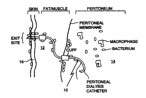
Fig. 1 shows a diagrammatic representation of a catheter according to the
invention
located within human tissue;
SUBSTTTUTE SHEET (RULE 26)
CA 02243649 1998-07-20
WO 97/29778 PCT/CA97/00089
12
Fig. 2 shows a diagrammatic representation of a standard peritoneal dialysis
catheter with
a coating of a bioresponsive pharmacologically-active polymer according to the
invention;
Fig. 3 shows a diagrammatic representation of a peritoneal dialysis catheter
made of
bioresponsive pharmacologically-active polymer;
Fig. 4 shows a diagrammatic representation of a vascular graft spun on a
mandrel with
bioresponsive pharmacologically-active polymer.
Fig. 5 shows a diagrammatic representation of a fibre according to the
invention;
Fig. 6 shows a diagrammatic representation of a tape according to the
invention;
Fig 7 shows a diagrammatic representation of a filter membrane according to
the
invention; and
Fig. 8 shows a diagrammatic representation of a tube insert according to the
invention.
Figs. 9 - 17, and 19-24 show gel permeation chromatograms (GPC), high
performance
liquid chromatograms (HPLC), or UV spectral charts; and
Fig. 18 is a chart showing ciprofloxacin release before and after an
incubation period for
various systems.
DETAILED DESCRIPTION OF PREFERRED EMBODIlVIENTS
Fig. 1 shows generally as 10 a peritoneal dialysis catheter embedded in the
fat/muscle 12 and peritoneum 14 through the skin 16. Catheter has a surface
coating of a
bioresponsive pharmacologically-active polymer incorporating ciprofloxacin.
Figs. 2 and 3 show a standard peritoneal dialysis catheter 20 with a coating
of
bioresponsive pharmacologically-active polymer and a peritoneal dialysis
catheter 30
made of bioresponsive pharmacologically-active polymer, respectively.
Fig. 4 shows a vascular graft 40 spun on a mandrel with bioresponsive
pharmacologically-active polymer.
Fig. 5 shows a reel or bobbin 50 holding a thread, fibre or dental floss 52
formed
of a bioresponsive pharmacologically-active polymer for use as an insert into
body areas,
which areas may be particularly susceptible to bacterial growth, such as
periodontal
pockets of the oral cavity; or as use as a suture.
SUBSTITUTE SHEET (RULE 26)
CA 02243649 1998-07-20
WO 97/29778 PCT/CA97/00089
13
Fig. 6 shows a reel or dispenser 60 holding a tape 62 made from a
bioresponsive
pharmacologically-active polymer for use as an insert into body areas which
are
particularly susceptible to bacterial growth, i.e. between toes, to release
anti-fungal agents
against athletes foot. Another example of a use for such a tape is use as a
seal for screw
devices, such as a dental implant, wherein bacteria often migrate into the
implant along
the screw hole. The tape provides both a seal between the screw and the screw
hole,
while simultaneously providing antimicrobial activity.
Fig. 7 shows a filter or other membrane 70 formed of a bioresponsive
pharmacologically-active polymer for use in sterilizing analytical solutions
or body fluids,
various blood products during preparation thereof, such as intravenous fluids
and the like.
Another example of use of the membrane is as a wound healing patch, which
requires air
and fluid to pass through the membrane of the patch, but which also needs
continuous
treatment of antimicrobial agents to eliminate bacteria from the open wounds.
Fig. 8 shows a bioresponsive pharmacologically-active polymer formed of an
insert 80 into body areas, which areas are particularly susceptible to
bacterial growth, i.e.
ear tubes 80 used for drainage of fluids from the ear canal.
Experimental Methods
Examples of diisocyanates of use in the practice of the present invention are
1,6,diisocyanatohexane and 1,12-diisocyanatododecane, which can be reacted
with the
antimicrobial agent, ciprofloxacin, with or without oligomeric molecules to
form
polymeric materials of the invention. These pharmacological compounds have
prolonged
efficacious activity against access device-related bacteria. The different
chain lengths of
these two diisocyanates allow for varying material structure to permit
tailoring of
biodegradation rates. Thus, biodegradable or bioerodable polymers incorporates
selected
antimicrobial drugs as monomer units by reaction with the diisocyanates. The
molecular
chains and material morphology are such that when the inflammatory response in
the
tissue of the mammal is turned on, subsequently, by upregulation of the
inflammatory
response as a result of device implantation or other inducers of inflanunation
(e.g.,
bacterial or fungal infection), the polymeric material is biologically and
specifically
enzymatically, degraded to release the antimicrobial drug from the polymer
chain.
SUBSTITUTE SHEET (RULE 26)
CA 02243649 1998-07-20
WO 97/29778 PCT/CA97/00089
14
Release of antimicrobials proceed at effective rates until the levels of
released hydrolytic
enzymes are significantly lowered as a result of the diminished host response
associated
with tissue healing. Diagrammatic rendering of the synthesis and execution of
the
bioresponsive pharmacologically-active polymers, may be represented, thus:-
10
R~ Con Phm- T:mam t
OGY.{Cfiyn-NCO HOOC DRIJ+G -~ COOH _
H2N NH2 PAD TubiAit
P. -t',JWC
ocx.~ccs~RUC-Kc~y.-xc~
ACM
PA5Xjbine..
espoame co mzymadc wtiwoa
+ba=NW adw
~P~fioa'a~
-4DM "~ =dmiaobial actirity
The antimicrobial polymers are synthesized in, for example, either pre-dried
toluene, tetrahydrofuran, dimethylsulfoxide or other such solvents, depending
on which is
SUBSTITUTE SHEET (RULE 26)
CA 02243649 1998-07-20
WO 97/29778 PCTICA97/00089
most appropriate based on yield and the desired polymer molecular weight. A
typical
bioresponsive pharmacologically-active polymer made from 1,6
diisocyanatohexane and
ciprofloxacin is shown, thus:-
5
'I'Cd3Cz3QiCX2CN:CH2
L n
10 The stoichiometry of the polymer components is varied depending on the
desired
structure, drug activity and the method of application onto AD tube surfaces.
If the
antimicrobial polymer is applied to polyurethane surfaces as a coating or by
solvent
bonding, then the stoichiometry is such as to favor terminal antimicrobial
drug monomers
i.e. no free diisocyanate. Quenching of remaining free diisocyanates can be
carried out
15 with traces of inethanol.
If the AD tube material (polyurethane or silicone) requires pre-activation
with
carboxylic acid, amine or hydroxyl groups using gas plasma or other known
surface
activation methods, to ensure a strong bond of the antimicrobial polymer to
the tubing,
then the stoichiometry is such as to favor end group diisocyanates. This
diisocyanate
prepolymer solution is then able to form covalent bonds with the surface-
activated tubing
material by reaction of free diisocyanates with active carboxylic acid, amine
or hydroxyl
groups.
If the polyurethane, preferably, a commercial product, used to manufacture the
AD surfaces is soluble in solvents that are compatible with the antimicrobial
polymers,
then coatings of various thickness can be applied directly to the surface of
the
polyurethane via solvent casting/bonding processes.
Textured or foamed surfaces may be prepared by casting a solution of the
antimicrobial polymer, containing either polyvinylpyrrolidone (PVP),
polyethylene glycol
(PEG), or other foaming agents, on the tubing material. The PVP or PEG is then
leached
out by water extraction and washing. The foam surfaces allow for the
assessment of the
SUBSTI'TUTE SHEET (RULE 26)
CA 02243649 1998-07-20
WO 97/29778 PCT/CA97/00089
16
effect of changing sample processing and sample morphology on the release of
antimicrobial drugs.
Physicochemical Characterization of the Polymers. Molecular weight analysis of
the antimicrobial polymers prior to surface-bonding is carried out using gel
permeation
chromatography. Differential scanning calorimetry and dynamic mechanical
analysis of
the polymers may be performed. These latter techniques provide information on
the
physical properties of the polymer crystalline structure. Fourier transform
infra-red
(FTIR) spectroscopy of the polymers may be carried out to provide structural
information
on the bioresponsive pharmacologically-active polymers. Scanning electron
microscopy
of prepared bioresponsive pharmacologically-active polymers may be carried out
in order
to defme surface topography of the prepared materials. Tensile strength
testing of the
bioresponsive pharmacologically-active polymers tubes may be evaluated using
standard
ASTM methods. This ensures that the mechanical properties of the original
tubes remain
similar to the original, unmodified tubing.
Chemical Surface Characterization. In order to estimate the type and degree of
modification at all stages in the reaction protocols and preparation of the
bioresponsive
pharmaceutically-active polymers, it is necessary to characterize the surface
using modem
surface analytical methods, such as by X-ray photoelectron spectroscopy (XPS)
and
secondary ion mass spectrometry (SIMS). XPS gives detailed information on the
type
and ainount of chemical species present, while static SIMS provides detailed
mass
information and can usually distinguish between related species from
differences in the
fragmentation pattern. Further information on the degree of surface
modification can be
obtained by the use of angle-resolved XPS, which allows the depth of
modification to be
probed in a non-destructive manner.
In vitro evaluation of antimicrobial release and biodegradation kinetics.
These
studies may be performed in order to assess the rates of degradation for the
different
antimicrobial polymer formulations. In these studies the polymers are
incubated with
enzyme and solutions are recovered for separation of degradation products
(47).
Hydrolytic enzymes related to monocyte macrophages, specifically cholesterol
esterase,
and neutrophils (elastase), within a pH 7 phosphate buffered saline solution
may be used
for in vitro tests over a 3-week time frame. Both cell types are
representative of the
SUBSTITUTE SHEE'T (RULE 26)
CA 02243649 1998-07-20
WO 97/29778 PCT/CA97/00089
17
chronic and acute inflamrnatory response to tissue damage. Degradation
products may be
characterized using High Performance Liquid Chromatography (HPLC), combined
with
mass spectroscopy.
The degradation of bioresponsive pharmacologically-active polymer surfaces may
be evaluated with the above enzymes in suspensions of urine, cryoprecipitate
and
complement inactivated serum for varying periods of time under static and
dynamic flow
conditions. Time-course fluorometric and/or HPLC measurements are made of
antibiotic
and other polymer degradation products in the bulk-phase solutions (48). These
experiments simulate conditions of the in situ PAD environment over a period
of 3-6
weeks.
Colonization Efficiency. In vitro evaluations of the bioresponsive
pharmacologically-active polymers and bioresponsive pharmacologically-active
polymer
formulations described hereinabove, along with native polyurethane and
silicone
substrata are challenged with clinically relevant bacterial strains, e.g.
Staph lococcus
epidermidis, Pseudortxonas aerutZinosa, and Escherichia coli. Surfaces are
colonized in
laminar-flow adhesion cells (49) in various suspensions, including urine and
complement-inactivated serum and challenged for varying periods of time in the
presence
and absence of the enzymes described above. Both dynamic and static flow
conditions
are used in the challenge assays. Colonization is followed on a real-time
basis via an
image analysis system interfaced with a light niicroscope. Immediately
following their
removal from the flow system, the test materials are subjected to a gentle
rinsing
procedure to remove non-adherent and loosely adherent organisms. Bacteria are
extracted from the bioresponsive pharmacologically-active polymers surfaces
via a
sonication procedure in ice-cold phosphate-buffered saline (50).
Bacteria are enumerated via standard plate counts as well as by a direct count
procedure (51). Cells are incubated for 1 h. at 37 C in a solution of 5-cyano-
2,3-ditolyl
tetrazolium chloride (CTC). Respiring bacteria reduce the CTC to a fluorescent
formazan
salt which can be visualized under epifluprescent microscopy. The suspension
is then
counterstained in a solution of 4',6-diamidino-2-phenylindole (DAPI), which
stains both
viable and non-viable bacteria and is visualized via epifluorescence
microscopy. This
SUBSTiTUTE SHEET (RULE 26)
CA 02243649 1998-07-20
WO 97/29778 PCT/CA97/00089
18
technique is well-suited to detecting viable and viable/non-culturable
bacteria in the
presence of antimicrobial agents.
Effects on Bacterial Metabolic Activitv. The effects of the bioresponsive
pharmacologically-active polymer antibiotics on biofilm metabolic activity are
determined under conditions designed to simulate those of the in situ
environment. This
study determines whether cells which colonize the bioresponsive
pharmacologically-
active polymers are active and, therefore, potentially capable of acting as a
nidus of
infection. The laminar-flow adhesion cells described above are used to
colonize
clinically relevant bacteria described above on the bioresponsive
pharmacologically-
active polymers. Following an initial growth-curve study to determine dilution
rates,
cells are maintained in continuous culture and dosed continuously during an
initial
colonization phase of ca. 12 h. Bacteria are pulse-labelled under the in situ
flow regime
using an appropriate 14C radiolabeled substrate e.g. glucose. Uptake
experiments are
conducted on biofilms developed at 12 h. intervals up to 48 h. following the
initial
colonization period. Label is then recirculated over the intact biofilms for
60 min. The
substrata is immediately removed for extraction and activity measurements are
carried out
as described below. Experiments are performed in the presence and absence of
the
enzymes described above. Bulk-phase bacteria are also removed at 12 h.
intervals
following the initial colonization period and subjected to a pulse-labeling
procedure as
well as quantitative assays described below.
Ilnrnediately following the pulse-labeling procedure, suspensions are placed
in a
lipid-extraction solution, fractionated, and the lipid portion analyzed for
DPM via liquid
scintillation counting. The uptake in terms of carbon assimilation per cell
per h. is
determined for cells colonizing the bioresponsive pharmacologically-active
polymers.
Biofilm and bulk-phase bacteria are counted using viable-count and direct-
count
epifluorescent assays.
Analysis of cell membrane lipids is performed on biofilm extracts and bulk-
phase
suspensions. Phospholipid fatty acid analysis is conducted to determined
differences in
membrane lipid biomarkers (52), e.g., the ratio of saturated:monounsaturated
fatty acids
provides an indication of inembrane "fluidity" and may be important in
antimicrobial
diffusion through cell membranes.
SUBSTITUTE SHEET (RULE 26) -
CA 02243649 1998-07-20
WO 97/29778 PCT/CA97/00089
19
SEM Analysis. Following colonization experiments, bioresponsive
pharmacologically-active polymer materials are fixed in 2.5% glutaraldehyde,
washed in
PBS, dehydrated in an ethanol series, air-dried, and gold sputter-coated. The
prepared
specimens are then examined directly under the SEM. Both colonized and
uncolonized
specimens are examined and photographs taken of representative areas.
In-vitro Toxicology Studies. Preliminary, tiered, toxicological analyses are
conducted on the degraded polymer products, i.e. either oligomeric products,
diamine or
drugs. The cellular response of human fibroblasts and epithelial cells
cultured in the
presence of the polymers and potential degradation products are assessed by
measuring
doubling times and determining total cell count and cell death with routine
dye exclusion
methods. Cells are inoculated onto test substrate surfaces or into altered
media (no
substrate) containing degradation products and compared to empty (control)
culture wells
with standard media. Furthermore, the presence of degradation products in the
cells are
assessed by scintillation counting of the cell lysate.
In-vivo Animal Studies. Two types of in vivo studies are performed on
substrates,
devices or articles according to the invention formed in whole or in part of
antimicrobial
composite material or polymer (ACM) according to the invention as follows.
1. Antimicrobial efficacy Challenge. This study involves perimeatal
inoculation of
catheterized rabbits. The '3g vivo efficacy studies of the ACM in preventing
UTI are
evaluated in a previously described rabbit UTI model (53,54). New Zealand male
rabbits (3.5-4 kg) are used. Initial sedation is achieved by an intramuscular
injection
(0.7 ml/kg) of a ketamine/xylazine mixture (29.2 mg/ml ketamine, 8.3 mg/ml
xylazine). Halothane inhalation general anesthesia is then administered. A
saline drip
is established via cut-down to the external jugular vein. Animals are infused
with 60
ml/h, in order to establish an adequate urine flow. The penis and periurethral
area are
cleaned with Povidone iodine solution prior to catheter insertion. The
external
genitalia are exposed by separating the legs and then painted with a Povidone-
iodine
solution. Identical 10 F silastic catheters, with and without antimicrobial
polymer
coatings, are used in all animals. A water soluble lubricant is used to
facilitate catheter
insertion and minimize trauma to the urethra. A lactose negative, streptomycin
resistant E. coli isolate is used in the inoculated groups. This isolate has
previously
SUBSTITUTE SHEET (RULE 26)
CA 02243649 1998-07-20
WO 97/29778 PCT/CA97/00089
been shown to induce cystitis in a similar rabbit model. Following
catheterization and
connection of urine collection bags, the animals are inoculated with 100 L of
the
washed bacterial suspension. A 0.5 mL syringe is used to drip the inoculum at
the
interface between the catheter and the urethral meatus. The inoculations are
repeated
5 on days two and three at approximately the same time each morning. No
inocula are
given on days 4-7. Animals are housed in a barrier isolation room within
Plexiglas
restraint cages and fed and watered ad :'biturn over the seven-day
experimental period.
The animals are euthanized with a Euthanol bolus at the first appearance of
the E. coli,
or at the end of the seven-day study period. Endpoints for the ACM treatments
are
10 time to establishment of E.coli in the urine, bacterial bioburden, and
tissue
inflammation score.
2. Biocompatibility assays. Two types of biocompatibility assays are
performed. The
first utilizes the same rabbit model described above, without a bacterial
challenge. In
this model, any inflammatory changes occurring in the urethra are evaluated.
15 Histological changes in the urethra are evaluated using a previously
established
inflammatory index (58) as described below. The second bicompatability assay
involves a longer-term implantation of antimicrobial polymer coated onto
tubing and
control surfaces (i.e. uncoated tubing) in the paraspinal region of pigs. Male
Yorkshire-Landrace pigs (18-20 kg) are used in the studies (55). All animals
receive a
20 mixture of Ketamine, atropine, and acepromazine prior to anaesthesia with
nitrous
oxide/Halothane. The animals are intubated and allowed to breathe
spontaneously.
Transverse incisions are made along the paraspinal region. Small tunnels are
made to
create a space for antimicrobial polymer coated onto tubing and control
surface
placement into the subcutaneous tissue or the paraspinal muscle with minimal
disruption of the tissue immediately surrounding the material. Incisions are
closed
with resorbable polyglycolic acid sutures. Six control and six antimicrobial
polymer
coated tubes are implanted along each side of the paraspinal region. The
catheters are
retrieved every two weeks over a 6 week period. A small cross-section of the
material
and surrounding tissue is submitted for histological analysis. Specimens are
fixed in
buffered formalin and stored at 4 C. Specimens for histological examination
are
paraffm-embedded and thin-sectioned prior to hematoxylin-phloxine-safranin
(HPS)
SUBSTITUTE SHEET (RULE 26)
CA 02243649 2008-02-29
21
staining. The stained thin sections are evaluated in a blinded fashion.
Inflammatory
zone size, giant cell and PMN infiltration, and lymphocyte numbers are used as
histological inflammation endpoints as previously described (55). The tissue
inflammation score for the antimicrobial polymer coated onto tubing and
control
surfaces are compared using a non-paired Students-t test.
Examples
The following examples illustrate the preparation of bioresponsive
pharmacologically-active polymers according to the invention.
Examgle I
This is an example (S 1) of a bioresponse-pharmacologically active copolymer
(BR-PAC) synthesized with hexamethylene diisocyanate (HDI) (0.4 grams),
ciprofloxacin
HCI (0.4 grams) and Jeffamine-900T"" polyether terminated with amines, (1.08
grams).
The BR-PAC was made using a 2:1:I stoichiometric combination of the above
respective
compounds, combined with dibutyltin dilaurate catalyst (6 mg). Synthesis was
carried
out by dissolving ciprofloxacin HCI in dimethylsulfoxide solvent and heating
to 70 C.
Subsequently, the ciprofloxacin was reacted with HDI for two hours at 70 C,
Jeffamine-
900 polyether diamine was added and the material reacted overnight, under
nitrogen in a
tin foil- covered reactor to reduce the degradation of the pharmacological by
light. As the
polymerization proceeded the polymer precipitated out. At the end of the
reaction the
precipitated polymer was filtered, washed with distilled water and dried at 50
C in a
vacuum oven. The final polymer was dark yellow, hard and brittle. It had a
polystyrene
equivalent molecular weight of 2.3 x 10 .
Example 2
This example (S2) is similar to S 1 with the exception of the order in which
reactants were combined during the reaction.
Ciprofloxacin HCI was dissolved as was described in Example I and added to a
reaction mixture of HDI and Jeffamine-900'm polyether diamine which had been
reacted at
45 C for 2 hours. The resultant mixture was reacted for two hours at 60 C and
then
CA 02243649 2008-02-29
22
cooled. While the reaction solution was maintained at 60 C, no precipitation
of polymer
was observed. Upon cooling below 60 C, the polymer precipitated out of
solution. In
Example Sl it was hypothesized that the initial product of the reaction
between HDI and
ciprofloxacin HCI precipitated out of solution as the reaction proceeded
because
formation of extended HDI/ciprofloxacin sequences became insoluble. The
synthesis of
S2 shows the effect of using a more soluble diisocyanate, i.e. the product of
HDI and
Jeffamine-900T"'polyether diamine, 2:1 molar ratio, respectively to react with
ciprofloxacin
HCI. This has the effect of reducing the size of the HCl-ciprofloxacin
segments in the
polymer to increase the overall solubility of the polymer. The final polymer
was light
yellow, hard and brittle. The polystyrene equivalent molecular weight was 1.5
x 1 If
ExamAle 3
This example (S3) is similar to S2 with the exception that the polymerization
was
carried out overnight in order to assess if precipitation occurred when the
reaction was
carried out for an extended period of time, i.e. over 13 hours. The polymer
resembled the
materials generated in Example S2 and precipitated out only upon cooling from
60 C.
Preliniinary antimicrobial MIC testing with this material showed that it was
able to
effectively kill P. aeruainosa bacteria using the methods described below in
Example 7.
Exampie 4
This example (S4) is similar to S 1 with the exception that the addition of
Jeffamine-900T"" polyether diamine occurred immediately following the mixing
of HDI and
ciprofloxacin HCI. Rather than the HDI and ciprofloxacin HCI reacting for 2
hours at
70 C, these materials were allowed to react only for five minutes and then
Jeffamine-900
polyether diamine was added. As in Example 3, this effectively reduces the
size of the
HD1/ciprofloxacin component in the fuial polymer because there is less time
for these two
reagents to react prior to having the Jeffamine-900T"" polyether diamine
molecules
competing with ciprofloxacin for reaction with the isocyanate sites in HDI.
Following the
addition of Jeffamine-900T"" polyether diamine the reaction proceeded for 22
hours at 65 C.
The polymer did not precipitate until it was cooled, indicating that the size
of the
HDI/ciprofloxacin component was controlled, as for polymer S3 in Example 3.
Example 5
CA 02243649 2008-02-29
23
This example (S5) demonstrates that the BR-PAC can be synthesized with
diisocyanates differing from those used in examples S 1-S4. Dodecyl-
diisocyanate (DDI)
was used (0.61 g) to react with Ciprofloxacin HCI (0.4 grams) and Jeffamine-
900T""
polyether diamine (1.08 grams). The BR-PAC was made using a 2:1:1
stoichiometric
combination of the above respective compounds, combined with dibutyltin
dilaurate
catalyst (6 mg). The material synthesis was carried out by first dissolving
ciprofloxacin
HCI in dimethylsulfoxide solvent and heating to ?0 C. Subsequently,
ciprofloxacin was
reacted with HDI for a few minutes and Jeffamine-900T"" polyether diamine was
immediately added. The resultant material was left to react overnight at 60 C.
Complete
reaction was carried out in a tin foil-covered reactor in order to reduce the
degradation of
the drug by light. During the overnight reaction period, this polymer had
precipitated out
of the reaction solution at 60 C. Synthesized materials were then washed with
distilled
water and dried at 50 C in a vacuum oven. The final polymer was yellow and
more
elastomeric than BR-PAC synthesized with HDI (Examples 1-4). The polymer was
easily
precipitated out of water and thus produced higher yields of product. These
two
observations reflect the greater chain length of the diisocyanate and its
ability to
significantly influence both water solubility and mechanical properties.
The average molecular weight values of S5 are given in the gel permeation
chromatogram shown in Fig. 9. The number average molecular weight is
approximately
1.3 x 10⁴ and the weight average molecular weight is approximately 1.8 x 10⁴.
The
chromatogram also shows that the polymer has a bimodal peak.
Exam le 6
This example (S6) is similar to S5, but used a 10-fold increase in catalyst
concentration (60 mg). Changing the catalyst concentration did not influence
the
appearance of the polymer synthesized in Example S5. Following the addition of
Jeffamine-900 polyether diamine, the mixture was reacted for 26 hours. The
polymer
remained in solution for 1.5 hours prior to precipitation at 60 C. The final
polymer was
yellow and elastomeric in nature.
Example 6A
This example (S 12) demonstrates that the BR-PAC can be synthesized with
different oligomeric components incorporating hydrolysable linkages and
differing from
CA 02243649 1998-07-20
WO 97/29778 PCT/CA97/00089
24
those used in examples S1-S4. Dodecyl-diisocyanate (DDI) was used (0.57 g) to
react
with ciprofloxacin HCl (0.4 grams) and polycaprolactone diol (PCL) of
molecular weight
2000 (2.06 grams). The latter molecule is an oligomeric polyester compound
with
terminal hydroxyl groups. The BR-PAC was made using a 2:1:1 stoichiometric
combination of the above respective compounds, combined with 6 mg of the
catalyst
(dibutyltin dilaurate). The material synthesis was carried out by first
forming a
prepolymer by reaction of PCL of molecular weight 2000 with DDI and tin
catalyst, at 65
OC for 3 hours in DMSO. Subsequently the ciprofloxacin HCl was dissolved in
dimethylsulfoxide solvent, heating to 70 C and then adding triethylamine as
acid
scavenger. The ciprofloxacin solution was reacted with the prepolymer for 40
hours at
65 C. The complete reaction was carried out in a tin foiled covered reactor in
order to
reduce the degradation of the drug in the presence of light. This polymer
remained in
solution until the reaction vessel was cooled to room temperature. The
synthesized
materials were then washed with distilled water and dried at 50 C in a vacuum
oven. The
fmal polymer was yellow and elastomeric in nature. The polymer was easily
precipitated
in water, and yields greater than 70% were obstained. The weight average
polystyrene
equivalent molecular weight was 2.4 x 10⁴.
Polymer S 12 was then used to evaluate the ability of the an hydrolytic enzyme
to
degrade the material and preferentially release drug. Polymer S 12 was coated
onto small
glass cylinders, then incubated in the presence and absence of hydrolytic
enzyme (i.e.
cholesterol esterase for up to 28 days at 37 C. At various time intervals
standard aliquots
were removed from the polymer S 12 incubation solution and assayed via high
pressure
liquid chromatography (HPLC). Standard aliquots of pure ciprofloxacin HCl were
run
through an HPLC system and the UV spectrum of each of the compounts were
acquired.
Figure 10 shows the HPLC chromatogram for a standard sample of ciprofloxacin
HCl
drug. The peak of the standard shows up at approximately 17 minutes. Figure 11
shows
the HPLC chromatogram for samples isolated from the buffer control solution.
This
chromatogram was recorded at 280 nm wavelength from the tN spectrum. Figure 12
shows the HPLC chromatogram for samples isolated from the enzyme solution. The
peak
area for the drug peak (at 17 minutes) is approximately 10 times greater for
samples
SUBSTITUTE SHEET (RULE 26)
CA 02243649 1998-07-20
WO 97/29778 PCT/CA97/00089
incubated with enzyme (Figure 12) than for buffer controls (Figure 11). This
clearly
illustrates the potential for drug delivery under conditions representing the
hydrolytic
action of the body's host response.
The same S 12 polymer incubation solutions assayed via HPLC were also
5 evaluated for antimicrobial activity using a biological assay. A
macrodilution minimum
inhibitionary concentration (MIC) assay was employed to determine the
concentration of
antimicrobial (ciprofloxacin) that would inhibit the growth of a pathogen
often associated
with device-related infections, PseudomoDas aeruginosa. The MIC for this
organism and
ciprofloxacin was determined to be 0.5 g/mL. Incubation solutions from both
enzyme
10 and buffer control treatment of the polymer S 12 were used in a biological
assay matrix
that was designed to estimate the concentration of ciprofloxacin as a function
of
incubation time and treatment. The data are presented in Table 1.
Antimicrobial activity
was not detected in any of the replicate S 12 polymers exposed to buffer
(control)
incubation solutions. However, the enzyme-treated S 12 polymers released
clinically
15 significant (>MIC levels) of antibiotic over the 28 day incubation period.
These
biological assay data show a significant correlation with the HPLC data
described above.
The results of these experiments demonstrate that the antibiotic agent is
released from
S 12 polymer under enzymatic activation, and that the antibiotic has
antimicrobial activity
against a clinically significant bacterium. Furthermore, clinically
significant
20 concentrations (_>MIC levels) of the antibiotic are released over an
extended period of
time, 28 days.
Tab 1. Ciprofloxacin antimicrobial levels in S12 polymer incubation solutions.
Concentration values (in g/mL) were determined from MIC assays.
25 Treatment (Replicate #) Incubation Time, Days
0 7 14 21 28
S 12 Control (1) <0.5 <0.5 <0.5 <0.5 <0.5
S 12 Control (2) <0.5 <0.5 <0.5 <0.5 <0.5
S12 Control (3) <0.5 <0.5 <0.5 <0.5 <0.5
SUBSTITUTE SHEET (RULE 26)
CA 02243649 1998-07-20
WO 97/29778 PCT/CA97/00089
26
-------------------------------------------------------------------------------
----------------------------
S 12 Enzyme (1) <0.5 0.5 0.5 0.5 0.5
S 12 Enzyme (2) <0.5 1 1 1 1
S 12 Enzyme (3) <0.5 1 1 1 1
Exalmple 7
This example (S7) is similar to the material in Example S5 except that 60 mg
of
catalyst were used and the reaction was carried out in nitrogen controlled
atmosphere. In
addition, a temperature controller device was used to maintain more control on
the
reaction vessel at 60 C. During reaction, polymer began to precipitate out of
the reaction
solution two hours after the addition of Jeffamine-900 polyether diamine. The
fmal
polymer was yellow and elastomeric and was purified by washing/precipitation
steps and
then tested for its anti-microbial effect on P. aeru inosa, a significant
clinical pathogen
associated with AD.
Biological Method
A series of borosilicate glass cylinders (1 cm length, 2 mm I.D., 3 mm O.D)
were
coated with antimicrobial co-polymer using a solution of DMAC and polymer for
the
coating step. Uncoated glass cylinders were employed as substrate controls.
The coated
and uncoated cylinders were placed in a minimal volume of either physiological
saline,
pH 7.2 or an enzyme/physiological saline solution. Coated cylinders in the
"test
solution", were stored at 37 C for up to 24 days. At various time intervals,
an aliquot of
the test solution was removed to polystyrene microtitre plates containing
Mueller-Hinton
broth. The remaining test solution volumes were archived in polypropylene
tubes and
frozen at -70 C. until required for liquid chromatography analysis. A similar
volume of
either physiological saline or enzyme solution was used to replenish the
respective coated
cylinder solutions.
The incubation solutions, with and without enzyme for time zero and a standard
aliquot of pure ciprofloxacin HCl were run through a high performance liquid
chromatographic system and the UV spectrum of each of the compounds was
acquired.
Fig. 13 shows a chromatogram for the incubation solutions with (peak 2) and
without
SUBSTtTUTE SHEET (RULE 26)
CA 02243649 1998-07-20
WO 97/29778 PCT/CA97/00089
27
(peak 3) enzyme at time 0 and 37 C. These are compared to a ciprofloxacin
standard
(peak 1). There is only one peak present for the pure standard and the
incubation samples,
at a wavelength of 280 nm. The peak of the standard is slightly shifted
relative to that of
the incubation samples and this shift is related to slight differences in the
chromatograph
mobile phases from one day to the next. The results show that at time zero,
the amount of
ciprofloxacin found in both incubation solutions is similar. Incubation
solutions obtained
from the buffer incubations (no enzyme) at time 0, 72 hours, 9 days and 18
days were run
on the HPLC and the data are shown in Fig. 14. The drug polymer shows its
highest
release of ciprofloxacin at 72 hours, wherein the equivalent of 40 g/mL of
ciprofloxacin
is released. After 72 hours, the amount of ciprofloxacin release is lower, but
is still
significant.
Sample Name Cipro Ret Time (min.) Area (uV*sec) Amount
s7con-18d 4.437 1156538 11.380
s7con-9d 4.453 1941569 18.982
s7con-0 4.455 1563129 15.317
s7con_72 4.755 4149669 40.364
Incubation solutions obtained from enzyme incubations at time 0, 72 hours, 9
days
and 18 days were also run on the HPLC and the data are shown in Fig. 15. Also
included
in Fig. 15 is a glass enzyme control which was incubated for 9 days. This
sample
contained no drug polymer but was replenished with enzyme at the same time as
the
enzyme incubated polymer solutions. While the enzyme processing cleaned up the
bulk
of the enzyme, it is apparent that some residual low molecular weight material
remained
and accounted for the peaks at 3 and 3.7 minutes. Since this sample had no
drug
polymer, there is no observable peak at >4 minutes, associated with
ciprofloxacin. Again,
the polymer incubated for 72 hours shows the highest ciprofloxacin peak at 4.5-
5 minutes
and the amounts of observed ciprofloxacin were similar in magnitude to those
of the
buffer incubated solutions, for all time points.
SU8ST1'TUTE SHEET (RULE 26)
CA 02243649 1998-07-20
WO 97/29778 PCT/CA97/00089
28
Ret. Time (min.) Area (uV*sec) Amount Sample Name
4.220 no peak no peak enzyme control
4.438 1257141 12.354 polymer/CE 0 days
4.455 1254240 12.326 polymer/CE 9 days
4.470 698737 6.947 polymer/CE 18 hours
4.471 3522680 34.292 polymer/CE 72 hours
These data indicate that this particular formulation of the drug polymer is
hydrolytically
degraded by the buffer solution at 37 C, and that the enzyme was not
preferentially
degrading the polymer.
Example 8
This example (S8) is similar to the material in Example S7 except that the
reaction was carried out in the presence of an acid scavenger, namely,
triethylamine
(TEA). TEA takes-up the HCI from the ciprofloxacin as the polymerization
proceeds.
This increases the molecular weight of the polymer, eliminates the bimodal
peaks
observed in example S5 and increases the amount of ciprofloxacin incorporated
into the
polymer. The polymer was precipitated in water and washed three time with
distilled
water with ovennight stirring. The fmal polymer was a yellow elastomeric type
material.
Its gel permeation chromatogram is shown in Fig. 16. The weight average
molecular
weight is approximately 3.0 x 10⁴ and the polymer no longer has a bimodal
peak.
Following the various washing steps, the wash solutions were analysed by HPLC
for the
presence of ciprofloxacin (Fig. 17).
Ret. Time (min.) Area (uV*sec) Amount Sample Description
4.287 38554 0.554 After 3rd wash
4.288 54868 0.712 After 2 d wash
4.335 191775 2.038 After 3rd wash
SUBSTITUTE SHEET (RULE 26)
CA 02243649 1998-07-20
WO 97/29778 PCT/CA97/00089
29
As is shown in Fig. 17 the amount of unreacted ciprofloxacin is dramatically
reduced
after the first wash. These levels suggest that following washing there are
approximately 10 g of residual ciprofloxacin per gram of drug polymer.
Incubation
experiments and assessment of antimicrobial action, similar to those carried
out in
Example 7, generated in excess of 64 g/mL of ciprofloxacin after 48 hours of
incubation time (Fig.18) for 0.35 grams of drug polymer. Based on Fig. 17, the
maximum amount of residual free drug that could be leached from all of the
drug polymer
for 0.35 grams of drug polymer in 3 mLs of incubation solution (amount
contained in the
vacutainers) is approximately 1 g/mL. This value is significantly less than
the detected
amount of >64 g/mL and clearly suggests that hydrolysis of the polymer and
subsequent
release of covalently bound drug is occurring. The effect of the released
antimicrobial
agent on the P. aeruginosa bacteria (Fig. 18) is similar to that observed in
Example 7 and
provides further evidence of the ability of the released drug to kill the
bacteria.
A MIC assay was performed on the test solutions using previously published
method (16). Two-fold dilutions of the test solution were made in Mueller-
Hinton broth.
An inoculum consisting of a 24 h broth culture of a clinical isolate of P.
aeruginosa was
added to each of the test solution dilutions. The inoculated test solutions
were incubated
24 h at 37 C. Following the incubation period, the broth solutions were
observed for
growth as shown by the development of a visible turbidity. The highest
dilution of test
solution showing no growth was defined as the minimum inhibitory concentration
(MIC).
Control experiments with this test organism and ciprofloxacin established a
minimum
inhibitory concentration of 0.5 g/mL.
Results
The results of the antimicrobial efficacy analyses are shown in Table 2 and
give a
concentration range related to equivalents of ciprofloxacin HCI present, based
on the
serial dilution of incubation media re~uired to correspond to the MIC for
ciprofloxacin
HCI. The data indicate that the BR-PAC exhibited efficacy against_P. ae inosa
for a
minimum of 24 days. The presence of hydrolytic enzyme activity did not
significantly
influence the anti-microbial efficacy.
SUBSTtI"'UTE SHEET (RULE 28)
CA 02243649 1998-07-20
WO 97/29778 PCT/CA97/00089
Table 2. Concentration of antibiotic present in test solutions as a function
of
exposure time.
5 Exposure Time, Uncoated Glass + Coated Glass + Coated Glass +
days at 37 C Enzyme solution, Saline, g/ml Enzyme Solution,
mg/mL antibiotic antibiotic gJmL antibiotic
0 < 2 32-128 32-128
3 < 2 32-128 32-128
10 10 < 2 32-128 32-128
17 < 2 16-32 4-8
24 < 2 8-16 8-16
15 Example 9
This example (S14) is similar to polymer S12 except that the diisocyanate was
substituted for HDI. hexamethylene -diisocyanate (HDI) was used (0.35 g) to
react with
ciprofloxacin HCl (0.4 grams) and polycaprolactone-diol (PCL) of molecular
weight
2000 (2.08 grams). The BR-PAC was made using a 2:1:1 stoichiometric
combination of
20 the above respective compounds, combined with 6 mg of the catalyst
(dibutyltin
dilaurate). The material synthesis was carried out by first forming a
prepolymer by
reaction of PCL with DDI and tin catalyst, at 65 C for 3 hours in DMSO.
Subsequently
the ciprofloxacin HCI was dissolved in dimethylsulfoxide solvent, heating to
70 C and
then adding triethylamine. The ciprofloxacin solution was reacted with the
prepolymer
25 for 40 hours at 65 C. The complete reaction was carried out in a tin foiled
covered
reactor in order to reduce the degradation of the drug in the presence of
light. This
polymer remained in solution until the reaction vessel was cooled to room
temperature.
The synthesized materials were then washed with distilled water and dried at
50 C in a
vacuum oven. The fmal polymer was yellow and elastomeric in nature. The
polymer was
SUBSTITUTE SHEET (RULE 26)
CA 02243649 1998-07-20
WO 97/29778 PCT/CA97/00089
31
easily precipitated in water, and yields greater than 70% were obstained. The
weight
average polystyrene equivalent molecular weight was 2.4 x 10⁴.
Polymer S 14 was then used to evaluate the ability of a hydrolytic enzyme to
degrade the material and preferentially release drug. The incubation solutions
(with and
without enzyme (CE)) after 7 days incubation and a standard aliquot of pure
ciprofloxacin
HCl were run through a HPLC system and the UV spectrum of each of the
compounds
were acquired. Figure 19 shows the HPLC chromatogram for the enzyme treated
sample. This chromatogram was recorded at 280 run wavelength from the UV
spectrum.
In this Figure it is clearly demonstrated that the polymer is breaking down
into several
products and must contain the drug component since it is the only monomer
component
of the polymer that absorbs W at 280 nm. Figure 20 shows the UV spectrum of a
ciprofloxacin HCl standard and Figure 21-24 show the UV spectrum of four
dominant
peaks from Figure 19. Figures 21, 22, 23 and 24, show the peaks of 17.34 min.,
21.19
min., 25.72 min. and 29.15 min., respectively, from Figure 13. These latter UV
peaks are
all similar to the standard (Figure 20) and support the claim that the polymer
is being
degraded by the enzyme which is ultimately resulting in the formation of
several products
that contain the drug. The presence of pure drug at the retention time of the
standard (i.e.
17 minutes in Figure 19) is indicative that the degradation products
ultimately degrade to
release the pure drug.
Although this disclosure has described and illustrated certain preferred
embodiments of the invention, it is to be understood that the invention is not
restricted to
those particular embodiments. Rather, the invention includes all embodiments
which are
functional or mechanical equivalence of the specific embodiments and features
that have
been described and illustrated.
SUBSTITUTE SHEET (RULE 26)