Note: Descriptions are shown in the official language in which they were submitted.
I CA 02244058 1998-09-09
., .
CAl~ K LOCAL~SATlON SYSTEM
The present invention relates to catheter localisation systems which operate
to determine a position of a head of a catheter with reference to a part of the
human or animal body.
Catheters provide a means for accessing a remotely located part of the
human or animal body via a vein or artery so as to provide a means to perform
medicaI diagnostics or treatment of that part of the body. The term catheter as
used herein therefore includes any medical tool which serves to provide remote
access to a part of a human or animaI body via a convenient conduit such as a vein
by a substantially invasive surgical technique. The term includes endoscopes andsimilar tools for remote access of the body.
A catheter may be provided with an electric sensor disposed on a tip of the
catheter which may further comprise an elongated flexible member arranged to
move axially within an outer sheath. The tip including the electric sensor may be
disposed and arranged to pass through a vein or artery. By moving the elongated
flexible member within the vein or artery positioning of the tip of the catheter at or
near a part of the body is facilitated. The sensor thereafter provides signals
representative of electrical activity of the part of the body. In such operations it is
an important requirement to be able to d~tellnille a position of the catheter tip so as
to provide correct interpretation of the diagnostic infor~nation and correct
positioning of the catheter during tre~t~nent A process wherein the position of the
catheter tip is deterrnined with reference to the part of the body being treated is
known as localisation.
Electrocardiography is a process for recording electrical signals created by
the heart using electrodes applied externally and more particularly electrodes
, CA 02244058 1998-09-09
-2-
positioned on tips of catheters inserted within the heart. In known endocardial
catheter technology, great use is made of in-theatre fluoroscopy to locate and
guide catheters to positions within the heart where me~ rements are required.
The use of fluoroscopy has a disadvantage in that inevitably theatre staff and
patients are exposed to X-ray radiation. ~n known catheter loc~li.c~tion systems, a
reference catheter is used to provide a predete~ ed position at a known locationinserted into the body under guidance of X-ray fluoroscopy. The X-ray
fluoroscopy is used to facilitate positioning of the lefe~ ce catheter at a
predete~ ed position throughout the procedure in a convenient and anatomically
well defined position such as a coronary sinus. The lefelellce catheter serves to
provide to a certain level, some compensation for the movement caused by the
patient's breathing and the beat of the heart. This is effected, as a result of
movement of both reference and measurement catheters being substantially the
same for both catheters. Having regard to known catheters and catheter tracking
systems, improving the direction and location of catheters to desired positions
represents a technical problem, which is addressed by the catheter loc~lis~tion
system according to cIaim 1, and method according to claim 9.
According to the present invention there is provided a catheter loc~lis~tion
system for detellni~ g a position of a catheter within a part of the human or
animal body, said catheter loc~li.s~tion system comprising at least one catheterhaving a plurality of acoustic transducers disposed on said at least one catheter in a
spaced apart relationship, each of which acoustic transducers is arranged to
transmit or receive acoustic signals and a signal proces~ing unit coupled to said
plurality of acoustic transducers and arranged to selectively energise said
transducers and consequent upon acoustic signals received by said acoustic
transducers generate a plurality of acoustic transfer functions represent~tive of an
effect of said part of said body on said acoustic signals, which signal processing
CA 022440~8 1998-09-09
.
-3 -
unit further operates to generate data appertaining to a three ~limen~ional
representation of said part of the human body in accordance with said plurality of
transfer functions.
By arranging for at least one of the acoustic transducers to generate a known
acoustic signal and by determining and measuring reflections of the acoustic signal
from the wall of the part of the body in which the catheter head is disposed, anacoustic transfer function may be detellnilled for each acoustic signal generated by
one of the acoustic transducers and received by another of the acoustic
transducers, or by the same transducer.
Advantageously the signal processing unit will operate to synthesise a
model of the part of the human body and consequent upon said plurality of
acoustic transfer functions generated from acoustic signals received by said
acoustic transducers operate to adapt said model so as to provide a substantially
close fit bet~veen the acoustic transfer functions and said synthesised model of the
part of the body.
By arranging for a signal processing unit to correlate the acoustic transfer
functions with an estimated model of a surface wall of the part of the human body,
and providing an iterative process of fitting this model to said plurality of acoustic
transfer fimctions, the signal processing unit will serve to generate a three
dimensional map of the inside of the part of the human or animal body. From
known or subsequently generated distance measurements, a position of the
acoustic transducers along the catheter may be determined, thereby providing in
combination with the three dimensional map information appertaining to a
position of the catheter within the body part. The catheter position may therefore
be dete~ hled with a substantial reduction in an amount of X-ray fluoroscopy.
CA 022440~8 1998-09-09
Advantageously the acoustic signals generated by said acoustic transducers
may be mo~ te~l so as to proYide contemporaneous detection of said acoustic
signals. This provides substantially contemporaneous generation of said acoustictransfer functions, from which said three (limen~ional map of said body part maybe generated, independently of any movement of said body part.
According to a first aspect of the present invention there is provided a
method of det~. "~ g a position of a catheter with reference to a three
~1imen~ional map of a part of a human or animal body, comprising the steps of,
generating at least one acoustic signal from acoustic transducers acting as
acoustic signal sources within the part of the human or animal body
detecting acoustic signals reflected via walls of the part of the body, with a
plurality of acoustic signal detectors,
dete~ illg a plurality of acoustic transfer functions corresponding to the
detected acoustic signals by correlating said detected signals with said generated
acoustic signal
synthesising a three dime~.~ional model of the part of the human or animal
body, and
adjusting the model of the human or animal body in accordance with the
acoustic transfer functions so that a three ~imen.~ional model of the part of the
human or animal body is synthesised, from which information as to the location of
the catheter head may be delell~ ed.
One embodiment of the present invention will now be described by way of
example only with reference to the accompanying drawings wherein
FIGURE 1 is an illustrative block diagram of a catheter localisation system,
CA 022440~8 1998-09-09
FIGURE 2 is a schematic diagram representing the generation of an acoustic
transfer function from scattered and reflected acoustic signals,
FIGURE 3 is a set of three waveform diagrams providing an illustration of
the generation of the acoustic transfer function, and
FIGURE 4 is an illustrative schematic diagram of a catheter with extensible
arms deployed in a patient's heart.
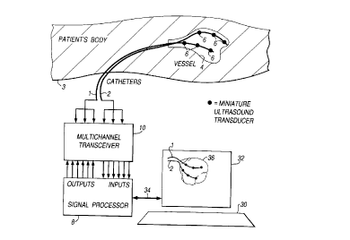
A catheter localisation system in accordance with one embodiment of the
present invention is presented in Figure 1. In Figure 1 two catheters 1, 2, are
shown to be inserted into a patient's body 3 and to be extended into the heart of
the patient. Although the illustrative embodiment sho~,vn in Figure I is provided
with two catheters 1, 2, it will be readily a~pl~ciated that other embodiments may
be arranged such that acoustic transducers 6 disposed on the catheters 1 and 2 may
be disposed on a single catheter so that the catheter tracking arrangement can be
effected with ~efelellce to one catheter only.
The catheters 1 and 2 are coupled to a signal processing unit 8 via a multi-
channel transceiver 10. The signal processing unit 8 operates to excite the
acoustic transducers selectively such that by either temporarily or phase
modulating acoustic signals generated by each of the acoustic transducers 6,
acoustic signals from any one of the acoustic transducers 6 may be uniquely
identified. Acoustic signals generated by one of the acoustic transducers 6 are
thereafter detected by others of the acoustic transducers 6 and for each acoustic
signal generated and for each acoustic transducer receiving this acoustic signal, an
acoustic transfer function may be deterrnined as will shortly be described. Signals
representative of the acoustic signals detected by each of the acoustic transducers
6 are fed to the signal processor via the multi-channel transceiver 10. The multi-
channel transceiver 10 serves to provide separation and determination of signaIs
CA 02244058 1998-09-09
-6-
representative of detected acoustic signals from each of the acoustic transducers 6.
The multi-channel transceiver 10, thereby serves to provide a facility through
which acoustic tr~n~ cers may be selectively excited and signals detected thereby
may be selectively fed to the signal proces.~ing unit 8.
The signal processing unit 8, operates to dete~ e a three flimen.~ional map
of the part of the hl1m~n body 4 in which the catheters I and 2 are disposed.
Generation ofthe three ~imen~ional map will now be described with lefe~cllce to
Figures 2 and 3 where parts also appearing in Figure 1 bear identical numerical
designations. In Figure 2 acoustic transducers 6 associated with the catheters 1and 2 are shown to be disposed within the heart 4. One of the transducers 6' is
shown to generate an acoustic signal which is thereafter received by another of the
acoustic tr~n.~dllcers 6". The acoustic signal is received along a direct path 12 and
along a plurality of indirect paths 14 which indirect paths represent paths taken by
the acoustic signals reflected via the heart wall 4. The diagram shows only someof many indirect paths. Acoustic signals received by the receiver acoustic
transducer 6" are illustrated in Figure 3. ~n Figure 3 the acoustic signal generated
by the acoustic transducer 6' is shown as waveform signal 16. The signal
representative of the acoustic signals received by the receiving acoustic transducer
6" is shown as waveform 18. The waveform 18 is shown to be comprised of a
signal from the direct path 12 and a signal ~om the indirect paths 14 which are
designated waveform 20 and 22. The waveform 22 is shown to be comprised of a
plurality of scattered signals which are formed from reflections of the acousticsignal from the heart wall. For each of the acoustic transducers receiving acoustic
signals generated by the acoustic tr~n.~ cer 6', the signal processing unit 8
operates to generate an acoustic transfer function. rn Figure 3 an acoustic transfer
function generated for the received waveform 18 in accordance with the
transmitted waveform 16 is shown to be illustrated by a signal waveform diagram
CA 022440~8 1998-09-09
24. By correlating the transmitted acoustic signal with the acoustic signals
received at the receiving acoustic transducer 6", the acoustic transfer function 24 is
generated. This is again shown to be comprised of a direct component 26 and a
scattered component 28.
After generating a plurality of acoustic transfer functions from the acoustic
transducers disposed within the vessel 4, the signal processing unit operates togenerate a three dimensional map of the inside of the heart wall 4 from which a
location of the catheters 1 and 2 may be dete~ ined. This is achieved by
generating a three dimensional synthesised model of the inside of the heart wall 4
and iteratively adapting this model to fit the acoustic transfer functions measured
by the acoustic transducers. In the catheter localisation system shown in Figure 1
the signal processing unit 8, operates to generate the shape of the heart wall since
the heart wall will be a strongly reflecting feature which can be modelled as a
surface ~ullowlding the acoustic transducers. The surface shape is then adjusted to
match the calculated reflections in accordance with a best fit image of the heart
wall corresponding to the plurality of acoustic transfer functions generated. This
is known as a holographic im~ging technique. The accuracy of the holographic
im~ging technique is increased by employing a more detailed model of the image
and by making more observations of wave scattering in the region. The greater the
distances between the transducers from which acoustic transfer fimctions are
generated, the more accurate the three dimensional map of the inside of the heart
wall will be. Hence, the acoustic transducers can be spaced apart accordingly
along the length of the catheter. To effect the holographic im~ging, it is necessary
that the relative positions of all the acoustic transducers along the catheter head are
known. However, in a case where the transducers are mounted on a flexible
catheter the position of the acoustic transducers may move with respect to each
other. This means that the relative positions of the acoustic transducers 8, must be
CA 02244058 1998-09-09
-
-8-
continually measured as the catheters move. One way to effect this operation is to
measure a direct unscattered sound tr~nsmi~sion between tr~n~ cPrs. A distance
between two acoustic transducers is equal to the product of the time taken for the
acoustic signal to travel between one to the other acoustic sensor and the average
speed of the acoustic signals in the medium between them. A direct sound
propagation time between a pair of acoustic transducers can be de~ellllhled at the
same time as the scaLl~led sound. The average sound speed can thereafter be
estimated from the material through which the acoustic signals have passed. Thusdistances between pairs of acoustic tr~ns(ll1cers may be dete-l,lhled in accordance
with this method which is known as sonomicrometry. By dete~"~;"ing the
distances between several pairs of acoustic tr~n.~ cers, the relative positions of all
of them may be d~te~ ed.
In the catheter loc~ tion system as hereinbefore described it is
advantageous that the acoustic transducers are mini~hlre and are capable of
transmitting and receiving acoustic signals subst~n~i~lly omnidirectionally.
Although the embodiment of the invention has been shown with acoustic
tr~n~llcers disposed on two separate c~thetçrs, it will be readily appreciated that
the acoustic transducers could be disposed at different sites on a signal catheter.
In yet a further embo~iment acoustic transducers may be mounted on
extensible arms or similar structures and arranged to mechanically extend and
spread the position of the acoustic transducers apart once the catheter head is
disposed inside the part of the body which is to be investigated. This is illustrated
in Figure 4, where catheter l is shown to have arms 38, deployed with acoustic
transducers 6 disposed on a distal end thereof.
An image of anatomical structures in a region in the vicinity of the acoustic
transducers is synthesised from the acoustic transfer fimctions measured. Ihis
CA 022440~8 1998-09-09
_9_
may be achieved using a computer with an associated user interface which is
coupled to the signal proces~in~ unit 8, which serves to display images dele~ i,led
by the signal processing unit, and to accept comm~nds to adjust system
parameters. As will be appreciated by those skilled in the art, the images
displayed on the computer may include indications of position of the catheter
mounted transducers and will therefore provide a means for guiding the cathetersrelative to the ~ullo~ ding anatomy. This is shown in Figure 1 with the computer30 and display monitor 32 coupled to the signal processor via interface 34.
As aforementioned, the three dimensional map of the structure of the part of
the body being investigated is syntllesi.~ed from the acoustic transfer functions
measured by the acoustic transducers. This is illustrated in Figure 1 by a threedimensional synthesised model 36, shown on the computer display 32. This may
be effected using a software model of the anatomical structures which surround the
acoustic transducers. Such a model can assume a wide range of anatomical
structures and can predict the acoustic transfer function between two acoustic
transducers or correspondingly the self transfer function between a single
transducer and itself. For each measured acoustic transfer function a
corresponding prediction is made using an initial estimate of the anatomy. Each
predicted acoustic transfer function is compared with its measured acoustic
transfer function and the degree of overall fit between the predictions and the
measured transfer functions is assessed. Hence the model of the anatomy is then
correspondingly adjusted and new predictions are made and compared with the
transfer functions measured. This process is iteratively repeated until an overall
best fit between predicted acoustic transfer functions and measured acoustic
tran~fer functions is effected. Hence the model of the anatomy which provides a
best fit is then displayed on the computer display 32, as shown. Relative positions
CA 02244058 1998-09-09
-10-
of the acoustic transducers may be determined along with the shape of the
anatomy.
As will be readily appreciated by those skilled in the art, various
modifications may be made to the embodiments hereinbefore described without
departing from the scope of the present invention. In particular the acoustic
transducers may be ultra sound transducers and the acoustic si$nals may be ultrasound signals. Furthermore the acoustic transducers may be disposed on any part
of the catheter including the elongated member.