Note: Descriptions are shown in the official language in which they were submitted.
CA 02244163 1998-07-23
W O 97/287;34 PCTrUS97/01745
ENDOSCOPIC ROBOTIC SURGICAL TOOLS AND METHODS
T~hle of Contents
I. BACKGROUND OF THE INVENTION . . . . . . . . . . . . . . 3
A. Field of the Invention . . . . . . . . . . . . . . 3
B. State of the Art . . . . . . . . . . . . . . . . . 3
II. SUM ~Y OF THE INVENTION . . . . . . . . . . . . . . . 6
III. BRIEF DESCRIPTION OF THE DRAWIN~ . . . . . . . . . . 11
IV. DETAILED DESCRIPTION OF THE PREFERRED EMBODIMENTS . . 14
A. Overview . . . . . . . . . . . . . . . . . . . . 14
B. The Encoder . . . . . . . . . . . . . . . . . . . 18
1. Exoskeleton . . . . . . . . . . . . . . . . 18
a. Electromechanical Using Potentiometers 18
b. Other Transducers . . . . . . . . . . . 23
c. Photoelectric Transducers . . . . . . . 24
2. Optical Without Exoskeleton . . . . . . . . 24
a. Laser Encoders . . . . . . . . . . . . 25
3. Suspended Encoders . . . . . . . . . . . . . 25
C. T]~e Control Circuit . . . . . . . . . . . . . . . 25
1. Potentiometers to Servo Motors . . . . . . . 26
2. Other Control Circuits . . . . . . . . . . 27
D. The Servo System . . . . . . . . . . . . . . . . . 27
1. Servo Motors with Pulleys and Tendons . . . 28
2. Direct Drive and Pullwire . . . . . . . . . 30
3. Jack Screws In Lieu of Pulleys . . . . . . . 31
E. The Robot Arms . . . . . . . . . . . . . . . . . . 31
1. Socket and Clevis Arrangement . . . . . . . 31
2. Rotational and Flexional Pulleys and Tendons 34
3. Path of Tendons, Direct Drive Shoulder,
Pullwire for Grippers . . . . . . . . . . 36
4. Other End Effectors . . . . . . . . . . . . 37
5. Interchangeable End Effectors . . . . . . . 37
CA 02244163 1998-07-23
W O 97~28734 PCT~US97/01745
2 ----
F. Feedback Means . . . . . . . . . . . . . . . . . . 37
1. Visual Feedback to the Practitioner . . . . 37
a. Fiber Optics and Video Camera . . . . . 37
b. Stereoscopic . . . . . . . . . . . . . 38
c. Non-Visible Spectrum . . . . . . . . . 38
2. Servo Position Feedback . . . . . . . . . . 39
3. Encoder Sensory Feedback . . . . . . . . . . 39
~. The Multi-lumen Tube . . . . . . . . . . . . . . . 40
H. Remote Communications . . . . . . . . . . . . . . 40
Claims . . . . . . . . . . . . . . . . . . . . . . . . . . 42
ABSTRACT OF THE DISCLOSURE . . . . . . . . . . . . . . . . 52
CA 02244l63 l998-07-23
W 0971287.34 PCTrUS97/01745
3 ----
I. BACKGROUND OF TH~ IN~rENTION
A. Field of the Invention
.
~ he invention relates to endoscopic surgical tools and
methods. More particularly, the invention relates to endoscopic
methods and devices having robotic capabilities.
B. State of the Art
Endoscopic surgery is widely practiced throughout the world
today and its acceptance is growing rapidly. In general,
endoscopic surgery involves one or more incisions made by trocars
where trocar tubes are left in place so that endoscopic surgical
tools may be inserted through the tubes. A camera or magnifyin~
lens is o~ten inserted through one trocar tube, while a cutter,
dissector, or other surgical instrument is inserted through
another trocar tub ~or purposes of manipulating and/or cutting
the interral organ. Sometimes it is desirable to have several
trocar tu~es in place at once in order to receive several
surgical instruments. In this manner, organ or tissue may be
grasped with one surgical instrument, and simultaneously may be
cut with another surgical instrument; all under view of the
surgeon via the camera.
By 1~96t it is expected that more than two million
additional endosurgeries will be performed per year that, in
1990, were done via open surgery ~MedPRO Month, I:12, p.178).
The advantages of endoscopic surgery are clear in that it is less
invasive, less traumatic and recovery is typically quicker. As a
result, many new tools and devices for use in endosurgery are
introduced every year. For example, it is now known how to use a
powered microsurgical tool for intravascular surgery such as that
disclosed in U.S. Patent Number 5,059,203 to Husted. Husted
teaches a miniature rotatable work wheel having a cutting blade
that is integrally formed with a drive hub and axle and mounted
CA 02244163 1998-07-23
W O 97128734 PCTrUS97/01745
4 ----
for rotation at the end o~ a casing member. The casing member is
a multi-lumen tube. A single monofilament drive line is looped
around the drive hub and extends through separate lumens of the
multi-lumen tube to a source of drive power. The drive hub also
has a cupped cross section ~or inhibiting the monofilament drive
line from riding off the hub. A drag load is applied to the
payout end of the monofilament drive line close to the drive hub.
The rotating wheel may be a cutting wheel or an abrading wheel,
and is used primarily for opening occluded blood vessels.
Endoscopic surgical tools with articulate end effectors are
also now known. U.S. Patent Number 4,880,015 to Nierman
discloses a biopsy forceps for use in a flexible fiberoptic
bronchoscope. Nierman's forceps are provided with an increased
range of operability when obtaining tissue samples due to a hinge
joint located between the forceps grippers and the cable from
which the grippers extend.
Meanwhile, the art of robotics is also developing rapidly.
While originally conceived of in fiction, modern robotics
involves discrete specialized applications often in the area of
manufacturing, but also in the areas of research and development
and ~here hazardous environments must be traversed. In this
latter application, robotics often involves electro-mechanically
activated articulate members emulating human arms and hands which
are operated by an encoding device responsive to the articulation
of human arms and hands of an practitioner. Known encoding
devices generally include an exoskeleton or sleeve which fits
over the hu~an arm of the practitioner. The encoding sleeve is
provided with joints corresponding to the joints in a human arm
~nd one or more ring bearings all of which translate motion by
the human arm of the practitioner into mechanical, electrical, or
electro-mechanical signals. These signals generated by the
encoder are then transmitted to a robotic arm which responds to
the signals from the encoder. The robotic arm is usually moved
by servo motors which are located in the robotic arm joints. The
object of this type of arrangement is to provide a mechanical or
CA 02244163 1998-07-23
W O 97/28734 PCT~US97/01745
----
electro-mechanical arm which will mimic the movements o~ the
human arm of an practitioner. Robotic arms are also usually
provided with some type of gripper which is activated by hand
movement of the practitioner. These types of robotic arm
arrangements are most often used in hazardous environments such
as the handling of nuclear materials, the disarming of bombs, and
in space exploration (where the signals from the encoder to the
robotic arm are transmitted over long distance by radio waves).
Early robotic arms were clumsy and useful only for
completing gross tasks such as lifting, pouring, twisting and the
like. It was difficult to obtain an arrangement of encoder and
robot arm which had the responsiveness of a human arm. This
difficult~ stemmed from the quality of the servo motors, the
difficult~ in encoding the articulations of the human arm, and
the lack of feedbac~ to the encoder. State of the ar~ robotlc
arms, however, have overcome many of the difficulties encountered
by the early robotic arms. It is now possible to construct
robotic arms which have a wide range of movements closely
mimicking the articulations of the human arm and which pr~ovide
feed~ack ~o the encoder to more correctly mimic the movements of
the practitioner. State of the art robotic arms are capable of
performing sophisticated tasks such as welding, soldering,
sewing, manipulation of a variety of tools, handling of small
pieces, etc. Encoders are now commonly provided with feedback
mechanisms which offer the practitioner varying resistance
corresponding to the resistance encountered by the robotic arm.
Recent developments in robotics and in telecommunications
have crea~ed a new art called "virtual presence". In virtual
presence, an encoder with audio, video, and tactile feedback is
worn by a human "participant" and is connected through a
transceiver to a robotic apparatus having audio, video and
tactile sensors at a distant location. The object of virtual
presence is to allow the human participant to act and feel as if
the participant is actually present at the distant location.
Virtual presence technology has been applied in the aerospace
CA 02244l63 l998-07-23
W O 97/28734 PCT~US97/0174
6 ----
industry for controlling remote space probes, in oceanography for
controlling deep sea probes, and in environmental sciences for
the handling of hazardous materials.
While endoscopic surgery is ever gaining in acceptance, it
is still often more difficult to perform than open surgery
insofar as the surgeon must view the surgical site through an
endoscope or a camera rather than viewing it directly through a
large incision. In this sense, endosurgery using video cameras
is similar to virtual presence since the participant (in this
case the surgeon) does not see the surgical site directly, but
rather sees a virtual representation of the surgical site on a
television screen. However, while virtual presence virtually
places the participant in a distant scene, in endosurgery, the
surgeon must reach into the televised scene using tools which
function unlike human arms and hands. In state of the art
endosurgery the surgeon is limited to tasks for which tools are
available, while in open surgery the surgeon can still apply the
wide range of articulation available to human arms and hands.
Nevertheless, in either type of surgery, the surgeon must be
within arm's length of the patient.
Despite advances in robotics, the robotic techniques have
heretofore never been used in endoscopic surgical instruments or
procedures, probably because the robotic instruments are
relat}vely large and the endoscopic instruments are relatively
tiny.
II. SUMMARY OF THE INVENTION
It is therefore an object of the invention to provide an
endoscopic tool having a plurality o~ rotational and flexional
joints.
It is also an object of the invention to provide an
endoscopic tool having a pair of articulate robotic arms.
CA 02244163 1998-07-23
W 097/28'~34 PCTA~S97/0174S
7 __
It is another object of the invention to provide robotic
endosurgical tools which are small enough to extend through
trocar twbes but which provide a surgeon with substantially all
of the articulation available in open surgery.
It is also an object of the invention to provide an
endoscopic tool having articulate robotic arms which are operable
by servo motors.
It is another object o~ the invention to provide endoscopic
robotic arms which are coupled to servo motors using tendons and
puLleys.
It is also an object of the invention to provide endoscopic
robotic arms which are flexional and rotational through the
movement of tendons.
It is also an object of the invention to provide and
endoscopic robotic arm with small diameter tendons which are
durable and which are positively coupled to the arm.
It is still another object of the invention to provide
ro~otic e~doscopic instrument arms which closely mimic the
movements of human arms.
It is another object of the invention to provide a robotic
endoscopic instrument arm which is controlled by an encoder worn
by a surgeon.
It is also an object of the invention to provide a robotic
encoder for controlling an endoscopic tool which is adaptable to
fit any size surgeon and which is easy to put on.
It is another object of the invention to provide ~eedback
mechanisms in the robotic arms so that the arms are more
responsive to movements registered by the encoder.
CA 02244163 1998-07-23
W O 97/28734 PCTAUS97/01745
It is also an object of the invention to provide tactile
feedback means in the robotic grippers so that the surgeon can
tell the amount of force applied at the grippers.
It is yet another object of the invention to provide a
coupling mechanism whereby robotic endoscopic instrument arms may
be coupled to an encoder so that the instrument arms are
disposable and the encoder is reusable.
It is also an ob~ect of the invention to provide a multi-
lumen tube through which robotic endoscopic instrument arms and
an endoscopic camera extend and through which supplies and the
like are delivered to the surgical site for use by the robotic
arms.
It is still another object of the invention to provide
multidimensional viewing means and remotely coupled encoder means
so that a surgeon may operate and view the operation from a
location remote from the patient.
In accord with these objects which will be discussed in
detail belowr the methods and devices of the present invention
include an encoder, an endoscopic robotic instrument, and an
encoder/robotic instrument interface. A preferred embodiment of
the encoder has a chest/shoulder plate provided with telescoping
tubes and joints. Each joint is provided with a direct drive
potentiometer to monitor movement and provide a corresponding
signal. The chest plate is preferably adaptable to a large range
of human chest si~es and the telescopic segments are strapped to
the arms of the practitioner at the elbows. A pistol grip is
provided at the wrist end of the telescopic segments. According
to the presently preferred embodiment, the encoder encodes
flexion and rotation at the shoulder, elbow and wrist of each arm
in addition to gripping at each hand.
The encoding device is coupled to a circuit which operates a
servo system. The servo system includes a series of servo motors
CA 02244163 1998-07-23
W O 97/28734 PCT~US97/01745
_ _
to move to positions correlating to the potentiometer positions
which correspond to the position of the arm of the practitioner
wearing the encoder. The servo motors are mounted in an
interface housing with the rotational axes of their shafts
parallel. The rotational shaft of each servo motor is provided
with a quick connecting end. A series of pulleys corresponding
to the number of servo motors are arranged in a single tray-like
housing. Each pulley is provided with a self-aligning socket
designed to mate with a corresponding servo motor shaft. All of
the pulleys are mounted on all of the shafts simultaneously and
quickly by coupling the tray-like housing to the servo motor
housing and are similarly quickly disconnectable ~rom the servo
motors. ~ccording to the presently preferred embodiment, for
each arm, seven servo motors and corresponding pulleys are
pro~ided ~or responding to flexion and rotation at the shoulder,
elbow and wrist of each arm in addition to gripping at each hand.
Fourteen servo motors and corresponding pulleys are provided for
a pair of arms and hands.
The robotic instrument preferably comprises two arms mounted
at the distal end of a multi-lumen tube. Each arm has rotational
and flexional joints corresponding to the shoulder, elbow, and
wrist of the practitioner. Tendons are coupled to the pulleys of
the servo motors and are fed through the multi-lumen tube to the
joints of the two arms. The endoscopic robotic arms preferably
mimic human arms having movements for shoulder rotation, shoulder
flexionr upper arm rotation, elbow flexion, lower arm rotation
and wrist flexion. In addition, grippers are mounted at the
distal en~ of the robotic arms to provide a limited hand
movement.
~ Preferred aspects of the encoder include its adjustability
to ~it di~ferent size users, its light weight, and the placement
of potentiometers to compensate for differences in the movement
of the exoskeleton relative to the movement of the user's arm.
The potentiometers are preferably supplied with op-amp followers
an~ are opto-isolated from the control circuit.
CA 02244163 1998-07-23
W O 97/28734 PCTrUS97/01745
-- -
Preferred aspects of the control circuit include its
adjustability to the arm movements of different users and the
adjustability of the relative movement of each robot arm ~oint
relative to the encoded movements of the user's arm joints. The
presently preferred control circuit includes, for each
potentiometer in the encoder, a pair of adjustable voltage
regulators and a pulse generator formed ~rom an astable and a
monostable timer. The voltage regulators supply an upper and a
lower voltage to the potentiometer and the potentiometer selects
a voltage between the upper and lower voltage and supplies that
voltage to the pulse generator. The pulse generator generates a
pulse train in which the length of the pulses is proportional to
the voltage selected by the potentiometer. The upper and lower
voltages for each potentiometer are independently adjustable and
determine the range of movement of the corresponding robot arm
joint relative to movement of the encoder ~oint.
Preferred aspects of the servo system include providing the
servo motors with self-aligning splined shafts and axially
offsetting some of the pulleys in the pulley tray relative to
other pulleys so that the tendons may be easily threaded through
the pulley tray. Each pulley preferably includes a radial slot
and an axial screw. A tendon is attached to a pulley by
threading its ends through the radial slot and around the axial
screw. ~ightening the axial screw secures the tendon to the
pul~ey. The pulley tray is preferably provided with ramped parts
which deflect the tendons upward for easier assembly of the tray.
The robotic arms preferably include alternating rotational
and flexional joints. Each flexional joint preferably includes a
clevis having a cylindrical stem and each rotational joint
preferably includes a socket in which a corresponding clevis stem
is rotationally mounted. Each socket distal of the most proximal
socket has a stem which is mounted between arms of a respective
clevis. The most proximal rotational joint is preferably
directly driven and the remainder of the rotational joints are
preferably pulley driven. The flexional joints are preferably
CA 02244163 1998-07-23
W 097128734 PCT~US97/0174
11
pulley driven. The preferred pulley driven rotational joint
includes a two layered pulley mounted on a socket with its axis
o~ rotation perpendicular to the longitudinal axis of the socket.
Th~ clevis stems preferably include stepped twist drums. The
tendon for a rotational joint wraps approximately ninety degrees
around a first layer of the layered pulley, approximately half
way around a first step of the twist drum, approximately half way
around a second step of the twist drum, and approximately ninety
degrees around a second layer of the layered pulley. An axial
bore between the first step and the second step of the twist drum
facilitates location of the tendon and secures the tendon against
slippage. The preferred pulley driven flexional joints include a
pulley which is mounted on a socket stem which is mounted between
arms of a clevis. The tendon for a flexional joint wraps
approximately ninety degrees around the top of the pulley, wraps
around the socket stem and wraps approximately ninety degrees
around the bottom of the pulle~. A screw in the clevis stem
secures t~e tendon to the clevis stem and prevents slippage.
The tendons are preferably encased by individual coiled
sheaths and are threaded around the robot arms so as to avoid
interference with each other and with movement o~ the robot arms.
Each pair of grippers is preferably spring biased in the open
pcsition and is operable by a single pull wire in a protective
coil sheath.
Addi~ional objects and advantages of the invention will
become apparent to those skilled in the art upon reference to the
detailed description taken in conjunction with the provided
figures.
III. BRIEF DESCRIPTION OF THE DRAWING
Fi~ure 1 is a schematic illustration of one embodiment of an
endoscopic robotic surgical tool according to the invention;
CA 02244163 1998-07-23
W O 97/28734 PCT~US97/01745
12
Figure 2 iS a rear view of the left portion of an
exoskeleton encoder;
Figure 3 is a top view of the left portion of the encoder of
Figure 2;
Figure 4 is a broken side elevation view of the left lower
arm portion of the encoder of Figure 2;
Figure 5 is a broken top view o~ the left lower arm portion
of the encoder shown ln Figure 4;
Figures 6 through 11 are schematic diagrams showing axes of
upper arm movement;
Figure 12 is a schematic diagram of a control circuit of the
encoder;
Figure 13 is a top view of a pulley tray;
Figure 14 is a transparent side elevation view of the pulley
tray of Figure 13;
Figure 15 is a transparent side elevation view of a single
pulley of the pulley tray of Figure 13;
Figure 16 is a cross sectional view along line 16-16 of
Figure 15;
Figure 17 is an exploded perspective view of a pulley and a
tendon coupling screw;
Figure 18 is a top view of a pulley and a tendon;
Figure 19 is a top view of two pulleys and tendons,
Figure 20 is a bottom view of a top servo motor array;
CA 02244163 1998-07-23
W O 97/28734 PCTrUS97/01745
13
Figure 21 is a top view of a bottom servo motor array;
Figure 22 is a transparent side elevation view of the top
and bottom servo motor arrays and the pulley tray,
Figure 23 is a plan view of an assembled servo motor tray,
multilumel~ tube, and robot arms,
Figure 24 is a schematic plan view of a jack screw coupled
to a servo motor and a tendon loop;
Figure 25 is a broken side elevation view of a robot arm
according to the invention;
Figure 26 is a view similar to Figure 25, but rotated 90~
about the shoulder axis;
Figure 27 is a view similar to Figure 26 with two flexional
joints flexed and grippers opened;
Figure 28 is a broken side elevation view of a portion of a
robotic rotation joint of Figures 25-27;
Figure 29 is a broken cross sectional view along line 29-29
in Figure 28;
Figure 30 is a broken top view of the robotic rotation joint
of Figures 28 and 29;
Figure 31 is an enlarged side elevation of a rotation
pulley;
Figure 32 is a broken side elevation view of a robotic
flexion joint according to the invention;
Figure 33 is a top view of the robotic flexion joint of
Figure 32;
CA 02244l63 l998-07-23
W 097/28734 PCTrUS97/0174S
14 -
Figure 34 iS a broken perspective view of two robotic arms
extending from the distal end of a multi-lumen tube;
IV. DETAILED DESCRIPTION OF THE PREFERRED EMBODIMENTS
A. Overview
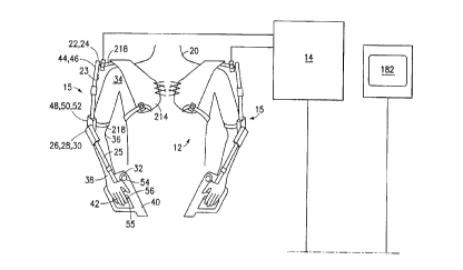
Turning now to Figure 1, a first embodiment of an endoscopic
robotic tool 10 according to the invention generally includes an
exoskeleton encoder 12, a control circuit 14, a servo system 16,
and a pair of remote robot arms 18. The exoskeleton encoder 12
fits over the shoulders and chest, and attaches to the arms of a
practitioner 20. Rotational and flexional joints 22, 24, 26, 28,
30, 32 are provided on the encoder 12 for each shoulder 34, elbow
36 and wrist 38 of the practitioner, while a pistol grip 40 is
provided for each of the hands 42 of the practitioner. In total,
seven transducers 44, 46, 48, 50, 52, 54, 56 are provided in each
arm of the encoder to register rotational and flexional movements
of the shoulders 34, elbows 36, and wrists 38 as well as gripping
movement of the hands 42 of the practitioner. The transducers
are all coupled to a control circuit 14 which in turn provides
outputs to an array of fourteen servo motors 60, 62, 64, 66, 68,
70, 72, 74, 76, 78, 80, 82, 84, 86. The servo motors are coupled
respectively to pulleys 90, 92, 94, 96, 98, 100, 102, 104, 106,
108, 110, 112, 114, 116 which in turn are coupled to tendon loops
120, 122, 124, 126, 128, 130, 132, 134, 136, 138, 140, 142, 144,
146, i.e., one tendon loop per motor. The tendons are fed through
a multi-lumen tube 150 to the remote robot arms 18 which are
mounted at the distal end of the tube 150.
Each robot arm is provided with three rotational joints 160,
164, 168 and three flexional joints 162, 166, 170, and the distal
end of each robot arm is provided with a gripper 172. Thus, the
fourteen tendon loops 120, 122, 124, 126, 128, 130, 132, 134,
136, 138, 140, 142, 144, 146, are each coupled to a respective
one of the seven joints and the gripper on each arm. However, as
discussed in more detail below, the shoulder rotational joint 160
may be controlled by a torsion tube rather than a tendon loop,
CA 02244163 1998-07-23
W O97128~134 PCTrUS97/01745--
and the gripper 172 may be controlled by a tendon pull-wire
rather than a tendon loop. Moreover, while each tendon is
preferably encased in a coil sheath, the shoulder flexional joint
may be ccntrolled by a tendon which is not sheathed since the
path taken by the tendon is a relatively straight line through
the multilumen tube and the tendon does not bend through a path
as the other joints are flexed.
The distal end of the multi-lumen tube 150 is also provided
with a camera lens 180 which is optically coupled to a video
camera (not shown). Output from the video camera is transmitted
by either wired or wireless communications to a monitor 182
viewable by the practitioner 20. Additional lumens 152, 154 are
preferably provided in the multi-lumen tube 150 for the delivery
of supplies to the surgical site, and/or for suction, irrigation,
and the like. The pulleys 90, 92, 94, 96, 98, 100, 102, 104,
106, 108, 110, 112, 114, 116 are preferably arranged in a tray
118 which is detachable from the array of servo motors 60, 62,
64, 66, 68, 70, 72, 74, 76, 78, 80, 82, 84, 86. The outputs from
the control circuit 14 may be transmitted to the array of servo
motors 60, 62, 64, 66, 68, 70, 72, 74, 76, 78, 80, 82, 84, 86 by
wired or wireless communications and the distance between the
control circuit and the servo motors may be several feet or
several thousand miles. The distance between the encoder 12 and
the control circuit 14 is preferably only several feet and the
length of the multi-lumen tube 150 is also preferably onIy
several feet. The outer diameter of the multi-lumen tube 150 is
small enough to fit through a trocar tube (not shown) and the
grippers 172 on the robot arms 18 are similar in size to the
grippers of known endoscopic instruments.
As will be discussed in detail below, the encoder may
include more or fewer transducers, and the transducers may take
any of many forms such as potentiometers, photoelectric sensors,
Hall effect sensors, inertial devices, or sonic sensors. The
pistol grips may include controls for suction, irrigation, and/or
cautery. The exoskeleton of the encoder may take different
CA 02244163 1998-07-23
W O 97/28734 PCTrUS97/~1745
16
forms, as well. For example, positions of the practitioner's
arms could be detected by sonar, IR or visible laser scanning, in
which case, the exoskeleton could be replaced by a series of
reflectors attached to the practitioner's arms. In order to ease
fatigue, the practitioner's arms could be suspended and the
suspension assembly used to encode arm movement.
Those skilled in the art will appreciate that the
configuration of the control circuit will depend to a greater or
lesser degree on the configuration of the encoder and the type of
servo system used. In this regard, it will be understood that
the servo system need not utili~e rotary motors with pulleys and
cables, but may use other drive means such as motorized jack
screws, hydraulic, or pneumatic drive means. The coupling of the
robot arms to the servo system will also depend on what type of
drive means is used.
The robot arms may be controlled in part with a direct drive
in lieu of tendons and pulleys. The path of the tendons from the
robot arms to the servo system may be varied according to other
considerations which will be understood from the discussion
below. Hydraulics or pneumatics may be used to control the robot
arms instead of tendons and pulleys. The grippers at the ends of
the robot arms may be cutters or other types of end ef~ectors and
the robot arms may be provided with removable, replaceable end
effectors. In one embodiment, the robot arms are retractable
into the multi-lumen tube and the tube is provided with means for
the practitioner or an on-site nurse or assistant to change end
effectors during the course of a procedure.
It is also preferable to provide various types of feedback
in the system. Generally, visual feedback to the practitioner
will always be provided, but such visual feedback may be
configured in various ways. Using the proper optics,
stereoscopic visual feedback can be provided. Some practitioners
may find it useful to have the video image transposed
horizontally so that the sensory impression is that of looking
CA 02244163 1998-07-23
W O 97/28734 PCTAUS97/01745
17 _
into a mirror. It is also possible to provide visual feedback
from infrared, ultrasound or other sensors located appropriately
relative to the surgical site. Tactile feedback is desirable at
least in t:he trigger portions of the encoder so that the
practitioner can judge how much force is applied by the grippers.
Other sensory feedback to the practitioner is also possible with
the appropriate transducers. Positional feedback from the robot
arrns to the control circuit is desirable in most instances. For
example, ~hen tendons are capable of kinking, stretching, or
slipping, it is advisable that the control circuit determine
whether the signal to the servo system has indeed effected the
desired movement of the robot arm. Various means for providing
these kincls of feedback are discussed in detail below.
The basic operation of the endoscopic robotic tool, shown in
Figure 1, is as follows. The practitioner 20 who is to per~orm
an endoscopic procedure dons the exoskeleton encoder 12 and turns
on the vicleo monitor 182. An assistant (not shown), who is in
c<~mrnlln; cation with the practitioner 20, incises the patient (not
shown) with a trocar (not shown). The assistant couples the
pulley tray 118 to the array of servo motors 60, 62, 64, 66, 68,
70, 72, 74, 76, 78, 80, 82, 84, 86 and inserts the distal end of
the multi-lumen tube 150 through a trocar tube (not shown) and
locates the robot arms 18 in the vicinity of the surgical site.
The tray of servo motors 16 is located a convenient distance from
the surgical site with the flexible sheathed tendons extending to
the multilumen tube which holds the robot arms. The servo motor
tray may be supported by an adjustable clamping means connected
to the operating table or other support. The practitioner 20 may
direct the assistant to relocate the robot arms 18 according to
information provided via the video monitor 182. When the control
circuit 14 is activated, movement of the practitioner's arms 34,
36, 38 is replicated in the robot arms 18. When the practitioner
grips one of the pistol grips 40, the gripper 172 on a
corresponding robot arm 18 is closed. Thus, the tool 10 provides
the practitioner 20 with a virtual presence of two arms and hands
and vision at the surgical site.
CA 02244163 1998-07-23
W O 97128734 PCT~US97/01745
18
From the foregoing, those skilled in the art will appreciate
that a practitioner wearing the encoder and viewing the video
monitor is equipped to perform an endoscopic procedure at a
location remote from the surgical site. The encoder, control
circuit and video monitor may be located many thousands of miles
from the surgical site and coupled to the servo motors and video
camera by any telecommunications device such as a wireless
transceiver or a telephone modem. As mentioned above, however,
there must be an assistant near the surgical site initially to
locate the robot arms according to instructions from the
practitioner. Those skilled in the art will appreciate that the
assistant is preferably provided with a simultaneous video
display for locating the robot arms. Hands free audio-visual
communication means is preferably provided between the
practitioner and the assistant. It will also be appreciated that
the assistant will be called upon during the course of the
endoscopic procedure to relocate the robot arms and to supply
materials through a lumen in the multi-lumen tube or through an
additional trocar tube to the surgical site for use by the robot
arms.
The following discussion deals with each major component of
the endoscopic robotic tool and explains in detail the various
embodiments of each component. In addition, the use of remote
communications systems with the endoscopic robotic tool is
discussed in detail. Methods of operating the endoscopic robotic
tool are addressed and yet additional alternate embodiments of
the tool are disclosed.
B. The Encoder
1. Exoskeleton
a. Electromechanical Using Potentiometers
Turning now to Figures 1 ~hrough 3, two pre~erred
embodiments of an encoder 12r 212 each include a pair of
chest/shoulder plates 214 and a respective pair of articulating
arms 15, 215, each of which terminates with a pistol grip 40. It
CA 02244163 1998-07-23
W O 97128734 PCTrUS97/01745
19
will be appreciated that each chest/shoulder plate and
articulating arm assembly 1s a mirror image of the other. The
following discussion of one chest/shoulder plate and articulating
arm, therefore applies to each of them. The chest/shoulder plate
214 is adaptable to fit a variety of chest sizes and the
articulating arms 15, 215 are adjustable in length. The encoder
designs described herein are adjustable to fit virtually any
practitioner weighing between 100 and 300 pounds and having an
arm length (neck to wrist) between twenty-four and thirty-six
inches. The chest/shoulder plate 214 extends over the shoulders
of the practitioner and is strapped or laced across the chest,
across the back and under the arm pits as seen best in Figure 1.
The articulating arms 15, 215 are attached to respective arms of
the practitioner by straps 218 located just above the elbow 36.
The hands 42 of the practitioner fit into the pistol grips 40.
A first preferred embodiment of the encoder 212 is shown in
Figures 2 through 5. Starting at the left shoulder plate 214, a
horizontal member 217 extends outward from the rear of the
shoulder plate 214 and terminates at a point behind the shoulder
34 o~ the practitioner. A ninety degree curved member 219 is
rotationally attached to the end of the horizontal member 217 at
rotation point 222 and a shoulder rotation transducer 244 is
coupled to the horizontal and curved members. A telescoping
upper arm member 223 is rotationally coupled to the curved member
219 at rotation points 224 and 226 so that it is rotatable about
two orthogonal axes relative to the curved member 219. A
shoulder ~lexion transducer 246 and an upper arm rotation
transducer 248 are coupled to the upper arm member 223 and the
curved member 219. A telescoping lower arm member 225 is
rotationally coupled to the lower end of the upper arm member 223
at rotation points 228 and 230 so that it is rotatable about two
orthogonal axes relative to the upper arm member 223. An elbow
flexion transducer 250 and a wrist rotation transducer 252 are
coupled to the upper and lower arm members 223, 225 at the elbow.
A spring-hiased pistol grip 40 is rotationally coupled at
rotation point 232 to the lower end o~ the lower arm member 225
CA 02244l63 l998-07-23
W 097/28734 PCT~US97/01745
-- -
and a wrist flexion transducer 254 iS coupled to the lower arm
member 225 and the pistol grip 40. A grip transducer 256 is
coupled to the trigger 255 in the pistol grip 40. Each of the
telescoping arm members 223, 225 iS provided with a locking
collar 227, 229 so that the length of the arm members may be
ad~usted and locked.
In sum, each arm, therefore, is provided with seven
transducers 244, 246, 248, 250, 252, 254, 256. The transducers
are preferably potentiometers which are directly coupled to the
members described above. Six of the potentiometers 244, 246,
248, 250, 252, 254 register changes in the position of the arms
of the practitioner and the seventh potentiometer 256 registers
the grip of the practitioner.
Returning to Figure 1, a slightly different second preferred
embodiment of the encoder is shown. Starting at the shoulder
plate 214, a horizontal member 218 extends outward from on top of
the shoulder plate 214 and terminates at a point above the
shoulder o~ the practitioner. A freely telescoping upper arm
member 23 iS rotationally coupled to the horizontal member 218 at
rotation points 22, 24 so that it is rotatable about two
orthogonal axes relative to the horizontal member 218. A
shoulder rotation transducer 44 and a shoulder flexion transducer
46 are coupled to the upper arm member 23 and the horizontal
member 218. A telescoping lower arm member 25 is rotationally
coupled to the lower end of the upper arm 23 member at rotation
points 26, 28 so that it is rotatable about two orthogonal axes
relative to the upper arm member 23. An upper arm rotation
transducer 48, an elbow flexion transducer 50 and a wrist
rotation transducer 52 are coupled to the upper and lower arm
members at the elbow. The remainder of the encoder of Figure 1
is the same as the encoder of Figures 2 through 5.
Figures 6 through 12 illustrate the range of upper arm
movements registered by the encoder. The first potentiometer 44,
244 mentioned above registers "shoulder rotation" which is
CA 02244163 1998-07-23
,_
W097/28734 PCT~S97/01745
21
defined as the movement of the arm in a first vertical plane
which touches both shoulders. The preferred range of shoulder
rotation is shown in Figures 7 and 8 and is approximately 90~.
The second potentiometer 46, 246 registers "shoulder flexion"
which is movement of the arm through a second vertical plane
which is ~erpendicular to the first vertical plane and which
touches only the one shoulder being flexed. Figures 9 and lO
show the pre~erred range of shoulder flexion which is also
approximately 90~. The third potentiometer 48, 248 mentioned
abo~e registers "upper arm rotation" which is illustrated in
Figures 6 and ll and is preferably approximately 180~. The
fourth potentiometer 50, 250 mentioned above registers "elbow
flexionl' which preferably has a range of approximately 120~ as
indicated in Figure 9.
Figures 7 through lO also illustrate that the shoulder
rotation and flexion transducers in the encoder of Figure l
provide a slightly inaccurate registration of the arm movements
of the practitioner. For example, as seen by comparing Figures 7
and 8, the center of rotation of the shoulder transducers is
offset from the center of rotation of the practitioner's shoulder
and because of this, the transducers do not register the exact
amount of arm movement. Likewise, a comparison of Figures 9 and
lO show that a flexional upper arm movement of 90~ will register
as less than 90~ by the transducer 46, 246. Neverthe~ess, the
encoder of Figure l is more adaptable to different sized
practitioners and the slight loss in accuracy of the encoder is
negligible and can be corrected electronically if desired.
The encoder of Figure l has additional advantages in that it
is more comfortable and less restrictive for the practitioner
than the encoder of Figure 2. In particular, the potentiometers
for registering "shoulder flex" and "shoulder rotation" can be
mounted together in one small box above the practitioner's
shoulder as shown in Figure l. This helps make the encoder a
"one size fits all" system. Any "throw" differences from one
CA 02244163 1998-07-23
W O 97/28734 PCT~US97/0174S
22 --
practitioner to another can be compensated for by electronic
adjus~ment. Twist motions will result in some extraneous input
to other adjacent encoders. For example, a wrist twist results
in some elbow flex motion. Upper arm twist results in some
shoulder flex and rotation, etc. The amount of accuracy
sacrificed by this encoder system is rapidly compensated for by
the practitioner's brain. As will be discussed in detail below,
it will also be noticed that the position of these transducers
above the shoulder requires that the upper arm member telescope.
The elbow is moved closer to the transducers when the arm is
rotated from the position in Figure 7 to the position in Figure 8
and when the arm is flexed from the position in Figures 9 to the
position in Figure 10. The freely telescoping upper arm member
compensates for this.
The registration of upper arm rotation is registered by a
potentiometer 48, 248 mounted along side the elbow as seen in
Figure 1 or by a potentiometer mounted alongside the shoulder as
seen in Figure 2. In both embodiments, the potentiometer is
slightly offset from the true axis of upper arm rotation as seen
best in Figure 6. This results in a small inaccuracy in
registration of the upper arm rotation as seen in Figure 11. As
the upper arm rotates from P1 to P2 through an angle ~, the elbow
follows an arc path from point E1 to point E2. The angle between
the upper and lower arm members increases from ~1 to ~2 This
error is slight, and is compensated for by the practitioner. The
result is that the robot follows the motion of the encoder, which
is a close approximation.
Both the elbow and the wrist flexion potentiometers are
directly in line axially with the biological joints o~ the
practitioner as seen in Figures 1, 4, and 5. In order to encode
any twisting action in the forearm without adding excessive
movements to either flex joint, the distance between the elbow
potentiometers and the wrist potentiometer must not change when
the arm is flexed. This is accomplished by making the length of
CA 02244163 1998-07-23
W097/28734 PCT~S97/01745
23
the telescopic lower arm member lockable once its length is
adjusted to fit the practitioner. The use of an L-shaped ~orearm
connection to the pistol grip (seen best in Figures 2 and 5)
allows the forearm rotation joint to register +90~ wlthout
interferlng with the practitioner's arm movements. This geometry
also m;n;m;zes disturbance of the elbow flex joint when rotating
the forearm about a stationary axis.
The trigger 255 in the pistol grip 40 is preferably coupled
to a solenoid which provides tactile feedback to the
practitioner. The solenoid receives a signal based on the
current drain in the servo motor which closes the gripper, e.g.
172 (Figure l). As the gripper encounters resistance and more
force is applied by the servo motor, the current drain across the
servo motor increases. The solenoid is arranged to provide a
variable resisting force at the trigger which is proportional to
the resistance encountered by the gripper.
While the encoder of Figure l is the presently preferred
embodiment, other types of encoders can be used. Since the
encoder is reusable and separable from the remainder of the
apparatus, a surgeon may prefer to use a more customized encoder.
The preferred encoder takes advantage of direct drive
potentiometers, but other encoders may use different means for
registering the position of the arms of the practitioner.
b. Other Transducers
In lieu of potentiometers which are directly driven by the
joints of the exoskeleton, the shafts of the potentiometers may
be coupled to weights. As the exoskeleton moves the
potentiometers relative to the earth, gravity holds the weight
downw~rd and the shaft of the potentiometer is thereby rotated.
For example, a potentiometer having a base portion and a
rotatable shaft portion is mounted by its base to an arm member
of an exoskeleton at the rotational axis of the arm member. A
vertically downward extending weight is attached to the shaft
,
CA 02244163 1998-07-23
W O 97/28734 PCT~US97/01745
24 __
portion of the potentiometer. As the arm member is rotated about
the rotational axis, the base of the potentiometer is also
rotated. The weight on the shaft of the potentiometer remains
vertically disposed, however, due to the action of gravity, and
maintains the angular orientation of the shaft of the
potentiometer constant. The base of the potentiometer is
therefore rotated relative to the shaft of the potentiometer,
which is equivalent to rotating the shaft of the potentiometer
relative to the base of the potentiometer. The relative angular
movement of the arm member is thereby encoded by the relative
angular movement of the base of the potentiometer relative to the
shaft of the potentiometer.
Another transducer using weights may be constructed from two
concentric spheres with an interposed droplet of mercury or a
freely moving weight. The relative position of the mercury
droplet can be detected by capacitance, conductive strips, or by
optical means. This type of transducer can detect position in
multiple axes.
c. Photoelectric Transducers
A photoelectric transducer can be made from a rotatable disc
having an optical gradient density surface and a peripheral
weight. A photodetector aimed at the optical gradient density
surface detects the angular position of the detector relative to
the disc which is held stationary by the weight. The disc and
the detector are mounted in a gimballed enclosure to keep the
shaft of the disc horizontal.
2. Optical Without Exoskeleton
There are several possible embodiments of an encoder which
does not require an exoskeleton. These embodiments of the
encoder use optical sensors and an image processor to determine
the movements of the arms of the practitioner and encode them for
use by the servo system.
CA 02244163 1998-07-23
W O 97/28734 PCTrUS97/01745
a. Laser Encoders
A first embodiment of an optical encoder without an
exoskeleton includes a series of reflectors which are attached to
the arms o~ the practitioner at the shoulder, elbow, wrist, and
hand. At least two orthogonally disposed photodetectors are
placed a~,ove and alongside the practitioner. A source of laser
light is provided with a rotating mirror s~nn;ng device which
directs the laser light at the reflectors and scans an area
through which the reflectors are expected to move. As the arms
of the practitioner move through space, the laser light detected
by the photo-detectors varies. An image processor interprets the
signals output from the photo-detectors and operates the servo
motors to move the robotic arms.
3. Suspended Encoders
A suspended encoder according to the invention is similar to
the electromechanical exoskeleton encoder described above.
However, the arms of the practitioner are suspended in the air by
cables which are attached to pulleys and dollies mounted in a
frame above the practitioner. The pulleys and dollies are
provided with transducers which detect their movement. As the
arms of practitioner move~ the ca~les translate this movement to
movement of the pulleys and dollies and the transducers encode
the movement. An advantage of this embodiment is that it can
reduce practitioner fatigue.
As mentioned above, the encoder may be provided with sensory
feedback for the practitioner. Various ways of providing such
feedback are discussed in detail below.
C. The Control Circuit
The encoder 12 is coupled to the servo system 16 through the
control circuit 14. The coupling of the encoder to the servo
system may be wired or may be wireless. In a presently preferred
CA 02244163 1998-07-23
W O 97/28734 PCT~US97/01745 26
embodiment, the encoder 12 is coupled by wires to the control
circuit 14 and the output of the control circuit is coupled to
the servo system 16 by wireless transmission. Those skilled in
the art will appreciate that many different modes of coupling the
encoder to the servo system are possible. It will also be
appreciated that the type of control circuit utilized will depend
in part on the type of encoder used and the type of servo system
used.
1. Potentiometers to Servo Motors
For the encoder described above with reference to Figures 1
and 2, each potentiometer is coupled to two regulated reference
voltages and provides a variable voltage output which is coupled
to two timers which generate a pulse output for controlling a
digital proportional servo motor.
An exemplary control circuit is shown in Figure 12 which
represents one portion of the control circuit 314 for one
potentiometer. It will be appreciated that for an encoder with
fourteen potentiometers, the circuit of Figure 12 will be
replicated fourteen times. The exemplary circuit 314 has two
parts: a reference voltage generator 316; and a pulse code
generator 318. The reference voltage generator 316 includes two
LM317 voltage regulators 320, 322 which are independently
adjustable by variable resistors 324, 326 to produce a high
reference voltage VA+B and a low reference voltage VA from a single
source voltage Vin. The potentiometer 328 from the encoder is
connected to the high and low reference voltages and is provided
with an op-amp voltage follower 330. The output of the voltage
follower 330 is coupled to an opto-isolator 332. As the
potentiometer 328 registers movement, a voltage between VA+B and VA
is selected and fed through the opto-isolator 332 to produce an
output voltage for the pulse code generator 318. The pulse code
generator 318 includes two LM555 timers 334, 336, one for
generating a pulse frequency and the other for generating a pulse
width. ~he output from the opto-isolator 332 is coupled to the
CA 02244163 1998-07-23
W O 97/28734 PCTrUS97/0174527 --
timer 336 which generates the pulse width, and a pulse output is
produced where the width of the pulses is proportional to the
encoder position as determined by the potentiometer 332.
According to the presently pre~erred embodiment, the first
voltage regulator 320 is adjustable by a variable resistor 324
and provides an output VA which is also coupled to ground through
a resistor 325. The second voltage regulator 322 is adjustable
by a variable resistor 326 which is coupled to ground through the
output VA of the first regulator and a second resistor 327. The
second voltage regulator thereby produces and output VA+B. The
output of the first timer 334 is a pulse train having a
particular fre~uency and the output of the second timer 336 is a
pulse traln having the particular frequency and a pulse width
proportional to the encoder position.
The high and low reference voltages are selected for each
servo motor individually depending on the range of movement which
will be required for the particular motor. Thus, the reference
voltages supplied to different potentiometers in the encoder will
be different. Moreover, depending on the reference voltages
supplied, the ranges of the potentiometers will be different as
well.
2. Other Control Circuits
It will be appreciated that other control circuits may be
used with the potentiometer encoder and that different encoders
may require different control circuits.
D. The Servo System
The encoders and control circuits described above may be
used with several different types of servo systems. These
include servo motors with pulleys and tendons, direct drive servo
motors, jack screws, hydraulics, and pneumatics, for example.
CA 02244163 1998-07-23
W O 97/28734 PCT~US97/01745
28
1. Servo Motors with Pulleys and Tendons
Turning now to Figures 13 through 23, the servo system 16 is
seen to include a disposable aluminum or injection molded plastic
pulley tray 402 and an upper and lower array of servo motors 404,
406. The pulley tray 402 contains fourteen pulleys 90, 92, 94,
96, 98, lO0, 102, 104, 106, 108, 110, 112, 114, 116 supported by
bearings (not shown). Seven pulleys 90, 92, 94, 96, 98, 100, 102
are engaged by the upper servo motor array 402 and seven pulleys
104, 106, 108, 110, 112, 114, 116 are engaged by the lower servo
motor array 406. The pulleys sit in bushings and are sandwiched
between the upper and lower servo motors.
As seen best in Figures 15 through 18, each pulley, e.g.
108, has a first cylindrical part 108a with a screw receiving
bore 108b, a second cylindrical part 108c with a servo motor
shaft receiving bore 108d and a pulley wheel 108e with a grooved
rim 108f. The pulley wheel 108e is longitudinally offset from
the center of the pulley, being closer to the first cylindrical
part 108a and is provided with a radial slot 108g which extends
from the groove 108f in the wheel rim to the screw receiving bore
108b. The screw receiving bore 108b has a stepped diameter, the
larger part for receiving a screw head lO9a and the smaller part
being threaded. The pulleys are arranged in the pulley tray as
shown in Figures 13 and 14 so that half of the pulleys have their
shaft receiving bore (e.g. 108d) facing up and half have their
shaft receiving bore facing down. The pulley tray has
asymmetrical upper and lower surfaces so that the top and bottom
of the tray are not confused. Tendons, e.g. 138, are attached to
the pulleys, e.g. 108, by threading the ends of the tendon around
the groove 108f in the pulley wheel, through the radial slot 108g
and around the tendon locking screw 109 as seen best in Figures
17 and 18. The locking screw 109 is then tightened against the
tendon 136.
The tendons are threaded through the pulley tray as shown in
Figure 14 and exit the pulley tray through the multi-lumen tube
CA 02244163 1998-07-23
W O 97/28734 PCT~US97/0174S; 29 --
150 which is preferably rigidly attached to the pulley tray as
shown in Figure 23. It will be appreciated that the of~set
pulley wheels on oppositely mounted pulleys provide upper and
lower space between tendons to ease the threading of the tendons
through the tray and to thereby save space.
The pulley tray 402 is engaged by two servo motor arrays
404, 406. An upper servo motor array 404, which is shown in
Figures 20 and 22, has seven servo motors 60, 62, 64, 66, 68, 70,
72 and a lower servo motor array 406, which is shown in Figures
21 and 22, has seven servo motors 74, 76, 78, 80, 82, 84, 86.
Each servo motor has a splined shaft 60a, 62a, 64a, 66a, 68a,
70a, 72a, 74a, 76a, 78a, 80a, 82a, 84a, 86a which engages the
shaft receiving bore, e.g. 108d, of a respective pulley. The
splined shafts and the shaft receiving bores are "self-aligning".
The servo system is assembled by placing the lower surface of the
pulley tray on top of the upper surface of the lower servo motor
array so that the splined shafts of the servo motors engage the
shaft receiving bores of the pulleys. The lower surface of the
upper servo motor array is then placed on top of the upper
surface of the pulley tray so that the splined shafts of the
servo motors engage the shaft receiving bores of the pulleys.
The sandwiched assembly of servo motor arrays and pulley tray is
then locked together to provide the assembly as shown in Figure
23.
~ s seen best in Figure 22, the upper surface of the pulley
402 tray is provided with a keyway 402a and the lower surface of
the upper servo motor array 404 is provided with a key 404a which
engages the keyway 402a. Similarly, the upper surface of the
lower servo motor array 406 is provided with a keyway 406a and
the lower surface of the pulley tray 402 is provided with a key
402b which engages the keyway 406a. Thus, it is impossible to
couple ~he servo motor arrays to the pulley tray incorrectly.
As mentioned above, the described servo system permits a
portion of the robotic tool to be reusable while another portion
CA 02244163 1998-07-23
W O 97/28734 PCTrUS97/01745
-
may be disposable, if desired. In particular, the encoder, the
control circuit, and the servo motors are reusable. The pulleys,
tendons, multi-lumen tube and robot arms which will be in contact
with human fluids, may be uncoupled from the servo motors and
disposed of, if desired. In addition, the described servo system
permits the use of several different types of robot arms with the
same encoder. For example, one type of robot arms may have two
grippers whereas another type of robot arms may have a gripper
and a cutter, etc. The self-aligning feature of the servo system
permits rapid coupling and uncoupling of the servo motors and the
pulleys so that different types of robot arms can be used with
the same encoder during a single endoscopic procedure.
2. Direct 3rive and Pullwire
According to a presently preferred embodiment, the shoulder
rotation joint 160 (Figure 1) of each robot arm (which is
described in detail below) is coupled to a respective servo motor
by a direct drive instead of by a pulley and tendon. This
simplifies operation and a direct connection is better for these
joints which have the highest loads. In addition, while the
tendons described above are "endless loops", the tendon which
controls the ~ripper 172 is preferably a single pull wire which
is described in detail in the following discussion of the robot
arms.
As indicated previously herein, the use of pulleys in the
servo assembly may require positional feedback from the robot
arms ~discussed in detail below) to compensate for slippage and
stretch and requires the careful alignment of the servo motors
with the pulleys ~using the self-aligning splined shafts
discussed above). The need for positional feedback and careful
alignment of the servo motors may be avoided through the use of
jack screws in place of pulleys.
CA 02244163 1998-07-23
W O 97/28734 PCT~US97/01745
. 31 __
3. Jack Screws In Lieu of Pulleys
As shown in Figure 24, a jack screw 508 has a rotational
shaft 510 mounted for rotation on two bearings 512, 514. One end
of the shaft has a self-aligning coupling 516 for removable
coupling with a servo motor 78. Half for the shaft 510a is left
hand threaded and the other half 510b of the shaft is right hand
threaded. Each half of the shaft has a screw jack nut 518, 520
threaded to it and an indexing track (not shown) engages the nuts
518, 520 to prevent them from rotating when the sha~t 510 is
rotated. Each screw jack nut has a tendon coupling clamp 518a,
520a and the two ends of a tendon loop 136 are coupled to
respective screw jack nuts by means of the tendon coupling
clamps. h~en the shaft is rotated in one direction, the screw
jack nuts are driven towards the center 510 of the shaft.
Conversely, when the shaft is rotated in the other direction, the
screw jack nuts are driven outward from the center 510c of the
shaft to the ends of the shaft. The use of screw jacks may
obviate the need for positional feedback since the input signal
is in 1:1 proportion with the position; however, it may still be
desirable to use positional feedback from the actual end effector
to compensate for slack or stretch in the connecting tendons or
linkages.
With jack screws, the interface with the servo motors is
less critical than with pulleys. This is because several
rotations of the servo motor are required to effect an
appreciable joint movement, depending on the screw pitch.
E. The Robot Arms
1. Socket and Clevis Arrangement
Figures 25 through 27 show a presently preferred embodiment
of one o~ the two robot arms 18. The robot arms approximate the
geometry of the encoder which approximates the geometry of the
CA 02244l63 l998-07-23
W O 97/28734 PCTrUS97/~174
32
arms of the practitioner. Each ro}~ot arm generally includes a
shoulder 600~ an elbow 602, a wrist 604, and a pair of grippers
172 (172a, 172b). The shoulder 600, elbow 602 and wrist 604 each
have a rotational joint 160, 164, 168 and a flexional joint 162,
166, 170. The axis of rotation of each rotational joint is
always perpendicular to the axis o:E flexion of the corresponding
flexional joint, regardless of their rotational or flexional
position. There are, therefore, three rotational joints and
three flexional joints. The presently preferred joints are
configured as alternating socket and clevis members. A clevis is
mounted for rotation in a socket and a socket is mounted for
flexion in a clevis. A presently preferred embodiment of this
joint configuration is described as follows.
With reference to Figures 25-27, the first joint in the
robot arm is the shoulder rotational joint 160 which is
proximally coupled to a direct drive torque tube (not shown) and
has a distal shoulder clevis 161. The shoulder rotational joint
160 has a cylindrical bore 160a which extends into the shoulder
clevis 161.
The second ioint is the shoulder flexional joint 162 which
is formed by mounting an elbow socket 606 in the shoulder clevis
161. The elbow socket 606 has a stem 608 which is mounted
between the arms of the shoulder clevis 161. A shoulder flexion
pulley 610 is mounted on the elbow socket stem 608 between the
arms of the shoulder clevis 161 and is rotatable about an axis
which is perpendicular to the axis of the shoulder rotation. A
shoulder flexion tendon 120 is wrapped around the shoulder
flexion pulley 610 and around the stem 608 of the elbow socket
606 as described in more detail below. The tendon 120 extends
proximally through the bore 160a in the shoulder rotational joint
160 back to the pulley tray described above.
The third joint is the elbow rotational joint 164 which is
formed by the elbow socket 60 6 and an elbow clevis 612 having a
stem 614 which is rotationally mounted in the cylindrical bore
CA 02244163 1998-07-23
W O 97/28734 P~TrUS97/01745
33 --
616 of=th~ elbow socket 606. An elbow rotation pulley 618 is
mounted on the elbow socket 606 and is rotatable about an axis
perpendicular to the axis of rotation of the elbow clevis 612.
An elbow rotation tendon 122 is wrapped around the elbow rotation
pulley 618 and around the elbow clevis stem 614 as described in
more detail below.
The fourth ~oint is the elbow flexional joint 166 which is
formed by mounting a wrist socket 620 in the elbow clevis 612.
The wrist socket 620 is similar to the elbow socket 606 and has a
stem 622 r~hich is mounted between the arms of the elbow clevis
612. An elbow flexion pulley 624 is mounted on the wrist socket
stem 622 between the arms of the elbow clevis 612 and is
rotatable about an axis which is perpendicular to the axis of the
elbow rotation. An elbow flexion tendon 124 is wrapped around
the elbow flexion pulley 624 and around the stem 622 of the wrist
socket 620 as described in more detail below.
The fifth joint is the wrist rotational joint 168 which is
formed by the wrist socket 620 and a wrist clevis 626 having a
stem 628 which is rotationally mounted in the cylindrical bore
630 of the wrist socket 620. A wrist rotation pulley 632 is
mounted on the wrist socket 620 and is rotatable about an axis
perpendicular to the axis of rotation of the wrist clevis 626. A
wrist rotation tendon 126 is wrapped around the wrist rotation
pulley 63 and around the wrist clevis stem 628 as described in
more detail below.
The sixth ~oint is the wrist flexional ~oint which is formed
by mounting a pair of grippers 172 between the arms of the wrist
clevis 626. A wrist flexion pulley 634 is mounted on one of the
grippers, e.g. 172a, between the arms of the wrist clevis 626 and
is rotata]~le about an axis which is perpendicular to the axis of
~ the wrist rotation. A wrist flexion tendon 128 is wrapped around
the wrist flexion pulley 634 as described in more detail below.
CA 02244163 1998-07-23
W O 97/28734 PCT~US97/01745
34 __
The grippers 172a, 172b are biased to the open position as
shown in Figure 27 by a coil spring 172c. Each gripper is
provided with a bore for receiving a gripper tendon 129 which is
axially movable in a tendon sheath 129a. The gripper tendon 129
passes freely through the bore in the first gripper 172a and the
coil spring 172c and is fixed inside the bore o~ the second
gripper 172b. The tendon sheath 129a abuts the outer surface of
first gripper 172a. When the gripper tendon 129 is pulled
axially through the tendon sheath 129a in a proximal direction,
the distal end of the gripper tendon and the distal end of the
tendon sheath move the grippers together against the force of the
spri~g to the closed position shown in Figure 26. When the
gripper tendon is released, the spring returns the grippers to
the open position shown in Figure 27. The first gripper 172a is
analogous to the palm of the surgeon's hand and the second
gripper 172b is analogous to an articulating thumb.
The robot arms 18 have an overall thickness of approximately
6.25 mm and the relative size of each of the joints is
proportional to the si2e of corresponding parts of a human arm.
The tendons are preferably thin multistranded wires The
proximal joints may use stronger wires than the distal ~oints.
The flexion joints may use thicker wires than the rotation
joints. Each tendon preferably has its own sheath except for the
shoulder flexion tendon 120 which has a straight run from the
pulley tray to the shoulder ~lexion joint. All of the tendons
other than the shoulder flexion tendon are prefera~ly carried in
lumen of the multi-lumen tube and enter to the pulleys on the
robot arm though bores in the respective joints.
2. Rotational and Flexional Pulleys and Tendons
Figures 28 through 31 show details of the presently
preferred embodiment of the elbow and wrist rotational joints.
As seen best in Figure 28 and 31, the rotation pulley 618 has two
layersl with an upper tendon groove 618a and a lower tendon
groove 618b, and is mounted tangentially to the socket 606 with a
CA 02244163 1998-07-23
W O 97/28~734 PCTAUS97/01745
--
screw 618c which passes through a bore 618d in the pulley 618.
The pulle~ pre~erably has an overall diameter of approximately
.180". The grooves 618a, 618b are approximately .015" wide, and
the diameter of the bottom portion of the pulley is approximately
.140".
The stem 614 of the clevis 612 has a circumferential
mounting groove 614a which is used to hold the stem in the socket
606 and tLle end of the screw 618c may engage the groove 614a for
this purpose. A twist drum 614b is provided on a portion of the
stem external of and immediately adjacent to the socket 606. The
twist drum is formed by two collars 614c, 614d and a flange 614e
of lncreasing outer diameter. The diameter of the first collar
614c is preferably approximately .170" and it extends for a
length of approximately .030". The diameter of the second collar
614d is preferably approximately .20" and it extends for a length
of approximately .040". The diameter of the flange 614e is
preferably approximately .250". The flange 614e has a first
longitudinal bore 614f and a second longitudinal bore 614g which
are spaced apart from each other radially as seen best in Figures
28 and 2g. The second collar 614d has a longitudinal bore 614h
which is radially aligned with~the second bore 614g in the flange
614e. These bores form a path for the rotation tendon as
descri~ed below.
The rotation tendon 122 loops approximately 90~ around the
lower tendon groove 618b o~ the pulley 618, turns at a
substantially right angle, and loops approximately 180~ around
the first collar 614c of the twist drum 614b. The tendon then
passes into the bore 614h of the second collar and through the
bore 614g of the ~lange. The tendon bends approximately 180~ and
passes through the bore 614f in the flange and 1QOPS
approximately 180~ around the second collar 614d of the twist
drum in a direction opposite to the loop around the first collar
of the tw~st drum. The tendon exits the twist drum with a
subs~antially right angle turn and loops approximately 90~ around
the upper tendon groove 618a of the pulley. The layers of the
CA 02244163 1998-07-23
WO 97/28734 PCTrUS97/01745
36 --
pulley and the increasing diameters o~ the twist drum prevent the
tendon 122 from crossing over itself and suf~ering premature wear
from frictional contact. The bores in the twist drum anchor the
tendon so that it does not slip off the second collar onto the
first collar.
From the foregoing and the description of the servo system
above, those skilled in the art will appreciate that rotation of
the tendon loop 122 at the servo system end results in rotation
of the clevis in the robot arm. This design allows a rotation of
the rotational joints up to about 270~.
Figures 32 and 33 show details of the presently preferred
embodiment of the shoulder, elbow and wrist flexional joints.
The clevis arms 512a, 612b are provided with a clevis pin 612c
~screw~ upon which the stem 622 o~ a corresponding socket 620 is
rotatîonally mounted. A flexion pulley 624 is also mounted
between the clevis arms 612a, 612b and is coupled to the socket
stem 622. The socket stem 622 is provided with a threaded hole
622a having a tendon locking screw 622b which is located between
the flexion pulley 624 and the socket 620. The flexion tendon
124 wraps approximately 90~ around one side of the flexion pulley
624, approximately half way around the socket stem 622, is looped
around the tendon locking screw 622b, wraps around the other half
of the socket stem 622 and wraps approximately 90~ around the
other side of the flexion pulley 624.
3. Path of Tendons, Direct Drive Shoulder, ~ullwire for
Grippers
As mentioned above, each of the tendons 122-129 is protected
by its own sheath and extends through a lumen in the multilumen
tube 150 as shown in Figure 34. The shoulder flexion tendon 120
is preferably deli~ered to the shoulder flexion joint directly
through the torque tube 160 which forms the shoulder rotation
~oint. It will also be appreciated that when the grippers are
_
CA 02244l63 l998-07-23
W O 97/28734 PCTrUS97/01745
37 --
activated by a pullwire arrangement, the tendon 129 will not be a
tendon loop like the other tendons.
4. Other End Effectors
.
Whi~e the robotic arms described above have been shown with
gripper end effectors, it will be appreciated that the arms could
be provided with any type of end effector such as a cutter,
dissector, bioptome, etc. Moreover, it will be further
appreciated that the end effectors could easily be provided with
cautery capability, either monopolar or bipolar. In addition,
either the end effectors or the multilumen tube could be provided
with suction and/or irrigation capabilities.
5. Interchangeable End Effectors
As mentioned above, the end effectors may be interchanged
during the course of a procedure by detaching the pulley
tray~multilumen tube/robot arms assembly ~rom the servo motor
arrays. In addition, however, it is possible to provide
interchangeable end effectors at the distal ends of the robot
arms so that the robot arms may be configured for a particular
procedure. For example, since the gripper is controlled by a
single ter~don pull-wire, the gripper can be removably coupled to
the wrist joint and the pull-wire can be removably coupled to the
gripper.
F. Feedback Means
1. Visual Feedback to the Practitioner
~ a. Fiber Optics and Video Camera
As mentioned in the overview section above, one embodiment
of the endoscopic robotic tool includes a lens at the distal end
of the multi-lumen tube 150 (Figure 1) which is optically coupled
to a tele~-ision camera. Typically, the lens is a "fish eye" or
CA 02244163 1998-07-23
W O 97/28734 PCTrUS97/Q1745
38
other type of wide angle lens 180 and the optical coupling is
through fiber optics or a rigid relay-lens system. A relay lens
system is optically coupled to the lens and extends through the
tube to the proximal end of the tube where it is optically
coupled to a CCD video detector or similar device. A fiber optic
bundle is optically coupled to a light source and ex~ends through
to the distal end o~ the tube below the fish eye lens. The image
formed on ~he CCD is processed by a video circui~ and transmitted
to a video display 182 for viewing by the practitioner.
Preferably, an additional video display is provided for the
practitioner's assistant.
b. Stereoscopic
The basic video feedback described above can be enhanced in
several ways. For example, the video circuit may be provided
with means for horizontally transposing the image so that the
sensory effect of viewing the surgical site is like looking in a
mirror. Some practitioners may find this transposed view easier
to coordinate robot arm movements. Moreover, a stereoscopic
visual feedback can be provided using a second lens, relay lens
and CCD arrangement or by processing the image formed by one
lens. For example, given a sufficiently high resolution CCD,
different portions of the image formed on the CCD may be selected
and displayed on separate video displays~ one left and the other
right, to simulate a stereoscopic view of the surgical site. In
addition, the stereoscopic view may be further enhanced by using
"goggle type" video displays which place a small high resolution
video display directly in from of each of the practitioner's
eyes. This also allows the practitioner to assume a comfortable
head position during the procedure and reduces fatigue during a
lengthy procedure.
c. Non-Visible Spectrum
It will also be appreciated that the visual feedback need
not be based on visual information or on information which is
,
CA 02244163 1998-07-23
W O 97128734 PCTrUS97/01745
39 --
visible to the human eye. The video display may include numeric
data relating to the patient's vital signs, coordinates
indicating the location of the robot arms inside the patient's
body, and other data. These data may be displayed
alphanumerically or graphically. The visual information from the
endoscopic lens may include that seen in visible light, infrared,
and ultra~iolet through the use of a broad band CCD and
electronic video filters. In addition, a radiography or
sonograph~ equipment may be placed over the surgical site to
provide a visual "road map" of the patient's body. The radiogram
or sonogram may be superimposed over the visual image supplied by
the endoscopic lens. The contents and format of the video
display may be preconfigured by the practitioner according to
individual preference by setting the video circuit through a
keyboard for example. Selection among different display contents
and/or formats during the course of a procedure can be "toggled"
by a foot switch or by a switch on the pistol grip of the
encoder.
2. Servo Position Feedback
It may be necessary or desirable to provide positional
feedback for the servo motor system. Those skilled in the art
will appreciate that each joint described above may be provided
with a position transducer which provides feedback to the control
circuit so that the robot arms are indeed moved to the desired
position by the servo motors, tendons, and pulleys.
3. Encoder Sensory Feedback
As mentioned above, it may also be advantageous to provide
sensory feedback to the practitioner via the encoder unit. The
most useful type of ~eedback is the force feedback to the trigger
as described above. However, other feedback such as temperature
fee~back and tensile ~eedback may also be useful.
CA 02244l63 l998-07-23
W O 97/28734 PCTrUS97/01745
G. The Multi-lumen Tube
Figures 1, 23 and 34 show the distal end of the multi-lumen
tube 150 with the two robot arms 18 extending therefrom. One of
the lumens of the multi-lumen tube is provided for an endoscope
180. At least one other lumen is provided for the delivery of
supplies such as needles, sutures, hemostats, etc., for use by
the robot arms. Other lumen may be provided for irrigation
and~or suction, and for illumination.
The multi-lumen tube is preferably rigid, approximately 15mm
in diameter and is preferably covered with a TEFLON sheath along
substantially its entire length. However, those skilled in the
art will appreciate that the multiLumen tube could be made
flexible with some minor adjustments to the system described.
H. Remote Communications
As mentioned above, the robotic tools described herein may
be utilized in a manner in which the practitioner is located a
relatively great distance from the patient. Since the encoder
and the feedback system are electronically linked to the servo
system, endoscope, etc., there is virtually no limit to the
distance which may separate the practitioner from the patient.
Thus, in an emergency situation, an expert practitioner can be
made available to a patient who is many miles away. Also, where
a patient is located in dangerous surroundings, such as a battle
field, a practitioner need not be exposed to the same hazards in
order to effect a procedure utilizing the tools described herein.
Moreover, in cases of extremely contagious disease, the tools
described herein allow the practitioner to be safely isolated
from the patient. Those skilled in the art of telecommunications
will appreciate how easily the communications link between the
practitioner and the robotic tools can be established.
CA 02244l63 l998-07-23
W O 971287'34 PCTrUS97/01745
41 --
There have been described and illustrated herein several
embodiments of endoscopic robotic surgical tools and methods.
While particular embodiments of the invention have been
described, it is not intended that the invention be limited
thereto, as it is intended that the invention be as broad in
scope as the art will allow and that the specification be read
likewise. It will therefore be appreciated by those skilled in
the art that yet other modifications could be made to the
provided invention without deviating from its spirit and scope as
so claimed.