Note: Descriptions are shown in the official language in which they were submitted.
CA 02245397 2004-07-26
74015-8
SPECIFICATION
1. TITLE OF THE INVENTION
Antitumor agents
2. BACKGROUND OF THE INVENTION
(a) FIELD OF THE INVENTION
The present invention relates to a group of antitumor agents
which by themselves are not toxic and exhibit an improoved
tumor selective cytotoxic action due to their preferred accumulation
in tumor tissue only after subsequent administration of nontoxic
substrate components, and a method for treatment cancer by
administration of the antitumor agents.
More specifically, it relates to enhance the use of a
non-toxic antitumor agent comprising oxidoreductase, such
as xanthine oxidase, which is chemically conjugated with a
polymer, and time lapse injection of its substrate, such as
hypoxanthine or xanthine.
(b) DESCRIPTION OF THE PRIOR ART
Several antitumor agents such as mitomycin and doxorubicin
have been found to exhibit their antitumor effect based on
their capability of generating reactive oxygen molecular species.
2o An earlier work by R. Bray and his co-workers (Nature, vol.
182, p. 1144-1146, 1958) and more recently T. Yoshikawa and
his co-workers (Cancer Res., vo1.55: p. 1617-1620, 1995) reported
that the antitumor activity of xanthine oxidase (hereinafter
referred to as "XO ") is achieved probably via the generation
CA 02245397 2004-07-26
74015-8
of reactive oxygen molecular species. However, a more critical
reevaluation of the antitumor effect of native XO by R. Bray
and J. C. Swarm showed that the effect was insignificant
(Structure and Bonding, vol. 11, p. 107-144, 1972, published
by Elsevior, a note added in footnote of page 112). This vvas
also confirmed again by the inventors of the present invention.
Reactive oxygen molecular species generated from those antii:urnor
drugs exhibit an antitumor effect based on their highly cytotoxic
nature. However a systemic distribution of those drugs causes
to undesirable side effects (J. Clin. Invest. , vol. 98, p. 1253-:L2fi0,
1996). For instance, native XO leads for binding to blc>od
vessels after the administration into blood due to its high
binding affinity to vascular endothelial cells (Biochem., :1.,
vol. 289, 523-527, 1993). The binding of XO to blood vessels
is expected to cause serious side effects such as . i) a superoxide
anion radical generated from XO would oxidatively damage
blood vessels ; ii) a reaction between the superoxide a.nd
endogenously formed nitric oxide leads for dilatation of i:he
blood vessels and lowers the blood pressure or thus regulates
2o the blood pressure (Pharmacol. Rev., vo1.43, p. 109-142, 1991),
which would cause hypertension due to lowered level of nitric
oxide in the blood vessels (Proc. Natl. Acad. Sci. USA,
vol. 88, p. 10045- 10048, 1991 ), iii ) a reaction product of
superoxide and nitric oxide, namely the peroxynitrite (ONOO )
further oxidatively damages the blood vessels. Therefore
it is not advantageous to apply native XO for clinical use.
In addition, endogenous anti-XO antibody (Brit. J. Biome~d.
Sci. , vol. 51 > 124- 127, 1994) may reduce the activity of XO
after intravenous injection.
30 To enhance the drug efficacy while reducing the systemic
-z-
CA 02245397 2004-07-26
74015-8
side effects, it is necessary to deliver this antitumor enzyme
selectively to the tumor tissue. The inventors of this invention
previously found that macromolecular drugs and lipids preferably
accumulate in the tumor tissue compared with other n~or~mal
organs, and furthermore they are retained in the tumor tissue
for a longer period. This phenomenon is called as the E;PR
effect (enhanced permeability and retention effect, Cancer Re~s. ,
vol. 46, p. 638-792, 1986). The enhanced therapeutic efficacy
and the reduction of side effects could be achieved by increasing
to the molecular weight of antitumor agent (J. Controlled Re~l. ,
vol 19, p. 315-324, 1992).
An object of the present invention is to provide a group
of antitumor agents which exhibit an improved tumor selective
accumulation and therefore an improved tumor selective cytotoxicity.
This object is met by the present invention according to which
antitumor effect is generated by a combination of an oxidoreductase,
which is chemically conjugated with a biocompatible polymer,
and its substrate.
The antitumor agent according to the present invention is
20 a combination of an active enzyme component (A) and of its
substrate (B). The active enzyme component (A) is an oxidoreduct~ase
which is chemically conjugated with a polymer. Upon administration
of (A) and later on of (B), an active molecular species(C;),
such as a peroxide, is formed.
The active enzyme component(A), by its polymer conjugation,
possesses a tumor targetting character. Namely, the antitumor
agent exhibits a selective accumulation in tumor tissue and
exerts an antitumor action if a known substrate (B) for the
active enzyme component (A) is injected thereafter. Due to
30 the enzyme reaction, active free radical components (C)
- 3 -
CA 02245397 1998-08-21
(02 ~ and H202) are formed. Both xanthine oxidase conjugated
with poly ( ethylene glycol ) ( A ) and its substrate ( B ) show
no toxicity by themselves. The potent antitumor activity is
only apparent when its substrate (B) is separately administered
later. By doing so, less systemic toxicity is seen while exhibiting
a remarkable antitumor activity. Thus the present invention
offers great benefit.
3. SUMMARY OF THE INVENTION
The present inventors have found a significant enhancement
of the tumor accumulation of XO in tumor tissue after the
XO has been chemically conjugated with a polymer, like poly
(ethylene glycol) (hereinafter called "PEG"; chemically conjugated
XO with PEG is hereinafter called "PEG-XO "), and hence the
remarkable antitumor effect of such conjugates, like PEG-X0.
Conjugation of PEG to the ~ -amino group of lysine residues
on the molecular surface of XO would reduce the binding
affinity to endothelial cells which contain a high level of
anionic charges. The masking of cationic amino groups with
PEG reduces the binding of PEG-XO to endothelial cells resulting
in an enhanced blood circulation time and hence in an accumulation
of PEG-XO in the tumor by the EPR effect. By administrating
hypoxanthine, the substrate of XO, subsequent to the administration
of PEG-XO, a tumor selective antitumor action can be accomplished
by the product of this enzyme reaction, which is the peroxide
(Fig. 1 ).
As mentioned below, the above effects can be accomplished
also by using other oxidoreductases besides XO with the subsequent
administration of their appropriate substrates. The oxidoreductases
- 4 -
CA 02245397 1998-08-21
can be chemically conjugated with various polymers other
than PEG.
When using the invention in practice, a therapeutically effective
amount of the oxidoreductase chemically conjugated with a
polymer is administered to a patient. Subsequently, a substrate
of the oxidoreductase is administered additionally.
4. DESCRIPTION OF THE PREFERRED EMBODIMENT
As the oxidoreductases used in the present invention, there
may be cited, for example, xanthine oxidase, D-amino acid
oxidase, glucose oxidase, galactose oxidase, etc. , among which
xanthine oxidase is preferably used.
If xanthine oxidase is used as an oxidoreductase, its substrate
is hypoxanthine or xanthine. Substrates of D-amino acid oxidase,
glucose oxidase and galactose oxidase are D-amino acids, glucose
and galactose, respectively.
These oxidoreductases are chemically conjugated with polymers.
Although a preferred polymer moiety for conjugation in the
present invention is PEG, both naturally occurring and synthetic
polymers which show little antigenicity or immunoreactivity
may be utilized, i. e. , polysaccharides such as pullulan, chitosan,
hyarulonic acid, heparin, heparan sulfate or their derivatives,
etc. , gelatin / collagen and their derivatives, copolymers of
D-glutamic acid and L- or D-lysine, and/or other amino acids
such as D/L-alanine and poly (aspartic acid) derivatives, their
copolymers and other polypeptide containing appropriate amino
acids, copolymers of styrene and malefic acid, poly (lactic acid),
hydroxypropyl- or isopropyl-methacrylamide copolymer (HPMA
copolymer), poly ( vinyl alcohol ) ( PVA ), poly ( vinylpyrrolidone ),
pyran copolymers, etc. , and combinations thereof .
- 5 -
CA 02245397 2004-07-26
74015-8
The chemical conjugation of oxidoreductases with any modifier
can be carried out by conventional methods previously described
which use various compounds having functional groups such
as cyanurylchloride, carbodiimides, acidanhydrides, aldehyd~es,
acylchlorides, succinimide, isothiocyanates, etc.. The reaction
can aim at amino, carboxyl, thiol, etc and will be carried
out under relatively mild conditions, low temperature, neutral
to slightly alkaline pH in an aqueous solution to avoid denaturation
of the enzyme. It could be carried out also in solvents including
liquid ammonia. Furthermore, a modification of amino acid
residues) involving enzyme activity should be avoided. As
a preferred example, conjugation of XO with PEG is described
below.
XO is an oxidoreductase which takes hypoxanthine or xanthine
as the substrate and produces uric acid and reactive oxygen
molecular species including peroxide (02~ ) and H202 though
for a small extent. XO can be easily obtained from bovine
milk, however the source of XO is not limited to the bovine
milk in the present invention.
2o According to the previous findings by the present inventors,
PEG is one of the suitable polymer to be used for conjugation.
It is well known that PEG is biocompatible and reduces immunogenicity
of foreign proteins upon conjugation and further enhances
the blood circulation time of the conjugates (cf : in Poly (ethylene
glycol) Chemistry : Biotechnical and Biomedical Applications,
by Harris, J. M. Ed., Plenum: New York, 1992, p. 153-1.69).
Activated PEG can be obtained by several methods such as
condensation reaction between carboxylated PEG and N-hydroxysuccinimide.
Other than ordinary single chain PEG, biantennary PEG,
-s-
CA 02245397 1998-08-21
which has double branched PEG chains at a single conjugation
point can be also used. This biantennary PEG is synthesized
using succinimidyl branched PEG. It has several advantages
as a modifier of oxidoreductase, such as more reduction of
immunogenicity and increased stability against temperature
or various proteases.
Conjugation of XO with PEG is carried out in 50 mM sodium
phosphate buffer pH 7.4, at room temperature or lower, for
30 to 60 min in this case. The extent of conjugation can
be controlled by changing the feed ratio of activated PEG
to lysyl residues in X0. In the present example, XO conjugated
to 17-50 % of lysine residues with succinimidyl PEG was obtained
by adding a 1.2-6.7 molar excess of PEG to 1 mole of lysine
in X0.
PEG-XO or other biocompatible macromolecules can be selectively
delivered to a solid tumor due to the EPR effect as described
earlier (or see Cancer Res. , vol. 46, p. 6387-6392, 1986). By
administration of hypoxanthine or xanthine by injecting intravenously
after the adequate accumulation of PEG-XO in the tumor,
but after clearance in the general circulation or in normal
organs, XO at the tumor site generates reactive oxygen molecular
species such as 02 ~ and H202, and exerts a unique antitumor
action without systemic side effects.
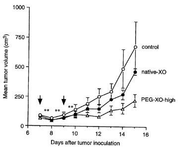
The results of this therapeutic tactics using XO and hypoxanthine
against S- 180 solid tumor model in mice demonstrates the
significant suppression of tumor growth (Fig. 1 and 2). These
results suggest that . i ) reactive oxygen molecular species,
which are generated by the reaction between XO and hypoxanthine,
have a potent antitumor activity, and ii) the reaction between
XO and hypoxanthine occurs in the solid tumor or around
7 _
CA 02245397 1998-08-21
its periphery. In contrast thereto, native XO showed no significant
antitumor activity under the conditions used (Fig. 1 ).
Systemic side effects of PEG-XO/hypoxanthine therapy, which
were evaluated by using the body weight as a parameter,
seem to be not so significant. The results showed only a
transitory body weight loss, on day 8-9, but recovered on
day 10 (Fig. 3). No serious or significant hematotoxicity or
liver toxicity was seen.
For treating cancer by using the antitumor agents of the
present invention, a therapeutically effective amount of a
chemically conjugated oxidoreductase is first administrated,
and of ter allowing a lapse of adequate time for the accumulation
of the chemically conjugated oxidoreductase in the tumor,
its substrate is administrated. The effective and low-toXicity
dosage of the substrate is 10- 100 mg/kgbody weight/day.
The proper time to administrate the substrate is preferably
6 to 100 h after the administration of the chemically conjugated
oxidoreductase.
The antitumor agents of the present invention may be administered
in the form of an injection or an oral dosage. In the case
of injection, any subcutaneous, intramuscular, intravenous,
intraarterial or local/direct injection is applicable. The type
of preparation for oral administration may be selected optionally.
Some examples of this type are tablets, granules, pills, liquid
medicines, either of the oil or the aqueous type syrup, troches,
and drops.
_8_
X4015-8
CA 02245397 1998-08-21
For preparing the preparation, as well known in the
art, pharmaceutically acceptable carriers or diluents may be
used. In addition, other conventionally used additives may be
optionally incorporated.
In an embodiment, the present invention provides an
antitumor agent kit for a cancer patient. The kit comprises:
(a) a preparation comprising an antitumor effective
amount of an oxidoreductase which is chemically conjugated
with a biocompatible polymer in admixture with a
pharmaceutically acceptable carrier or diluent,
(b) a preparation comprising a substrate for the
oxidoreductase in admixture with a pharmaceutically acceptable
carrier or diluent, and
(c) a written matter which states that the preparation
(b) should be taken by the patient after the preparation (a).
EXAMPLES
The present invention is described by examples shown
below
- 8a -
74015-8
CA 02245397 2004-07-26
74015-8
in detail which do not limit the scope of the invention.
Procedure of Synthesis
Example 1: Synthesis of PEG-XO
XO from bovine milk (Sigma Chemicals, St. Louis, MO, USA)
was first purified by ultrafiltration and concentrated with
the use of an Amicon~ system with a PM 30~ membrane (cutoff
size 30, 000). The concentration of the XO solution was adj'us'ted
to 10 mg/ml protein with 50 mM sodium phosphate bufi'er
(pH 7.4). To the XO solution, succinimide activated-PEG (Mw
l0 5000, Shearwater Polymers, Huntsville, AL) was added at molar
ratios of PEG over the ~ -amino group of lysine in X0, of
1. 2 and 6.7, respectively, to prepare PEG-XOs having a low
and a high extent of PEG conjugation.
Unreacted PEG derivatives with functional groups, decomposed
components, and other impurities were removed similarly by
ultrafiltration using a PM-10 membrane as mentioned above.
The conjugates thus obtained were stored in 50 mM sodium
phosphate buffer (pH 7. 4) containing 1 mM sodium salicylate
at 4 °C.
2o Physicochemical and Biochemical Characteristics
Example 2: Determination of the extent of the PEG conjugation
The extent of the PEG conjugation was determined by the
loss of free amino groups as a result of the PEG-coupling.
2, 4, 6-Trinitrobenzenesulfonic acid was used to quantify the
free amino groups of PEG-XO spectroscopically as described
by Fields (Methods Enzymol. , vol. 25, p. 464-468, 1972). Glycine
was used as a standard amino acid. The protein concentrations
*Trade-mark
- 9 -
CA 02245397 2004-07-26
74015-8
of both native XO and PEG-XO were quantified by using
the DC Protein Assay kit (Bio-Rad Laboratories, Hercules,
CA, USA). The excess feed molar ratio of succinimidyl PEG
at 1. 2 6. 7 to ~ -amino groups oflysine in XO resulted
or
in 17 S6 49 6 conjugation of PEG toX0, respectively. PEG
or
-XO having a portion of PEG of 17 ~ or 49 6 modified amino
groups by the conjugation are hereinafter referred to as "PEG
-XO-low" and "PEG-XO-high", respectively.
The molecular weight of these PEG-XO conjugates were 383
kDa or 543 kDa, respectively, which were estimated on the
basis of the conjugation degree, ie. , of the loss of amino
groups as obtained by the TNBS assay. The results are shown
in the Table 1.
Table 1. Physicochemical and Biochemical Characteristics of
Native-XO and PEG-XO
Feed ratio % Mw XO activity
(PEG/amino group conjugated(kDa) (U/mg
molar ratio) protein)
Native-XO - 0 298 2.12
PEG-XO-low 1.2 17 383 2.40
PEG-XO-high 6.7 49 543 1.15
Example 3: Size exclusion chromatography
The increase of the molecular size of XO after PEG conjugation
was demonstrated by means of size exclusion chromatography
using the FPLC system (Pharmacia LKB, Uppsala, Sweden)
equipped with a Superose'~ 6 HR 10/30 column (Pharmacia LKB)
using a mobile phase of 50 mM sodium phosphate buffer (pH
*Trade-mark '
- 10 -
CA 02245397 1998-08-21
7.4). Elution of the conjugates were detected at 280 nm (Fig.
4 ).
Example 4: Enzyme activity of PEG-XO
The enzyme activity was determined by quantifying the formation
of uric acid from hypoxanthine by measuring the increase
of absorbance at 290 nm, an absorption maximum of uric acid.
The initial concentration of the substrate hypoxanthine was
50 ,u M. The enzyme reaction was carried out in 50 mM sodium
phosphate buffer (pH 7. 4) at room temperature. One unit of
XO activity is defined as the velocity of the formation of
1 a mol of uric acid per min. The results are shown in the
Table 1.
PEG-XO-low showed slight increase of the activity ( 110 °6)
compared with native XO. PEG-XO-high, even after a 49
conjugation of the amino group, retained 54 ~ of the original
enzyme activity of native XO.
Pharmacokinetic studies
Example 5: In vivo distribution of PEG-XO conjugate after
intravenous injection
In vivo distribution of native XO and PEG-XO-high was examined
by using radioemitting 1251-labeled derivatives. Both radiolabeled
native-XO and PEG-XO-high were prepared by the chloramine
T method.
Sarcoma 180 tumor cells were implanted subcutaneously with
2 x 10⁶ cells in male ddY, 6-week-old mice, weighting 30-35 g,
from SLC Inc. , Shizuoka, Japan. The organ or tissue distribution
study was performed on day 7-10 after the tumor inoculation,
when the tumors were 5-7 mm in diameter, but contained
-il-
CA 02245397 1998-08-21
no necrotic region.
1251-Labeled native-XO or PEG-XO-high was administered
to mice via the tail vein (100,u 1/mouse). After 24 h, the
mice were sacrificed, and blood samples were drawn by cardiac
puncture, and they were then subjected to reperfusion with
heparin containing saline to remove blood components in the
blood vessels of the tissues. The tumor tissue as well as
normal tissues including the brain, liver, spleen, muscle, skin,
heart, lung, colon, and kidney were collected and weighed.
The radioactivities of those tissues were measured by a gamma
counter.
As shown in Fig. 5, PEG-XO-high was found to significantly
improve both the blood and the tumor accumulation compared
with that of native-X0, whereas slight or negligible increase
in accumulation in other normal organs was observed for PEG
-XO-high. Furthermore, less accumulation of PEG-XO-high
in the kidney was observed than with native-XO.
Example 6: Time course of tumor accumulation of PEG-XO coniugate
The time course of the tumor accumulation of PEG-XO-high
was examined by measuring the enzyme activity derived from
PEG-XO-high in the tumor tissue. Tumor bearing mice were
prepared as described above. PEG-XO-high (2U; ml, 100 a 1)
was injected intravenously ( i. v. ) to the mice. Af ter a given
period, the tumor tissue was removed as described above.
The tumor tissue was then homogenized with three volumes
of 20 mM potassium phosphate buffer pH 7. 6 which contained
2 mM ethylenediaminetetraacetic acid, 2 mM amidinophenylmethanesulfonyl
fluoride, 10 mM dithiothreitol, 0. 5 ,u g / ml of leupeptin. The
homogenates were centrifuged at 10, 000 g for 20 min, and
- 12 -
CA 02245397 1998-08-21
each supernatant was applied to a FPLC system with a Superose
6 HR 10/30 column similar to the previous section. The enzyme
activity of the PEG-XO-high fraction was determined fluorometrically,
i. e. , the formation of fluorescent isoxanthopterin from pterin
was measured with an excitation at 345 nm and an emission
at 390 nm, in which hypoxanthine was replaced with pterin
as substrate. The quantification was made using the calibration
curve of the authentic isoxanthopterin (Aldrich Chemical, Milwaukee,
WI).
The tumor accumulation of PEG-XO-high with its enzyme
activity was demonstrated by measuring the XO activity of
the homogenate of the tumor before and 24 hrs after the PEG
-XO high injection. The results are shown in Fig. 6.
In S- 180 solid tumor tissue without the administration of
PEG-XO-high, XO activity appears only in a fraction corresponding
to native X0. This means that small amount of XO had existed
in the tumor tissue endogenously (Fig 6A). On the other hand,
with the solid tumor tissue after PEG-XO-high injection (0. 2 U/mouse),
a new large peak of XO activity was observed at the molecular
weight range different from native XO. This new peak consisting
of fraction numbers of 11, 12, and 13 corresponds to the
molecular weight range of PEG-XO-high as demonstrated in
Fig. 4.
Thus, Fig. 6A and 6B show that the XO activity in tumor,
corresponding to the molecular weight range of PEG-XO-high,
appeared after the PEG-XO-high administration.
In addition, the PEG-XO-high activity increased in a time
dependent manner (Fig. 7).
Antitumor activity in vivo
- 13 -
CA 02245397 1998-08-21
Example 7 : Antitumor activity of PEG-XO in vivo
Sarcoma 180 were implanted subcutaneously with 2 x 10⁶ cells
in male ddY, 6-week-old mice, weighting 30-35 g. When the
tumors became palpable (5-7 mm in diameter), usually 7 days
after implantation, the treatment was resumed.
After native-XO or PEG-XO-high was intraveneously injected
( 2 times, 0. 6 U / mouse, the first time 7 days and the second
time 9 days after the tumor inoculation) to the mice. Hypoxanthine
( 13. 3mg / kg ) was injected intraperitoneally six times, each
time more than 6hrs after the last time, as indicated by asterisks
(*) in the Fig. 1 to 3. A significant (p < 0. 05) suppression
of the tumor growth was observed in mice administered with
PEG-XO-high. However, a similar treatment by native XO
showed no significant reduction of the tumor growth (Fig.
1). The weights of tumor 15 days after the tumor inoculation
were 0. 36 ~ 0. 12 g (control), 0. 31 ~ 0. 03 g (native XO treatment),
and 0. 22 ~ 0. 05 g (PEG-XO-high treatment), respectively,
which indicates that 39 °6 inhibition of the tumor growth was
achieved by only twice administration of PEG-XO-high.
By three administrations of PEG-XO-high (on 7, 8, and 9
days after the tumor inoculation, 0. 6 U / mouse) and subsequent
6 intraperitoneal injections of hypoxanthine ( 13. 3mg/kg) as
indicated by asterisks (*), a remarkable antitumor activity
of PEG-XO-high was observed (Fig. 2). 13 days after the
tumor inoculation, the weights of the tumor were 0. 5'7 ~ 0. 24
g (control) and 0. 13 ~ 0. 08 g (PEG-XO-high treatment), corresponding
to a 77 °6 inhibition of tumor growth by PEG-XO-high.
Example 8: Systemic side effect of PEG-XO
- 14 -
CA 02245397 1998-08-21
In order to examine the systemic side effect of PEG-XO-high
administration, the change of the body weight after the PEG
-XO-high administration was investigated. PEG-XO-high was
intravenously injected 3 times (on 7, 8, and 9 days after
the tumor inoculation, 0. 6 U/mouse). Hypoxanthine (13. 3
mg/kg) was intraperitonealy injected 6 times as indicated by
the asterisks (*).
As shown in Fig. 3, a slight but significant decrease of
the body weight was observed on day 8 and day 9, but then
the body weight recovered to normal level on about day 10
or later. Thus, toxicity is reversible and transitory.
5. BRIEF DESCRIPTION OF THE DRAWING
Fig. 1 shows the effect of native-XO and PEG-XO-high on
the growth of S-180 solid tumor in ddY mice (2 times administration).
Fig. 2 shows the effect of PEG-XO-high on the growth of
S-180 solid tumor in ddY mice (3 times administration).
Fig. 3 shows the body weight change of ddY mice with and
without a treatment with PEG-XO-high/hypoxanthine.
Fig. 4 shows a size exclusion chromatography of native-XO
and PEG-X0.
Fig. 5 shows the body distribution of 1251-labelled native-
XO and PEG-XO-high after intravenous injection to ddY mice
bearing S- 180 solid tumor.
Fig 6A and 6B shows the XO activity of S-180 solid tumor
tissue before and 24 hrs after a PEG-XO high injection, respectively
Fig. 7 shows time dependent accumulation of PEG-XO-high
in tumor tissue.
- 15 -