Note: Descriptions are shown in the official language in which they were submitted.
CA 0224~712 1998-08-07
W O 97/30749 PCTrUS97/02264
TRANSD13~MAL TR~NSPORT USING ULTRA~ONIC STANDING
WAVES
Field of the Invention
The disclosure relates to the transdcrmal sampling of extracellular
fluid. The present disclosure provides an apparatus and process for the
e~hanced transdermal transport of drugs or other substances using
ultrasound standing waves.
Background of the Invention
Conventional sampling methods for collecting body fluids typically
involve invasion of the organism (e.g., physical disruption of the skin).
Such invasive processes are both painful and messy. The difficulty and
pain involved with the process provides a disincentive to the patient to
p~rform the procedure.
Several techniques have been reported that involve little or
minim~l invasion of the skin. Exemplary such techniques are
sonophoresis, iontophoresis and vacuum.
The use of iontophoresis requires using electrodes containing
2 0 o cidation-reduction species as well as passing electric current through the skin. Iontophoresis has also been used to increase skin
permeability. Despite the effective use of iontophoresis for skin
permeation enhancement, there are problems with irreversible skin
damage induced by the transmembrane passage of current.
2 5 Vacuum has been reported to draw fluid transcutaneously while
avoiding the complications of invasive procedures. The use of vacuum
to extract fluid across the skin is limited because of the relative
impermeability of the stratum corneum.
The art discloses methods of using ultrasound traveling waves to
enhance the rate of permeation of a drug medium into a selected area
of contact of an individual or to enhance the rate of diffusion of a
substance through the area of contact of an individual. The use of
ultrasound traveling waves may induce localized skin heating.
r Thus, there continues to be a need to provide a process and
3 5 apparatus for sampling extracellular fluid across the skin of an Anim~l.
.. The present disclosure provides ultrasonic standing waves toenhance permeation and mass transport through skin. While prior art
te:chniques use ultrasonic traveling waves to enhance permeation of the
skin, traveling waves do not enhance mass transport of the interstitial
CA 0224~712 1998-08-07
W 097/30749 PCT~US97/02264
fluids. Standing waves on the other hand may promote permeation as
well as mass transport. High velocity gradients exhibited by a standing
wave sound field can provide enhanced mass transport specifically at
boundary layer and at air-fluid interfaces within the structures of sl~in.
Purthermore, standing waves differ from traveling waves in
radiation force. As understood in the art, radiation force is the time-
average force exerted on a rigid spherical object immersed in a sound
field over a number of cycles. In other words, the radiation force of a
traveling wave is the gradient of the l~inetic energy density minus the
gradient of the potential density plus a phase factor. In contrast, the
sum of the kinetic and potential energy density of a standing wave is
independent of distance, and so their gradients are equal in magnitude
but opposite in sign. The phase factor equals zero since it is constant
with distance. Thus, the force for the standing wave is a constant times
the gradient of the potential energy density whose maximum is equal to
twice the potential energy density.
For example, in a traveling wave of pressure amplitude A, a
particle is acted on by a small steady state force in the direction o~ the
wave. If the wave is uniform, then the force is the same independent of
2 0 the particle's position. However, in a standing wave the total pressure
amplitude varies in space or position. The maximum amplitude is 2~
and occurs in planes spaced at a half-wavelengths apart. The radiation
force on the particle varies in both magnitude and direction. The force
reverses direction every quarter wavelength.
2 5 The ratio of the maximum standing wave radiation force to the
traveling wave value is approximately (1/kR)3, where R is the particle
radius and k is 6.28 divided by the wavelength. The wavelength in soft
tissue is about 1.5/f millimeters, where f is the frequency in MHz (e.g.
at 1 MHz the wavelength is 1.5 millimeters). If the radius of a particle is
3 0 .01 mm and the wavelength is 1.5 mm, one obtains 0.042 for kR,
.000073 for (kR)3, and 13,600 for (1/kEi~3. As is apparent, the radiation
force produced by a standing wave relative to a traveling wave is
significant. While the radiation force is calculated for rigid spherical
particles, the relationship is applicable to small biological particles such ,~
3 5 as blood cells, intracellular bodies such as chloroplasts, and
mitochondria, as these cells and organelles exist in vivo, since these "
structures in which they are located are comparable to a suspending
medium. Thus~ the radiation force is applicable to biological structures
existing within animal s~in.
CA 0224~712 1998-08-07
W 097/30749 PCTAUS97/02264There are several advantages to the use of standing waves in
enhancing skin permeability and mass transport for diagnostic
sampling. First, the energy required for diagnostic sampling is less than
th!at required for traveling wave techniques. This is evident with the
5 fact that the radiation force generated by a standing wave is larger in
comparison to a traveling wave of the same energy. Second, a standing
wave using significantly less intensity but effectively producing the
necessary permeability and, in addition mass transport effects, would
alleviate the danger of bioacoustic effects. In addition, acoustic sources
10 of low energy typically require less electrical power and are more
amenable to miniaturization. Finally, the acoustic effect of standing
waves can be localized within the stratum corneum, which is the rate-
limiting barrier to transport in skin, while low frequency traveling
waves tend to penetrate deeply into skin significantly beyond the
15 stratum corneum. This can potentially cause undesirable bioeffects at
bone-tissue interfaces that produce discomfort to a subject undergoing
treatment, e.g., drug delivery or extracellular-fluid-extraction for
di agnostic purposes.
The present disclosure provides, in part, a surface-acoustic-wave
2 0 (SAW) device to generate standing waves within the stratum corneum
region as a means for enhancing permeability and mass transport of
analytes across the skin. A SAW device provides safe-coupling of sound
field adjacent to skin since the electrodes needed to excite the waves
are mounted on the opposite side of the acoustic device away from skin.
Brief Description of the ~rawin~s
In the drawings, which form a portion of the specification:
Figure 1 illustrates one embodiment of an apparatus of the
invention for the transdermal transport of extracellular fluid.
~,nmmary of the Invention
In one aspect, the present invention provides a process of
sampling extracellular fluid across the skin of an animal comprising
,. establishing an ultrasonic standing wave across the skin and collecting
3 5 fluid transudate.
In a preferred process of the invention, the standing wave is
generated by a surface acoustic wave device.
In alternate processes of the invention, the standing wave is
established by generating an ultrasonic wave of a given wavelength
CA 0224~712 1998-08-07
W 097/30749 PCTrUS97/02264
~rom an ultrasound transducer located at a first position on the external
surface of the skin and reflecting that wave from an ultrasound
reflector located at a second position on the surface of the skin, wherein
the half-trip distance of ultrasonic wave travel between the first and
S second location is equal to integer multiple number of half-
wavelengths. Optionally, the pressure on the surface of the skin in the
vicinity of the ultrasonic standing wave may be reduced, preferably by
applying a partial vacuum to the surface of the skin.
The invention further provides an apparatus for the transdermal
10 sampling of extracellular fluid. In a preferred apparatus of the
invention, the standing wave is generated by a surface acoustic device
and includes means for collecting transudate.
In alternate embodiments, the apparatus includes means for
generating an ultrasonic wave through the skin of an animal, means for
15 reflecting the ultrasonic wave sonically aligned with the means for
generating the wave such that when the apparatus is positioned on the
skin the half-trip distance of ultrasonic wave travel between the means
for generating and the passive reflector is equal to integer multiple
number of half-wavelengths, and means for collecting transudate.
2 0 Optionally, the pressure on the surface of the skin in the vicinity of the
ultrasonic standing wave may be reduced, preferably by applying a
partial vacuum to the ~urface of the skin.
In preferred embodiments of this aspect of the invention, thc
reflector may be a passive reflector sonically aligned with the
2 5 transducer such that, when the apparatus is positioned on the skin, the
half-trip distance of wave travel between the transducer and the
passive reflector is equal to integer multiple number of half-
wavelengths. In other preferred embodiments, the reflector is a second
transducer, sonically aligned with the first transducer such that, when
3 0 the apparatus is positioned on the skin, the half-trip distance of
slltrasonic wave travel between the two transducers satisfies the
multiple number of half-wavelength resonant condition.
Detailed Description
3 ~ The present disclosure provides an apparatus and process for
enhancing permeability and mass transport through skin, preferably
the skin of a human. The apparatus comprises a device suitable to
produce standing waves within the region of the stratum corneum. The
process includes steps of establishing standing waves within skin and
CA 0224~712 1998-08-07
W 097/30749 PCT~S97/02264
transport of compounds contained in fluid transudate to appropriately
positioned sensors for detection and/or analysis of the compounds.
Any compound which can be delivered to the body through the
skin or can be sampled from the body via the skin is suitable for use or
5 detection by the processes and devices disclosed herein. Among such
compounds well known in the art are compounds of clinical and/or
therapeutic significance, such as glucose, cholesterol, insulin, estradiol,
and other hormones or proteins, potassium, sodium, calcium, etc. The
preferred compound is glucose.
As used here, the term "ultrasound" means ultrasonic radiation of
a frequency above 20 kH~. Ultrasound used for most medical purposes
employs frequencies ranging 50kHz to 100 MHz.
The term "standing wave" means that acoustic wave forms
e~hibited within a medium remain fixed in position while the amplitude
of the waves fluctuate repetituously from maximum to minimum over
the total operating distance of the wave. The distance between adJacent
nodes or antinodes is equal to integer multiples of half-wavelength.
The phases of wave form~ between two nodes or antinodes are
constant .
2 ~ In contrast, traveling waves have amplitudes that remain
constant. A traveling acoustic wave can be characterized by a
parameter of intensity. ~ntensity is the average power transported per
Wlit area and is defined in a traveling acoustic wave. The intensity in a
standing wave is zero.
2 5 In accordance with the present invention, ultrasound frequencies
greater than 20 kHz and less than 300 MHz are preferable. The most
preferable frequencies are those which, when converted to
wavelengths, are comparable to single or multiples of cellular
dimensions. Such frequencies will provide oscillation and mechanical
3 0 mlotions within the fine-structure of cells and lipid-bilayers that
irlfluence movements or fluid motions within the stratum corneum.
The time period during which the standing wave is generated is
t~pically from about 30 milliseconds to 60 minutes, more preferably
r from about 10 seconds to 20 minutes. The most preferred time is 30
seconds to 3 minutes.
Any type of device can be used to :l~lminister the ultrasound,
which can be pulsed or corltinuous. The ultrasound is preferably
continuous at lower frequencies and pulsed at very high frequencies to
dissipate generated heat.
CA 0224~712 1998-08-07
W097/30749 PCTrUS97/02264
The preferred intensity of the applied ultrasound is less than
about ~.0 W/cm2, more preferably from about 0.2 to about 5.0 W/cm2,
and most preferably from about 0. ~ to about .03 W/cm~ .
In the process of the invention, the standing wave is established
5 by generating an ultrasonic standing wave of a given frequency and
distributing the wave over the surface of the skin. The "foot print" of
the ultrasonic standing wave is not critical to the invention and is
typically in the form of a circular or rectangular surface area.
~n ultrasonic standing wave càn be established in a number of
10 ways. One preferred apparatus capable of establishing a standing wave
is a surface-acoustic-wave (SAW) device. SAWs are commercially
available and are well suited for enhancing the permeation of the
stratum corneum since the surface waves travel parallel to the surface
and do not penetrate the skin to any significant degree, e.g., at most to
15 about 1 00 micrometers. A SAW device is compact and can constructed
so as to elimin~te direct electrical contact with the skin by placing the
electrical contacts on a side of the device away from the sl~in. A ~AW
device is typically characterized by an electrically excited surface
acoustic wave in a piezoelectric single-crystal plate substrate by use of
2 0 a metallic (e.g. aluminum) interdigital transducer (IDT) structure. As is
understood in the art, an IDT structure comprises a row of metallic
electrodes laying parallel and adjacent, but not touching each other.
Each electrode has an alternating applied voltage potential. Typical
substrates are quartz, lithium niobate~ and lithium tantalate, but other
2 5 substrate materials are known and are suitable for use in the invention,
e.g., piezoceramics such as lead-zirconate-titanate ~PZT), zinc oxide
(ZnO), and polyvinylidene-fluoride ~PVDF). The specific operating
characteristics of these materials, such as direction of particle
displacement of the wave, is defined by the cut of the substrate. The
3 0 anisotropy of the piezoelectric crystals allows different angles of cut
with very different properties.
An alternate method of providing a standing wave is a transverse
vibrating wire. The wire is secured at each end as to satisfy the
stand}ng wave resonant condition and is caused to vibrate at a desired
3 5 frequency. The device is applied parallel to the skin and the field
emanating ~rom the side of the wire is used as previously described,
e.g., to enhance permeation and mass transport. Structures resembling
wires can also be fabricated from silicon or other suitable materials
using microfabrication techniques well known in the electronic
:
CA 0224~712 1998-08-07
W O 97/30749 PCT~US97/02264
industry. ~uch structures can be made to operate analogous to metallic
wires and can be incorporated and operated in a similar manner
previously described for mating and extraction with skin.
In further embodiments, a combination of ultrasonic transducers
and reflectors is arranged on the surface of a patient extremity such
that the necessary spacing of multiple number of half-wavelength of
ultrasonic wave to establish a standing wave is satisfied.
Single transducers can be positioned perpendicularly to the
interface of different layers of skin or at the tissue-bone plane.
Multiple transducers and reflectors can be positioned on the same
plane. In one embodiment of a process of the invention, a transducer
and a passive reflector are utilized to establish the standing wave.
In yet another embodiment of the invention, a second transducer
can be used as a reflector. The two emitting transducers establish the
standing wave when they are operated at the same frequency and are
positioned, as is well known in the art, to satisfy the multiple number of
ha,lf-wavelength resonant conditions.
As is well known in the art, the location of the transducer with
respect to the reflector depends on the frequency of excitation needed
2 0 to establish a standing wave via the interior of the skin. The half-trip
distance of ultrasonic wave travel between the transducer and reflector
should be equal to integer multiple number of half-wavelengths.
A sinusoidal voltage at a given frequency is applied to the
tr~msducer to produce an ultrasonic wave that propagates from the face
2 5 of the transducer. The wave travels through the interior of the skin
and exits at the reflector of the same diameter but is reflected back to
the source transducer.
The half-trip distance between the transducer and reflector
causes the wave to resonate and be confined between the transducer
and reflector. The amplitude of the wave is controlled by the amplitude
of the sinusoidal voltage signal.
As is well known, the oscillation of the transducer can be
stabilized to compensate for drift using the current-voltage phase
relation occurring at the resonant response of the transducer.
3 5 By changing the phases of the signals applied to each transducer,
. complex movement of tissues and microcirculation between tissues can
occur creating more fluid flux through the skin (transudation). The
transudate can then be analyzed using appropriate sensors and/or
detectors. In preferred embodiments, an absorbent pad or material
CA 0224~712 1998-08-07
W 097130749 PCTrUS97/02264
receives the iluid from which the content of fluid can be analyzed using
appropriate sensors.
In the processes and apparatus of the invention, impedance
mismatches can be reduced by applying a coupling agent to the surface
5 of the transducer and reflector.
The coupling agent should have an absorption coefficient similar
to that of water, be non-staining, non-irritating to the skin, and slow
drying. It is clearly preferred that the coupling agent retain a paste or
gel consistency during the time period of ultrasound ~lministration so
l 0 that contact is maintained between the ultrasound source and the skin.
Exemplary and preferred coupling agents are mixtures of mineral
oil, glycerin, and propylene glycol, oil/water emulsions, and a water-
based gel. A solid-state, non-crystalline polymeric film having the
above-mentioned characteristics can also be used.
The description and operation of a particular embodiment of the
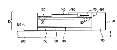
invention maybe understood with reference to ~igure 1. As shown in
Figure l, the device 10 includes a housing 20 which surrounds the
internal mechanism and provides one or more attachment sites 1 8 0
(two shown) for fixing the device 10 to the patient's skin 200. The
2 0 device 10 is configured to fit snugly on the surface of the skin. Within
the device 10 is located the ultrasound source, e.g., fabricated from a
thin PZT-5A piezoelectric crystal substrate 110. Typically, it is
anticipated that the ultrasonic source 110 will be operating in the
region of 1 to 3 MHz. Thus the thickness of a device operating at 1 M~Iz
2 5 is approximately 0.4 mm thick. A representative area of a substratc
100 is 1 cm by l cm. Pairs of opposing metal electrodes 120 are
deposited on the top surface of the PZT substrate. The distance
between the electrodes 12 0 is determined by the operating fre~uency,
in this example multiples of 2.26 mm. The metal electrodes 120 can be
3 0 approximately l micron wide and approximately l mm long.
Alternatively, multiple finger-interdigital electrodes (not shown) can
also be utilized. In such, embodiments, the electrode fingers are
typically spaced at fractions of the operating wavelength from each
other and two opposing IDTs are excited by sinusoidal inputs from a
3 5 function generator and power amplifier. A battery 170 powers an
electronic chip 140 capable of providing memory and control functions.
In addition, the electronic chip 140 can provide sinusoidal outputs
amplified in an appropriate manner as an alternative to the individual
generator and amplifier. A display 160 provides means by which the
CA 0224~712 1998-08-07
W 097/30749 PCT~US97/02264
operation of the device, its functioning and results are provided to the
user. Optionally, the device can include a port (not shown) allowing
connection to an external computer and thus allowing the health care
provider the ability to more closely monitor the patient's condition.
The excitation frequency of the SAW device might drift due to
external conditions and operating environments. Therefore the
electronic chip should contain a close-loop portion such a Phase-Lock-
Loop (PLL). Since the SAW has two opposing ID~s, one of them can
provide the feed~ack sensing input to the PLL. The chip 140 is also
1 Q capable of providing a variety of other excitation functions such as
s~uare pulses. The chip 140 can also provide different modulation and
phase shift functions to the SAW device. These modulation and phase
shifts can provide additional bioeffects to the stratum corneum regions
of the skin 200. By providing excitation to the IDTs 120, an acoustic
l 5 beam is caused to propagate between the IDTs and a resulting wave is
established within the region. The opposite side of PZT substrate is
coated with a thin layer typically equivalent to about a one-quarter
w avelength of material, typically glass 100, to maximize coupling to a
coupling agent 1~0 between the substrate 110 and skin 200.
2 0 The coupling agent also functions as a means for transporting
e:~tracted fluids containing the compound of interest (metabolites,
dîffusing species, etc.) to a sensor for detection. Permeation through the
coupling membrane can take place by two mechanisms; viscous flow
alld diffusive flow. The viscous flow mechanism can facilitate the
2 5 rnovement of extracted transudate and the diffusive flow can facilitate
the diffusion of metabolites. A hydrated polymeric membrane or
hydrogel can be formulated as to have the capacity to absorb more fluid
in proportion to the solid proportion of the hydrogel. A specific volume
of extracted transudate can then be transported to the hydrogel for
3 0 subsequent analysis by appropriate detection means. As stated above,
the coupling agent medium should have a similar impedance with skin
when placed between the sound source and skin in order to provide
efficient transfer of acoustic energy into skin. Since the acoustic
. impedance of skin is similar to the impedance of water, it is assumed
3 5 that the acoustic wave is propagating in water and therefore the
optimal configurations and characteristics of the extraction apparatus is
designed to operate with a water interface.
Optimally, the components of the device are housed in a thin
molded plastic device 20, e.g., a patch. As shown in Figure 1, the
CA 0224~712 1998-08-07
WO 97130749 rCT~US97/02264
excitation or control electronic chip 140 and the batteries 170 are
preferably stored within a separate compartment or layer 19 0 of the
patch. In this way the functional elements and the control electronics
can be physically separated such that the sound source is attached to
the skin and the electronics are contained in small package, similar to a
electronic paging devices, that can be located elsewhere near or on the
body, e.g., attached to a belt. Optionally, the excitation of a SAW device
110 can be performed in a wireless fashion since the SAW is capable of
receiving an excitation wave from a wave propagating through free
l 0 space at specific wavelengths. In this embodimentt a separate sending
unit containing electronic controls and transmitter provides the
excitation wave. Preferably, the sending unit is located near the
vicinity of the SAW-containing patch so as to minimi7e transmission
requirements. The patch is attached to the surface of the skin via one
or more attachment sites 1 80 using bioadhesives. In addition or
alternatively, the patch can be further secured to the skin in the form
of a bracelet or watch.
When a standing wave from the sound source is applied to the
surface of skin it is physically transferred into the skin through the
2 0 glass 100 and coupling agent 150 layers. While not wishing to be
bound by any one theory, the penetration of the wave into the skin is
limited to within a few wavelengths beyond the thickness of the
stratum corneum, which is approximately 15 micrometers. It is
believed that the deepest penetration is approximately 100
2 5 micrometers. As exhibited in any standing waves, cells and their
constituents such as lipid-bilayers will migrate toward pressure nodes.
The amplitude of displacement is determined by the elastic nature of
the fine-structure of the stratum corneum. The stratum corneum will
exhibit dense regions as well as regions sparse in materials. The cells in
3 0 the stratum corneum such as keratenocytes that are asymmetric in
shape, being long in one axis and short in another axis, will rotate to
align with the preferred axis of the standing wave to minimi7e energy
with the acoustic field. The lipid-bilayer channels between corneocytes
provide regions capa~le of producing acoustic microstreaming near
3 5 boundary layers. The microstreaming generate high velocity gradients
w~}ich enhance mass transport of compounds within the extracellular
flui~ (ECF). The effect of the standing wave is thus to create transient
intercellular pores through the stratum corneum. In conjunction with
the surface standing wave, secondary standing waves are produced
1 0
CA 0224~712 1998-08-07
W 097130749 PCTrUS97/02264
within the corneocytes. These secondary waves arise from flexural
coupling modes of the corneocytes. Corneocytes contain approximately
half water and half keratin and thus have boundaries defined by
diifferences in densities. As material density is a parameter of the
5 p~ropagation of acoustic waves, velocity gradients are produced within
the corneocytes which provide enhanced mass transport of E(~F and
subsequent intracellula~ permeation. The combined effect of the
s~lrface standing wave and the secondary standing wave is to produce a
region of skin which exhibits enhanced permeability and convective
10 transport of molecular species and fluids from one side of the stratum
corneum to the other.
The extracellular fluid (ECF) is extracted and diffuses through the
transport medium. The transport medium is then analyzed for the
presence or amount of the compound of interest. Of course, the
15 particular analytical technique utilized will be selected depending on
the compound of interest, ease of use, sensitivity, etc., and other factors
well known to the clinician or diagnostician. The detection and analysis
can be accomplished in situ or somc or all of the transport medium can
be removed from the device for analysis. Several different methods
2 0 are know which are suitable for use in the method and apparatus of the
invention, e.g., amperometric and optical detectors.
Alternatively, a SAW device is used in the form of a mass sensor.
A layer of the SAW substrate is coated with biologics, such as receptors
or antibodies, reactive to ECF compounds or metabolites, and the
2 5 presence of the compound is detected by changes in the SAW
generated, e.g., a shift in the resonance frequency, a phase shift of the
acoustic wave, or a shift in the amplitude of an acoustic wave. Since the
preferred embodiments utilize a SAW device to extract E~C~F, it is
adlvantageous to incorporate a portion of the substrate within a region
30 capable of providing detection. In this embodiment, another pair of
IDTs can be incorporated onto the SAW substrate in combination with a
detection portion of the fluid transport/coupling medium. This portion
of the fluid transport medium is coated with biologics providing
sp~ecificity to the metabolites of interests. A separate set of electronic
35 contrvls provides excitation of the second set of IDTs as well as
detection of the frequency, phase, or amplitude shifts due to the
presence of the analytes. The extraction and detection functions can
operate at the same frequency or at combinations of frequencies. The
spacing arrangements of the detection IDTs with respect to the
1 1
CA 0224~712 1998-08-07
WO 97f30749 PCT/US97/02264
operating surface provides optimal extraction and detection means.
When appropriate electronics are included, the S~W device can detect
.he presence of increase or decrease in fluid flow by a mass sensing
region and then compensate by the excitation region. Thus, such a S~W
device can provide a complete system capable of controlling fluid
extraction and feedback to as to optimize the EC~ extraction.
Further alternate embodiments include a transducer, e.g., PZT
sandwiched between two thin isolated electrodes, and a reilector. Any
passive reflector can be used in conjunction with the transducer to
establish an ultrasonic standing wave. The passive reflector is
positioned in the apparatus such that, when the apparatus is contacted
with skin, the half-trip distance of ultrasonic wave travel between the
transducer and reflector is equal to multiple of a half-wavelength.
The size of the reflector contacting the slcin is preferably the same
as the size of the transducer that contacts the skin.
In another embodiment, the reflector is a second transducer. The
second transducer is stimulated at the same frequency as the first
transducer to create a standing ultrasonic wave. The position of the
second transducer in the apparatus is such that, when the apparatus is
2 0 contacted with the skin, the distance separating the transducers
satisfies the multiple number o~ half-wavelength resonant condition.
Procedures for establishing such a distance are well known in the art.
Of course, these emhodiments also include coupling agents/fluid
transport medium as previously described.
The present invention has been described with reference to
preferred embodiments. Those embodiments are not limiting of the
claims and specification in any way. One of ordinary skill in the-art can
readily envision changes, modifications and alterations to those
embodiments that do not depart from the scope and spirit of the
3 0 present invention.
1 2