Note: Descriptions are shown in the official language in which they were submitted.
CA 02245888 2005-10-20
30582-17
1
ISOLATION AND AMPLIFICATION OF NUCLEIC ACID MATERIALS
FIELD OF THE INVENTION
The invention relates to the field of purification
and amplification of nucleic acids from nucleic acid
containing starting materials, especially from biological
materials such as urine, faeces, sperm, saliva, whole blood,
serum or other body fluids, fractions of such fluids such as
leucocyte fractions (buffy coats), cell cultures and the
like, but also samples from the environment such as soil,
water and the like.
BACKGROUND OF THE INVENTION
Until recently isolation and/or purification of
nucleic acids from complex mixtures as described above was a
laborious, multi-step procedure. In EP 0389063 a simple and
rapid purification of nucleic acid material from a complex
mixture is disclosed. This procedure comprises treating the
complex mixture, such as whole blood with a chaotropic agent
in the presence of a nucleic acid binding silica solid phase
material under conditions that allow for binding of all
nucleic acid material to said solid phase and separating
said solid phase from the mixture. The reference shows that
both single stranded and double stranded nucleic acids are
bound to the solid phase if present in a mixture. The
reference also discloses amplification (PCR) of a certain
nucleic acid with a known sequence, suspected to be present
in a mixture.
Thus said reference teaches a simple and rapid
detection method for known nucleic acids suspected to be
present in a sample.
CA 02245888 2005-10-20
30582-17
2
In many cases the nature of the target nucleic
acid (double stranded or single stranded) may not be known
beforehand, or there may be many different targets necessary
to be analyzed. In these cases the rapid but rather crude
method described above may not be sophisticated enough and
further separations of the crude material may be wanted.
Fractionation of mixtures of double- (ds) and single-
stranded (ss) nucleic acids (NA) into single- and double-
stranded forms is frequently needed e.g. in the separation
of labelled ss-NA probes from ds-hybrids, in the separation
of in vitro transcripts from ds-DNA templates, and in the
separation of genomic DNA from mRNA. Currently, the
separation of different kinds of nucleic acids can be
accomplished by several techniques. Electrophoresis can be
used to fractionate different forms of nucleic acids,
because of differences in size and shape (1-3).
Centrifugation takes advantage of differences in
density (4), and more recently the technology of high-
performance liquid chromatography (HPLC) has been applied to
separate and purify single- and double-stranded DNA and RNA
molecules (5-8).
RNA purified from eukaryotic cells by the
currently most widely used procedure (9) appears to contain
significant amounts of genomic DNA, an adaptation which
reduces genomic DNA contamination of the ss-RNA fraction has
recently been described (10).
It is not possible to look at single stranded
and/or double stranded material separately using the method
of EP 0389063 because the method does not discriminate
between the two.
CA 02245888 2005-10-20
30582-17
2a
SUNMARY OF THE INVENTION
In one aspect, the invention provides a method for
separating single stranded nucleic acid material from double
stranded nucleic acid material and isolating the single
stranded nucleic acid material, said method comprising:
contacting a mixture of said single stranded nucleic acid
material and said double stranded nucleic acid material with
a first liquid comprising a chaotropic agent and a first
nucleic acid-binding solid phase that is capable of
reversibly binding nucleic acid, wherein said first liquid
has a composition such that said double stranded nucleic
acid material binds to said first solid phase and a
substantial amount of said single stranded nucleic acid
material does not, and separating said solid phase from said
first liquid, whereby a supernatant containing said single
stranded nucleic acid material is formed; and treating said
supernatant containing said single stranded nucleic acid
material with a second liquid comprising a chaotropic agent
and a second nucleic acid binding solid phase also capable
of reversibly binding nucleic acid, wherein the second
liquid has a composition such that the resulting mixture of
said supernatant and said second liquid allow for binding of
said single stranded nucleic acid material to said second
solid phase, whereby said single stranded nucleic acid is
isolated.
BRIEF DESCRIPTION OF THE DRAWINGS
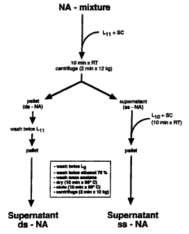
Figure 1. Outline of protocol R.
Recovery of ds-NA takes place from the initial
pellet (R-pellet), recovery of ss-NA takes place from the
initial supernatant (R-sup). L11, L10, L6 and L2 are GuSCN
based-buffers, SC is silica particle suspension. For
details see Materials & Methods section.
CA 02245888 2005-10-20
30582-17
2b
Figure 2. Separation of ds-DNA and ss-DNA.
NA was purified (in duplicate) by protocol R from
a mixture of ds-DNA (phage lambda, HindIII digest, 1 ug) and
ss-DNA (phage M13 DNA, 500 ng). Final elutions were in
50 ul TE and 25 p1 were electrophoresed through a 1% agarose
gel (containing ethidiumbromide) which was subsequently
photographed under UV-illumination. Lane 1, 100% recovery
marker for ds-DNA fragments; lane 2, 100% recovery marker
ss-DNA; lane 3, 100% recovery marker mixture ds-DNA/ss-DNA.
Lanes 4 and 5, output protocol R-pellet; lanes 6 and 7,
output protocol R-sup.
Figure 3. Separation of ds-RNA and ss-RNA.
NA was purified (in duplicate) by protocol R from
a mixture of ds-RNA (Rotavirus ds-RNA) and ss-RNA (phage MS2
RNA, 800 ng). Final elutions were in 50 pl TE and 25 ul
were electrophoresed through a 1% agarose gel (containing
ethidiumbromide) which was subsequently photographed under
UV-illumination. Lane 1, 100% recovery marker for ds-RNA
fragments; lane 2, 100% recovery marker ss-RNA; lane 3, 100%
recovery marker ds-RNA/ss-RNA mixture. Lanes 4 and 5,
output protocol R-pellet; lanes 6 and 7, output protocol
R-sup.
Figure 4. Separation of ds-DNA and ss-RNA.
NA was purified (in duplicate) by protocol R from
a mixture of ds-DNA (750 ng phage lambda digested with
HindIII) and ss-RNA (phage MS2 RNA, 800 ng). Final elutions
were in 50 pl TE and 25 ul were electrophoresed through a 1%
agarose gel (containing ethidiumbromide) which was
subsequently photographed under UV-illumination. Lane 1,
100% recovery marker for ds-DNA; lane 2, 100% recovery
marker for ss-RNA; lane 3, 100% recovery marker for
CA 02245888 2005-10-20
30582-17
2c
ds-DNA/ss-RNA mixture. Lanes 4 and 5, output protocol
R-pellet; lanes 6 and 7, output protocol R-sup.
Figure 5. Separation of ds-DNA and ss-RNA.
NA was purified by protocol R-sup from a mixture
of ds-DNA (1000 ng linearized pHC624, 2 kb) and ss-RNA
(phage MS2 RNA, 800 ng). Final elution was in 50 ul TE, and
25 pl or tenfold serial dilutions of the ss-NA fraction were
electrophoresed through a 1% agarose gel (containing
ethidiumbromide) which was subsequently photographed under
UV-illumination.
Panel A: Upper row: lane 1, HindIII digested phage
lambda DNA; lane 2, 100% recovery marker for ds-DNA and
ss-RNA and serial tenfold dilutions thereof (lanes 3-6).
Bottom row, output of protocol R-sup (lane 2) and tenfold
serial dilutions (lanes 3-6).
Panel B: Ds-DNA was subsequently transferred to a
nitrocellulose filter and hybridized with a 32P-labelled
probe homologous to input ds-DNA. ds-DNA and ss-RNA are
indicated.
Figure 6. Separation of genomic DNA from ss-RNA.
How to deal with trapping of ss-RNA. E. coli
bacteria were directly used as input material for duplicate
extractions by protocol R (lanes 6 and 7, R-pellet; lanes 8
and 9, R-sup). Alternatively, total NA was first purified
by protocol Y using diatoms as NA-carrier (which causes
shearing of genomic DNA). The purified nucleic acids were
subsequently used as input for protocol R (lanes 2 and 3,
R-pellet; lanes 4 and 5, R-sup). Final elutions were in
50 pl TE and 25 ul were electrophoresed through a 1% agarose
CA 02245888 2006-10-12
30582-17
2d
gel (containing ethidiumbromide) which was subsequently
photographed under W-illumination.
Markerlanes 1 and 10 500 ng phage lambda DNA,
(HindIII digested). 23S and 16S rRNA, and ds-DNA molecular
weight markers (23 kb and 2.0 kb) are indicated.
Figure 7 depicts a cDNA synthesis reaction.
Figure 8
Single-stranded nucleic acid was purified from
samples containing HIV-1 RNA and TE (negative control) by
protocol R-sup. and subsequently amplified with the
non-selective RT-PCR.
Panel A: lane 1, 100 bp DNA ladder; lanes 2 and 3
negative extraction controls; lanes 4 and 5 non-selectivily
amplified HIV-1 RNA; lanes 6, 7, 8 and 9 600, 60, 6 and 0
molecules resp. of pHCrec (positive PCR control).
Panel B: Southern blot hybridization with
32 P-labelled HIV-1 probes (containing the GAG, POL and ENV
genes of HIV-1) of the samples shown in panel A. After
overnight hybridization at 65 C in 6 x SSC, 0.1% SDS, 10%
Dextran Sulphate and 50 g/mi salmon sperm DNA, the filter
was subsequently washed under high stringency conditions
with 0.1 SSC/0.1% SDS at 65 C, and autoradiographed on X-ray
film for two hrs. at -70 C. This experiment showed that
most of the bands visible on the ethidiumbromide stained
agarose gel originated from the HIV-1 genome.
DETAILED DESCRIPTION
The present invention therefor provides a method
for separating single stranded nucleic acid material from
double stranded nucleic acid material comprising contacting
CA 02245888 2005-10-20
30582-17
2e
a mixture of the both with a liquid comprising a chaotropic
agent and a nucleic acid binding solid phase, whereby the
liquid has a composition such that double stranded nucleic
acid binds to the solid phase and a substantial amount of
single stranded nucleic acid does not and separating the
solid phase from the liquid. Suitable circumstances to
arrive at such a separation can be determined by the person
skilled in the art.
Circumstances under which double stranded material
binds to the solid material and single stranded material
will vary, however important parameters to obtain such
differential binding are the concentration of the chaotropic
agent, which should roughly be between 1-10 M, preferably
between 3-6 M and particularly about 5 M; the concentration
of chelating agent, which in the case that EDTA is applied
should be equal to or
CA 02245888 2006-10-12
30582-17
3
greater than 10 mM and preferably not higher than 1 M; the pH
of the aqueous solution in which the separation is carried out
should be above 2 when a thiocyanate is used as chaotropic
agent and it should be below 10 because otherwise there is a
risk that the ds material will become ss. The temperature at
which the process is carried out seems to be non-critical,
however, it is probably best to keep it between 4 C and 60 C.
An important aspect of the process is of course that the ds
material remains double stranded during the separation. Under
the circumstances as disclosed above this will normally be the
case if the ds nucleic acid is at least 50 bp long at 40% GC
basepairs. The skilled artisan knows how this length may vary
with lower or higher GC content. In Van Ness et al (26) and/or
Thompson et al (27) it is shown that the whole process depends
on intricate interactions between the factors mentioned
above. Using this disclosure and the cited references the
skilled artisan will be able to adjust the circumstances to
his or her particular process.
Chaotropic agents are a very important feature of the
present invention. They are defined as any substance that can
alter the secondary, tertiary and/or quaternary structure of
nucleic acids. They should have no substantial effect on the
primary structure of the nucleic acid. If nucleic acids are
present associated with other molecules, such as proteins,
these associations can also be altered by the same or
different chaotropic agents.Many chaotropic agents are
suitable for use in the present invention, such as sodium
iodide, potassium iodide, sodium (iso)thiocyanate, urea or
guanidinium salts, or combinations thereof. A preferred class
of chaotropic agents according to the invention are
guanidinium salts, of which guanidinium thiocyanate is most
preferred.
By serendipity we found that ss-nucleic acid did not bind
to silica particles or diatomeous earth in the presence of
buffer L11 (see examples), whereas ds nucleic acid did.
Experiments with different circumstances showed that addition
of Mg2+ or other positive (bivalent) ions to the unbound
CA 02245888 1998-08-13
WO 97/30062 PCT/NL97/00063
4
fraction was of great importance. The best results were
obtained with a concentration of bivalent ion (Mg2+) about
equal to the concentration of the chelating agent (EDTA).
The solid phase to be used is less critical. Important is
that it should bind nucleic acids reversibly.
Many such materials are known, of which a number are
silicium based, such as aluminium silicate and the like,
preferably silica. Silica is meant to include Si02 crystals and
other forms of silicon oxide, such as diatom skeletons, glass
powder and/or particles and amorphous silicon oxide. The solid
phase may be present in any form, it may even be the vessel
which contains the nucleic acid mixtures or a part of such a
vessel. It may also be a filter or any other suitable
structure. Apart from silicium based materials other materials
will also be suitable, such as nitrocellulose (filters), latex
particles and other polymeric substances. A preferred form of
the solid phase is a particulate form, which allows for easy
separation of bound and free material, for instance by
centrifugation. The particle size of the solid phase is not
critical. Suitable average particle sizes range from about
0.05 to 500 pm. Preferably the range is chosen such that at
least 80, preferably 90 % of the particles have a size between
the values just mentioned. The same holds true for the
preferred ranges of which the average particle sizes are
between 0.1 and 200 pm, preferably between 1 and 200 pm. The
binding capacity of a given weight of the particles increases
with decreasing size, however the lower limit of the size is
when particles cannot easily be redispersed after separation
through for instance centrifugation. This will be the case in
starting material rich in nucleic acids containing many
nucleic acids of a higher molecular weight. The particles and
the nucleic acids may form aggregates in these cases. The
person skilled in the art will be able to choose the right
particle size for the particular application envisioned. The
formation of aggregates may be avoided by using fractionated
silica or diatomaceous earth in a number of applications.
CA 02245888 1998-08-13
WO 97/30062 PCT/NL97/00063
A further embodiment of the present invention is a method
for isolating single stranded nucleic acid material from a
mixture of nucleic acid material, comprising the steps of
subjecting the mixture to a method as described hereinabove
5 and treating the supernatant containing the single stranded
nucleic acid material with a second liquid comprising a
chaotropic agent and a second nucleic acid binding solid
phase, whereby the second liquid has a compositon such that
the resulting mixture of supernatant and second liquid allow
for binding of the single stranded nucleic acid material to
the second solid phase.
This way the double stranded nucleic acid material is
removed from the crude mixture and the single stranded nucleic
acid is purified from the remaining still crude mixture in
another single step. Both the double stranded material and the
single stranded material are reversibly bound to the
respective solid phases, so that they may be easily eluted
from said solid phases to undergo further analysis or other
treatments. A very useful further treatment is the
amplification of the (double or single stranded) nucleic acid
material.
Both types can be amplified, or both types may be
converted into one another so that they can be amplified. The
present invention provides in yet another embodiment a method
for amplifying single stranded nucleic acid material
comprising the steps of hybridizing the single stranded
nucleic acid with primers and elongating the probes using an
enzyme which adds nucleotides to the primer sequence using the
hybridized single strand material as a template, whereby at
least one primer comprises a random hybridizing sequence and
an amplification motif.
Single-stranded nucleic acids purified in accordance with
the invention were used as input for a cDNA synthesis reaction
using primers with random 3' ends (tagging primers) for the
first and second strand synthesis (see the outline in Fig. 7).
These tagged cDNAs are then amplified by using only one
PCR primer homologous to the PCR motif present in both tagging
CA 02245888 1998-08-13
WO 97/30062 PCT/NL97/00063
6
primers. The tagging primer used in the first strand synthesis
(TAG 20) has been especially designed to facilitate subsequent
direct sequencing of the resultant PCR products.
In contrast with most other protocols (16-22) the
described method does not need any sequence data at all, and
the majority of amplified products can be visualized on
ethidiumbromide stained agarose gels as discrete bands, which
makes isolation and direct sequencing of the amplified cDNA
feasible. The criteria for amplification are well known in the
art. The length of suitable primers, suitable buffers,
suitable melting temperatures for separating strands, suitable
hybridization conditions can all be determined using standard
handbooks in the field.
Of course the sequences which are exemplified can be
varied without departing from the present invention. It is not
so much important what sequence is used as an amplification
motif, as long as it is suitable for hybridization and primer
extension purposes. Suitable limits depend on the conditions
which can be varied by the person skilled in the art. Usually
primers will be at least 10 bases long and not much longer
than 100 bases.
The amplification embodiments of the invention are
exemplified using PCR (polymerase chain reaction). Other
amplification methods are of course equally suitable.
The exemplified label (or tag) on the primers is DIG
(digoxygenin). However other labels are available and well
known in the art.
The invention will now be explained in further detail in
the following detailed description.
Separation / Isolation
MATERIALS AND METHODS
Source of nucleic acids.
Phage MS-2 ss-RNA (3569 nt), E. coli rRNA (16 and 23S;
1,7kb and 3,5kb respectively), phage M13 ss-DNA (7599 nt) and
CA 02245888 2005-10-20
30582-17
7
HindIiI digested phage lambda ds-DNA were purchased from
Boehringer (Mannheim, Germany). Rotavirus ds-RNA was purified
from feces of an infected individual by protocol Y/SC (11).
Plasmid DNA was purified from E. coli HB101 as described by
Ish-Horowicz and Burke (13) followed by column chromotography
with Sepharose CL2B (Pharmacia, Inc. Uppsala, Sweden). Total
NA was purified from E.coli by protocol Y/D (11).
Chemicals.
Guanidiniumthiocyanate (GuSCN) was obtained from Fluka
(Buchs, Switzerland).
EDTA (Titriplex) and MgC12.6H20 were obtained from Merck
(Darmstadt, Germany). TRIS was obtained from Boehringer
(Mannheim, Germany). The preparation of size-fractionated
silica particles (silica coarse, SC) and diatom suspension has
been described (11). Triton X-100 was from Packard (Packard
Instrument Co., Inc., Downers Grove, Ill).
Composition of buffers.
The lysis/binding buffer L6, washing buffer L2, and TE
(10mM Tris.HCI, 1 mM EDTA; pHs8.0) have been described (11).
0.2M EDTA (pH 8.0) was made by dissolving 37.2 g EDTA (Merck,
Germany) and 4.4 g NaOH (Merck, Germany) in aqua in a total
volume of 500 ml. Lysis/binding buffer Lil was made by
dissolving 120 g of GuSCN in 100 ml 0.2M EDTA (pHs8.0).
Binding buffer L10 was prepared by dissolving 120 g GuSCN in
100 m1 0.35M TRIS.HC1 (pH 6.4); subsequently 22 ml 0.2M EDTA
(pH 8.0) and 9.1 g Triton*X-100 were added and the solution
was homogenized; finally 11 g of solid MgC12.6H20 was added.
The final concentration of MgClz in L10 is about 0.25M. L10 is
stable for at least 1 month when stored at ambient temperature
in the dark.
Fractionation of ds-NA and ss-NA by protocol R.
The procedure is outlined in Figure 1. A 50p1 specimen
(containing a mixture of NA-types in TE buffer) was added to a
mixture of 900 1 L11 and 40 1 SC in an Eppendorf"tube and was
*Trade-mark
CA 02245888 2005-10-20
30582-17
8
subsequently homogenized by vortexing. After 10 min. binding
at room temperature, the tube was centrifuged (2 mia. at
approx. 10.000 x g) which resulted in a silica/ds-NA pellet
("initial silica pellet") and a supernatant containing ss-NA.
To recover.ss-NA forms (protocol R-sup), 900p1 of the
supernatant were added to a mixture of 400p1 Li0 and 40 1 SC
and ss-NA was bound during a 10 min. incubation at room
temperature. The tube was subsequently centrifuged (15 sec. at
approx. 10.000 x g), and the supernatant was discarded (by
suction). The resulting pellet was subsequently washed twice
with 1 ml of L2, twice with 1 ml ethanol 700 (vol/vol) and
once with 1 ml acetone. The silica pellet was dried (10 min.
at 56 C with open lid in an Eppendorf*heating block) and
eluted in 50p1 TE buffer (10 min. at 56 C; closed lid). After
centrifugation (2 min. at approx. 10.000 x g) the supernatant
contains the ss-NA fraction.
To recover ds-NA forms (protocol R-pellet) from the
initial silica-pellet, the remaining supernatant was
discarded, and the silica pellet was washed twice with L11 to
remove unbound ss-NA. The resulting silica pellet was
subsequently washed twice with L2, twice with ethanol 70%,
once with acetone, dried and eluted.as described above. After
centrifugation (2 min. at approx. 10.000 x g) the supernatant
contains the ds-NA fraction.
In the complete procedure (which takes about one hour)
for fractionation of NA by protocol R, only two Eppendorf*
tubes are used.
Fractionation of genodic DMA and sa-NA.
Due to trapping of ss-NA into high-molecular-weight
genomic DNA, protocol R as described above gives only low
yields of ss-NA. This can be circumvented by first isolating
total NA by protocol Y/D (11), which causes some shearing of
the high-molecular-weight genomic DNA, sufficient enough to
prevent trapping of the ss-NA. Total NA thus purified can
subsequently be used as input for protocol R.
*Trade-mark
CA 02245888 1998-08-13
WO 97/30062 PCT/NL97/00063
9
Gel electrophoresis.
In all experiments, NA was electrophoresed (8 to 10 V/cm)
through neutral agarose slab gels containing ethidiumbromide
(l g/ml) in the buffer system (40mM TRIS-20 inM sodium acetate-
2mM EDTA adjusted to pH 7.7 with acetic acid; ethidium bromide
was added to a concentration of 1 g/ml of buffer) described by
Aaij and Borst (14).
Hybridization.
DNA fragments were transferred to nitrocellulose filters
by the procedure of Southern (15) and hybridized with
[alpha-32P]dCTP labelled pHC624 (16) prepared by random
labeling (Boehringer, Germany). Hybridization conditions were
as described previously (12).
RESULTS
Comparison of different GuSCN-containing lysisbuffers
with respect to the binding of different NA-types to silica
particles revealed that only doublestranded forms were bound
when using LIi (which is about 100 mM for EDTA) as binding
buffer; on the other hand both double- and single-stranded
forms were bound in binding buffer L6 (which is about 20 mM
for EDTA) (Table 1). These observations formed the basis for
the development of a protocol (Protocol R) for the
fractionation of single-stranded nucleic acids and double-
stranded nucleic acids (Fig. 1)
Once double-stranded nucleic acid is bound by silica
particles in Lil, a brief centrifugation will separate the
silica/ds-NA pellet from the supernatant containing the
single-stranded forms. Additi.on of this supernatant to a
mixture of silica particles and binding buffer L10 (which is
about 250 mM for Mg2+) the binding of single-stranded nucleic
acids to the silica particles is restored. Double-stranded and
single-stranded forms can subsequently be purified by washing
and eluting the silica-NA complexes (protocol R). Double-
stranded nucleic acid is recovered from the initial silica-
CA 02245888 1998-08-13
WO 97/30062 PCT/NL97/00063
pellet (protocol R-pellet), whereas single-stranded forms are
recovered from the initial supernatant (protocol R-sup).
For optimization of protocol R we performed
reconstruction experiments in which previously purified or
5 commercially available, nucleic acids were mixed and
subsequently fractionated by protocol R.
Fractionation of a mixture of doubl.e-stranded DNA and
single-stranded DNA.
10 The fractionation of a ds-DNA/ss-DNA mixture, into double
stranded- and single stranded forms is shown in Figure 2. The
recovery estimated from the band intensity of the ethidium
bromide stained gel for ss-DNA was about 50%, the estimated
recovery of ds-DNA in the range of 500 bp to 4,6 kb was
80%-90% [similar recoveries were obtained for ds-DNA fragments
in the range of 100-500 bp (not shown)], larger fragments were
significantly sheared as noted before (11). At the level of
detection by Uv-illumination, fractionation into ds- and ss-
forms was complete.
Fractionation of a mizture of double-stranded RNA and
single-stranded RNA.
Figure 3 shows the fractionation of a mixture of ds-RNA
(human Rotavirus genome segments 1-11; for review see 14) and
ss-RNA (phage MS2 RNA) into double stranded- and single
stranded forms. The estimated recovery of ds-RNA and ss-RNA
was at least 80%. At the level of detection by UV-
illumination, fractionation into ds- and ss-forms was
complete.
Fractionation of a mixture of double-stranded DNA and
single-stranded RNA.
In Figure 4 it is shown that ds-DNA can also efficiently
be separated from ss-RNA.
Again are the recoveries for both fractions at least 80%.
Similar results were obtained when E.coli rRNA (23S and 16S)
was used as ss-RNA input (not shown).
CA 02245888 1998-08-13
WO 97/30062 PCT/NL97/00063
11
In the experiments described above, fractionation of ds-
and ss-NA forms (as judged by visual inspection of band
intensities after ethidiumbromide staining and IN
illumination) appeared to be complete. In order to establish
the performance of the fractionation procedure for a mixture
of ds-DNA and ss-RNA into ss- and ds-forms, NA purified by
protocol R-sup from such a mixture was studied by Southern
blotting and hybridization with a 32P-labelled DNA probe,
homologous to the ds-DNA used as input for fractionation. This
experiment revealed that the ss-NA fraction contained less
than 0,1% of the ds-DNA input (figure 5).
Fractionation of a mixture of genomic DNA and single-
stranded RNA.
When we investigated the separation of high-molecular-
weight (genomic) dsDNA and ss-RNA by direct fractionation
using E. coli as input for protocol R, it appeared that the
ds-DNA fraction was heavily contaminated with rRNA (Fig. 6,
lanes 6 and 7), and ss-RNA recovery was low (Fig. 6, lanes 8
and 9). This was likely due to trapping of RNA into high-
molecular-weight (genomic) ds-DNA when silica/NA complexes
were formed. On the other hand no genomic DNA was observed in
the ss-RNA fraction. Total nucleic acid, which was first
isolated using the standard protocol Y/D (11), and hereafter
used as input material in protocol R showed significantly
higher recoveries of the ss-RNA fraction (Fig. 6, lanes 2 and
5).
CA 02245888 1998-08-13
WO 97/30062 PCTINL97/00063
12
Amplifications
MATERIALS AND METHODS
Source of nucleic acids.
HIV-1 RNA was isolated from a virus culture (23), phage
MS-2 RNA was purchased from Boehringer (Mannheim, Germany) and
the 7.5 Kb Poly(A)Tailed RNA and the 100 bp ladder used as a
marker were purchased from Life Technologies (Gaithersburg,
Maryland, USA). The PCR TA3 cloning vector was obtained from
Promega (Madison, USA). Plasmids 5' NOT Hxb2ENN (24)
[containing the GAG and POL genes of HIV-1 from nucleotide
638-4647} and 168.1 RTN (24) [containing the ENV gene of HIV-1
from nucleotide 5674-8474) were purified as described by
Ish-Horowicz and Burke (13) followed by protocol R-pellet as
described in the examples. The plasmid pHCrec used as a
positive control in the PCR experiments was made by a low
annealing PCR on lambda DNA (Boehringer) using PCR primer RB 8
(see below). The discrete PCR products were purified using
protocol Y/D (11) and subsequently cloned in a PCR III vector
(Invitrogen) . The revealing plasmid, pHCrec with a
approximately 600 bp insert was subsequently purified from
E.coli HB101 as described by Ish-Horowicz and Burke (13)
followed by column chromotography with Sepharose CL2B
(Pharmacia, Inc. Uppsala, Sweden).
Chemicals and enzymes
EDTA, KC1, MgC12.6H2O, NaCl and tri-Sodium citrate
dihydrate were obtained from Merck (Darmstadt, Germany).TRIS
and BSA were obtained from Boehringer (Mannheim, Germany).
Triton X-100 was obtained from Packard (Packard Instruments
Co., Inc., Downers, Ill, USA). Sodium Dodecylsulfate (SDS) was
obtained from Serva (Heidelberg, Germany).
The dNTP's and Dextran Sulphate were obtained from
Pharmacia (Uppsala, Sweden).
The chemicals used in protocol R have been described
herein.
CA 02245888 2005-10-20
30582-17
13
Reverse transcriptase SuperScript*II was purchased from
Life Technologies (Gaithersburg, Maryland, USA). DNA
polymerase Sequenase"2 was obtained from Amersham (United
Kingdom). Ampli-Taq*DNA polymerase was obtained from Perkin
Elmer (Norwalk, USA). RNAse H was obtained from Boehringer
(Mannheim, Germany). Salmon sperm DNA was obtained from sigma
(St. Louis, USA).
Composition of buffers and solutions.
The preparation of the buffers used in protocol R have
been described herein, except that the lysis buffer and
washing buffers (L10, Lii, and L2) used in protocol R for the
isolation of nucleic acids were filtered through a column
packed with Diatoms (11) in order to remove any endogenous
nucleic acids in the lysis buffer and washing buffers.
The 10 x reverse transcription buffer (CHB1) consists of
100 mM Tris.HCl (pH 8.5), 500 mM KC1 and 1$ Triton X-100.
The 10 x PCR buffer consists of 500 mM Tris.HC1 (pH 8.3),
200 mM KC1 and 1 mg/mi BSA.
The elution buffer Tris/EDTA (TE, pH 8.0) consists of
10 mM Tris.HC1 (pH 8.0) and 1 mM EDTA (pH 8.0).
oligonucleotide8.
The first strand primer TAG 20:
5' GA AGAATGCCG1e-AATGACCCCNNNNNG 3'
The second strand primer TAG 7:
5' DIG-r,ACAGAATGCCGAAATGAI?NNNNG3'
The PCR primer RB 8:
5'rACAGAA .CCGAAATGA3'
*Trade-mark
CA 02245888 1998-08-13
WO 97/30062 PCT/NL97/00063
- 14
underlined: PCR motif
bold: motif for direct sequencing
N= A, T, C, or G
Protocol for first strand synthesis.
ss-RNA, present in the commercially available reverse
transcriptases, appeared to produce unwanted side products
when used in first strand synthesis, To overcome this problem
reverse transcriptase was first pretreated in a mixture for
cDNA synthesis lacking exogenously added primers:
1 l SuperScript II (200 U/ l)
1 l CMB1 (10 X)
0.5 L MgC12 (100 mM)
0.4 L dNTP's (25 M each)
7.1 L H20
Incubate 15 min. at 37 C
Nucleic acids (20 l) purified by protocol R-sup were
incubated for 5 min. at 60 C and thereafter quenched on ice.
Subsequently the following mixture was added:
3 l. CMB1 (10 x )
1 l TAG 20 (100 ng/ l)
1.5 p.L MgC12 (100 mM)
1.2 l dNTP's (25 mM each)
3.3 .1. H20
Finally 10 gl of the preincubated Superscript II (SS II)
was added and the resulting mixture was incubated for 30 min.
at 42 C.
After the reverse transcription reaction SS II was
inactivated by incubating the mixture for 5 min. at 80 C, and the mixture was
subsequently cooled down to room temperature.
In order to convert the RNA/DNA hybrids into single-stranded
cDNA twenty units of RNAse H were added to the mixture and
incubated for 60 mi-n. at 37 C. The single-stranded cDNA was
subsequently isolated using protocol R-sup. The single-
CA 02245888 1998-08-13
WO 97130062 PCT/NL97/00063
stranded cDNA was eluted in 40 l TE and 20 l was used as
input for second strand synthesis.
Protocol for second strand synthesis.
5 To twenty microliter of single-stranded cDNA, the
following mixture was added (on ice):
4 41 CbIDl (10 x)
1 l TAG 7-DIG* (100 ng/ l)
10 2 91 MgC12 (100 mM)
1.6 l dNTP's (25 mM each)
0.2 l Sequenase 2 (13 U/ l)
11.2 l H20
15 The mixture was incubated for 10 min. on ice, and
subsequently for 60 min. at 37 C. After the second strand
synthesis the double-stranded cDNA was isolated using protocol
R-pellet. The double-stranded cDNA was eluted in 40 l TE.
Twently microliter was taken of and 2 l was used as input for
PCR. The remaining 18 l was stored at -20 C.
Protocol for the polymerase chain reaction.
Two microliters of double-stranded cDNA was added to 48 l
of a PCR mixture consisting of:
18 l TE (pH 8.0)
1 l RB 8 (100 ng/ l)
5 l PCR buffer (10 x)
0.9 l MgC12 (100 mM)
0.2 l dNTP's (100 pm)
0.1 g1 dUTP* (25 }1M )
0.3 l Ampli Taq (5 U/ l)
22.5 l H20
After incubation for 5 min. at 95 C the sample was
subjected to 45 cycles of amplification in a DNA thermal
cycler (type 480; Perkin Elmer Cetus)-. A cycle consisted of
CA 02245888 1998-08-13
WO 97/30062 PCT/NL97/00063
16
denaturation for 1 min. at 95 C, annealing for 1 min. at 63 C,
and elongation for 2 min. at 72 C. After cycling the sample
was incubated for 8 min. at 72 C, and subsequently the
temperature was lowered to 4 C. Twentyfive microliter of the
PCR product was examined by agarose gel electrophoresis and
ethidiumbromide staining. In every experiment TE was used as a
negative extraction control and as a negative PCR control.
*Partial substitution of dTTP with dUTP provides a methodology
for ensuring that products from previous PCR reactions cannot
be reamplified. Products of PCR amplifications will be uracil-
containing deoxyribonucleic acids. Possible contaminating PCR
products from a previous PCR amplification will be eliminated
by excising uracil bases using the enzyme Uracil N-glycosylase
(UNG) prior to PCR (25)
Gel electrophoresis.
In all experiments, the nucleic acids were
electrophoresed (8 to 10 V/cm) through neutral agarose slab
gels containing ethidiumbromide (1 g/ml) in the buffer system
as described by Aaij and Borst (14)
Hybridization.
DNA fragments were detected after Southern blotting (15)
by hybridization with 32P-labelled probes representing the
entire GAG, POL, and ENV genes of HIV-l. {plasmid 5' NOT Hxb2ENN
and plasmid 168 1 RTN)(10).
RESULTS
In parallel, 105 molecules of HIV-1 RNA (23) and negative
controls (TE) were extracted using protocol R-sup. The
resulting single-stranded nucleic acids were amplified by the
non-selective RT-PCR as disclosed above, resulting in a
discrete banding pattern for HIV-1 RNA, and no amplification
products in the TE controls (Fig. 8). The variation between
the duplicates is a reflection of the non-selectivity of the
procedure. As a control for the efficiency of the PCR part of
CA 02245888 1998-08-13
WO 97/30062 PCT/NL97/00063
_ 17
the procedure we used an input of 0, 6, 60 and 600 molecules
of the plasmid pHCrec.
In order to confirm the HIV-origin of the bands visible
in figure 8 A, we performed a Southern blot hybridization
under high stringency conditions with 32P-labelled probes
encompassing almost the entire HIV-1 genome (Fig. 8 B). This
experiment showed that most of the bands visible by
W-illumination hybridized with the HIV-1 probe. The bands
that did not hybridize with the probe might be homologous to
parts of the HIV-1 genome other than those present in the
probe or might originate from single-stranded RNA present in
the HIV-1 RNA preparation (e.g. cellular mRNA) or ss-RNA
present in Superscript II, which was not converted to
ds-hybrids during the preincubation of the SuperScript II.
Similar results were obtained with other single-stranded
RNAs such as hepatitis C virus RNA, phage MS2 RNA, and the 7.5
Kb Poly(A)-Tailed RNA (results not shown).
It iS cOncl.uded that the descri.bed procedure can be used
to amplify single stranded RNA targets (present in e.g. serum)
to a series of discrete bands in agarose gels. The discrete
bands can be purified from agarose gels, cloned in e.g. a
bacterial vector and the clones can subsequently be sequenced.
Due to the fact that one of the tagging primers (TAG 20)
harbours a sequence motif it is possible to sequence the
discrete bands without cloning, after the bands are purified
from gel. The method described here is useful in isolating and
characterizing unknown sequences present in clinical samples
(e.g. viral sequences) or for the amplification of cDNAs from
transcripts without having any sequence data.
CA 02245888 1998-08-13
WO 97/30062 PCT/NL97/00063
18
REFERENCES
1. Serwer, P.(1989) Electrophores.is, 10 (5-6), 327-331.
2. Shain, D.H., Yoo,J., Slaughter, R.G., Hayes, S.E. and
Tae H.J. (1992) Anal. Biochem., 200, 47-51.
3. Sheer, D.G., Yamane, D.K., Hawke, D.H. and Yuan, P.
(1990) Biotechniques, 9, 486-495.
4. Mirkes, P.E. (1985) Anal. Biochem., 148 (2), 376-383.
5. Hoistege, C.P., Pickaart, M.J. and Louters, L.L. (1988)
J. Chromatogr., 455, 401-405.
6. Thompson,J.A. (1986) Biochromatographry, 1, 68-80.
7. Stowers, D.J., Keim, J.M., Paul, P.S., Lyoo, Y.S.,
Merion, M. and Benbow, R.M. (1988) J. Chromatogr., 444,
47-65.
8. Liautard,J.P. (1992) J. Chromatogr., 584, 135-139.
9. Chomczynski,P. and Sacchi, N. (1989) Anal. Biochern., 162,
156-159.
10. Siebert, P.D. and Chenchik, A. (1993) Nuclclei.c Acids
Res., 21, 2019-2020.
11. Boom, R., Sol, C.J.A., Salimans, M.M.M., Wertheim van
Dillen, P.M.E. and van der Noordaa, J.(1990) J. Clin.
Microbiol., 28, 495-503.
12. Boom, R., Sol, C.J.A., Heijtink, R., Wertheim van
Dillen, P.M.E. and van der Noordaa, J.(1991) J. C2in.
Microbiol., 29, 1804-1811.
13. Ish-Horowicz, D. and Burke, J.F.(1981) Nucleic. Acids
Res., 9, 2989-2998.
14. Aaij, C., and Borst, P.(1972) Bioclhim. Biophys. Acta.,
269, 192-200.
15. Southern, E.M.(1975) J. Mol. BioL, 98, 503-517.
16. Froussard, P. (1992) Nucleic Acids Res., 20, 2900.
17. Fritz, J.D., Greaser, M.L. and Wolff, J.A. (1991) Nucleic
Acids Res., 19, 3747.
18. Frohman, M.A., Dush, M.K. and Martin, G.R. (1988)
Proc. Natl. Acad. Sci., 85, 8998-9002.
CA 02245888 1998-08-13
WO 971-30062 PCTINL97/00063
19
19. Schlayer, H.-J., Peters, T., Preisler, S., Fehr, J.,
Gerok, W. and Rasenack, J. (1992) J. Viro1. Methods, 38,
333-341.
20. Trout, A.B., McHeyzer-Williams, M.G., Pulendran, B. and
Nossal, J.V. (1992) Proc. Natl. Acad. Sci., 89, 9823-
9825.
21. Akowitz, A. and Manuelidis, L. (1989) Gene, 81, 295-306.
22. Fukuoka, S.-I. and Scheele, G.A. (1991) Nucleic Acids
Res., 19, 6961.
23. Layne, S.P., Merges, M.J., Dembo, M., Spouge, J.L.,
Conley, S.R., Moore, J.P., Raina, J.L., Renz, H.,
Gelderblom, H.R. and Nara, P.L. (1992) Viro.togy, 1.89,
695-714.
24. de Jong, J., Goudsmit, J., Keulen, W., Klaver, B., Krone,
W., Tersmette, M. and de Ronde, T. (1992) J. of Virol.,
66, 757-765
25. Varshney, U., et al. (1988) J. Biol. Chem., 263,
7776-7784.
26. Van Ness et al. Nucleic Acids Research, vol. 9, NO. 19,
pp. 5143-5151.
27. Thompson et al. Anal.Biochem., vol. 3, pp. 281-291, 1987.
CA 02245888 1998-08-13
WO 97/30062 20 PCT/NL97/00063
NA-type binding in L6 binding in Lil
ds-DNA + +
ds-RNA + +
ss-DNA +
ss-RNA + _
Table 1.
Binding of different NA-types to silica particles in different lysisbuffers;
similar results were obtained using diatoms rather than silica particles (data
not shown).