Note: Descriptions are shown in the official language in which they were submitted.
CA 02246687 1998-11-12
-
Title: A method and apparatus for making an image of a lumen
or other body cavity and its surrounding tissue.
The present invention relates to a method for making an
image of a lumen or other body cavity and its surrounding
tissue in a body wherein a transducer is inserted in the
cavity and ultrasound signals are emitted by said transducer
and directed towards a wall of said lumen or cavity, after
which echo signals are collected and processed in a manner
known per se for ultrasound techniques to form an image of
- the cavity and its surrounding tissue, whereas from the
emitted and collected signals also information is derived
concerning the stiffness of the tissue around the cavity,
which information is displayed in the image that has been
formed. The invention also relates to an apparatus for
making an image of a lumen or other body cavity and its
surrounding tissue in a body comprising a transducer
arranged for insertion in the cavity and for emitting
ultrasound signals to be directed towards a wall of said
lumen or cavity, and for collecting echo signals, and
comprising processing means arranged for processing echo
signals in a manner known per se for ultrasound techniques
to form an image of the cavity and its surrounding tissue,
and to derive information concerning the stiffness of the
tissue around the cavity from the emitted and collected
signals, which information can be displayed in the image
that has been formed.
A method and an apparatus of this kind are disclosed in
an article in Ultrasound in Med. & Biol., Vol. 20, pp. 759-
772, 1994. In said article under the title "Spectral Tissue
Straln: A new technique for imaging tissue strain using
intravascular ultrasound" a method and apparatus for imaging
tissue strain using intravascular ultrasound is described.
The strain information is displayed superimposed over the
conventional echo image in the form of a colored bar graph.
Both color and height of the bars indicate the average
strain along one radius. Strain information is displayed on
CA 02246687 1998-11-12
the periphery of the conventional echo image. The
measurement of strain for each radius is performed for the
entire depth of tissue, and therefore there is no depth
resolution. The method of strain measurement is based on the
change in mean scatterer spacing in tissue as a result of
pulsation. Stiff tissue will deform little due to a given
source of pulsation and therefore the spacing between the
scatterers within that tissue will remain approximately
constant. Softer tissue will deform more and the mean
scatterer spacing will change correspondingly with
pulsation.
The purpose of the current invention is to provide a
method and apparatus of the kind as described with which it
is possible to measure more accurate over a well defined
layer of tissue and with a better resolution.
Elastography, is another approach related to the current
invention (see for instance the articles of Céspedes et al.
in Seminars in International Cardiology, 2, 55-62 and of de
Korte et al. in Ultrasound in Medicine and Biology 23, 735-
746 (1997). In these manuscripts, a method to produce anindependent image of tissue elasticity is described. Due to
the presentation of elasticity information in a separate
image, a different set of advantages and limitations apply
to elastography than to the method described herein. The
methods and material utilized in these papers are the same
ones used in the experiments presented herein. However, the
algorithm and goal of the methods are substantially
different.
The above goal is attained by a method in which the
following steps are comprised:
a) obtaining one or more echo signals from tissue in a
chosen direction, said tissue being at a given state of
mechanical stress;
b) obtaining one or more echo signals from said tissue in
said chosen direction, after the given state of mechanical
stress has changed;
CA 02246687 1998-11-12
c) determining in a manner known per se of the extent of the
cavity in said chosen direction in order to identify the
lumen-tissue boundary;
d) comparing the echo signals from steps a) and b) starting
at the lumen-tissue boundary and for a finite depth in the
tissue to obtain a parameter indicative of tissue
stiffness of the inner layer of tissue in said chosen
direction;
e) simultaneously or successively or intermittently
performing the steps a) to d) for a number of directions;
f) deriving and displaying in a manner known per se the
conventional echo image of the cavity or lumen and its
surrounding tissue from the echo signals obtained in step
a);
g) superimposing in the image obtained in step f) the tissue
stiffness as a suitable coded line along the lumen-tissue
boundary or at other suitable, non-obstructive positions,
and with an apparatus of which the transducer and the
processing means are arranged to perform the steps of:
a) after insertion of the transducer in a lumen or cavity
surrounded by its tissue obtaining one or more echo
signals from tissue in a chosen direction, said tissue
assumed to be at a given state of mechanical stress;
b) obtaining one or more echo signals from said tissue in
said chosen direction, after the given state of mechanical
stress has changed;
c) determining in a manner known per se of the extent of the
cavity in said chosen direction in order to identify the
lumen-tissue boundary;
d) comparing the echo signals from steps a) and b) starting
at the lumen-tissue boundary and for a finite depth in the
tissue to obtain a parameter indicative of tissue
stiffness of the inner layer of tissue in said chosen
direction;
e) simultaneously or successively or intermittently
performing the steps a) to d) for a number of directions;
CA 02246687 1998-11-12
f) deriving and displaying in a manner known per se the
conventional echo image of the cavity or lumen and its
surrounding tissue from the echo signals obtained in step
a);
g) superimposing in the image obtained in step f) the tissue
stiffness as a suitable coded line along the lumen-tissue
boundary or at other suitable, non-obstructive positions.
According to the invention a one-dimensional method is
proposed to measure and display local deformation of the
-~ 10 inner layer of the vessel wall. When possible , the
thickness of this layer will encompass no less than the
entire vessel wall (typically 0.5 to 1.5 mm). Additionally
it is proposed to integrate the strain or elastic modulus
over the entire lumen to obtain a single indicator of cross-
sectional arterial stiffness. Thus, both global (cross-
sectional) and local elasticity information can be obtained
from IVUS (intravascular ultrasound) palpation.
Radial strain is measured from echo signals obtained at
two stages of intraluminal pressure and displayed as a coded
(preferably colored) profile coincident with the location of
the lumen-vessel interface. The corresponding image is
termed the strain palpogram. Alternatively, based on
knowledge of the acting incremental pressure, the stress
(pressure) to strain ratio can be calculated to obtain a
quantity that resembles an elastic modulus. The
corresponding image is termed the modulus palpogram.
In an embodiment of the method of the invention the
given state of mechanical stress of the tissue is actively
changed by expansion of a balloon in said lumen or cavity.
It should be mentioned here that in USP 5,265,612 the use of
a balloon is described to expand in endoluminal elasticity
assessment.
In a different embodiment in which the transducer is
inserted in an artery the steps a~ and b) of the method of
the invention are performed at different pressures in a
cycle of pressure pulsation of the artery.In the above
CA 02246687 1998-11-12
identified article by Talhami et al. also a one-dimensional
approach to imaging arterial elasticity is described. Main
differences between the known method and the now proposed
IVUS palpation technique are the following.
IVUS palpation can measure strain by direct assessment
of the deformation of tissue, whereas in Talhami's approach
tissue strain is measured indirectly from the change in
average spacing of the scatterers in tissue. IVUS palpation
also can be used with a decorrelation approach which is
indirect.
In IVUS palpation the strain or elasticity of a well
defined layer of tissue is measured. The thickness of this
layer is taken to include the thickness of the vessel wall
or an equivalent thickness of the diseased area (plaque).
This is an essential difference because the ability to
measure local strain in regions of stress concentration
supports one of the fundamental uses of this technique.
According to the article Talhami measures the average strain
for the entire thickness of tissue in the echo image.
However, in the Talhami article it is mentioned that
advancing the approach described to RF processing will allow
to achieve some degree of depth resolution.
Probably due to the lack of depth resolution, Talhami et
al. do not mention the possibility to detect (measure) areas
of high stress concentration since this strain in such small
areas would get averaged into the strain estimate for the
entire depth.
In IVUS palpation the strain information is displayed
directly at the inner surface of the artery where the
measurement is performed or at other suitable, non-
obstructive positions. This provides automatic and exact
association of the elasticity information with respect to
the echo image.
While the mechanical properties of the arteries have
been extensively studied in the past, precise quantitative
knowledge of this subject is still lacking. The complete
CA 02246687 1998-11-12
description of the mechanical properties of tissue is very
complicated and involves a large number of elastic
constants, which are difficult or impossible to measure. In
order to obtain realizable estimates of arterial elasticity,
a number of simplifying assumptions are commonly accepted:
the vessel wall is considered to be isotropic, homogeneous,
incompressible and linearly elastic. Based on these
assumptions, three prevailing measures of arterial
elasticity are compliance, distensibility and pressure-
strain elastic modulus. All of these measurements provide aglobal measure of the stiffness of the arterial cross-
section since they are based on the relationship between a
pressure gradient and the resulting change in luminal area.
By definition these measures of arterial stiffness do not
take into consideration the thickness of the arterial wall
or plaque. However, their prevalence implies that reasonably
useful information on the elasticity of the artery can be
obtained even when the thickness is ignored.
In the following measures of the elastic properties of
the arterial wall incorporating a finite thickness of tissue
are described. The local incremental pressure-strain elastic
modulus is defined as
E= - , (1)
where ~P is the pressure change and ~ iS the resulting
strain. Since in general the stress strain ratio is
nonlinear, this incremental p-s modulus is a function of the
mean value of the stress.
The local radial strain in the arterial wall is given by
~R2-~RI (2)
R2 -R,
CA 02246687 1998-11-12
where ~RI is the displacement of the inner layer of wall
tissue, ~R2 is the displacement at a deeper location in
arterial wall and R2 - Rl is the distance between range
gates. The above elastic modulus refers to the stiffness of
the artery wall per se, while other mechanical measures such
as compliance and distensibility refer to the stiffness of
the artery as a hollow structure.
In practice, most arteries requiring ultrasonic
evaluation are abnormal to some extent and contain focal or
diffuse atherosclerotic disease or arteriosclerosis. Except
for few cases, the thin-walled tube approximation is not
valid and local stress and strain values can be
disproportionally large. Nevertheless, useful conceptual
information can be obtained from idealized models of blood
vessels. In an isotropic tube, the radial component of stain
lS glven by
I
~r=(E ~r-v~ -v~z), (3)
where az, ~, and az are the stress components in the radial
circumferential and longitudinal directions, respectively.
Notice that radial strain is compressive and circumferential
and longitudinal stresses are tensile. This equation
indicates that when any stress component increases a
corresponding increase in radial strain can be expected.
Thus, the strain palpogram has the potential to be a good
indicator of increased focal, omnidirectional stress.
Global measures of elasticity in diseased arteries will
deviate from normal values as a result of focal disease.
Consequently, in order to obtain a unified quantitative
measure of the overall stiffness of the cross-section we
define the integrated pressure-strain modulus as
CA 02246687 1998-11-12
~P
Eavg = ~ (4)
Eavg
where
~avg = 2~ )
In general, the integrated pressure-strain modulus does not
directly correspond to any conventional measure of
elasticity, but in the case of a uniform, isotropic, elastic
artery it would correspond to the Young's modulus.
Nevertheless, the mechanical properties of a plaque can be
expected to inflict a noticeable change in the cross-
sectional measure of arterial stiffness. Because Eavg is an
overall measure of the local elastic modulus of the artery
wall per se, it may be a more reliable indicator of the
mechanical properties of the constituent tissues than
compliance or distensibility which totally ignore the
contribution of wall thickness.
The invention will be described in details with
reference to the accompanying drawings, in which
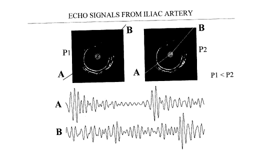
Fig. 1 shows IVUS images of an iliac artery specimen
obtained at two levels of intraluminal pressure ~pressure
change of 2 mm Hg) with rf echo signals;
Fig. 2 shows IVUS image and strain palpogram of a vessel
phantom of uniform elasticity and echogenicity;
Fig. 3 shows IVUS image and strain palpogram of a vessel
phantom containing a soft plaque with echogenicity contrast;
Fig. 4 shows strain palpograms of vessel phantom
containing a hard plaque obtained using three different
regions of interest (layer thickness: 0.4, 0.8 and 1.2 mm);
Fig. 5 shows IVUS images and strain palpograms of an iliac
artery specimen with a stiff plaque at three intraluminal
pressure differentials (1, 2 and 3 mm Hg), and
CA 02246687 1998-11-12
.
Fig. 6 shows IVUS images and pressure-strain modulus
palpograms of an iliac artery specimen with a stiff plaque
at three levels of intraluminal pressure differentials (1, 2
and 3 mm Hg)
In order to evaluate the feasibility of intravascular
ultrasonic palpation, an experimental framework was utilized
based on a clinical IVUS system. The set-up was used to
image gel-based vessel mimicking phantoms and an artery
specimen. These were scanned in the water tank at several
states of static intraluminal pressure. These intraluminal
pressures can be interpreted as the pressure state at
specific time intervals during arterial pulsation or as the
pressure state at different pressurizations of a balloon.
Vessel phantoms were constructed from solutions of agar
and gelatin in water with carborundum (SiC) particles used
for scattering. Combining plastic tubes of different
diameters, gels were molded into vessel-like structures. The
phantom materials and construction method have been
described elsewhere. Additionally, a 45 mm long human iliac
artery specimen was dissected and frozen. The specimen
contained a plaque that was palpable externally. After
thawing, the iliac specimen was scanned at room temperature.
Following standard procedure, histology slides were obtained
by staining and demonstrate a fibrous atheroma.
The experimental set-up consisted of a water tank
equipped with sheaths (8F) at two opposite sides to which
the phantom or specimens were securely attached. A 4.3F, 30
MHz, intravascular catheter (EndoSonics, The Netherlands)
was inserted through one sheath and into the lumen of the
phantom. This sheath was also connected to a variable water-
column system for intraluminal pressurization of the
phantom. Static pressurization levels were used for
compression: a strain on the order of 1% was achieved by a
change in pressure of 4 mm Hg. The artery specimen was
pressurized from 95 to 98 mm Hg in 1 mm Hg steps.
. CA 02246687 1998-11-12
..
The IVUS catheter was connected to a modified Intrasound
IVUS scanner (EndoSonics, The Netherlands) with a stepper-
motor unit that was set to scan the vessel at 400
steps/revolution. The radiofrequency (rf) echo signals were
digitized at 100 MHz, 8 bits using a digital oscilloscope
(LeCroy 9400, LeCroy, Spring Valley, NY) and stored in a
personal computer for off-line processing and display.
Generally, temporal shifts of the echo signals are
related to the corresponding displacements of the tissue
originating the echoes. Under the effect of an intraluminal
pressure differential, stiff tissues will deform less than
softer tissues. This is illustrated in fig. 1, where two
IVUS images of the iliac artery specimen obtained at
different intraluminal pressures are shown. The echo signals
from two selected angles corresponding to normal wall and
stiff plaque are also shown. As a result of the pressure
increment, the distance between echoes from within the
normal arterial wall appears to be compressed, while the
echoes from stiffer plaque show little change in relative
position. Note that this overt difference in the position of
the echoes is practically undetectable on the IVUS images.
Strain, pressure-strain modulus and the average pressure
strain modulus are calculated using Eqs. (1) to (4). Two rf
range gates along a line of sight extending the vessel wall
or sufficiently long to include the vessel wall and an
equivalent part of the plaque are utilized to estimate the
time shift. These range gates can be chosen to be
overlapping or not, and from contiguous or disjoint regions
of the artery. The difference between the time shift divided
by the distance between the range gates is the wall strain.
The beginning of tissue is easily detected in vitro since
blood is replaced by echo free saline. Then, a simple
amplitude thresholding algorithm is used to identify the
lumen. Starting at the lumen, the default analysis thickness
was 1 mm. Additionally, to investigate the dependence of the
palpogram on the analysis thickness, the results from one
CA 02246687 1998-11-12
-
11
phantom were obtained using tissue layers of 0.4 mm, 0.8 mm
and 1.2 mm.
On the echo images of fig. 1 two angles which correspond
to relatively normal vessel wall (labeled A) and fibrous
plaque (labeled B) are identified with dashed lines. The rf
echo signal pairs at those angular position are also shown.
It is clear that while the echoes from direction A appear
compressed, those from direction B have suffered little
change. These changes are not visible from the echo images.
Fig. 1 also shows that discrimination of softer and stiffer
tissue is possible with rf processing. The discussion will
be concentrated on rf processing. However, the robustness
and decreased precision of time delay estimation with
envelope processing may be well suited for IVUS palpation.
In fig. 2 an endoluminal image with strain palpogram of
a uniform vessel phantom void of lesions is shown. The
palpogram shows a constant strain in the phantom wall in
correspondence to the uniform stiffness of the phantom.
In fig. 3 an IVUS image with strain palpogram of a
vessel phantom with a soft plaque is shown. The plaque is
isoechoic and therefore cannot be seen in the echo image.
However, the palpogram demonstrates increased strain, easily
identifying the region of decreased stiffness.
Fig. 4 shows the palpograms of a vessel phantom with a
hard plaque calculated with depths (layer thickness) of 0.4
mm, 0.8 mm and 1.2 mm. A small, albeit noticeable change in
the location of the plaque as a function of the analysis
thickness can be noticed. However, all palpograms correctly
identify the plaque as stiffer than the rest of the phantom
increased strain, thus satisfying the objective of the
imaging approach.
Fig. 5 shows the IVUS strain palpograms obtained at
increasing pressures. Notice the increase in strain,
particularly in the normal wall. Little change is visible in
the stiff plaque. Regions of increased strain are shown
around the edges of the plaque. Strain increases with
CA 02246687 1998-11-12
increased luminal pressure, particularly in the plaque-free
region of the wall.
In fig. 6, the corresponding pressure-strain modulus
palpograms are shown. Note that after conversion to the
modulus the presentation of the palpogram is very similar at
all three pressurizations. The respective average pressure
strain modulus were 78, 83 and 97 kPa. Note the clear
identification of the fibrous plaque as stiff. Although the
plaque is also easily identified in the echo image,
biomechanical information is added without disturbing the
original presentation of the IVUS image.
The ultimate goal of elasticity imaging is to assess the
local deformation and/or modulus of elasticity of tissue. In
general, the linear elasticity properties of tissue is fully
characterized by several elastic constants which can only
be calculated with knowledge of the three-dimensional state
of stress and strain; furthermore, tissue is viscoelastic
and nonlinear. In practice, the internal stress in tissue
cannot be measured and ultrasonic measurement of strain is
limited to precise estimation of the strain component along
the direction of the ultrasound beam. Thus, any practicable
approach must support a number of assumptions with a
resulting accuracy consistent with the expected deviations.
In the past, strain imaging has been performed as a
practical substitute for elastic modulus imaging because the
local stress components were unknown. However, modulus
imaging was considered the optimal technique because it
depicts a basis property of the tissue. Here, an important
clinical application is identified where the strain, rather
than the modulus, is important. Therefore, in the context of
imaging of vulnerable plaques, the strain palpogram is
advantageous over the elastic modulus palpogram. For
characterization of plaque composition, the modulus
palpogram may be more adequate.
Endoluminal ultrasound is routinely used in several
nonvascular applications, e.g., transrectal, endovaginal,
CA 02246687 1998-11-12
endoesophageal, transurethral. With advances in the
miniaturization of endoluminal devices, applications of
ultrasound diagnosis from within one body are certainly on
the rise. Analogously to the situation in intravascular
imaging, additional information on the stiffness of
pathologies is useful in urologic and gastrointestinal
applications. Although the technique of ultrasonic palpation
has been described in the context of intravascular
application, the same principles can be utilized in other
areas. In fact, the phantoms presented which are intended to
emulate blood vessels could also be construed to be scaled
versions of other cavities such as the ureter or the
esophagus. Lacking the pressure source provided by the
pulsation of blood and the acoustic contact provided by
blood, a fluid-filled balloon should be utilized to apply
the probing deformation of the tissues and acoustic
coupling. Thus, ultrasonic palpation of the ureter or the
esophagus, for example, would be a feasible and interesting
area of investigation. In fact, a similar situation arises
in the assessment of esophageal tumors where ultrasound
endoscopy is able to identify the extent but not the
characteristic of the disease.
Strain palpograms of an iliac artery specimen
demonstrate the ability of IVUS palpation to measure local
deformation of the vessel wall and atheroma. Despite the low
magnitude of the effect, regions of stress concentration can
be identified at the shoulders of the plaque. In plaques
with lipid contents, identification of areas of stress
concentration is one main indicator of plaque vulnerability
and no method to obtain this information in vivo is
presently available.
The simplicity and robustness associated with the
ultrasonic palpation concept may allow advancement to a
real-time implementation with which the true potential of
the method can be adequately explored in the clinical
environment.