Note: Descriptions are shown in the official language in which they were submitted.
CA 02249064 1998-09-17
W O 97/32623 PCT~US97/03543
CANNULA AND METHOD OF MANUFACTURE AND USE
BACKGRO ~ D OF THE INrVENTION
The present invention is directed to reinforced
hollow tubes and their methods of manufacture and use. A
specific application of the present invention is for arterial
and venous cardiopulmonary bypass (CPB) cannulas. The
present invention is particularly useful as the arterial
return cannula for the cardiopulmonary bypass system described
in co-pending U.S. Patent Application No. 08/282,192 which is
incorporated herein by reference. The CPB system has an
arterial return cannula which is used to return oxygenated
blood to the patient. An aortic occlusion catheter passes
through the arterial cannula. The aortic occlusion catheter
is used to block blood flow through the ascending aorta and
deliver cardioplegic fluid to arrest the heart for performing
surgery on the heart and great vessels. The aortic occlusion
catheter is inserted through the same lumen in the arterial
cannula which is used to return arterial blood to the patient
so that the arterial blood essentially passes in the annular
space between the aortic occlusion catheter and the arterial
return cannula.
An advantage of the CPB system described above is
that only one opening in the patient's arterial system is
required for both delivery of cardioplegic fluid and return of
arterial blood. In order to achieve optimum blood and
cardioplegic fluid flow rates, the wall of the arterial
cannula must be minimized while retaining enough structural
integrity to prevent kinking and/or cracking. The present
invention is particularly useful in providing a thin walled
cannula which may be used as an arterial return cannula for
the system described above.
CA 02249064 1998-09-17
W097/32623 PCT~S97/03543
A known method of making a reinforced cannula is to
dip a mandrel in a polymer solution and wrap a metal wire over
the polymer. The mandrel is then dipped again to encase the
metal wire between two layers of polymer.
Another known method of making a reinforced cannula
is to extrude a polymer tubing, wrap a metal wire around the
polymer tubing, and extrude another polymer layer over the
metal wire.
A problem with the known methods of manufacturing a
reinforced cannula is that the spacing between adjacent wires
must be relatively large to ensure that the polymer flows
between adjacent coils so that the two polymer layers bond
together to form an integrated structure. Unfortunately, the
relatively large spacing requires a relatively thick polymer
layer to provide the necessary strength since the wire has a
large pitch. The relatively thick polymer layer is also
re~uired to ensure that a sufficient amount of polymer is
provided to fill the relatively large space. The resulting
cannula has a relatively thick wall.
Thus, a specific object of the present invention is
to provide a new method of manufacturing reinforced tubing
and, in particular, cannulas for venous withdrawal and
arterial return of blood for a cardiopulmonary bypass system.
SUMMARY OF THE INVENTION
The present invention solves the problems associated
with prior art cannulas by providing a reinforced, thin-walled
cannula and a method of manufacturing the reinforced, thin-
walled cannula.
An elongate member, such as a steel or polymer wire,
is coated with a coating, preferably a polymer, thereby
forming a coated elongate member. A preferred method of
coating the material is to coextrude the material over the
elongate member. The coated elongate member is then wound
helically around a mandrel and heated so that the coating on
adjacent parts of the elongate member bond together. The
coated elongate member is then mounted to a cannula body.
CA 02249064 1998-09-17
W O 97/32623 PCTrUSg7/03543
In a preferred method, the coated elongate member is
formed so that opposing sides of the coated elongate member
engage one another when the coated elongate member is wrapped
around the mandrel. A preferred cross-sectional shape is
substantially square. An advantage of the present invention
is that the coating does not need to flow between adjacent
portions of the helically-wound member since the coated
elongate members are configured to have sides which engage one
another. In another aspect of the invention, the coated
elongate member is compressed after being wound around the
mandrel. The coated elongate member is preferably compressed
with a heat shrink tube placed over the coated elongate member
before heating. The shrink tube compresses the polymer to
further ensure bonding between adjacent portions of the coated
elongate member.
In another aspect of the present invention, a layer
is positioned over and/or below the coated elongate member.
The layer is preferably positioned over the coated elongate
member and is applied as a tube of material having a larger
inner diameter than the largest outer diameter of the coated
elongate member. The tube is expanded, preferably by
inflating the tube, and the coated elongate member is
positioned inside the tube. The tube is then deflated so
that it contracts around the coated elongate member. The tube
and coated elongate member are then heated to fuse the
elongate member and tube together to form an integrated
structure. Although it is preferred to apply the layer as a
tube, the layer may also be applied by dipping the coated
elongated member in a suitable solution.
An advantage of the cannula of the present invention
is that the cannula has a thin-walled construction while
providing a lumen having a relatively large inner diameter.
- The lumen is particularly suited to receive an aortic
occlusion catheter while still providing enough annular area
between the catheter and lumen wall for return of arterial
blood to sustain full CPB.
CA 02249064 1998-09-17
W097/32623 PCT~S97/03543
These and other aspects of the invention will become
apparent with the following description of the preferred
embodiments.
BRIEF DESCRIPTION OF THE DRAWINGS
Fig. 1 is a front view of an arterial cannula and
introducer sheath for use with an endoaortic occlusion
catheter.
Fig. 2 is a cross sectional view of a hemostasis
fitting for the arterial cannula and introducer sheath of
Fig. 1.
Fig. 3 illustrates the cannula of Fig. 1 with the
endoaortic occlusion catheter introduced into the catheter
insertion chamber.
Fig. 4 illustrates the cannula of Figs. 1 and 2 with
the endoaortic occlusion catheter introduced into the
patient's femoral artery.
Fig. 5 illustrates a multifunction embodiment of the
endoaortic occlusion catheter combined with the arterial
cannula and introducer sheath.
Fig. 6 is a cross-sectional view of a cannula having
a reinforced section coupled to a body.
Fig. 7 is a cross-sectional view of a coated
elongate member wrapped around a mandrel.
Fig. 8 is a cross-sectional view of the coated
elongate member of Fig. 7 after heating and removal from the
mandrel.
Fig. 9 is a cross-sectional view of a second
construction for the reinforced section.
Fig. 10 is a cross-sectional view of a third
construction for the reinforced section.
Fig. 11 is a cross-sectional view of a fourth
construction for the reinforced section.
Fig. 12 is a cross-sectional view of a fifth
construction for the reinforced section.
Fig. 13 is a cross-sectional view of a sixth
construction for the reinforced section.
CA 02249064 1998-09-17
W097/32623 PCT~S97/03~3
Fig. 14 is a cross-sectional view of a seventh
construction for the reinforced section.
Fig. 15 is a cross-sectional view of a eighth
construction for the reinforced section.
Fig. 16 is a cross-sectional view of a ninth
construction for the reinforced section.
Fig. 17 shows an exploded view of another arterial
return cannula.
Fig. 18 shows the distal end of the arterial return
cannula of Fig. 17 before heating.
Fig. 19 shows the distal end of the arterial return
cannula of Fig. 18 after heating.
Fig. 20 shows an enlarged view of the distal end of
an obturator used with the arterial return cannula of Fig. 17
along line A-A.
DESCRIPTION OF THE PREFERRED EMBODIMENT
The invention is directed to cannulas and their
methods of manufacture. A particularly useful application of
the present invention is for arterial and venous
cardiopulmonary bypass cannulas.
Referring to Figs. 1-4, an endoaortic occlusion
catheter 95 is coupled to a cannula 50 that is configured to
serve as an arterial bypass cannula and an introducer sheath
for introduction of the endoaortic occlusion catheter 95. By
configuring the catheter 95 and cannula 50 in this manner,
both devices are inserted through the same arterial opening
which minimizes trauma to the patient. Use of the cannula 50
to receive an aortic occlusion catheter is merely an example
of a use of the present invention and the cannula 50 may be
used for any other purpose. Furthermore, the term cannula as
used herein refers to any hollow body structure, such as a
- catheter or trocar, which is inserted into a patient~s
vascular system. The cannula 50 is coupled to a
cardiopulmonary bypass system (not shown) for delivering
oxygenated blood to the patient~s arterial system. The aortic
occlusion catheter 95 has a lumen which is coupled to a source
of cardioplegic fluid (not shown). The lumen is coupled to an
CA 02249064 l998-09-l7
W 097/32623 PCT~US97/03543
outlet which is distal to the balloon 96. Cardioplegic fluid
is delivered through the lumen and outlet for arresting a
patient's heart when the patient is on full cardiopulmonary
bypass. The balloon 96 occludes the ascending aorta to
prevent oxygenated blood from reaching the coronary arteries
and starting the heart prematurely.
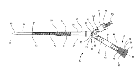
The cannula 50 has a body 51 which is preferably
made of a transparent, flexible, biocompatible polyurethane
elastomer or similar material. In one preferred embodiment,
the body 51 has a 45~ beveled distal end 53, a proximal end
52, a blood flow lumen 57 extending between the proximal end
52 and the distal end 53, and an outflow port 91 at the distal
end 53. Alternatively, the body 51 can have a straight cut
distal end with a chambered or rounded edge. Optionally, a
plurality of additional outflow ports may be provided along
the length of body 51, particularly near distal end 53. The
body 51 is tapered from the proximal end 52 to the distal end
53 and, in one preferred embodiment, the tapered body 51 is
reinforced with a coil of flat stainless steel wire 54
embedded in the wall of the body 51. Adjacent to the proximal
end 52 of the body 51, proximal to the reinforcing coil 51, is
a clamp site 51 which is a flexible section of the body 51
that can be clamped with an external clamp, such as a Vorse
type tube occluding clamp, forming a hemostatic seal to
temporarily stop blood flow through the lumen 57 of the
cannula 50. In a preferred embodiment, the body 51 has a
length between about 10 cm and 60 cm, and preferably between
about 12 cm and 30 cm. In one particular embodiment, the body
51 has a distal external diameter of approximately 7 mm or 21
French (Charrière scale) and a distal internal diameter of
approximately 6.0 mm or 18 French. In a second particular
embodiment, the body 51 has a distal external diameter of
approximately 7.7 mm or 23 French (Charrière scale) and a
distal internal diameter of approximately 6.7 mm or 20 French.
Preferably, the proximal end 52 of the body 51 of either
embodiment has an internal diameter of approximately 3/8 inch
or 9.5 mm. The choice of which embodiment of the cannula 50
to use for a given patient will depend on the size of the
CA 02249064 l998-09-l7
W097t32623 PCT~S97/03543
patient and the diameter of the artery chosen for the arterial
cannulation. Generally, patients with a larger body mass will
require a higher infusion rate of oxygenated blood while on
cardiopulmonary bypass, therefore the larger arterial bypass
cannula 50 should be chosen if the size of the artery allows.
An adapter assembly 65 iS connected to the proximal end
52 of the body 51. In one preferred embodiment, the adapter
assembly 65 and the body 51 are supplied preassembled as a
single, sterile, ready-to-use unit. Alternatively, the
adapter assembly 65 can be packaged and sold as a separate
unit to be connected to the body 51 at the point of use. The
adapter assembly 65 has a Y-fitting 58 which is connected to
the proximal end 52 of the cannula body 51. The Y-fitting 58
has a first branch ending in a barbed connector 59 which is
configured for fluid connection to tubing 92 from a
cardiopulmonary bypass system, as shown in ~ig 4. To prepare
the arterial bypass cannula 50 for insertion into a peripheral
artery, such as a patient's femoral artery or brachial artery,
by an arterial cutdown or by a percutaneous Seldinger
technique, a connector plug 71, which is molded of a soft,
elastomeric material, is placed over the barbed connector 59.
A tapered dilator 67 iS passed through a wiper-type hemostasis
seal 72 in the connector plug 71. The wiper-type hemostasis
seal 72 iS a hole through the elastomeric connector plug 71
that has a slight interference fit with the external diameter
of the dilator 67. A series of ridges can be molded within
the hemostasis seal 72 to reduce the sliding friction on the
dilator 67 while maintaining a hemostatic seal. It is
understood that any other type of hemostasis seal 72 may be
used with the present invention. The dilator 67 has a tapered
distal tip 69, a proximal hub 70 with a luer lock connector,
and a guidewire lumen 79, sized for an 0.038 inch diameter
guidewire, that runs from the distal tip 69 to the proximal
hub 70. The diameter of the dilator 67 iS such that the
dilator 67 substantially fills the cannula lumen 57 at the
distal end 53 of the cannula body 51. The length of the
dilator 67 iS such that the distal tip 69 of the dilator 67
extends approximately 2 to 5 cm, and more preferably 4 to 5
CA 02249064 1998-09-17
W097/32623 PCT~S97/03543
cm, beyond the beveled end 53 of the body 51 when the dilator
hub 70 is against the connector plug 70. The dilator 67 may
assume a bend 73 in it at the point where the dilator 67
passes through the Y-fitting 58 when the dilator 67 is fully
inserted. One or more depth markers 74, 75 can be printed on
the dilator 67 with a nontoxic, biocompatible ink. One depth
marker 74 may be placed so that, when the marker 74 is just
proximal to the hemostasis seal 72 on the elastomeric
connector plug 71, the tapered distal tip 69 of the dilator 67
is just emerging from the beveled end 53 of the body 51. In
one particular embodiment, the tapered dilator 67 is made of
extruded polyurethane with a radiopaque filler so that the
position of the dilator can be verified fluoroscopically.
A second branch of the Y-fitting 58 is connected to an
extension tube 62 which terminates in a hemostasis valve 76
configured to receive the endoaortic occlusion catheter 95
therethrough (Figs. 3 and 4). The extension tube 62 has a
flexible middle section which serves as a proximal clamp site
64 that can be clamped with an external clamp, such as a Vorse
type tube occluding clamp, forming a hemostatic seal to
temporarily stop blood flow through the lumen 63 of the
extension tube 62. The lumen 63 of the extension tube 62
between the proximal clamp site 64 and the hemostasis valve 76
serves as a catheter insertion chamber 66, the function of
which will be more fully explained in connection with Fig. 3.
The hemostatic seal may, of course, be any other type of seal.
In a preferred embodiment of the arterial bypass
cannula 50, the hemostasis valve 76 is a type of compression
fitting known in the industry as a Tuohy-Borst adapter,
however, any other suitable seal may be used. The adapter 76
is shown in greater detail in Fig. 2. The adapter 76 has a
compressible tubular or ring-shaped elastomeric seal 83 that
fits within a counterbore 79 in the fitting body 77. The
elastomeric seal 83 is preferably made from a soft, resilient,
self-lubricating elastomeric material, such as silicone rubber
having a hardness of approximately 20-50 and preferably 40-50
Shore A durometer. The elastomeric seal 83 has a central
passage 84 with a beveled entry 85 on the proximal end of the
CA 02249064 1998-09-17
W O 97/32623 PCTrUS97/03543
passage 84. The elastomeric seal 83 has a beveled distal
surface 86 angled at about 45~ which fits against a tapered
seat 80 in the bottom of the counterbore 79 that is angled at
about 60~. A threaded compression cap 87 screws onto the
fitting body 77. The threaded cap 87 has a tubular extension
89 which fits within the counterbore 79 in the fitting body
77. An externally threaded section 88 on the proximal end of
the tubular extension 87 engages an internally threaded
section 81 within the proximal end of the counterbore 79.
When the threaded cap 87 is screwed down onto the fitting body
77, the tubular extension 89 bears on the elastomeric seal 83
forcing it against the tapered seat 80 of the counterbore 79.
The resultant force on the elastomeric seal 83 squeezes the
elastomeric seal 83 inward to close off the passage 84 to make
a hemostatic seal. When the threaded cap 87 is unscrewed
again from the fitting body 77, the central passage 84 of the
elastomeric seal 83 opens up again. The deliberate 15~
mismatch between the angle of the beveled distal surface 86 of
the elastomeric seal 83 and the tapered seat 80 of the
counterbore 79 prevents the elastomeric seal 83 from binding
and causes the passage 84 to open up reliably when the
threaded cap 87 is unscrewed from the fitting body 87. An
internal ridge 90 within the threaded cap 87 engages in a snap
fit with an external ridge 82 on the proximal end of the
fitting body 77 to keep the threaded cap 87 from being
inadvertently separated from the fitting body 77 if the
threaded cap 87 is unscrewed to the point where the threads
88, 81 are no longer engaged.
In one particular embodiment, the central passage 84 of
the elastomeric seal 83 has an internal diameter of about 5 mm
to allow clearance for inserting a catheter 95 with a shaft
diameter of 3-4 mm through the adapter 76 without damaging the
occlusion balloon 9~ mounted on it. The adapter 76 is
adjustable through a range of positions, including a fully
open position for inserting the balloon catheter 96, a
partially closed position for creating a sliding hemostatic
seal against the shaft 97 of the catheter 95, and a completely
closed position for creating a hemostatic seal with no
CA 02249064 l998-09-l7
W 097132623 PCTAUS97/03543
catheter in the passage 84. In an alternative embodiment, the
passage 84 of the elastomeric seal 83 can be sized to have a
slight interference fit with the shaft 97 of the catheter 95
when uncompressed. In this embodiment, the adapter 76 has
positions which include a fully open position for creating a
sliding hemostatic seal against the shaft 97 of the catheter
95, and a completely closed position for creating a hemostatic
seal with no catheter in the passage 84. In a second
alternative embodiment, a separate ring-like wiper seal (not
shown) is added in series with the adapter 76 to create a
passive sliding hemostatic seal against the shaft 97 of the
catheter 95 without the necessity of tightening the threaded
cap 87. Additionally, the adapter 76, in either embodiment,
may have a tightly closed position for securing the catheter
shaft 97 with respect to the patient. In other alternative
embodiments, other known hemostasis valves may be substituted
for the Tuohy-Borst adapter 76 as just described.
In a particularly preferred embodiment, the internal
surface of the lumen 63 of the extension tube 62 and/or the
internal surface of the lumen 57 of the body 51 are coated
with a highly lubricious biocompatible coating, such as
polyvinyl pyrrolidone, to ease the passage of the endoaortic
occlusion catheter 95, and especially the occlusion balloon
96, through these lumens. Other commercially available
lubricious biocompatible coatings can also be used, such as
Photo-Link~ coating available from BSI Surface Modification
Services of Eden Prairie, MN; sodium hyaluronate coating
available from Biocoat of Fort Washington, PA; proprietary
silicone coatings available from TUA of Sarasota, FL; and
fluid silicone or silicon dispersions. Similarly, a distal
portion of the exterior of the body 51 can be coated with one
of these lubricious biocompatible coatings to facilitate
insertion of the arterial bypass cannula 50 into the artery at
the cannulation site. Furthermore, the endoaortic occlusion
catheter 95 itself, in any of the embodiments described
herein, can be coated with one of these lubricious
biocompatible coatings to facilitate its insertion and passage
through the arterial bypass cannula 50 and the patient's
CA 02249064 l998-09-l7
W097/32623 PCT~S97/03543
11
vasculature. Preferably, the occlusion balloon 96 of the
endoaortic occlusion catheter 95 should be free of any
lubricious coating so that there is sufficient friction
between the expanded occlusion balloon and the interior aortic
wall to prevent accidental dislodgement or migration of the
occlusion balloon 96.
In operation, the arterial bypass cannula 50 iS
prepared for insertion as shown in Fig. 1, with the tapered
dilator 67 in place in the blood flow lumen 57 of the body 51
and with the fitting 76 completely closed. An arterial
cutdown is made into an artery, preferably the patient's
femoral artery, at the cannulation site or a guidewire is
placed percutaneously using the Seldinger technique and the
dilator 67 and the distal end 53 of the body 51 are inserted
into the lumen of the artery with the bevel up. A suture 94
can be tied around the artery 93 where the body 51, as shown
in Fig. 3, inserts to avoid bleeding from the artery 93 at the
cannulation site. The dilator 67 iS then withdrawn from the
body 51, allowing blood to flash back and fill the lumen 57 of
the body 51. When the tip 68 of the dilator 67 iS proximal to
the distal clamp site 56 an external clamp is applied to the
distal clamp site 56 to stop further blood flow. The dilator
67 iS completely withdrawn and the connector plug 71 iS
removed so that a tube 92 from the cardiopulmonary bypass
system can be attached to the barbed connector 59 of the Y-
fitting 58, as shown in Fig. 33. Air is bled from the
arterial bypass cannula 50 by elevating the extension tube 62
and opening the fitting 76 slightly and releasing the external
on the distal clamp site 56 to allow the blood to flow out
through the fitting 76. Alternatively, air can be bled out of
the arterial bypass cannula 50, through an optional vent
fitting with a luer cap (not shown) that can be provided on
the Y-fitting 58 or an infusion line and a three-way stopcock.
The optional vent fitting can be also used as a port for
monitoring perfusion pressure within the arterial bypass
cannula 50. Once the air is bled out of the system, the
external clamp can be removed from the distal clamp site 56
the cardiopulmonary bypass system pump can be turned on to
CA 02249064 l998-09-l7
W 097/32623 PCTAUS97/03543
12
perfuse the patient's arterial system with oxygenated blood at
a rate of about 3 to 6 liters/minute, preferably at a pump
pressure of less than about 500 mm Hg.
To introduce the endoaortic occlusion catheter 95
into the arterial bypass cannula 50, an external clamp 91 is
placed on the proximal clamp site 64, as shown in Fig. 3, to
stop blood from flowing out through the extension tube 62 and
the adapter 76 is opened all the way by unscrewing the
threaded cap 87 to open up the passage 84 through the
elastomeric seal 83. The distal end of the endoaortic
occlusion catheter 95 with the occlusion balloon 96 mounted
thereon is inserted through the passage 84 of the adapter 76
into the insertion chamber 66 of the arterial bypass cannula
50. Optionally, first and second depth markers 98, 99 may be
printed on the shaft 97 of the endoaortic occlusion catheter
95 with a nontoxic, biocompatible ink. The first depth marker
98 on the catheter 95 indicates when the occlusion balloon 96
is entirely distal to the elastomeric seal 83. When the first
depth marker 98 is positioned just proximal to the threaded
cap 87, the adapter 76 should be tightened to create a
sliding, hemostatic seal around the catheter shaft 97. Now,
the clamp 91 can be removed to allow the catheter 95 to be
advanced distally through the arterial bypass cannula 50.
Before the endoaortic occlusion catheter 95 enters
the blood flow lumen 57 within the Y-fitting 58, the perfusion
rate from the cardiopulmonary bypass system pump should be
temporarily turned down to a rate of about 1 to 2
liters/minute to avoid hemolysis, tubing disruptions or other
complications due to the additional flow resistance caused by
the occlusion balloon 96 as it passes through the blood flow
lumen 57. The catheter 95 can now be advanced distally until
the occlusion balloon 96 iS distal to the distal end 53 of the
body 51. A second depth marker 99 on the catheter 95
indicates when the occlusion balloon 96 is entirely distal to
the distal end 53 of the body 51. When the second depth
marker 98 reaches the proximal end of the threaded cap 87, as
shown in Fig. 3, the perfusion rate from the cardiopulmonary
bypass system pump should be returned to a rate of about 3 to
CA 02249064 l998-09-l7
W O 97t32623 PCT~US97/03543
13
6 liters/minute. The endoaortic occlusion catheter 95 can now
be advanced into the ascending aorta for partitioning the
heart and inducing cardioplegic arrest according to the
methods described above. When the endoaortic occlusion
catheter 95 is in position within the ascending aorta the
adapter 76 can be tightened around the catheter 95 to act as a
friction lock to hold the catheter in place.
After completion of the surgical procedure on the
heart, the endoaortic occlusion catheter 95 can be removed
from the cannula 50 by reversing the sequence of operations
described above. The cannula 50 can remain in place until the
patient has been weaned from cardiopulmonary bypass, then the
cannula 50 can be removed and the arterial puncture site
repaired.
It should be noted that for the venous side of the
cardiopulmonary bypass system, a similar dual purpose venous
bypass cannula and introducer sheath with the above-described
features can be used for accessing the femoral vein and for
introducing a venting catheter or other devices into the
venous side of the circulatory system. In a venous
configuration the dual purpose venous bypass cannula and
introducer sheath preferably has an external diameter of about
21 to 32 French units, an internal diameter of about 18 to 30
French units, and a length of about 50 to 75 cm.
2s It should be noted that while several aspects of the
present invention have been illustrated and discussed
separately in the foregoing description, many of these aspects
can be advantageously combined into a single, multifunction
embodiment. As an illustrative example, Fig. 5 shows a
multifunction embodiment of the endoaortic occlusion catheter
160 combining several of the inventive aspects previously
discussed. As discussed above, however, any other aortic
occlusion catheter may be used and preferred aortic occlusion
catheters are described in U.S. Patent Application 08/692,992.
The shaft 164 of the catheter 160 has a coaxial construction
with an inner 161 and outer 162 tubular member. The shaft 164
may be made with varying degrees of stiffness along the length
of the shaft 164, culminating in a soft atraumatic tip 165
CA 02249064 l998-09-l7
W097/32623 PCT~S97/03543
14
which may be highly loaded with a radiopaque filler. The
shaft 164 may be made with a precurved distal portion 166 or
with a precurved distal portion 166 which is out of plane with
the proximal portion of the shaft 164. An expandable
occlusion balloon 163 iS mounted on the distal portion 166 of
the shaft 164. The balloon 163 preferably has a low profile
deflated state with an ellipsoidal shape. In addition, the
balloon 163 may have an eccentric or asymmetrical inflated
profile 163' which would also provide a steering means for the
distal tip of the catheter.
The occlusion balloon 163 iS mounted with its distal
balloon neck 167 attached to the inner tubular member 161 and
its proximal balloon neck attached to the outer tubular member
162. The inner tubular member 161 iS attached at its proximal
end to a first hub 171 and the outer tubular member 162 iS
attached at its proximal end to a second 169 hub 171 which are
axially slidably and/or rotatable with respect to one another.
An infusion fitting 177, such as a luer lock, on the first hub
171 is connected to an infusion lumen 178 which terminates at
the distal end of the catheter 160. An inflation fitting 170,
preferably a luer lock, on the second hub 171 iS connected to
an inflation lumen 179 defined by an annular space between the
inner 161 and outer 162 tubular members which communicates
with the interior of the occlusion balloon 163.
The second hub 169 may be moved proximal and/or
rotated with respect to the first hub 171 to minimize the
deflated profile of the occlusion balloon 163. The lower
deflated profile of the occlusion balloon 163 facilltates easy
insertion of the catheter 160 through a dual function arterial
cannula and introducer sheath 50. When the endoaortic
occlusion catheter 160 iS combined with the dual function
arterial cannula and introducer sheath 50, the shaft 164 of
the catheter 160 should be made with an additional 20-25 cm of
length for a total shaft length of approximately 100-115 cm.
The diameter of the catheter shaft 164 should also be
minimized as much as possible to reduce the amount of cross
sectional area the catheter shaft 164 takes up in the blood
flow lumen of the arterial cannula 50. To this end, this
CA 02249064 1998-09-17
W097/32623 PCT~S97/03543
combined embodiment is made with a distal pressure transducer
172 and a balloon pressure monitoring transducer 173 mounted
on the inner tubular member 161. The distal pressure
transducer 172 and the balloon pressure monitoring transducer
173 are electrically connected to an electrical connector 174
on the first hub 171. Also on the first hub 171 is a
fiberoptic connector 176 which connects to a fiberoptic bundle
175 which terminates with a means for directing a lateral beam
of light at the distal end of the catheter 160 for aortic
transillumination and/or for facilitating nonfluoroscopic
placement of the catheter 160. The fiberoptic bundle 175 may
also be made as a separate unit for insertion through the
infusion lumen 178 of the catheter 160 to further reduce the
catheter shaft diameter while maintaining maximum
functionality. The diameter of the catheter shaft 164 can
thus be reduced to as small as 8 to 10.5 French (2.7-3.5 mm
diameter).
Referring to Fig. 6, a cross-sectional view of
another preferred cannula 201 is shown. A specific
application of the present invention is for arterial and
venous cannulas for a cardiopulmonary bypass system. The
methods and devices described herein in connection with
arresting a patient's heart and placing the patient on
cardiopulmonary bypass are incorporated here for use with the
cannula 201 described below and any other cannula described
herein. The cannula 201 includes a body 203 and a reinforced
section 205. As will be discussed in greater detail below,
the reinforced section 205 has a thin wall which maximizes the
lumen size for a given outer diameter.
Referring to Fig. 7, an apparatus for forming the
reinforced section 205 is shown. The reinforced section 205
of the cannula 201 is preferably manufactured with an elongate
member 207 coated with a coating 209. The elongate member 207
may be made of any suitable material which has the requisite
structural characteristics such as stainless steel, nickel
titanium, or a polymer. A preferred material is 304V
stainless steel wire having a 0.008 inch diameter. The
CA 02249064 l998-09-l7
W 097/32623 PCT~US97103543
16
elongate member 207 may have any cross-sectional shape and a
preferred shape is circular.
The elongate member 207 is preferably coated with
the coating 209 by coextruding the elongate member and the
coating 209. Any suitable coating 209 may be used and
preferred coatings include polymers and specifically
polyurethane, PVC, polyether block amide which can be
purchased from Elf Atochem Inc. under the name PEBAX, and
styrene block copolymer which can be purchased from Shell
under the name KRATON. A preferred polyurethane is
polytetramethylene glycol ether which can be purchased from
Dow under the name Dow 2363 PELLETHANE 80AE.
The coating 209 is extruded over the elongate member
207 so that the coating 209 has opposing sides 211, 212 which
are configured to engage one another when the coated elongate
member 207 iS wrapped around a mandrel 213. A preferred shape
is a quadrangle, and specifically a square, however, any other
shape may be used including irregular shapes so long as the
opposing sides 211, 212 are configured to engage one another.
The square cross-sectional shape preferably has sides having
lengths between 0.010 and 0. 020 inch and more preferably
between 0.010 and 0.015 inch and most preferably 0.014 inch.
The relative dimensions for the thickness of the cannula has
been exaggerated as compared to the inner diameter for clarity
with the actual dimensions being provided herein.
The coated elongate member 207 is wrapped around the
mandrel 213 in a helical shape. The mandrel 213 iS preferably
coated with a lubricious coating such as TFE to prevent
sticking. An advantage of the present invention over other
methods of forming a cannula is that the coating 209 encasing
the reinforcing member 207 does not have to flow between
ad~acent portions of the elongate member 207 since the
elongate member 207 is coextruded to have a shape in which the
opposing sides 211, 212 already engage one another. A shrink
tube (not shown), preferably a heat shrink tube such as a
polyester or fluorinated ethylene propylene (FEP) tube, may
also be positioned around the elongate member 207 to
facilitate bonding. The shrink tube is preferably removed
CA 02249064 1998-09-17
WOg7/32623 PCT~S97/03543
17
after heating. The wound coated elongate member 207 may also
be dipped in a polymer solution such as polyurethane and
tetrahydrofuran (solvent) to enhance the structural
characteristics of the reinforced section 205. Furthermore,
the coating or tube may also be applied over the wound coated
elongate member. Alternatively, a tube may be positioned over
the mandrel 213 and the coated elongate member 207 may be
wound over the tube. The reinforced section 205 may be made
of more than one layer of the coated elongate member 207 and
the coated elongate member 207 may be wrapped in different
directions to increase the hoop and tensile strength.
Although it is preferred that the elongate member 207 has a
constant cross-sectional profile, the elongate member 207 may
also have differing sizes to provide stiff and flexible areas.
After the coated elongate member 207 has been
wrapped around the mandrel 213, the coated elongate member 207
is heated to melt the coating 209 and fuse adjacent portions
of the coating 209 together to form an integrated structure.
The coated elongate member 207 is preferably heated using an
oven, however, any other heating method may be used including
an IR lamp, heating the mandrel 213, or a combination thereof.
The coated elongate member 207 is then cooled and removed from
the mandrel 213 thereby forming the reinforced section 205 of
the cannula 201.
Referring to Fig. 8, the resulting reinforced
section 205 is shown. The coating 209 on the elongate member
207 fuses together so that the coating 209 forms a matrix
which is reinforced by the elongate member 207. Although it
is preferred to heat the coated elongate member 207 to fuse
the material together, the coated elongate member may also be
coated with a solvent before winding the coated elongate
member around the mandrel. The solvent would fuse the
- adjacent material together and would flash off leaving the
fused material.
Referring again to the cross-section of Fig. 6, the
reinforced section 205 has a lumen 215 therethrough for
delivering or withdrawing fluids from a patient. The
reinforced section 205 is attached to the body 203 by any
CA 02249064 l998-09-l7
W097/32623 PCT~S97/03543
18
method and is preferably bonded to the body 203 by insert
molding. The body 203 includes a lumen 217 which is fluidly
coupled to the lumen 215 of the reinforced section 205. The
body 203 has been simplified and may include valves, a Y-
connection, luer connections or any other features.
Furthermore, the body 203 iS preferably configured to engage a
3/8 inch fitting which is a standard size for cardiopulmonary
bypass systems. The lumen 215 of the reinforced section 205
may be any size but preferably has an internal diameter of at
least 0.180 and more preferably at least 0. 236 and most
preferably at least 0. 242 but no more than 0. 375 inch.
A distal end 219 of the cannula 201 has an
atraumatic tip 221 for introduction into the patient. The
atraumatic tip 221 iS preferably an integral extension of the
coating 209 (see Fig. 8) extending beyond the reinforced
section 205. The atraumatic tip 221 has a length of at least
0.050 and a thickness adjacent to the reinforced section which
is preferably the same as the reinforced section.
A proximal end 223 of the reinforced section 205 iS
flared outward slightly so that the proximal end 223 has a
larger lumen than the distal end 219. The proximal end 223
preferably forms an angle of between 2- and 10- and more
preferably between 4-and 6- with respect to a longitudinal
axis of the cannula 201.
The cannula 201 iS particularly useful for arterial
return and venous drainage cannulas for the cardiopulmonary
bypass system described above since the cannula 201 can be
manufactured with a thin wall. As such, the reinforced
section 205 preferably has a thic3~ness between 0.010 and 0 .025
inch and more preferably between 0. 013 and 0. 020 inch and most
preferably between 0.014 and 0. 017 inch. The preferred
thickness provides the necessary structural characteristics
while maximizing the lumen size so that flow rates through the
cannula are optimized. The cannula 201 of the present
invention also has a unique spacing between adjacent portions
of the coated elongate member. Referring to Fig. 8, a gap K
between adjacent portions of the elongate member 207 iS
preferably less than 0.019 inch and more preferably less than
CA 02249064 l998-09-l7
W097/32623 PCT~S97/03~3
19
0.006 inch and most preferably less than 0.004 inch. A
centerline spacing L between adjacent portions of the elongate
member 207 iS preferably less than 0. 022 inch and more
preferably less than 0. 018 inch and most preferably less than
0. 014 inch.
Referring to Fig. 9, a second preferred construction
is shown for the reinforced section 205. The elongate member
207 and coating 209 are preferably the same as described above
in connection with Figs. 7-8, however, another layer 225 iS
positioned either over the elongate member 207 or below the
elongate member 207 to increase the strength of the reinforced
section 205. When the layer 225 iS on the radially inner wall
of the cannula 201, the layer 225 may be applied by dipping
the mandrel 213 in a suitable solution, extruding the layer
over the mandrel 213 or positioning a tube over the mandrel
213. The coated elongate member 207 iS then wrapped around
the mandrel 213 and heated to fuse the coating 209 and layer
225 together. When the layer 225 iS on the radially outer
wall of the cannula, the layer 225 may be applied by dipping
the coated elongate member 207 in a suitable solution after
wrapping the coated elongate member 207 around the mandrel
213, extruding the layer 225 over the coated elongate member
207 wound around the mandrel 213, or positioning a tube over
the coated elongate member wound around the mandrel 213 and
fusing it to the coated elongate member. The coated elongate
member 207 and coating 209 have the same preferred dimensions
described above. The layer 225 has thickness of no more than
0.007 inch and more preferably between 0.001 and 0. 003 inch
and is preferably made of the same materials as the coating
209 described above. Fig. 9 depicts the reinforced section
205 before heating, however, after heating the polymer layer
225 and coating 209 fuse together to form an integrated
structure.
Referring to Fig. 10, a third preferred construction
for the reinforced section 205 iS shown. The reinforced
section 205 iS made according to the same procedure described
above except that a different elongate member 207A iS used.
The elongate member 207A iS preferably made of metal and has a
CA 02249064 1998-09-17
W097/32623 PCT~S97/03543
quadrangle shaped cross-section. A preferred elongate member
is a stainless steel flat wire having cross-sectional
dimensions of 0.005 inch by 0. 020 inch. The elongate member
207A is preferably coextruded with the coating 209 to a
5 thickness of 0. 003 all around although any thickness may be
used. A layer 225A, which is preferably the same as the layer
225 described above, may be positioned on the radially inner
or outer wall of the cannula. The resulting structure yields
an inner diameter of at least 0.180 inch, more preferably at
least 0. 236 inch, and most preferably at least 0. 242 inch and
no more than 0.. 375 inch. The resulting reinforced section
205 has a thickness of 0.011 inch without the layer 225A and
0.013 inch with the layer 225A. The reinforced section 205
may also be formed without the layer 225A so that the wall
thickness of the cannula is minimized. Fig. 10 depicts the
reinforced section 205 before heating, however, after heating
the layer 225A and coating 209 fuse together to form an
integrated structure.
Referring to Fig. ll, a fourth preferred
construction for the reinforced section 205 is shown. The
reinforced section 205 is made according to the same procedure
described above and has the same elongate member 207 as
described in connection with Fig. 70. The coating 209B has an
overlapping portion 227 which lies over an adjacent portion of
the coated elongate member 207B. The elongate member 207B is
a 0. 005 inch by 0.020 inch stainless steel flat wire, and the
coating has a width of 0. 003 inch all around the elongate
member 207. The overlapping portion 227 has a thickness of
0.005 inch and a length of 0.013 inch. The overlapping
portion 227 provides an interlocking relationship between
adjacent portions of the coated elongate member 207. Fig. 11
depicts the reinforced section 205 before heating, however,
after heating the material from adjacent portions of the
coating 209 and the overlapping portion 227 fuse together to
form an integrated structure.
Referring to Fig. 12, a fifth preferred construction
for the reinforced section 205 is shown. The fifth preferred
construction differs from the first through fourth preferred
CA 02249064 1998-09-17
W097/32623 PCT~S97/03543
21
constructions in that the elongate member 207C is not coated
before being wrapped around the mandrel. As discussed above,
a known method of manufacturing reinforced tubing is to
extrude a tube, mount the tube on a mandrel, wind a metal
coil around the tube and position another tube over the coil.
The tubes and coil are then heated so that the inner and outer
tubes bond together. A problem with the known method is that
relatively thick walled tubes are formed since the layers must
be relatively thick to ensure sufficient strength since the
wire must be spaced apart.
The elongate member 207C of Fig. 12 is made of a
polymer, preferably 75D polyurethane, so that radially inner
and outer polymer layers 229, 231 can fuse to the elongate
member 207C to form an integrated structure. Thus, the
polymer layers 229, 231 do not need to fuse together
completely to form an integrated structure which overcomes a
problem with prior art methods of forming reinforced cannulas.
The polymer layers 229, 231, preferably 80A polyurethane, are
positioned on opposite sides of the polymer elongate member
207C. The polymer layers 229, 231 are preferably softer than
the polymer used for making the elongate member 207C. The
elongate member 207C preferably has a diameter between 0.005-
0.020 inch and more preferably between 0.008 and 0.012 inch.
The layers 229, 231 preferably have a thickness of 0.002 to
0.015 inch and more preferably 0.005 to 0.10 inch. The
elongate member 207C is preferably wound so that adjacent
portions of the elongate member 207C contact one another,
however, the polymer elongate member 207C may be wound so that
a space exists between adjacent portions of the elongate
member 207C. Furthermore, although the elongate member 207C
preferably has a circular cross-sectional shape the elongate
member 207C may have any other shape. The polymer layers 229,
231 may be applied in any manner including coextrusion,
dipping or by simply using pre-formed tubes.
The polymer layers 229, 231 are preferably heated so
that they bond with the elongate member 207C. The polymer
layers 229, 23~ are preferably positioned on both sides of the
elongate member 207C before heating the layers 229, 231,
CA 02249064 1998-09-17
W097t32623 PCT~S97/03543
22
however, the layers 229, 231 may also be applied one at a
time. By constructing the reinforced section 205 in this
manner, the polymer does not need to flow completely between
each part of the elongate member 207C to provide an integrated
structure since the layers 229, 231 must simply bond to the
elongate member 207C rather than having to bond with the
opposing layer 229, 231. Fig. 12 depicts the reinforced
section 205 before heating, however, after heating the polymer
material from the layer 225A and coating 209 fuse together to
form an integrated structure.
Referring to Fig. 13, a sixth preferred construction
for the reinforced section 205 is shown with polymer and metal
elongate members 207D, 207E wound together. Two polymer
layers 229D, 231D are positioned on opposite sides of the
elongate members 207D, 207E and may be provided in any manner
described above. The polymer layers 229D, 231D are preferably
softer than the polymer elongate member 207D. A preferred
material for the polymer layers 229D, 231D is 75D polyurethane
and a preferred material for the polymer elongate member 207D
is 80A polyurethane. The soft polymer layers 229D, 231D are
melted to bond to the polymer elongate member 207D thereby
forming an integrated structure. The metal elongate member
207E provides structural strength and is preferably a
stainless steel wire although any metal may be used. Although
it is preferred that the elongate members 207D, 207E have
circular cross-sectional shapes, the elongate members may have
any other shape. Furthermore, although it is preferred that
the elongate members have the same cross-sectional shape, the
elongate members may also have different cross-sectional
shapes. Fig. 13 depicts the reinforced section 205 before
heating, however, after heating the material from the layers
229D, 231D and the elongate member 207D will fuse together to
form an integrated structure.
Referring to Fig. 14, a seventh preferred
construction for the reinforced section 205 is shown. A
polymer elongate member 207F is wound together with a flat
elongate member 207G. The polymer material for the polymer
elongate member 207F may be any polymer and is preferably 75D
CA 02249064 1998-09-17
W O 97/32623 PCT~US97/03543
23
polyurethane. The flat elongate member 207G is preferably the
same as the elongate member 207A described above in connection
with Fig. 10. Two layers of polymer 229F, 231F encase the
polymer and flat wire elongate members 207F, 207G. The
polymer layers 229F, 231F are preferably sof~er than the
polymer material of the elongate member 207F. The polymer
layers 229F, 231F are preferably 80A polyurethane, however,
any polymer may be used. The polymer layers 229F, 231F may be
applied in any manner described above. The polymer layers
229F, 231F preferably have a thickness between 0.002 and 0.010
inch and more preferably between 0.004 and 0.008 inch. The
polymer layers 229F, 231F are heated to bond to the polymer
elongate member 207. Fig. 13 depicts the reinforced section
205 before heating, however, after heating the layers 229F,
231F and elongate member 207F fuse together to form an
integrated structure.
Referring to Fig. 15, an eighth preferred
construction for the reinforced section 205 is shown. A first
elongate member 207H is preferably the same as the elongate
member 207A described above in connection with Fig. 10. A
second elongate member 207J is made of a polymer and has a
thickness between 0.003 and 0.008 inch and more preferably
0.005 inch. Two polymer layers 229H, 231H encase the elongate
members. The layers 229H, 231H are preferably 80A
polyurethane having a thickness between 0.002 and 0.010 inch
and more preferably between 0.004 and 0.008 inch. The polymer
layers 229H, 231H may be applied in any manner described
above. The polymer layers 229H, 231H are heated to bond to
the second elongate member 207J.
Referring to Fig. 16, a ninth preferred construction
for the reinforced section 205 is shown. A first elongate
member 207~ is wound around a mandrel 213 (not shown). The
first elongate member 207L is preferably made of polymer,
preferably 80A polyurethane, and has a T-shaped cross-
sectional shape. The T-shaped cross-sectional shape has a
width of 0.028 inch and a height of 0.008 inch. The first
elongate member 207L has a radial extension 233 having a width
of 0.008 inch. A second elongate member 207M, which is
CA 02249064 1998-09-17
W097/32623 PCT~S97103543
24
preferably the same as the elongate member 207A described
above in connection with Figs. 70, is wound over the first
elongate member 207L. A polymer layer 229L is then positioned
over the first and second elongate members 207T~, 207M and is
preferably 80A polyurethane having a thickness of 0.008 inch.
The polymer layer 229L may be applied in any manner described
above. The polymer layer 229L is then heated so that the
polymer layer 229L and the radial extension 233 bond to one
another to form an integrated structure.
Referring to Fig. 17, another preferred cannula 301
is shown. The cannula 301 is preferably used as the arterial
return cannula for the CPB system described above. The
cannula 301 includes the reinforced section 205 as described
above. A tube 303 connects the reinforced section 205 to a Y-
connector 305 which has first, second and third connections
307, 309, 311. The tube 303 is preferably a flexible tube
made of estane 58810 42D polyether polyurethane. When using
the cannula 301 for the CPB system described above, the first
connection 307 is coupled to a source of oxygenated blood (not
shown) while the second connection 309 receives an aortic
occlusion catheter (not shown). The aortic occlusion catheter
is used to occlude the ascending aorta and deliver
cardioplegic fluid for arresting the patient's heart. The
second connection 309 preferably receives the extension tube
62 and hemostasis valve 876 for receiving the aortic occlusion
catheter in the manner described above in connection with
Figs. 1-4.
A dilator 313 is used to facilitate introduction of
the cannula 301 into the patient's artery. A dilator seal 315
seals the space between the cannula 301 and dilator 313. The
dilator seal 315 and dilator 313 are removed after the cannula
301 has been introduced into the patient. Referring to Fig.
20, the end of the dilator 313 has an enlarged end 319 which
engages an interior wall of the reinforced section 205 when
passing through the cannula 301. The enlarged end 319 is
preferred so that the dilator 313 does not contact the cannula
301 throughout the length of the dilator 313 thereby reducing
CA 02249064 1998-09-17
W097/32623 PCT~S97/03543
the resistance to moving the dilator 313 through the cannula
301.
Referring to Fig. 18, the method of forming the
reinforced section 205is shown. The reinforced section 205
has an elongate member 207N coated with a coating 209N with
the elongate member 207N and coating 209N being any of the
members 207A-M and coatings 209A-M described above in
connection with Figs. 6-16. A preferred elongate member 207N
is a 0.008 inch stainless steel wire which is coated with 80A
durometer polyurethane to a 0.014 x 0.014 inch cross-section.
The elongate member 207Nis wrapped around a mandrel (not
shown), as described above in connection with Figs. 6-16, and
a soft tip 221Nis butted against the elongate member 207N.
The soft tip 221N preferably has the same thickness as the
coated elongate member 207N with a preferred material being
9OA polyurethane.
A layer 225N, which may be the layer 225 described
above, is positioned over the coated elongate member 207N and
the soft tip 221N. The layer 225NiS preferably a tube havin~
a thickness of 0.001-0. 005 inch, more preferably about 0. 003
inch, and is preferably made of the same material as the soft
tip 221N. Although it is preferred to provide the layer 225N
over the coated elongate member 207N it is understood that the
layer 225N may also be positioned on the radially inner
surface of the coated elongate member 207N (or not used at
all). When the layer 225Nis a tube, the tube has an inner
diameter which is slightly smaller than the smallest outer
diameter of the reinforced section 205. The tube is
positioned over the reinforced section by inflating the tube,
inserting the coated elongate member 207N into the tube, and
deflating the tube so that the tube contracts around the
helically wound coated elongated member 207N.By sizing the
layer 225N somewhat smaller than the helically wound elongate
member 207N, close contact between the layer 225N and elongate
member 207Nis ensured.
A heat shrink tube (not shown) is then positioned
over the layer 225N, coated elongate member 207N, and soft tip
221N. The layer 225N, coated elongate member 207N and soft
CA 02249064 l998-09-l7
W 097132623 PCTrUS97/03543
26
tip 221N are then heated to fuse the material together to form
an integral structure as shown in Fig. 19. The tip of the
reinforced member 205 is then trimmed and a tapered mandrel is
inserted into coated elongate member and a heat shrink tube
is recovered over the tip to form a bevel 317 at an end 319 of
the soft tip 221N which facilitates atraumatic insertion of
the cannula 301. The end 319 is curved inward slightly to
form a seal with the dilator 313.
The resulting reinforced section 205 preferably has
an internal diameter of at least 0.180 inch, more preferably
at least 0. 200 inch, more preferably at least 0. 236 and most
preferably at least 0. 242 but no more than 0. 375 inch. The
reinforced section 205 also preferably has a thickness of no
more than 0.0020 inches, more preferably no more than 0.018
15 inches, and most preferably no more than 0. 016 inch. When the
coated elongate member 207N has a 0. 014 X 0.014 inch exterior
surface and the layer 225N has a 0. 003 inch thickness the
resulting thickness is about 0. 0016 inch since about 0.001
inch is lost when the coated elongate member 207N and layer
225N are compressed with the shrink tube during heating. The
unique combination of inner diameter and wall thickness
provides an excellent cannula.
The methods and devices disclosed herein have been
described in conjunction with cannulas, however, it is
understood that the methods and apparatus may also be used for
constructing any other hollow tubes including catheters and
the like. While the above is a preferred description of the
invention, various alternatives, modifications and equivalents
may be used without departing from the scope of the invention.
For example, the opposing sides of the coated elongate member
207 may have an S-shape, and the reinforced section 205 may
have a varying wall thickness. Therefore, the above
description should not be taken as limiting the scope of the
invention which is defined by the claims.