Note: Descriptions are shown in the official language in which they were submitted.
CA 02249099 1998-09-30
OPTICAL IMAGING THROUGH SCATTERING MEDIA: FIT TO AN
INHOMOGENEOUS DIFFUSION MODEL FOR DIF"FERENTIATION
BACKGROUND OF THE INVENTION
Field of the Invention
The present invention relates to an optical method for
imaging through a scattering medium in which a fit is inade to an
inhomogeneous diffusion model. The method provides a simple
means to separate the absorption and scattering contributions of
inhomogeneities.
Background of the Invention
.The ability to optically imaging through a scattering
medium is of great interest. Potential applications are the non-
destructive localization of inclusions or defects in scattering
materials such as composites= or polymers and the detection of
parasites in fish or meat produce. A main target application is
breast cancer detection, which is currently carried out mostly
with X-'rays. X-rays provide good resolution images but with poor
contrast between healthy and cancerous tissues. They are also
considered as potentially hazardous to humans. This explains
that optical imaging through scattering media is an area of
research that has created enormous interest.
Obtaining optical images of the interior of a scatter-
ing medium such as a breast is complicated by the extensive
scattering of light in.such a medium, which results in blurring
of the image.. As a result of such scattering, the trajectory of
a photon (i.e. a light particle) can be predicted only on a
statistical basis, each photon propagating along a random-like
path as shown in Figure 1. In addition to being randomly
redirected by scattering events, each photon alsohas a probabil-
ity of being absorbed by the medium.
CA 02249099 1998-09-30
2
In a slab of material that is highly scattering and
weakly absorbing, such as the human breast, most photons are
reflected towards the entrance surface after traveling only a few
millimeters into the medium. Other photons are absorbed by the
medium or are transmitted to the output surface where they can
be detected. For a breast of typical thickness and optical
parameters, 0.01 to 1%- of the injected photons at a wavelength
around 800 nm are transmitted to the output surface.
The transmitted photons can be separated into three
categories: ballistic photons that reach the output surface
without be scattered, snake photons that are scattered slightly
and maintain a fairly rectilinear trajectory, and diffuse photons
that are widely scattered and cover a considerable volume element
before emerging. Figure 1 illustrates each of these categoriqs.
Ballistic photons do not experience any scattering event and
therefore have the potential to produce a very clear image of the
breast interior. Unfortunately, for typical breast thickness and
optical,parameters, no ballistic photons are transmitted. Snalce
photons have a nearly rectilinear trajectory, and are sufficient
in number to produce a.relatively clear image. Diffuse photons
provide image information of poor quality due to their degree of
scattering. Therefore, researchers have focused their efforts
on detecting the snake photons and excluding the diffuse photons.
Typically this has been done by utilizing time gating techniques.
Time gating is implemented by sending ultra-short laser
pulses inside of a scattering medium. When an ultra-short laser
pulse is injected at the surface of a scattering medium its
component photons propagate along different trajectories. The
different times.of propagation lead to the emergence from the
scattering medium of a temporally broadened pulse which is called
the diffusion pulse or the diffusion curve as illustrated in
CA 02249099 1998-09-30
3
Figure 1. For a breast of typical thickness and optical
parameters, the duration of the diffusion pulse can be as large
as several nanoseconds, which is more than 1000 times the width
of the entrance pulse, typically less than 1 picosecond. The
initial portion of the diffusion curve corresponds to the snake
photons with a shorter path, whereas the remainder corresponds
to the diffuse photons. By their shorter arrival time at the
detector, the snake photons can be isolated and used to construct
the image. This technique, known as tiine gating, created a
resurgence of interest in optical mammography in the early 1990s.
Using only snake photons allows the user to generate
images with better spatial resolution. However, the relative
noise level increases significantly because much fewer photons -
are detected in this way. This method also does not allow for
the determination of the scattering and absorption properties of
an inclusion detected within a scattering medium. In order to
overcome these limitations, researchers have looked for ways to
use the-information carried by all photons, i.e., by the whole
of the diffusion curve, to obtain images through scattering
media, as follows.
The shape and amplitude of the diffusion curve depend
on the scattering and absorption properties of the scattering
medium. A theoretical model, the diffusion model, can be used
to describe the diffusion curve for homogeneous and optically
thick slabs having uniform structure throughout. This model is
appropriate in the specific case of light transinitted through a
human breast. It involves a limited number of parameters that
characterize the scattering and absorption properties of the
scattering medium. The optical properties of scattering media
such as human tissues are usually characterized by three
parameters. The absorption coefficient a is the probability of
CA 02249099 1998-09-30
4
a photon being absorbed per infinitesimal pathlength. The,
scattering coefficient pH is the probability of the photon being
scattered per infinitesimal pathlength. Finally, the third
parameter is the anisotropy factor g which describes the average
change in propagation direction associated with the scattering
process. In addition to these three parameters, it is useful to
define the reduced scattering coefficient as
s' $ (1-g)
representing the average distance over which a photon sustains
a sufficient number of scattering events to randomize its
direction of propagation. The reduced scattering coefficient is
the isotropic equivalent of the scattering coefficient and is
applicable to the case of thick scattering media. The quantities
a and e' are the two main optical parameters that describe the
light propagation in thick scattering media. Those two parame-
ters appear in the diffusion model suitable for a homogeneous
scattering slab.
' Researchers have tried to extract imaging information
from the whole of the diffusion curve through curve fitting.
Curve fitting is a general numerical technique which includes
adjusting a mathematical expression on experimental data. The
idea of using curve fitting in optical mammography is not. new.
The diffusion model has been used as the analytical expression
(valid only in homogeneous cases), and curve fitting was employed
to smooth the experimental data to reduce the level of noise in
the time gating approach.
Since the output of the curve fitting is the parameters
of the analytical model, and since some of the parameters are the
two optical coefficients a and e', the process allows for the
separation of information about the scattering and absorption in
the probed region. Curve fitting can therefore be performed to
CA 02249099 1998-09-30
obtain those optical parameters and to plot their spatial
distributions. As a result, more information is obtained since
two output images are created instead of only one. Because the
analytical expression used in the curve fitting process is the
5 diffusion model, which is valid only in the homogeneous case,
problems develop. In particular, non-uniformity in an inhomoge- ~
neous medium results in non-unifoxmity in the spatial distribu-
tion bf the optical parameters a and B,. Since non-uniformity
is incorrectly described by the model, incorrect .8 and '
distribution are obtained. As a result, an actual spatial
variation of the scattering coefficient may result in a variation
of $ as outputted by data processing and vice-versa. The
foregoing method does not discriminate correctly between scatter-
ing and absorbing effects.
There is a need for a method which provides a simple
mathematical expression which describes the effect of inclusions
on the diffusion curves which will be applicable to generate
images of thick inhomogeneous scattering media.
CA 02249099 1998-09-30
6
SUMMARY OF THE INVENTION
The foregoing and other deficiencies of the prior art
are addressed by the present invention which is directed to an
optical method for imaging through a scattering medium in which
a fit is made to an inhomogeneous diffusion model. The method
facilitates good differentiation between scattering and absorp-
tion. The variation of the diffusion curve associated with the
presence of an inclusion is considered rather than the diffusion
curve itself. An empirical model is provided which describes the
variation of the diffusion curve. A line'ar curve fitting process
is performed to provide two parameters, one parameter associated
with the scattering property of the inclusion and the other
parameter associated with the absorption property of the inclu-
sion.
It is an object of the present invention to provide a
method for optical imaging through scattering medium in which fit
is made to an inhomogeneous diffusion model.
Another object of the present invention is to provide
a method which facilitates good differentiation between scatter-
ing and absorption.
Yet another object of the present invention is to
provide a method in which the variation of the diffusion curve _
associated with the presence of an inclusion is considered rather
than the diffusion curve itself.
Still another object of the present invention is to
provide an empirical model which describes the variation of the
diffusion curve.
Another object of the present invention is to provide
a linear curve fitting process which produces two parameters, one
associated with the scattering property of the inclusion and the
other associated with the absorption property of the inclusion.
CA 02249099 1998-09-30
7
Still another object of the present invention is to
provide significant advantages over previous curve fitting
techniques in that the mathematical expression is extremely
simple.
CA 02249099 1998-09-30
, :r 8
BRIEF ESCRIPTION OF THE IaR.A.WINGS
These and other objects and attributes of the present
invention will be described with respect to the following
drawings in which:
FIG. 1 is a drawing of typical trajectories for the
three categories of photons transmitted through a scattering
medium as known in the prior art;
FIGS. 2a = and 2b are graphs showing diffusion curves
measured through a scattering cell containing scattering and
absorbing inclusions;
FIGS. 3a and 3b are graphs showing the relative
transmissions calculated from the measurements represented in
Figures 2a and 2b;
FIGS. 4a and 4b are the theoretical transmissions
corresponding to homogeneous cases resulting from a uniform
increase of the scattering (a) and absorption (b) coefficients;
FIG. 5a is an iinaging result of a 5mm scattering
inclusion corresponding to the total time-integration of the
diffusion curves;
FIG. 5b is a graph depicting diffusion curves corre-
sponding to the image center and a reference bac}cground pixel;
FIG. 5c is 'an imaging result of a 5mm scattering
inclusion corresponding to the spatial distributions of the curve
CA 02249099 1998-09-30
9
fitted absorption coefficient using the homogeneous diffusion
model;
FIG. Sd is an imaging result of a 5mm scattering
inclusion corresponding to the curve fitted scattering coeffi-
cient using the homogeneous diffusion model;
FIG. 5e is an imaging result of a 5mm scattering
inclusion corresponding to the absorption FIDM parameter;
FIG. 5t is an imaging result of a 5mm scattering
inclusion corresponding to the scattering FIDM paraineter;
FIG. 6a is an imaging result of a 5mm absorbing
inclusion corresponding to the total 'time-integration of the
diffusion curves;
FIG. 6b is a graph depicting diffusion curves corre-
sponding to the image center and a reference background pixel;
FIG. 6c is an imaging result of a 5mm absorbing
inclusion corresponding to the spatial distributions of the.curve
fitted absorption coefficient using the homogeneous diffusion
model;
FIG. 6d is an imaging result of a 5mm absorbing
inclusion corresponding to the curve fitted scattering coeffi-
cient using the homogeneous diffusion model;
FIG. 6e is an imaging result of a 5mm absorbing
inclusion corresponding to the absorption FIDM parameter;
CA 02249099 1998-09-30
FIG. 6f is an imaging result of a 5mm absorbing
inclusion corresponding to the scattering FIDM parameter.
FIG. 7a represents an image of an absorbing and a
scattering inclusion embedded in a scattering medium, correspond-
5 ing to the total time-integration.
FIG. 7b represents an image of an absorbing and a
scattering inclusion embedded in a scattering medium, correspond-
ing to the absorption FIDM parameter.
FIG. 7c represents an image of an absorbing and a
10- scattering inclusion embedded in a scattering medium, correspond-
ing to the FIDM scattering parameter.
CA 02249099 1998-09-30
11
DETAILED DESC~tlPTION OF TFiE INVENTION
As discussed previously, because the diffusion model
is valid only in the case of a homogeneous slab of scattering
material, the spatial distributions of the scattering and
absorption coefficients do not reflect reality. A technique for
representing the spatial distributions of the actual coefficients
would be a significant asset. To achieve the foregoing, the
diffusion model must be enhanced to adequately take into account
the inhomogeneous nature of the turbid mediu[n.
The method of the present invention provides a simple
mathematical expression describing the relative change in the
diffusion curve measured in specific inhomogeneous cases. The
simple mathematical expression is an empirical model based on
diffusion curve measurements in different situations. A curve
fitting process using the inhomogeneous model allows for proper
separation of the absorption and scattering contributions on an
arbitrary inclusion. More particularly, the empirical model
describes the change in the diffusion curve resulting from the
addition of an inclusion in a homogeneous slab of scattering
medium. Considering only the variations associated with the
presence of the inclusion provides a simpler model. The present
invention is referred to as a Fit to an Inhomogeneous Diffusion
Model (FIDM). f
The empirical inhomogeneous diffusion model has been
based on limited geometries and measurement schemes. Inclusions
of different, sizes and optical parameters have been introduced
at the center of a homogeneous slab of scattering medium.
Diffusion curve measurements have been performed only when the
inclusion is on-axis, i.e. when it is along the source-detector
line.
CA 02249099 1998-09-30
12
Typical diffusion curves measured through a scattering'
cell containing scattering and absorbing inclusions are shown in
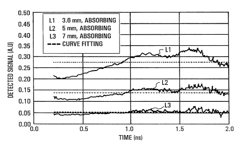
Figures 2a and 2b. As shown in these graphs, when the inclusion
is scattering, the beginning of the diffusion curve is signifi-
cantly changed and the tail remains unchanged. An absorbing
inclusion has the opposite effect, i.e., the tail of the
diffusion curve is strongly attenuated while the beginning is
less changed. The diffusion curves were obtained through a 20mm
thick scattering solution ( a' = 0.97 mm-1, a = 0.002 mm-1) con-
taining inclusions of different sizes. Figure 2a shows the
results for scattering inclusions with $' = 1.76 mm-1 . Figure
2b shows the results for absorbing inclusions with a = 0.029
mm-1. In Figure 2a, line L1 is the diffusion curve measured when
no inclusion is present while lines L2, L3 and L4 are the
diffusion curves measured wheri cylindrical scattering inclusions
having diameters and thickness of 3.6, 5 and 7 mm respectively
are placed in the center of the solution. In Figure 2b, line Ll
is the diffusion curve measured when no inclusion is present
while lines L2, L3 and L4 are the diffusion curves measured_when
cylindrical absorbing inclusions having diameters and thickness
of 3.6, 5 and 7 mm respectively are placed in the center of the
solution.
In order to highlight the effect of inclusions on the
diffusion curve, the relative transmission can be defined as
follows:
1', (t) = -ln (Tincl/Tref) i (1)
where Tinci is the diffusion curve when the inclusion is present,
and Tref is the diffusion curve without inclusion. The relative
transmissions ij(t), calculated from the measurement presented in
Figures 2a and 2b, are shown in Figure 3. From Figure 3, it is
clear that a temporal signature exists and facilitates the
CA 02249099 1998-09-30
13
differentiation between the two types of inclusion. Figures 3a
and 3b illustrate relative transmissions obtained through a 20
mm thick scattering solution ( s' = 0.97 mm-1, a = 0.002 mm-1)
containing inclusions of different sizes. Figure 3a shows the
results for scattering inclusions with e' = 1.76 mm-'. Figure
3b shows the results for absorbing inclusions with A. = 0.029
mm-1.
It is convenient to model the relative transmission
1j (t) by an analytical expression. Curve fitting will then be
possible on experimental measurements and few numerical values
will characterize the change in the diffusion curve. The model
must separate the scattering and absorption components and
ideally, only one parameter,should describe each effect. Such
an ideal situation is possible.
According to the present invention, when the inclusion
differs from the solution only by its scattering coefficient, the
function i)(t) can be modeled as follows:
1 1) (t) = Ad (to/t) 2 (2)
where Ad is a constant representing the amplitude of effect of
the inclusion, and to is an arbitrary constant that makes Ad
dimensionless. It is convenient to choose to to be approximately
equal to the time at which the maximum of the diffusion curve
occurs. The dotted lines on Figure 3a represents the curve
fitting of the equation (2) on the experimental data.
On the other hand, when the inclusion differs from the
solution only by its absorption coefficient, the function r)(t)
is almost time-independent and equal to Aa, a constant describing
the amplitude of the inclusion effect:
f~ (t) = .Aa. (3)
Once again the dotted lines on the Figure 3b represents
the curve fitting of equation (3) on the experimental data.
CA 02249099 1998-09-30
14
For an arbitrary inclusion, it has been assumed that
the function i)(t) can be properly modeled as follows:
(t) = Ad (to/t) a + Aa. (4)
where Ad and A. are associated to the diffusion and absorption
phenomenon respectively. Performing a curve fitting of this
analytical function on experimental data provides Ad and A. which
are proportional to the scattering and absorption properties
respectively.
It is important to point out that the method of the
present invention permits one to account for the inhomogeneous
nature of the geometry. For comparison, Figures 4a and 4b show
the theoretical relative transmissions i~(t) corresponding to
homogeneous cases, i.e, the r)(t) resulting from a u.niform
increase of the scattering and absorption coefficients. The
temporal signatures are significantly different from those
obtained experimentally for inhomogeneous cases. The theoretical
relative transmissions ij(t) are calculated for a 20 mm thick
homogeneous slab ( 8' = 0.97 mm-1, a = 0.002 mm-1) . Figure 4a
shows the effect of a homogeneous increase of the scattering
coefficient to a' = 1.10 mm-1. Figure 4b shows the effect of a
homogeneous increase of the absorption coefficient to a = 0.003
mm-1.
The technique has been successfully tested on data
obtained from the scanning of diffusing cells containing small
inclusions. Figures 5 and 6 show images corresponding to the
scanning of a diffusing cell containing a scattering and
absorbing inclusion, respectively. For each of the pixels, a
pair of values Ad and Aa is obtained from a curve fitting. Thus,
for each scan two images are generated: one representing the
spatial distribution of Ad values and the other representing the
spatial distribution of Aa values. Images generated using the
CA 02249099 1998-09-30
15 _
standard curve fitt.ing method, described previously, are also
shown for comparison.
Figures 5a-f show imaging results of a 5 mm scattering
inclusion ( s' = 1.76 mm-1, a = 0.002 mm-1) embedded at the center
.5 plane of the 20 mm thiclc scattering cell ( 8' = 1.13 mm-1, /j, =
0.002 mm-1). Figure 5a shows an image (40 x 40 mm) corresponding
to the total time-integration of the diffusion curves. Figure
5b shows the diffusion curves corresponding to the image center
(weaker curve M1) and a reference background pixel (stronger
curve M2). Figures 5c and 5d show the spatial distributions of
the curve fitted absorption coefficient, and the curve fitted
scattering coefficient, respectively, using the homogeneous
diffusion model. Figures 5e and Sf show the absorption FIDM
parameter Aa and scattering FIDM parameter Ad, respectively.
Figures 6a-f show imaging results of a 5 mma_bsorbing
inclusion ( H = 1.13 mm-1, a = 0.015 mm-1) embedded at the center
plane of the 20 mm thick scattering cell ( 8' = 1.13 mm'1,
0.002 mm;'). Figure 5a shows an image (40 x 40 mm) corresponding
to the total time-integration of the diffusion curves. Figure
6b shows the diffusion curves corresponding to the image center
(weaker curve Ni) and a reference background pixel (stronger
curve N2). Figures 6c and 6d show the spatial distributions of
the curve fitted absorption coefficient, and the curve fitted
scattering coefficient, respectively, using the homogeneous
diffusion model. Figures 6e and 6f show the absorption FIDM
parameter and scattering FIDM parameter, respectively.
From these Figure 5 and 6 it can be seen that the FIDM
method provides excellent inclusion type recognition, which can
not be achieved using previous methods. In the context of
imaging through human tissues, the present invention is a
significant step toward tissue differentiation since it properly
CA 02249099 1998-09-30
16
separates the scattering and absorption properties of a local
heterogeneity. Furthermore, the spatial resolution is better for
scattering inclusions than for absorbing inclusions. More
precisely, the size of an object appears smaller when it is a
scattering object. This is explained by the temporal shape of
ij(t): for scattering inclusions, the relative transmission ~(t)
is high only for first arrival photons which are thus favored by
the curve fitting process over the late arrival photons. For
absorbing inclusions, the function fj(t) is almost time-indepen-
dent and the curve fitting process does not favor the first
arriving photons. Thus the FIDM technique performs an intrinsic
time gating in the case of local variations of the scattering
coefficient.
The examples shown in Figures 5 and 6 illustrate the
power of the method of the present invention for inclusion type
recognition.
A further example is shown in Figure 7a-c where two
inclusioYas were embedded in a 50 mm-thick homogeneous scattering
medium (,uH' =1 . 13 mm-1 and ,u8 0. 002 mm-1) . The total image is 50 by
25 mm. The inclusion at the left is absorbing having optical
coefficients ,ue' =1 . 13 mm-1 and ,ua=0 . 015 mm-'-, it has a cylindrical
shape with a diameter of 7 mm and a thickness of 7 mm. The
inclusion at the right is scattering having optical coefficient
,uH=2.85mm-1 and ,ua=0.002 mm-1, it has a cylindrical shape with a
diameter of 10 mm and a thickness of 7 mm. The top image has
been obtained by performing a total time-integration of the
measured diffusion curves. The central and bottom images
correspond to the spatial distribution of the absorption FIDM
parameter Aa and the scattering parameter N respectively. The
result of figure 7 clearly illustrates the power of the present
CA 02249099 1998-09-30
17
invention for separating the absorption and scattering properties
of an inhomogeneous scattering medium.
Other examples are summarized in Table 1, where a
diffusion ratio defined as
R = IAdI/(IAdI + IAaI),
has been calculated for on-axis diffusion curve measurements
performed on a diffusing cell containing different inclusions.
The diffusion ratio R is in the range 0 to 1. An R value close
to 1 indicates that the inclusion is diffusing while an R value
close to 0 indicates an absorbing inclusion. As can be seen from
Table 1, the correlation between the R values and the inclusion
type is excellent . For the last three inclusions in Table 1,
which are three mixed inclusions of different sizes but with the
same optical parameters, approximately the same value has been
obtained.
The technique of the present invention also has
significant advantages over previous curve fitting techniques in
that the-mathematical expression is extremely simple. There is
a linear dependence of the two parameters Ad and Aa on experimen-
tal measurement 1), facilitating a linear curve fitting process
which can be calculated significantly faster. For example, the
images generated with the standard curve fitting method shown in
Figures 5 and 6 took 20 minutes to calculate while those obtained
with the FIDM technique took approximately 3 seconds.
CA 02249099 1998-09-30
18
TABLE I
Type ,ua l Ya dimension R
(mm 1) (mm-1) (mm)
diffusing 0.45 0.002 7 0.90
diffusing 0.45 0.002 5 0.98
diffusing 0.45 0.002 3.6 0.96
diffusing 1.36 0.002 7 0.97
diffusing 1.36 0.002 5 0.79
diffusing 1.36 0.002 3.6 0.94
diffusing 1.76 0.002 7 0.96
diffusing 1.76 0.002 5 0.93
diffusing 1.76 0.002 3.6 0.89
diffusing 2.85 0.002 7 0.95
diffusing 2.85 0.002 5 0.99
diffusing 2.85 0.002 3.6 1.00
absorbing 1.13 0.022 7 0.13
absorbing 1.13 0.022 5 0.11
absorbing 1.13 0.022 3.6 0.11
absorbing 1.13 0.015 7 0.07
absorbing 1.13 0.015 5 0.06
absorbing 1.13 0.015 3.6 0.02
absorbing 1.13 0.036 .7 0.06
absorbing 1.13 0.036 5 0.07
absorbing 1.13 0.036 3.6 0.09
mixed 1.76 0.022 7 0.21
mixed 1.76 0.022 5 0.24
mixed 1.76 0.022 3.6 0.23
CA 02249099 1998-09-30
19
Having described several embodiments of the method of
optical imaging through scattering media in accordance with the
present invention, it is believed that other modifications,
variations arnd changes will be suggested to those skilled in the
art in view of the description set forth above. It is therefore
to be understood that all such variations, modifications and
changes are believed to fall within the scope of the invention
as defined in the appended claims.