Note: Descriptions are shown in the official language in which they were submitted.
CA 02249525 1998-10-06
a
A DEVICE USED WITH A SURGICAL
RETRACTOR TO ELEVATE BODY PARTS
FIELD OF THE INVENTION
The present invention generally relates to surgery. More specifically, the
present invention relates to surgical retractors for temporarily providing
access to
portions of the internal anatomy such as the thoracic cavity.
to BACKGROUND OF THE INVENTION
In traditional methods for performing coronary artery bypass surgery, a
segment of a blood vessel is harvested from another portion of the body and is
used as an autogenous graft. the graft is typically sutured onto the coronary
artery
t5 so as to bypass the stenosed area and restore adequate blood flow distal to
or
downstream from the blockage. Often in such a procedures, the saphenous vein
is
harvested from the surgical patient's leg and subsequently used as the graft
vessel.
In a large number of cases, the wound created in the leg is slow to heal and
the
patient endures considerable pain and irritation. In addition, surgeons have
20 learned that, in general, an artery rather than a vein serves as a better,
long term
bypass graft.
Many surgeons prefer to use one of the internal mammary arteries (IMA)
as the bypass graft. The descending IMA's are located within the thoracic
cavity
25 of the patient along each side of the sternum of the rib cage. The IMA is
in close
proximity to the heart and therefore it is not necessary to completely remove
it
from the patient. To prepare the IMA, the side branches of the IMA are first
hemostatically severed and the main trunk of the vessel is occluded with a
clamp.
The IMA is then severed at a point just above to the patient's diaphragm so
that it
3o is mobilized. However, the IMA is never disconnected it from its original
blood
supply. The freed end of the IMA is then anastomosed to a coronary artery,
such
as the left anterior descending (LAD) coronary artery, just distal to the
stenosis.
This procedure requires significant access and visibility into the upper,
thoracic
EN D-475
CA 02249525 1998-10-06
-2-
cavity for the surgeon. The surgeon must free the IMA from the "ceiling" or
wall
of the internal thoracic cavity, while at the same time being very careful not
to
puncture or otherwise traumatize the IMA. The side branches of the IMA must be
located and transected, usually by using an electrosurgical device, with
minimal
s blood loss.
The most commonly used method of access to the thoracic cavity for the
mobilization of the IMA and the anastomosis of it to the LAD coronary artery
is a
medial sternotomy. For this procedure, a longitudinal incision is made through
io the patient's sternum on the midline of the chest. Then a surgical
retractor is used
to spread and hold apart the left and right rib cages, creating an opening
which is
about tour inches wide. The muscles and other tissues of the chest wall are
significantly traumatized by this procedure, and the post-operative healing
process
for the rejoining of the split sternum is sometimes very slow. As a result,
the
~s patient endures significant pain and the recovery time is long. In some
cases there
are significant complications and occasionally follow-up surgical procedures
are
required.
In recent years, new methods of access into the thoracic cavity have been
2o developed. One minimally invasive method is called a mini-thoracotomy and
involves access through an incision running intercostally (between two ribs)
of the
left chest wall. A surgical retractor, such as the one used for a traditional
stemotomy, is used, but in this case the superior and inferior rib cages of
the left
chest are only spread apart about two inches, thus resulting in much less
overall
2s trauma to the bones, muscles, and other tissues in the chest. Subsequently,
the
patient endures less pain and irritation following the surgery, and the
recovery
time is significantly decreased.
The mini-thoracotomy method of access to the thoracic cavity, however,
3o has propagated the need for new surgical tools and methods because the
opening
into the thoracic cavity is considerably smaller than for the sternotomy.
Also,
since the IMA is attached to the thoracic cavity wall, the angle of approach
the
surgeon must use through the opening is very difficult since the inferior rib
cage
EN D-475
CA 02249525 1998-10-06
-3-
tends to obstruct the manipulation of surgical devices used for the procedure.
Many of the new surgical retractors used in thoracic surgery have a rib
elevator,
which tilts the retractor at an angle so as to give the surgeon better access
to the
thoracic cavity. However, because of this change in the retractor to thoracic
5 surgery, hospitals must now stock both the new retractors and the
traditional
retractors used in medial sternotomies.
There has, therefore, been a need for a device that can elevate surgical
retractors at angles, but which are separate from and readily attachable to
such
to retractors. In addition, there has been a need for such a device which is
adaptable
for use with many of the commercially available surgical retractors.
Furthermore,
there is a need for such a device which is easy and quick to set-up, given the
importance of minimizing the length of time of the surgical procedure. Also,
considering the high cost of surgical procedures today, it is important that
such a
is device be easy to clean and sterilize for reuse, or that it be low cost and
disposable.
Finally, there is a surgical need for a device which can be attached to any
of numerous surgical retractors in use today, which can provide another means
for
2o support or attachment of other surgical devices used in the procedure.
Often the
surgeon wishes to hold or stabilize an organ or tissues within the cavity, and
attach
or support an ancillary holding tool on a fixed structure so that an assistant
does
not have to maintain the position of the holding tool throughout the
procedure. Yet
the surgical retractor arms are too far away from the organ or tissue of
interest to
25 be used as a platform. What is needed is a bar or bridge that can attach to
the
arms of the surgical retractor and cross over the opening nearer to the organ
or
tissue of interest. Then this bridge can be used as a platform for supporting
or
attaching the ancillary holding device.
END-475
CA 02249525 1998-10-06
-4-
SUMMARY OF THE INVENTION
In accordance with the present invention, there is provided a device for
pivoting a surgical retractor with respect to a patient it is being used on.
The
5 device includes a bridge having distal and proximal ends wherein a distal
coupling
is attached to the distal end of the bridge and a proximal coupling is
slidably
attached to the bridge proximal to the distal coupling. The proximal and
distal
couplings include a means for releasably attaching itself to a surgical
retractor.
The device further includes a lifting assembly attached to the bridge proximal
to
to the proximal coupling. The lifting assembly comprises a means for applying
an
upward force to the proximal coupling, whereby when the device is attached to
a
surgical retractor, the lifting assembly pivots the retractor upward about the
distal
coupling.
is BRIEF DESCRIPTION OF DRAWINGS
While the specification concludes with claims which particularly point out
and distinctly claim the subject matter fonming the present invention, it is
believed
that the invention will be better understood from the following description of
the
2o preferred embodiment taken in conjunction with the accompanying drawings
wherein:
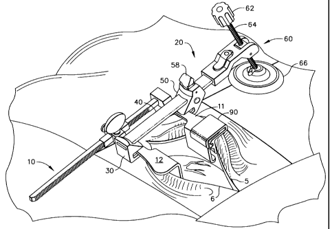
Figure 1 is a perspective view of the present invention as it is used in
conjunction with a surgical retractor on a chest wall incision on a surgical
patient;
Figure 2 is a perspective view of the rib lifting apparatus 20 of the present
invention depicted in Figure 1;
Figure 3 is a front elevational view of the rib lifting apparatus 20 depicted
3o in Figure 2;
Figure 4 is a top elevational view of the rib lifting apparatus 20 depicted in
Figure 2;
EN D-475
CA 02249525 1998-10-06
-5-
Figure 5 is a perspective view of the arm extender 90 of the present
invention depicted in Figure 1;
Figure 6 is a front elevational view of the arm extender 90 of the present
invention depicted in Figure 1;
Figure 7 is'a perspective view of an alternate embodiment of the present
invention, being used in conjunction with a surgical retractor on a surgical
patient;
to Figure 8 is a front elevational view of the bridge assembly 168 of the
alternate embodiment of the present invention depicted in Figure 7;
Figure 9 is a bottom elevational view of the bridge assembly 168 of the
alternate embodiment of the present invention depicted in Figure 7;
Figure 10 is a front elevational view of the tower of the alternate
embodiment of the present invention depicted in Figure 7;
Figure 11 is longitudinal sectional view 11-11 of the tower depicted in
2o Figure 10;
Figure 12 is transverse sectional view 12-12 of the tower depicted in Figure
10;
Figure 13 is a front elevational view of the elevator of the alternate
embodiment of the present invention depicted in Figure 7;
Figure 14 is a top elevational view of the elevator of the alternate
embodiment of the present invention depicted in Figure 7;
Figure 15 is longitudinal sectional view 15-15 of the elevator depicted in
Figure 13; and
EN D-475
CA 02249525 1998-10-06
-6-
Figure 16 is a perspective view of the bridge assembly and the arm
extender of the alternate embodiment of the present invention being used in
conjunction with a surgical retractor on a surgical patient.
s The drawings are not necessarily to scale.
DETAILED DESCRIPTION OF THE INVENTION
The present invention described herein can be used in conjunction with a
io number of commercially available, reusable, surgical retractors for
improving
access into the thoracic cavity. There is shown in Figure 1, a rib lifting
device 20
which serves as a lever for tilting retractor 10 at an angle. Device 20
comprises a
distal coupling, which in this embodiment is shown as hook 30, a bridge 40, a
slideable proximal coupling, which in this embodiment is shown as hook 50, and
a
is lifting sub-assembly 60. The distal hook 30 is attached to the distal arm
12 of the
surgical retractor 10 and serves as the fulcrum for the lever system. Bridge
40 is
attached to the proximal arm I1 of the surgical retractor 10, thereby
retracting the
superior and inferior rib cages S and 6, respectively. An upward force is
applied
to the proximal hook 50 by the lifting subassembly 60 so that the entire
system
2o pivots upward about the distal hook 30, and thereby lifts the superior rib
cage 5
above the inferior rib cage 6. It should be appreciated that the present
invention
could be used in the reverse manner, if the surgeon preferred, in which the
inferior rib cage 6 is lifted above the superior rib cage 5. It should also be
appreciated that the present invention can be used for a medial sternotomy as
well
2s as the thoracotomy. In Figure 1, the arm extender 90 is slideably attached
to the
proximal arm 11 of the surgical retractor 10, so that the blade 92 (see Figure
5) is
reliably supporting the superior rib cage 5 from underneath.
Still referring to Figure 1, it can be seen that the surgical retractor shown,
3o as for all commercially available surgical retractors of this type, has a
means for
mechanically adjusting the distance between the proximal and distal arms 12
and
11, respectively. Therefore it is necessary for the rib lifting device 20,
which is
attached to surgical retractor 10, to have also a means of adjustment of the
EN D-475
CA 02249525 1998-10-06
-?-
distance between the distal and proximal hooks 30 and 50, respectively. Also
it
can be seen that a means for adjusting the elevation of the superior rib cage
5 over
the inferior rib cage 6 has been provided so that the surgeon can adjust the
size of
the opening into the thoracic cavity with minimal trauma to the surgical
patient.
Knob 62 is turned by the surgeon or an assistant to advance the screw 64 while
the
foot 66 bears against the chest of the surgical patient. The foot 66 is
distanced
somewhat superior to blade 92 (see Figure 5) of the arm extender 90 so that an
effective lifting force can be applied to the proximal hook 50 by the lifting
subassembly 60.
The present invention may also be assembled to the surgical retractor 10 in
the reverse manner to that shown in Figure 1, without change to its usage or
function. The physical anatomy of the surgical patient and the requirements of
the
surgical procedure would dictate in which direction to assemble it.
~s
Turning now to Figure 2, the rib lifting device 20 is shown without the
extender 90 and the surgical retractor 10 for clarity. The rib lifting device
has
three actuators for its attachment and detachment to the surgical retractor: a
slide
lock lever 58 for locking the proximal hook 50 onto the bridge 40 or for
unlocking
2o it from the bridge in order to adjust the distance between the distal and
proximal
hooks, 30 and 50, respectively; a release button 82 for detaching the lifting
subassembly 60 from the proximal end 48 of the bridge 40; and a screw knob 62
for rotating screw 64 for lifting or lowering the proximal hook 50.
25 Figures 3 and 4 are front and top views, respectively, of the rib lifting
device depicted in Figure 2. Distal hook 30 may be attached to the distal end
46
of the bridge 40 by a press fit, by use of fasteners, or by a number of other
means
well-known to those skilled in the art. Integrally situated in distal hook 30
and
spaced at a optimal distance vertically beneath the bridge 40 is V-groove 32
for the
3o insertion of surgical retractor arm 12. Slideably mounted on the bridge 40
is
proximal hook 50 which also has a V-groove 52 directly opposing the V-groove
32
on the distal hook 30. The lever 58 is raised to an up-position to allow the
movement of the proximal hook 50 along the bridge 40. Indentations 54 (front
END-475
CA 02249525 1998-10-06
_8_
and back side of proximal hook) aid the surgeon in gripping the proximal hook
to
position it on the surgical retractor. When the retractor arms 11 and 12 (see
Figure 1) of the surgical retractor are captured within the opposing V-grooves
32
and 52, the lever 58 is pushed down to lock the position of the proximal hook
onto
s the bridge 40. Lever 58 pivots about lever pivot 56 and cams against the
posterior surface 42 of the bridge 40, thus locking the proximal hook to the
bridge.
Still referring to Figures 3 and 4, proximal end 48 of bridge 40 is inserted
io into lifting frame 80. An indentation (not visible) on bottom surface 44 on
the
proximal end 48 of the bridge latches with a projection (not visible) off of
button
82 which is spring biased in the latching position. This attachment may be
released by pressing button 82 and withdrawing the bridge 40 from the frame
80.
The ability of the rib lifting device to disassemble in this way is
advantageous for
is the shipping, handling, and cleaning of the device, and also for the use of
the
bridge and hooks separately as will be described later for the alternate
embodiment
of the present invention. Integral with lifting frame 80 is lifting frame fork
84
which holds swivel block 70. The swivel block pivots about swivel pins 72, 73
(pin 72 visible only) and contains an internal screw thread for receiving
screw 64.
2o As described earlier, knob 62 is attached to screw 64. On the opposite end
of the
screw 64 is affixed ball 68 which in turn is captured within a cup 69 integral
with
foot 66. The screw is constrained by the swivel block 70 to an optimal angular
variation within the plane defined by the longitudinal axis through it and the
bridge
40. The range of motion for the screw 64 with respect to the foot 66 is
generally
Zs conical due to the ball and cup attachment described. All of the components
for
the rib lifting device 20 described for Figures 3 and 4 may be made from
various
metals such as stainless steel, or from various, rigid, medical grade
plastics, or
from a combination of metal and plastics. The device can be manufactured to be
reusable or single-patient-use disposable.
Now referring to Figures 5 and 6, the arm extender 90 is seen to consist of
one piece which may be made of metal, preferably stainless steel, or of a
rigid,
medical grade plastic. Arm extender 90 is comprised of a blade 92, a. vertical
END-475
CA 02249525 1998-10-06
-
span 98, an arm wrap 100 forming an L-shape slot 94, and a fin 96. Blade 92 is
designed to extend underneath the rib cage (see Figure 1) so that an upward
force
can be applied by the rib lifting device without the arm extender slipping off
the
edge of the surgical incision in the chest wall. It also distributes the
lifting force
over a broad area of tissue and/or bones so as to minimize trauma to the
delicate
tissue lining the internal, thoracic cavity. Variation of the length of
vertical span
98, the length of blade 92, and the angle between, is advantageous to the
surgeon
for accommodating variations in the surgical patients. Therefore a set of
these
arm extenders, each having a different geometry in these aspects, may be
provided
io from which the surgeon may choose. The L-slot 94 is sized to fit slideably
over
many different sizes and kinds of commercially available, surgical retractors.
The
L-slot, together with the fin 96, prevent the arm extender from rotating about
the
arm of the surgical retractor, so as to transmit the upward force to the chest
wall.
is Referring now to Figure 7, an alternate embodiment of the present
invention is shown being used in conjunction with a surgical retractor on a
surgical
patient. This embodiment is much like the other in that it tilts the plane of
the
anatomical opening into the body cavity so that access and visibility within
is
enhanced. The primary difference of the alternate embodiment is that the same
2o function is accomplished as before, but with fewer components. As will
become
apparent, the alternate embodiment also has a different method of assembly
during
the surgical procedure. The alternate embodiment of the present invention is
the
rib lifting device 110 depicted in Figure 7, comprising a bridge 170, a tower
120,
an elevator 140, and an arm extender 90. Distal hook 172 of bridge 170 hooks
25 and passes beneath retractor arm 12 of surgical retractor 10. This junction
serves
as the fulcrum of the lever system of the present invention. Bridge 170 passes
also
beneath arm 11 of the surgical retractor 10 and thus is positioned to lift the
arm 11
and the superior rib cage 6 attached thereto above the inferior rib cage 5.
The
proximal end 174 of bridge 170 is supported within elevator 140 which in turn
is
3o adjustably mounted within tower 120. Preferably, bridge 170 can axially
rotate
about its longitudinal axis, extending between the distal and proximal ends,
with
respect to or independent of the lifting assembly. Base 122 of tower 120 bears
against the chest of the surgical patient. The elevator 140 contains a locking
END-475
CA 02249525 1998-10-06
-10-
feature to be described later which engages with ratchet teeth 176 of bridge
170
only when the tower 120 is tilted superior with respect to the bridge 170 at
an
angle of approximately thirty degrees past vertical, as is shown in Figure 7.
When
the tower 120 is vertical and its longitudinal axis is essentially
perpendicular to the
s longitudinal axis of the bridge 170, then it is possible to move the tower
along the
length of the bridge so as to position the base 123 of the tower on the chest
of the
surgical patient, or to remove the tower from the bridge 170. This adjustment
is
easily accomplished while the elevator 140 is in the lowered position within
tower
120, because the force of the bridge 170 against the retractor arm 11 is
minimal.
~o Once the base 123 of the tower 120 is properly located on the chest of the
surgical
patient, the elevator may be manually raised by the surgeon or surgical
assistant by
lifting up on the proximal end 174 of the bridge 170. A locking mechanism, to
be described later, of the elevator 140 engages with the ratchet teeth 128,
129 of
the tower 120 in order to maintain the vertical position of the elevator 140
during
is the surgical procedure. To release this lock, the release button 150 may be
pushed
downwardly and the elevator falls immediately to a lower position within the
tower 120 due to the downward force exerted by the arm 11 of the retractor 10.
At this point the tower 120 can be repositioned, and then the elevator 140
raised
again, or the device may be disassembled from the surgical retractor 10.
20
The arm extender 90 depicted in Figure 7 is identical in form and function
to that which is depicted in Figure 1.
Figures 8 and 9 show the bridge 170 depicted in Figure 7 assembled with
2s slide 180 (not shown in Figure 7), hereinafter referred to as the bridge
assembly
168. This arrangement provides the surgeon an option for use of a portion of
the
present invention as shown in Figure 16. Specifically, the bridge assembly 168
becomes an advantageously located platform for attaching other surgical
devices or
simply as a support for the hand of the surgeon or surgical assistant. Here
the
3o hooks 172 and 182 of the bridge assembly 168 are facing downward towards
the
surgical patient and capturing the arms 12 and 11 respectively of the surgical
retractor 10. Referring to Figure 8, the bridge 170 is inserted through a
rectangular, longitudinal hole in the slider frame 183. This hole is large
enough to
EN D-475
CA 02249525 1998-10-06
-11-
allow some angular movement of the bridge 170 within the slider 180 in the
vertical, longitudinal plane. When the slider is pushed against the retractor
arm 11
so that the arm presses firmly against hook 182, the slide lock pawl 184
engages
the bridge ratchet teeth 176 to lock the slider in place. The same result
occurs
s when the slider is held in place while the retractor anms 11 and 12 are
spread apart
slightly. The lock can easily be released by either adjusting the retractor
arms to a
smaller width than before, or by pressing down on the top of the slider 180 to
rock
the pawl 184 from engagement with the ratchet teeth 176. The bridge 170 and
the slider 180 may be made of a metal such as stainless steel, or from a
medical
io grade, rigid plastic such as a glass-filled polyetherimide. The slider 180
is not
intended for use on the bridge 170 while the tower 120 is attached.
Next is described the features of the tower 120 and elevator 140 which
work in concert to supply a upward holding force to the proximal end 174 of
the
~s bridge 170. These features can best be viewed in Figures 10 through 15. In
Figure 10 is a front view of the tower 120 which comprises a left column 124,
a
right column 126, joined at the top by cornice 130, and at the bottom by base
122.
The columns 124 and 125 form an essentially rectangular opening 125. On the
front of left column 124 is vertical left rail 134 which runs around cornice
130 to
2o join vertical right rail 136 on right column 126. Also on left column 124
is a
vertical array of ratchet teeth 128, and likewise on the right column are
ratchet
teeth 129. As can be seen in longitudinal cross section view 11-11 of Figure
11,
these teeth are designed to allow a pawl to slide freely when moving in the
upward
direction, but to lock in the downward direction. In Figures 10 and 11 the
base
2s 122 is shown to consist of a plurality of fins 123 which facilitate the
injection
molding of the tower 120 from a rigid, medical grade plastic such as glass-
filled
polyetherimide. It may also be made of a metal such as stainless steel.
Figure 12 is lateral cross-sectional view 12-12 depicted in Figure 10. In
3o this view are shown left and right second rails, 135 and 137, respectively,
which
serve to capture the elevator 140. Rails 134 and 136 are again shown to
indicate
the front of the tower 120 as the top of this cross-sectional view.
EN D-475
CA 02249525 1998-10-06
-12-
The elevator 140 is shown in Figures 13, 14, and 15 and comprises a frame
141 (Figure 13), extending from which is a T-beam 160, left wing 162, right
wing
163, left lever stop 152, right lever stop 153, upper projection 143, lower
projection 146, left lower latch 154, left upper latch 156, right lower latch
155,
s and right upper latch 157. Centered on frame 141 is bow-tie slot 142.
Extending
from the front of T-beam 160 is release lever 150, and extending from the back
of T-beam 160 is pawl rib 151.
The elevator 140 is slideably attached to tower 120 by the four latches,
io 154-157, which are flexible cantilevers. These latches are aligned and then
inserted into the front of opening 125 of the tower 120. The latches snap
around
the edges of rails 135 and 137 of the tower (Figure 12) so that guide edges
158
and 159 on the elevator 140 are closely interposed between rails 134 and 136
of
the tower. Left and right wing surfaces 186, 187, slide against left and right
~5 sliding surfaces 138, 139, respectively, of the tower 120.
Once assembled to tower 120, pawl rib 151 can engage with left and right
ratchet teeth 128, 129 of the tower to maintain the vertical position of the
elevator
140. The elevator can be raised in the tower most easily by pulling up on the
2o bridge 170 which is inserted through bow-tie slot 142. T-beam portion 161
flexes
as the pawl rib 151 rides over the ratchet teeth 128, 129. To release the pawl
rib
from the ratchet teeth, the surgeon or surgical assistant may press down on
the
release button 150 and the elevator will immediately fall to its lowest
position in
the tower. Left and right stop surfaces 166, 167 of the left and right lever
stops
2s 152, 153, respectively, serve to prevent over-flexure of the T-beam portion
161
when the release button 150 is depressed.
The proximal end 174 of bridge 170 (see Figure 8) fits loosely through
bow-tie slot 142 when the longitudinal axis of the bridge 170 is normal to the
3o plane of the elevator frame 141. The ratchet teeth 176 are to face upward
when
the bridge 170 is assembled with the elevator 140. (As previously noted, slide
180
of Figure 8 is not to be used with the elevator and tower.) Due to the bow-tie
slot
shape, the bridge is permitted to rotate slightly in both directions about its
EN D-475
CA 02249525 1998-10-06
-13-
longitudinal axis. This allows the surgeon a wide range of variation in the
assembly of the present invention to the surgical retractor 10, and is
necessary due
to the curvature of the chest of the surgical patient. As dexribed earlier,
when the
tower and the elevator contained within it are tilted about 30 degrees past
the
s perpendicular formed with the longitudinal axis of the bridge 170, the
elevator
becomes locked on the teeth 176 of the bridge. Bridge pawl 148 is seen in
Figures
13 and 15 and only engages the bridge teeth 176 at the angle described. Stop
surface 144 of the upper projection 143 and stop surface 147 of the lower
projection 146 serve to limit the maximum amount of tilt of the elevator 140
and
io tower 120. The elevator 140 may be made from a metal such as stainless
steel,
but the preferred material is a medical grade, rigid plastic such as
polyetherimide.
The method dexribed for locking the bridge to the tower by tilting the
tower thirty degrees from its perpendicular position to the bridge to allow
the
is bridge pawl 148 to engage the bridge teeth 176 on the bridge is only one of
various methods for doing so, as can be appreciated by those skilled in the
art.
For example, another method would be to fashion a spring biased release
mechanism on the elevator 140 which automatically engages the bridge teeth 146
when the tower is assembled to the bridge. The angle the tower makes with the
2o bridge would not matter, and in fact, an optimal, fixed angle of assembly
could be
incorporated. A pawl on this release mechanism on the elevator could ratchet
over
the bridge teeth as the tower is moved towards the distal end of the bridge,
but
could only move proximally upon actuation of the release mechanism.
2s The alternate embodiment of the present invention can 'also be made to be
reusable or single-patient-use disposable.
From the foregoing, it will be observed that numerous modifications and
variations can be effected without departing from the true spirit and xope of
the
3o novel concept of the present invention. It is to be understood that no
limitation
with respect to the specific embodiments illustrated herein is intended or
should be
inferred. The dixlosure is intended to cover by the appended claims all such
modifications as fall within the scope of the claims.
END-475