Note: Descriptions are shown in the official language in which they were submitted.
CA 02249531 1998-10-OS
G-9610 1 PATENT
The invention disclosed herein relates to an intraluminal
shunt device which is utilized in coronary bypass surgery to
avoid the usage of a heart/lung machine and the attendant hazards
of stopping the heart during the operation.
The intraluminal shunt of the present invention relates to a
device to be inserted within a blood vessel to allow blood flow
during an operation, such as a coronary bypass procedure, wherein
the procedure does not involve a heart/lung machine. A relatively
common operation in which anastomosis is employed is a coronary
artery bypass operation in which blood is routed about a blocked
portion of a coronary artery to restore and insure adequate blood
supply to the heart muscle. In a conventional heart bypass
operation, a short segment of a vein taken from another part of
the patient's body is used, with one end of this vein connected
to the aorta and the opposite end connected to the blocked
coronary artery beyond the blockage. The connection of this vein
between the aorta and the coronary artery serves as a bypass
around the blockage.
Standard operative technique for providing a coronary artery
bypass comprises first clamping off the aorta to occlude blood
flow to all the coronary arteries. The bypass connection is then
CA 02249531 1998-10-OS
G-9610 2 PATENT
made by suturing the vein in place. Many times multiple bypasses
are required and, as a result, it may be necessary for the aorta
to be clamped off for an extended period of time during which
there is no blood supply to the muscle tissue of the heart or
myocardium. The prolonged suspension of blood supply to the
heart can result in life threatening infarcts temporarily harming
the heart muscle.
To overcome this problem, the vast majority of coronary
artery bypass grafting procedures are performed with the
assistance of cardiopulmonary bypass (CPB) and cardiac
standstill. To stop the heart is traumatic to the patient and
may precipitate undesirable ischemic conditions for the patient
both during and after completion of the operation. However, an
advantage of the use of a heart/lung machine with the attendant
stoppage of the beating of the heart for the surgeon is that the
heart is motionless and blood-filled, making it technically
easier to fashion delicate sutured connections (anastomosis)
between the coronary sutures and vein grafts. Thus, the surgery
is less stressful and the results are consistent and reproducible
from one operation to the next.
In the last decade, there has been progress made in
performing bypass grafting without stopping the heart. To
accomplish this, a segment of the blocked artery is temporarily
occluded and a bypass graft is inserted. However, in these areas,
a condition occurs in the patient known as ischemia; i.e., there
is a lack of blood in that region from surgical control of the
target vessel. This can cause strain on the heart, with changes
CA 02249531 1998-10-OS
G-9610 3 PATENT
in the EKG, dangerous rhythm disturbances, or stoppage of the
heart beat. Between 15 and 30$ of coronary bypass operations
done on the beating heart are associated with EKG changes
resembling a heart attack. Fortunately, nearly all of these
changes are temporary and resolve upon restoration of blood flow
in the target vessel. Just the same, there is constant pressure
on the surgeon to finish quickly and get the bypass graft open as
quickly as possible.
To safely perform coronary bypass grafting without the need
to completely arrest the heart, a shunt device has been designed;
the shunt providing blood to the starving heart muscle while the
surgeon carefully and cautiously constructs the new bypass. The
shunt design presented here has many unique features which
provide major advantages to the surgeon during off-pump coronary
grafting.
S TMMARY 0 TH . T .NTTnN
The intracoronary shunt presented in this application is
intended to be inserted inside of the target coronary artery and
deliver blood to the heart muscle while, at the same time,
creating a relatively blood-free zone in the target vessel into
which the new bypass graft is connected by fine sutures. Since
the heart receives blood flow through the shunt, EKG changes and
other deleterious effects common in non-shunt surgical techniques
are avoided.
The current design is unique and specialized. Our design
features:
CA 02249531 1998-10-OS
G-9610 4 PATENT
1) A flexible tube of silicone, small enough to be inserted
completely inside the target coronary artery;
2) Expansion bulbs on each end which fit snugly against the
artery and prevent bleeding around the device; and
3) A side port which can be used to remove air, perfuse
with blood or specialized medications.
Insertion of this shunt into the target coronary artery
during off-pump grafting will:
1) Provide a relatively bloodless operative field;
2) Hold open the edges of the coronary artery, permitting
easier suturing;
3) Provide a small space between the bulbs and the main
shunt suitable for passing the suture needle without struggling
to work around the shunt;
4) Provide blood to the heart muscle during construction of
the bypass graft, preventing complications and deleterious
effects seen when blood flow is interrupted
5) Guarantees the sutured connection (anastomosis) is
properly constructed when the device slides out without any
appreciable resistance: and
6) Reduces the need for the surgeon to hurry, permitting
careful and precise construction of these delicate and
life-saving bypass grafts.
Our design has additional safety features. The side limb
permits the surgeon to connect up to a source of red (oxygen-
rich) blood to pump directly into the target coronary artery.
This option can be elected if the blood flow through the shunt
CA 02249531 1998-10-OS
G-9610 5 PATENT
body is not sufficient. In addition, medications such as blood
vessel dilating drugs could be infused into the heart to improve
regional function of the heart muscle or alleviate strain.
In addition, our design is adaptable for use during
minimally invasive coronary bypass procedures. These new
procedures involve construction of bypass grafts to the coronary
artery through small surgical incisions for tiny instrument parts
inserted into the chest. During these operations, exposure to
the heart is very limited,. sometimes with the chest cavity
entirely closed. Although this strategy is definitely less
traumatic to the patient overall, the suturing takes much longer
and is considerably more difficult. Our shunt design was created
to permit insertion using tiny instruments inserted through the
closed chest. Furthermore, a long side part is provided which
can exit the chest through a 5 mm. port and be connected to a
source of red (oxygen-rich) blood for pumping into the target
coronary vessel. This will deliver nutrients to the heart muscle
and permit the surgeon to carefully construct the anastomosis
even if it takes a long time. For minimally invasive bypass
operations, this will be a strong advantage since the technical
aspects of suturing through tiny incisions is more difficult and
time consuming.
The present invention relates to a new and novel
intraluminal shunt which comprises a primary perfusion tube and a
secondary perfusion tube integral with and intersecting the
primary tube at a generally right angle. An enlarged silicon
bulb or occluder is formed adjacent to each end of the primary
CA 02249531 1998-10-OS
G-9610 6 PATENT
tube to seal off the leakage of blood around the shunt as well as
stent the target vessel open. The bulbs provide isolation of the
artery and a blood-free working space, while the primary tube
allows blood flow therethrough and retains the artery widely
open, which facilitates suturing. The secondary perfusion tube
is fashioned to accept a luer connector which then locks onto a
source of secondary blood flow from another area of the patient's
body, such as the femoral or radial artery, or from an external
circulatory assist pump.
Another feature of the intraluminal shunt resides in two
differing designs of the shunt depending on a surgeon's suturing
technique. A first embodiment locates the secondary perfusion
tube at the midpoint of the primary perfusion tube with the bulbs
a set distance apart and each end of the primary tube having a
beveled tip. The secondary tube provides the ability to infuse
drugs through the secondary tube directly into the primary tube
and locally into the heart if the occasion requires. The free
end of the secondary perfusion tube is fashioned to accept a
luer connector or be provided with a Y-type connection with a
potential needle-less valve.
The present invention also provides a second embodiment of
intraluminal shunt wherein the secondary perfusion tube
intersects the primary perfusion tube to provide a
one-third/two-thirds ratio along the primary tube, thus providing
a "heel and toe" arrangement of the primary tube relative to the
secondary tube. In this embodiment, the enlarged bulbs are
positioned directly at the ends of the primary tube rather than
CA 02249531 1998-10-OS
G-9610 7 PATENT
provided with lead-in areas as in the first version. This
embodiment lends itself to locations in the target artery where a
blockage is very close to the suture line. In either version,
the secondary perfusion tube can be elongated to lead to the
exterior of the patient's body for use during minimally invasive
surgical techniques. The secondary perfusion tube also acts as a
handle to aid in removal of the shunt from the target vessel as
suturing of the graft is substantially completed.
Another feature of the present invention is that the
intraluminal shunt easily adapts itself to more modern techniques
where surgery has changed from a large incision to several small
incisions as in endoscopic surgery. Thus, a coronary bypass
operation can be accomplished through the use of several
incisions for an endoscope, an instrument to guide and manipulate
the intraluminal shunt and surgical instruments for providing an
incision in the artery and for suturing the graft onto the
incision during surgery.
BRI . D . RT TTnN O H . D AWTN S
FIGURE 1 is a schematic view of a patient for coronary
bypass surgery with positions for incisions in the chest wall
indicated for thoracoscopic bypass grafting.
FIGURE 2 is a schematic showing of the thoracoscopic
instruments extending through small incisions or ports in the
chest wall.
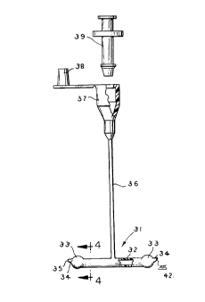
FIGURE 3 is a front elevational view of the first embodiment
of intraluminal shunt showing possible connection to external
instruments.'
CA 02249531 1998-10-OS
G-9610 8 PATENT
FIGURE 4 is a vertical cross sectional view of the shunt
taken on the line 4-4 of FIGURE 3.
FIGURE 5 is a schematic view of the shunt before insertion
into an incision.
FIGURE 6 is a schematic view similar to FIGURE 5, but with
the shunt inserted into the incision in the artery.
FIGURE 7 is a schematic view similar to FIGURE 6, but
showing the graft partially sutured to the artery.
FIGURE 8 is a front elevational view of a second embodiment
of intraluminal shunt.
FIGURE 9 is a top plan view of the shunt of FIGURE 8
inserted into the artery adjacent a blockage.
FIGURE 10 is an exploded view of the shunt and artery with
the artery partially in cross section
FIGURE 11 is a view similar to FIGURE 10, but showing the
shunt being inserted into the artery.
FIGURE 12 is a front elevational view of the artery
partially in cross section with the shunt further inserted
therein.
FIGURE 13 is a front elevational view similar to FIGURE 12,
but with the shunt completely inserted in the artery.
FIGURE 14 is a front elevational view of a Y-connector for
use in combination with a luer connector.
$ .ST MOD . O RRYTNC' 0 T TH T NTTC~N
Referring more particularly to the disclosure in the
drawings wherein are shown illustrative embodiments of the
present invention, FIGURE 1 discloses a candidate or patient P
CA 02249531 1998-10-OS
G-9610 9 PATENT
for coronary bypass surgery -with the location of four incisions
10, 11, 12 and 13 in the patient's chest wall 14 shown for use of
endoscopic instruments 15, 16, 17 and 18 (see FIGURE 2). Unlike
previous coronary bypass surgery where the heart is stopped and
the patient is kept alive by the circulation of his blood to the
brain and vital organs provided by a heart/lung machine, the
intraluminal shunt 31 of the present invention allows the heart
19 to remain beating with blood flow through the shunt. As seen
in FIGURE 2, thoracoscopic instruments 15, 16, 17 and 18 are
inserted through the incisions or ports 10, 11, 12 and 13 in the
chest wall 14 for access to the patient's heart 19. These
instruments include a thoracoscopic camera and fiber optic light
21, endoscope 22, instrument 23 to guide and manipulate the shunt
31, and instruments 24 for operating on the target vessel.
Once a blockage 26 of the target vessel 25 is located, an
incision 27 is made adjacent to the blockage 26 which is of
sufficient length to allow insertion of the intraluminal shunt 31
into the vessel. As seen in FIGURE 3, the intraluminal shunt is
formed as a short length of thin wall member or primary perfusion
tube 32 having an enlarged occluder or bulb 33 adjacent each end
34, with the opposite ends of the tubing having beveled surfaces
or tips 35 at an approximate angle of forty-five degrees.
Between the ends of the primary tube 32 is a secondary perfusion
tube 36 intersecting the primary tube at an angle of
approximately ninety degrees (right angle). In practice, the
primary tube is of a length of approximately 2.0 centimeters
(cm.) in length with each end 34 extending approximately 3.0
CA 02249531 1998-10-OS
G-9610 10 PATENT
millimeters (mm.) beyond its respective bulb 33, while the
secondary tube has a length of approximately 10.0 cm. (or 25 cm.)
and is provided with a connector 37 for an interface luer having
a closure cap 38 therefor.
To properly size the appropriate shunt for the vessel,
various sizes of Garrett probes can be inserted into the blood
vessel containing the blockage 26. The appropriate shunt and
occluders is selected from the diameter of probe found to be
appropriate for the vessel. Also, the secondary tube of a length
of approximately 10.0 cm. (or 25 cm.) provides the ability to
lock on a secondary blood supply from another area of the body or
from an exterior heart pump through a connection 39.
This intraluminal shunt 31 allows blood flow through the
target vessel as a graft 41 is sewn onto the incision 27 in the
artery and keeps the artery open. Thus, by allowing blood flow
and preventing backbleeding due to the bulbs or occluders 33, the
shunt increases safety of the coronary bypass operation by
allowing sufficient time for suturing the graft 41 onto the
incision 27 to reduce the stress on the surgeon performing the
operation, provide reproducibility of results from patient to
patient and reduce the possibility of ischemic reactions during
and after the operation. As the external diameter of the primary
tube is smaller than the internal diameter of the blood vessel,
suitable spacing is provided between the primary tube and vessel
wall to allow the sliding of the sutures into the vessel wall to
attach the graft to the incision. As the suturing of the graft
41 at 42 onto blood vessel at the incision nears completion, the
CA 02249531 1998-10-OS
G-9610 11 PATENT
intraluminal shunt 31 is gradually withdrawn through the incision
by traction on the side limb or secondary tube 36, the final
sutures are completed and the suture ends are tied. It has been
shown that the use of this shunt reduces suturing time in
off-pump coronary bypass procedures by approximately 50~.
Now considering FIGURES 8 through 13, a second embodiment of
intraluminal shunt 45 is shown. In this version of shunt, a
primary perfusion tube or shunt 46 is provided with enlarged
bulbs or occluders 48 at the opposite ends 47 of the shunt and a
secondary perfusion tube 49 intersects the primary tube at a
point providing a one-third/two thirds ratio resulting in a heel
51 and toe 52 configuration to the uneven ends of the tubing 46
projecting from the intersection of the secondary tube 49 to the
primary tube. As seen in FIGURE 9, a blood vessel 53 is provided
with an incision 54 adjacent a blockage 55 in the vessel.
FIGURES 10 through 13 illustrate the gradual insertion of the
shunt 45 into the incision 54 in the blood vessel to a position
where the occluders completely block the blood flow in the vessel
around the shunt but the interior passage formed in the primary
tube 46 allows blood flow through the tube so that the heart
remains beating during surgery.
As seen in FIGURES 10 through 13, the toe end 52 of the
shunt is initially inserted in the vessel incision 54 and
gradually worked forward into the interior of the vessel, then
the heel 51 is dropped in- and engaged by sliding the shunt
backward. This is an advantageous technique where the incision
must be made close to a blockage and space for insertion of the
CA 02249531 1998-10-OS
G-9610 12 PATENT
shunt is limited, with the heel end of the shunt nearest to the
blockage. In some cases, it is desirable that the end of the
suturing procedure be made in the least critical area and, in the
instance of the heel and toe arrangement, the sutures are
initially provided adjacent the heel portion for ease of removal
of the shunt beginning with the heel portion. Once the graft
(not shown) is sutured onto the edges of the incision and as the
gradual stitching occurs, the shunt is moved forward, the heel is
disengaged and the shunt body is withdrawn. Insertion and
withdrawal of the shunt is accomplished by manipulation of the
secondary perfusion tube or side limb.
The present invention thus discloses a method and device to
provide for substantial non-restrictive blood flow through a
blood vessel so that the heart is not stopped during surgery with
the attendant possibility of ischemic reactions during or after
the construction of a bypass graft. The primary perfusion tube
with the attendant occluders at the opposite ends keeps the blood
vessel open while allowing blood to flow through the vessel
during the operation; the shunt expanding the artery and
preventing backbleeding within the blood vessel. If the occasion
arises where additional blood is required or one or more drugs
are required in the heart, such as a blood thinner, the blood or
drug is inserted directly into the heart through the secondary
perfusion tube.
Considering FIGURE 15, a Y-type junction 61 is shown which
may be inserted into the secondary perfusion tube 36 and includes
an optional mixing chamber 62, a Y-connection 63 having one
CA 02249531 1998-10-OS
G-9610 13 PATENT
branch 64 providing an extension 65 of the secondary perfusion
tube 36 leading to the luer 'connector 37 which, in turn, may be
connected to a separate blood supply line 66. A second branch 67
of the Y-connection is connected to a drug delivery tubing line
68 that terminates in a second luer connector 69. Attached to
this connector may be a needle-less valve 71 so that drugs can be
injected into the valve without threat of contamination of the
line leading to the shunt. The configuration of the Y-connection
will be identical in either embodiment of the primary and
secondary perfusion tubes.
Although shown in the drawings for a minimally invasive
surgical procedure, either embodiment of the shunt may be
utilized for a coronary bypass procedure where surgery includes
the opening of the chest wall of the patient and direct operation
upon the heart of the patient.