Note: Descriptions are shown in the official language in which they were submitted.
Y t
S~
1
CA 02249635 1998-11-02
PATENT
P-3869
TITLE OF THE INVENTION
DETECTION OF NEISSERIA GONORRHOEAE BY
AMPLIFICATION AND DETECTION OF ITS NUCLEIC ACID
FIELD OF THE INVENTION
The present invention relates to methods for determining the presence or
absence of
Neisseria gonorrhoeae in patients. The method involves using nucleic acid
primers to amplify
specifically DNA of Neisseria gonorrhoeae, preferably using the technique of
Strand
Displacement Amplification (SDA), thermophilic Strand Displacement
Amplification (tSDA) or
fluorescent real time tSDA.
BACKGROUND OF THE INVENTION
Neisseria gonorrhoeae is the causative agent of the sexually transmitted
disease
gonorrhea. It is one of the most prevalent sexually transmitted diseases
reported in humans
despite antibiotic treatment. Diagnosis and detection of this organism is
still dependent on
overnight culture of clinical swabs followed by biochemical and/or microscopic
identification.
N. gonorrhoeae shares an extremely high degree of homology with other closely
related
Neisseria species. This poses a difficult problem when trying to design
primers that are specific
for N. gonorrhoeae. This invention describes the development of N. gonorrhoeae
specific
primers used in thermophilic Strand Displacement Amplification (tSDA).
Several N. gonorrhoeae specific DNA fragments were identified by Donegan et
al. via a
"sandwich hybridization" screen of an M 13 library derived from N. gonorrhoeae
genomic DNA
(Donegan et aL, Mol. Cell. Prob. 3:13-26 (1989); U.S. Patent No. 4,755,458).
One of these
fragments was further mapped and characterized in U.S_ Patent 5,108,895.
Oligonucleotide probe based assays suck as Southern hybridizations or dot
blots are
capable of returning a rapid result (i.e., in one day or less) for diagnosis
of bacterial infections.
Assays based on amplification of nucleic acids are usually more sensitive and
may provide even
more rapid results, often within hours. For diagnosis of N. gonorrhoeae
infections such methods
require development of oligonucleotide probes or primers which are specific
for this species.
The following terms are defined herein as follows:
An amplification primer is a primer for amplification of a target sequence by
extension of
the primer after hybridization to the target sequence. Amplification primers
are typically about
10-75 nucleotides in length, preferably about 15-50 nucleotides in length. The
total length of an
Express Mail Label No.
x '
CA 02249635 1998-11-02
PATENT
P-3869
amplification primer for SDA is typically about 25-50 nucleotides. The 3' end
of an SDA
amplification primer (the target binding sequence) hybridizes at the 5' end of
the target sequence.
The target binding sequence is about 10-25 nucleotides in length and confers
hybridization
specificity on the amplification primer. The SDA amplification primer further
comprises a
recognition site for a restriction endonuclease 5' to the target binding
sequence. The recognition
site is for a restriction endonuclease which will nick one strand of a DNA
duplex when the
recognition site is hemimodified, as described by G. Walker, et al. (1992.
PNAS 89:392-396 and
1992 Nucl. Acids Res. 20:1691-1696). The nucleotides S' to the restriction
endonuclease
recognition site (the "tail") function as a polymerase repriming site when the
remainder of the
amplification primer is nicked and displaced during SDA. The repriming
function of the tail
nucleotides sustains the SDA reaction and allows synthesis of multiple
amplicons from a single
target molecule. The tail is typically about 10-25 nucleotides in length. As
the target binding
sequence is the portion of a primer which determines its target-specificity,
for amplification
methods which do not require specialized sequences at the ends of the target
the amplification
primer generally consists essentially of only the target binding sequence. For
amplification
methods which require specialized sequences appended to the target other than
the nickable
restriction endonuclease recognition site and the tail of SDA (e.g., an RNA
polymerase promoter
for 3SR, NASBA or transcription based amplification), the required specialized
sequence may be
linked to the target binding sequence using routine methods for preparation of
oligonucleotides
without altering the hybridization specificity of the primer.
A bumper primer or external primer is a primer used to displace primer
extension
products in isothermal amplification reactions. The bumper primer anneals to a
target sequence
upstream of the amplification primer such that extension of the bumper primer
displaces the
downstream amplification primer and its extension product.
The terms target or target sequence refer to nucleic acid sequences to be
amplified. These
include the original nucleic acid sequence to be amplified, the complementary
second strand of
the original nucleic acid sequence to be amplified and either strand of a copy
of the original
sequence which is produced by the amplification reaction. These copies serve
as amplifiable
targets by virtue of the fact that they contain copies of the sequence to
which the amplification
primers hybridize.
Copies of the target sequence which are generated during the amplification
reaction are
referred to as amplification products, amplimers or amplicons_
-2-
K
CA 02249635 1998-11-02
PATENT
P-3869
The term extension product refers to the copy of a target sequence produced by
'
hybridization of a primer and extension of the primer by polymerase using the
target sequence as
a template.
The term species-specific refers to detection, amplification or
oligonucleotide
hybridization in a species of organism or a group of related species without
substantial detection,
amplification or oligonucleotide hybridization in other species of the same
genus or species of a
different genus.
The term assay probe refers to any oligonucIeotide used to facilitate
detection or
identification of a nucleic acid. For example, in the present invention, assay
probes are used for
detection or identification of Neisseria gonorrhoeae nucleic acids. Detector
probes, detector
primers, capture probes and primers as described below are examples of assay
probes.
SUMMARY OF THE INVENTION
The present invention provides oligonucleotides useful as amplification
primers and
assay probes for species-specific detection and identification of Neisseria
gonorrhoeae. Species
specificity means that the inventive primers amplify a target sequence in
Neisseria gonorrhoeae
nucleic acids with little or no detectable amplification of target sequences
of other species of
closely related microorganisms. The primers of the invention uniquely amplify
the target
sequence in Neisseria gonorrhoeae but not in other bacteria thereby allowing
sensitive detection
and identification of Neisseria gonorrhoeae. Optimization of the primers for
use in tSDA
permits increased amplification efficiency in shorter reaction times.
The oligonucleotides of the invention may be used after culture as a means for
confirming
the identity of the cultured organism. Alternatively, they may be used prior
to culture or in place
of culture for detection and identification of Neisseria gonorrhoeae nucleic
acids using known
amplification methods. In either case, the inventive oligonucleotides and
assay methods provide
a means for rapidly discriminating between the nucleic acids of Neisseria
gonorrhoeae and other
species of bacteria, allowing the practitioner to identify rapidly this
microorganism without
resorting to the time-consuming phenotypic and biochemical procedures
customarily relied upon.
Such rapid identification of the specific etiological agent involved in a
bacterial infection
provides information which can be used to determine appropriate therapy within
a short period of
time.
-3-
CA 02249635 2002-02-11 ... . . .. .... ...
PATENT
P-3869
BRIEF DESCRIPTION OF THE DRAWINGS
The various objects, advantages and novel features of the present invention
will be
readily understood from the following detailed description when read in
conjunction with the
appended drawings in which:
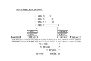
S Figure 1 illustrates the relative locations of the system GCIRS primers,
bumpers and
detectors across a 103 base pair region.
' Figure 2 illustrates the relative locations of the system GCIRSL primers,
bumpers and
detectors across a 100 base pair region.
Figure 3 illustrates the relative locations of the GC 02 primers, bumpers and
detectors
across a 98 base pair region.
DETAILED DESCRIPTION OF THE INVENTION
The present invention provides oligonucleotides, amplification primers and
assay probes
which exhibit Neisseria gonorrhoeae specificity in nucleic acid amplification
reactions. Also
I5 provided are methods for detecting and identifying Neisseria gonorrhoeae
nucleic acids using
the oligonucleotides of the invention. Preferred methods are to use the
oligonucleotides in tSDA
and homogeneous real time fluorescent tSDA reactions. These methods are taught
in U.S. Patent.
Nv. 5,S47, 861, U.S. Patent No. 5,648,21 l, U.S., Patent 5,928,869 and US
Patent 5,846,726.
The present invention provides three tSDA systems (GCIRS, GCIRSL and GC 02)
that
specifically amplify and detect N. gonorrhoeae genomic DNA. Several primer
combinations
were designed, for each system and tested in statistically designed
experiments. Specificity,
sensitivity and crossreactivity experiments were performed with the best
primer combination for
each system.
Sequence analysis was conducted on a 800 by region of N. gonorrhoeae genomic
DNA.
The 800 by region was generated using primers GC 1.3 S'-CTGATATCTGCATGGAGGCAA-
3'
(SEQ ID NO: 1) and GC 2.3 5'-GATCGTAATCTCCGCCTTTCTT-3' (S~Q ID NO: 2), and a
200 by region internal thereto was generated using primers IR.R.2 S'-
CCGCAGCATACGCGCAAATCAA-3' (SEQ ID . NO: 3) and IRLI 5'-
GGTATGGTTTCAAGACGCTTCA-3' (SEQ ID NO: 4). Mapping of the 800 by fragment
revealed several regions of complete specificity when tested with several N
gonorrhaeae strains
as well as other related Neisseria species. However, several regions of
crossreactivity with
-4-
CA 02249635 1998-11-02
PATENT
P-3869
Neisseria species were identified within this fragment. Based on this
information, the 3 tSDA
systems were designed.
Primers were designed based on the 800 by fragment of Neisseria gonorrhoeae
nucleic
acid. Primer combinations were screened for optimal conditions. Various
detector probes were
tested for specificity and sensitivity in tSDA reactions and in fluorescent
real time tSDA
reactions.
As nucleic acids do not require complete complementarity in order to
hybridize, it is to be
understood that the probe and primer sequences herein disclosed may be
modified to some extent
without loss of utility as Neisseria gonorrhoeae-specific probes and primers.
As is known in the
art, hybridization of complementary and partially complementary nucleic acid
sequences may be
obtained by adjustment of the hybridization conditions to increase or decrease
stringency (i.e.,
adjustment of hybridization temperature or salt content of the buffer). Such
minor modifications
of the disclosed sequences and any necessary adjustments of hybridization
conditions to maintain
Neisseria gonorrhoeae specificity require only routine experimentation and are
within the
ordinary skill in the art.
The amplification products generated using the inventive primers may be
detected by a
characteristic size, for example on polyacrylamide or agarose gels stained
with ethidium
bromide. Alternatively, amplified N. gonorrhoeae target sequences may be
detected by means of
an assay probe, which is an oligonucleotide tagged with a detectable label. In
one embodiment,
at least one tagged assay probe may be used for detection of amplified target
sequences by
hybridization (a detector probe), by hybridization and extension as described
by Walker, et al.,
Nucl. Acids Res., supra (a detector primer) or by hybridization, extension and
conversion to
double stranded form as described in EP 0 678 582 (a signal primer).
Preferably, the assay probe
is selected to hybridize to a sequence in the target which is between the
amplification primers,
i.e., it should be an internal assay probe. Alternatively, an amplification
primer or the target
binding sequence thereof may be used as the assay probe_
The detectable label of the assay probe is a moiety which can be detected
either directly
or indirectly as an indication of the presence of the target nucleic acid. For
direct detection of the
label, assay probes may be tagged with a radioisotope and detected by
autoradiography or tagged
with a fluorescent moiety and detected by fluorescence as is known in the art.
Alternatively, the
assay probes may be indirectly detected by tagging with a label which requires
additional
reagents to render it detectable. Indirectly detectable labels include, for
example,
chemiluminescent agents, enzymes which produce visible reaction products and
ligands (e.g.,
haptens, antibodies or antigens) which may be detected by binding to labeled
specific binding
-5-
' ,
CA 02249635 1998-11-02
PATENT
P-3869
partners (e.g., antibodies or antigens/haptens). Ligands are also useful
immobilizing the ligand- '
labeled oIigonucleotide (the capture probe) on a solid phase to facilitate its
detection.
Particularly useful labels include biotin (detectable by binding to labeled
avidin or streptavidin)
and enzymes such as horseradish peroxidase or alkaline phosphatase (detectable
by addition of
S enzyme substrates to produce colored reaction products). Methods for adding
such labels to, or
including such labels in, oligonucleotides are well known in the art and any
of these methods are
suitable for use in the present invention.
Examples of specific detection methods which may be employed include a
chemiluminescent method in which amplified products are detected using a
biotinylated capture
probe and an enzyme-conjugated detector probe as described in U.S. Patent No.
5,470,723. After
hybridization of these two assay probes to different sites in the assay region
of the target
sequence (between the binding sites of the two amplification primers), the
complex is captured
on a streptavidin-coated microtiter plate by means of the capture probe, and
the
chemiluminescent signal is developed and read in a luminometer. As another
alternative for
I S detection of amplification products, a signal primer as described in EP 0
678 582 may be
included in the SDA reaction. In this embodiment, labeled secondary
amplification products are
generated during SDA in a target amplification-dependent manner and may be
detected as an
indication of target amplification by means of the associated label.
For commercial convenience, amplification primers for specific detection and
identification of N. gonorrhoeae nucleic acids may be packaged in the form of
a kit. Typically,
such a kit contains at least one pair of amplification primers according to
the present invention.
Reagents for performing a nucleic acid amplification reaction may also be
included with the N.
gonorrhoeae-specific amplification primers, for example, buffers, additional
primers, nucleotide
triphosphates, enzymes, etc. The components of the kit are packaged together
in a common
container, optionally including instructions for performing a specific
embodiment of the
inventive methods. Other optional components may also be included in the kit,
e.g., an
oligonucleotide tagged with a label suitable for use as an assay probe, and/or
reagents or means
for detecting the label.
The target binding sequences of the amplification primers in conjunction with
detector
probes can confer species hybridization specificity on the oligonucleotides
and therefore provide
species-specificity to an amplification based assay. Other sequences, as
required for performance
of a selected amplification reaction, may optionally be added to the target
binding sequences
disclosed herein without altering the species-specificity of the
oligonucleotide. By way of
example, the N. gonorrhoeae-specific amplification primers of the invention
may contain a
-6-
CA 02249635 1998-11-02 -
PATENT
P-3869
recognition site for the restriction endonuclease BsoBI which is nicked during
the SDA reaction. '
It will be apparent to one skilled in the art that other nickable restriction
endonuclease
recognition sites may be substituted for the BsoBI recognition site, including
but not limited to
those recognition sites disclosed in EP 0 684 315. Preferably, the recognition
site is for a
thermophilic restriction endonuclease so that the amplification reaction may
be performed under
the conditions of thermophilic SDA (tSDA). Similarly, the tail sequence of the
amplification
primer (5' to the restriction endonuclease recognition site) is generally not
critical, although the
restriction site used for SDA and sequences which will hybridize either to
their own target
binding sequence or to the other primers should be avoided. Amplification
primers for SDA
according to the invention therefore consist of the 3' target binding
sequences, a nickable
restriction endonuclease recognition site S' to the target binding sequence
and a tail sequence
about 10-25 nucleotides in length S' to the restriction endonuclease
recognition site. 'The
nickable restriction endonuclease recognition site and the tail sequence are
sequences required
for the SDA reaction. For other amplification reactions, the amplification
primers according to
the invention may consist of the disclosed target binding sequences only
(e.g., for PCR) or the
target binding sequence and additional sequences required for the selected
amplification reaction
(e.g., sequences required for SDA as described above or a promoter recognized
by RNA
polymerase for 3SR).
In SDA, the bumper primers are not essential for species-specificity, as they
function to
displace the downstream, species-specific amplification primers. It is only
required that the
bumper primers hybridize to the target upstream from the amplification primers
so that when
they are extended they will displace the amplification primer and its
extension product. The
particular sequence of the bumper primer is therefore generally not critical,
and may be derived
from any upstream target sequence which is sufficiently close to the binding
site of the
amplification primer to allow displacement of the amplification primer
extension product upon
extension of the bumper primer. Occasional mismatches with the target in the
bumper primer
sequence or some cross-hybridization with non-target sequences do not
generally negatively
affect amplification efficiency as long as the bumper primer remains capable
of hybridizing to
the specific target sequence. However, the bumper primers described herein are
species-specific
for N. gonorrhoeae and may therefore also be used as target binding sequences
in amplification
primers, if desired.
Amplification reactions employing the primers of the invention may incorporate
thymine
as taught by Walker, et al., szrpra, or they may wholly or partially
substitute 2'-deoxyuridine 5'-
triphosphate for TTP in the reaction to reduce cross-contamination of
subsequent amplification
~ ,
CA 02249635 1998-11-02
PATENT
P-3869
reactions, e.g., as taught in EP 0 624 643. dU (uridine) is incorporated into
amplification '
products and can be excised by treatment with uracil DNA glycosylase (UDG).
These abasic
sites render the amplification product unamplifiable in subsequent
amplification reactions. UDG
may be inactivated by uracil DNA glycosylase inhibitor (Ugi) prior to
performing the subsequent
amplification to prevent excision of dU in newly-formed amplification
products.
Other systems were developed for performing tSDA using different combinations
of
primers, bumpers and detectors. However, these other systems were not
preferred for various
reasons such as lack of adequate specificity, narrow range of optimal
conditions and lack of
robustness.
The systems which were found to be useful for performing homogeneous nucleic
acid
amplification and real time detection of N. gonorrhoeae nucleic acid sequences
were developed
from the following primers and detectors.
GCIRS Primers and Detectors
Primers
Amp. Upstream Sequence
GCIR-AL5.1 5'CGATTCCGCTCCAGACTTCTCGGGGAACAGCTTGAAGTTTT3' (SEQ ID
NO: 5)
GCIR-AL5.2 5'CGATTCCGCTCCAGACTTCTCGGGGAACAGCTTGAAGTTT3' (SEQ ID
NO: 6)
GCIR-AL5.3 5'CGATTCCGCTCCAGACTTCTCGGGAACAGCTTGAAGTTTT3' (SEQ ID
NO. 7)
Amp. Downstream:
GCIR-AR5.1 5'ACCGCATCGAATGCATGTCTCGGGTCCTTGCAGTTAGGC3' (SEQ ID
NO: 8)
GCIR-AR5.2 5'ACCGCATCGAATGCATGTCTCGGGCCTTGCAGTTAGGC3' (SEQ ID
NO: 9)
GCIR-AR5.3 5'ACCGCATCGAATGCATGTCTCGGGTCCTTGCAGTTAGG3' (SEQ ID
NO: 10)
Bumpers:
GCIR-BL5.1 5'CGCAAATCATCAAAG3' (SEQ ID NO: 1 I)
_g_
CA 02249635 1998-11-02
PATENT
P-3869
GCIR-BRS. I 5'TCAAGACGCTTCACG3' (SEQ ID NO: 12)
-9-
CA 02249635 1998-11-02
PATENT
P-3869
Detectors.
GCIR-DIL 5'AAAGGAGAAGATAAAAG3' (SEQ ID NO: 13)
GCIR-D2L 5'AGCAGACGGAGAAG3' (SEQ ID NO: 14)
S Fluorescent Detectors.
Downstream
GCIRS-FD10 5'TAGCACCCGAGTGCTTTCTCCGTCTGCTCTTTTATCTTCTC3' (SEQ ID
NO: 15)
GCIRS-FD8 5'TAGCACCCGAGTGCTTTCTCCGTCTGCTCTTTTATCTTC3' (SEQ ID
NO: 16)
GCIRS-FD3 5'TAGCACCCGAGTGCTTTCTCCGTCTGCTCT3' (SEQ ID NO: 17)
Upstream
GCIRS-FD11 5'TAGCACCCGAGTGCTTAAAGGAGAAGATAAAAGAGCAG3' (SEQ ID
NO: 18)
GCIRS-FD6 5'TAGACCCGAGTGCTTAAAGGAGAAGATAAAAGAGC3' (SEQ ID NO: l9)
GCIRS-FD2 5'TAGCACCCGAGTGCTTAAAGGAGAAGATAAAAG3' (SEQ ID NO: 20)
GCIRS-FD I 5'TAGCACCCGAGTGCTTAAAGGAGAAGATAAAAGAGCAGACGGAGA3'
(SEQ ID NO: 21)
GCIRSL Primers and Detectors
Primers
Amp. Upstream Sequence
GCIRSL.APL1 5'-CGATTCCGCTCCAGACTTCTCGGGGAGAAGCCTAACTG-3'
(SEQ ID NO: 22)
GCIRSL.APL2 5'-CGATTCCGCTCCAGACTTCTCGGGAGAAGCCTAACTGCA-3'
(SEQ ID NO: 23}
Amp Downstream
GCIRSL.APR1 5'-ACCGCATCGAATGCATGTCTCGGGCTGCCTATTGCCGGT-3'
(SEQ ID NO: 24)
-10-
CA 02249635 1998-11-02
PATENT
P-3869
GCIRSL.APR2 5'-ACCGCATCGAATGCATGTCTCGGGTGCCTATTGCCGGTA-3' '
(SEQ ID NO: 25)
GCIRSL.APR3 5'-ACCGCATCGAATGCATGTCTCGGGTGCCTATTGCCGGT-3'
(SEQ ID NO: 26)
Bumpers
GCIRSL.BL 5'-GAGAAGATAAAAGAG-3' (SEQ ID NO: 27)
GCIRSL.BR 5'-ACAATACGGCTGCG-3' (SEQ ID NO: 28)
Detector
GCIRSL.DL1 5'-CAAGGAAGGCGTGAA-3' (SEQ ID NO: 29)
GCIRSL.DL2 5'-GCGTCTTGAAACCAT-3' (SEQ ID NO. 30)
Fluorescent Detector
GCIRSL.FD 1 5'-TAGCACCCGAGTGCTGGAAGGCGTGAAGCGTCTTGAAAC
CAT-3' (SEQ ID NO: 31)
GC 02 Primers and Detectors
Primers
Amp. Upstream Sequence
02AL44. I 5'-CGATTCCGCTCCAGACTTCTCGGGAGGCTGGAAGAAAAG-3'
(SEQ ID NO: 32)
02AL42.1 5'-CGATTCCGCTCCAGACTTCTCGGGGGCTGGAAGAAAAG-3'
(SEQ ID NO: 33)
Amp Downstream
02AR46.1 5'-ACCGCATCGAATGCATGTCTCGGGCGAGTTTACGCATCAA-3'
(SEQ ID NO: 34)
-II-
CA 02249635 1998-11-02
PATENT
P-3869
02AR42.1 5'-ACCGCATCGAATGCATGTCTCGGGGAGTTTACGCATCAA-3' '
(SEQ ID NO: 35)
Bumpers
02BL42.1 5'-TTT CCC CGA CTT CA-3' (SEQ ID NO: 36)
02BR42.1 5'-GTG ATA CGC AAT AAC-3' (SEQ ID NO: 37)
Detectors
02DL42.1 5'-AAG AAG CCT AAA AAA G-3' (SEQ ID NO: 38)
02DR42.1 5'-TCA TCA TCG CAG CA-3' (SEQ ID NO: 39)
Assays for Neisseria gonorrhoea were performed using the technique of
fluorescent real
time tSDA. The primers, bumpers, and detectors designed for GCIRS and GCIRSL
are shown in
Figure 1 and Figure 2, respectively. The annealing regions for the
amplification primers and
fluorescent detectors are represented by the rectangles above or below the DNA
sequence. The
conditions for fluorescent GCIRS and GCIRSL tSDA were continually modified to
achieve
optimal sensitivity. Set forth below is one such set of optimal conditions for
GCIRS and one
such set of optimal conditions for GC1RSL_
GCIRS:
7% glycerol
8% DMSO
45mM potassium phosphate
(0.1 mM) dATP, (0.1 mM) dGTP, (0.25mM) dUTP, (0.7mM) alpha-thin dCTP
SmM Magnesium acetate
100ug/ml BSA
I .82% Trehalose
360uM DTT
2400 ng human DNA
320 units BsoBI restriction endonuclease
20 units Bst polymerase
1 Unit Uracil-N-glycosylase
-12-
CA 02249635 1998-11-02
PATENT
P-3869
Units Uracil-N-glycosylase inhibitor
200 nM detector (FD10) (SEQ ID NO: 15)
500 nM amplification primers (GCIR-AL5.3 (SEQ ID NO: 7) and GCIR-AR5.1 (SEQ ID
N0:8))
SOnM bumpers (GCIR-BL5.1 (SEQ ID NO: 11) and GCIR-BR5.1(SEQ ID NO: 12))
5
100u1 reaction volume
Decontamination performed for 20 minutes at 45°C
Amplification performed for 60 minutes at 52°C
GCIRSL:
7% Glycerol
5% DMSO
25mM potassium phosphate
(0.2mM) dATP, (0.2mM) dGTP, (O.SmM) dUTP, (l.4mM) alpha thio-dCTP
6mM Magnesium acetate
100 ug/ml BSA .
1.82% Trehalose
360uM DTT
2000 ng human DNA
1 Unit Uracil-N-glycosylase
5 Units Uracil-N-glycosylase Inhibitor
480 units BsoBI
units Bst
200 nM detector (GCIRSL.FD1 (SEQ ID NO: 31))
25 500 nM amplification primers (GCIRSL.APL1 (SEQ ID NO: 22)and GCIRSL.APRl
(SEQ ID
NO: 24))
50 nM bumpers (GCIRSL.BL (SEQ ID NO: 27) and GCIRSL.BR (SEQ ID NO: 28))
I OOuI reaction volume
30 Decontamination performed for 20 minutes at 45°C
Amplification performed for 60 minutes at 52°C
As explained in greater detail in the Examples, the sensitivity of GCIRS and
GCIRSL in
fluorescent real time tSDA was assessed using the detectors GCIRS-FD 10 (SEQ
ID NO: 15) and
-I3-
CA 02249635 1998-11-02
PATENT
P-3869
GCIRSL-FD1 (SEQ ID NO: 31), with the plasmid GC10 as the target DNA source.
The plasmid '
GC 10 contains an 800 base pair region of the Neisseria gonorrhoeae genome
inserted into
pUC 18. Fluorescent tSDA was performed using a titration of GC I 0 plasmid:
250, I 00, 50, 25,
and 12 copies. GCIRS with FD 10 was capable of detecting down to 25 copies of
the GC 10
plasmid. The sensitivity of GCIRSL in real time tSDA was determined to be at
250 copies of the
GC 10 plasmid. Judgement of a reaction being positive was determined by
comparing the RFU
values of sample reactions with those from negative controls (no target DNA
added). If the
reactions with GC 10 produced RFU values greater than 2-3 times the average
RFU values for the
negative control, then it was considered positive.
Other detectors for GCIRS had been tested in separate experiments: FD1 (SEQ ID
NO:
21), FD2 (SEQ ID NO: 20), FD3 (SEQ ID NO: 17), FD6 (SEQ ID NO: 19), FD8 (SEQ
ID NO:
16), and FD11 (SEQ ID NO: 18). While FD8, FD3, and FDI were capable of
detecting the
GC10 plasmid at levels of 250 copies, others, such as FD11, FD6, and FD2,
produced lower
RFU values at this identical target concentration. This clearly demonstrates
the importance of
examining multiple detector sequences/lengths to achieve maximum sensitivity,
and as a result,
GCIRS-FD 10 (SEQ ID NO: 15) was chosen as the primary detector to be used in
real time
fluorescent tSDA.
Specificity and crossreactivity of GCIRS and GCIRSL were determined by testing
various Neisseria gonorrhoea strains, other Neisseria species, and non-related
bacteria and
viruses. The 12 Neisseria gonorrhoea strains were tested at 1 x 10⁴ genomes.
All of the
crossreactant DNAs were diluted to approximately 1 x 10~ genomic copies. A
summary of the
specificity and crossreactivity from multiple experiments is seen in Table 5.
Strand Displacement AmpliFcation (SDA) is an isothermal method of nucleic acid
amplification in which extension of primers, nicking of a hemimodified
restriction endonuclease
recognition/cleavage site, displacement of single stranded extension products,
annealing of
primers to the extension products (or the original target sequence) and
subsequent extension of
the primers occurs concurrently in the reaction mix. This is in contrast to
the polymerase chain
reaction (PCR), in which the steps of the reaction occur in discrete phases or
cycles as a result of
the temperature cycling characteristics of the reaction. SDA is based upon 1 )
the ability of a
restriction endonuclease to nick the unmodified strand of a
hemiphosphorothioate form of its
double stranded recognition/cleavage site and 2) the ability of certain
polymerases to initiate
replication at the nick and displace the downstream non-template strand. After
an initial
incubation at increased temperature (about 95°C) to denature double
stranded target sequences
for annealing of the primers, subsequent polymerization and displacement of
newly synthesized
-14-
CA 02249635 2002-02-11
PATENT
P-3869
strands takes place at a constant temperature. Production of each new copy of
the target '
sequence consists of five steps: 1) binding of amplification primers to an
original target
sequence or a displaced single-stranded extension product previously
polymerized, 2) extension
of the primers by a 5'-3' exonuclease deficient polymerase incorporating an a-
thio
.5 deoxynucleoside triphosphate (oc-thio dNTP), 3) nicking of a hemimodified
double stranded
restriction site, ) dissociation of the restriction enzyme from the nick site,
and 5) extension from
the 3' end of the nick by the 5'-3' exonuclease deficient polymerase with
displacement of the
downstream newly synthesized strand. Nicking, polymerization and displacement
occur
concurrently and continuously at a constant temperature because extension from
the nick
regenerates another nickable restriction site. When a pair of amplification
primers is 'used, each
of which hybridizes to one of the two strands of a double stranded target
sequence, amplification
is exponential. This is because the sense and antisense strands serve as
templates for the
opposite primer in subsequent rounds of amplification. When a single
amplification primer is
used, amplification is linear because only one strand serves as a template for
primer extension.
Examples of restriction endonucleases which nick their double stranded
recognition/cleavage
sites when an a-thio dNTP is incorporated are HincII, HindII, AvaI, NciI and
Fnu4HI. All of
these restriction endonucleases and others which display the required nicking
activity are suitable
for use in conventional SDA. However, they are relatively thermolabile and
lose activity above
about 40°C.
Targets for amplification by SDA may be prepared by fragmenting larger nucleic
acids by
restriction with an endonuclease which does not cut the target sequence.
However, it is generally
preferred that target nucleic acids having the selected restriction
endonuclease
recognition/cleavage sites for nicking in the SDA reaction be generated as
described by Walker,
et al. (1992, Nuc. Acids Res., supra) and in U.S. Patent No. 5,270,184.
Briefly, if the target sequence is double stranded, four primers :are
hybridized to it.
Two of the primers (S t and S,) are SDA amplification primers and two (B t and
B2) are external
or bumper primers. S ~ and S2 bind to opposite strands of double stranded
nucleic acids flanking
the target sequence. B1 and B2 bind to the target sequence 5' (i.e., upstream)
of S~ and S2,
respectively. The exonuclease deficient polymerase is then used to
simultaneously extend all
four primers in the presence of three deoxynucleoside triphosphates and at
least one modified
deoxynucleoside triphosphate (e.g., 2'-deoxyadenosine 5'-O-(1-
thiotriphosphate), "dATPaS").
The extension products of S, and SZ are thereby displaced form the original
target sequence
template by extension of B, and B2. The displaced, single stranded extension
products of the
amplification primers serve as targets for binding of the opposite
amplification and bumper
-15-
CA 02249635 1998-11-02
PATENT
P-3869
primer (e.g., the extension product of S~ binds S2 and B2). The next cycle of
extension and '
displacement results in two double stranded nucleic acid fragments with
hemimodified restriction
endonuclease recognition/cleavage sites at each end. These are suitable
substrates for
amplification by SDA. As in SDA, the individual steps of the target generation
reaction occur
concurrently and continuously, generating target sequences with the
recognition/cleavage
sequences at the ends required for nicking by the restriction enzyme in SDA.
As all of the
components of the SDA reaction are already present in the target generation
reaction, target
sequences generated automatically and continuously enter the SDA cycle and are
amplified.
To prevent cross-contamination of one SDA reaction by the amplification
products of
another, dUTP may be incorporated into SDA-amplified DNA in place of dTTP
without
inhibition of the amplification reaction. The uracil-modified nucleic acids
may then be
specifically recognized and inactivated by treatment with uracil DNA
glycosylase (UDG).
Therefore, if dUTP is incorporated into SDA-amplified DNA in a prior reaction,
any subsequent
SDA reactions can be treated with UDG prior to amplification of double
stranded targets, and
any dU containing DNA from previously amplified reactions will be rendered
unamplifiable.
The target DNA to be amplified in the subsequent reaction does not contain dU
and will not be
affected by the UDG treatment. UDG may then be inhibited by treatment with Ugi
prior to
amplification of the target. Alternatively, UDG may be heat-inactivated. In
thermophiIic SDA,
the higher temperature of the reaction itself (>_ 50°C) can be used to
concurrently inactivate UDG
and amplify the target.
SDA requires a polymerase which lacks 5'-3' exonuclease activity, initiates
polymerization at a single stranded nick in double stranded nucleic acids, and
displaces the
strand downstream of the nick while generating a new complementary strand
using the unpicked
strand as a template. The polymerase must extend by adding nucleotides to a
free 3'-OH. To
optimize the SDA reaction, it is also desirable that the polymerase be highly
processive to
maximize the length of target sequence which can be amplified. Highly
processive polymerases
are capable of polymerizing new strands of significant length before
dissociating and terminating
synthesis of the extension product. Displacement activity is essential to the
amplification
reaction, as it makes the target available for synthesis of additional copies
and generates the
single stranded extension product to which a second amplification primer may
hybridize in
exponential amplification reactions. Nicking activity is also of great
importance, as it is nicking
which perpetuates the reaction and allows subsequent rounds of target
amplification to initiate.
Thermophilic SDA is performed essentially as the conventional SDA described by
Walker, et al. (1992, PNAS and Nuc. Acids Res., supra), with substitution of
the desired
-16-
CA 02249635 1998-11-02
PATENT
P-3869
thermostable polymerase and thermostable restriction endonuclease. Of course,
the temperature '
of the reaction will be adjusted to the higher temperature suitable for the
substituted enzymes and
the HincII restriction endonuclease recognition/cleavage site will be replaced
by the appropriate
restriction endonuclease recognition/cleavage site for the selected
thermostable endonuclease.
Also in contrast to Walker, et al., the practitioner may include the enzymes
in the reaction
mixture prior to the initial denaturation step if they are sufficiently stable
at the denaturation
temperature. Preferred restriction endonucleases for use in thermophilic SDA
are BsrI, BstNI,
BsmAI, BsII and BsoBI (New England BioLabs), and BstOI (Promega). The
preferred
thermophilic polymerases are Bca (Panvera) and Bst (New England Biolabs).
Homogeneous real time fluorescent tSDA is a modification of tSDA. It employs
detector
oligonucleotides to produce reduced fluorescence quenching in a target-
dependent manner. The
detector oligonucleotides contain a donor/acceptor dye pair linked such that
fluorescence
quenching occurs in the absence of target. Unfolding or linearization of an
intramolecularly
base-paired secondary structure in the detector oligonucleotide in the
presence of the target
increases the distance between the dyes and reduces fluorescence quenching.
Unfolding of the
base-paired secondary structure typically involves intermolecular base-pairing
between the
sequence of the secondary structure and a complementary strand such that the
secondary
structure is at least partially disrupted. It may be fully linearized in the
presence of a
compIementary strand of sufficient length. In a preferred embodiment, a
restriction
endonuclease recognition site (RERS) is present between the two dyes such that
intermolecular
base-pairing between the secondary structure and a complementary strand also
renders the RERS
double-stranded and cleavable or nickable by a restriction endonuclease.
Cleavage or nicking by
the restriction endonuclease separates the donor and acceptor dyes onto
separate nucleic acid
fragments, further contributing to decreased quenching. In either embodiment,
an associated
change in a fluorescence parameter (e.g., an increase in donor fluorescence
intensity, a decrease
in acceptor fluorescence intensity or a ration of fluorescence before and
after unfolding) is
monitored as an indication of the presence of the target sequence. Monitoring
a change in donor
fluorescence intensity is preferred, as this change is typically larger than
the change in acceptor
fluorescence intensity. Other fluorescence parameters such as a change in
fluorescence lifetime
may also be monitored.
A detector oligonucleotide for homogeneous real time fluorescent tSDA is an
oligonucleotide which comprises a single-stranded 5' or 3' section which
hybridizes to the target
sequence (the target binding sequence) and an intramolecularly base-paired
secondary structure
adjacent to the target binding sequence. The detector oligonucleotides of the
invention further
-17-
CA 02249635 1998-11-02
PATENT
P-3869
comprise a donor/acceptor dye pair linked to the detector oligonucleotide such
that donor
fluorescence is quenched when the secondary structure is intramolecularly base-
paired and
unfolding or linearization of the secondary structure results in a decrease in
fluorescence
quenching. Cleavage of an oligonucleotide refers to breaking the
phosphodiester bonds of both
strands of a DNA duplex or breaking the phosphodiester bond of single-stranded
DNA. This is
in contrast to nicking, which refers to breaking the phosphodiester bond of
only one of the two
strands in a DNA duplex.
The detector oligonucleotides of the invention for homogeneous real time
fluorescent
tSDA comprise a sequence which forms an intramolecularly base-paired secondary
structure
under the selected reaction conditions for primer extension or hybridization.
The secondary
structure is positioned adjacent to the target binding sequence of the
detector oligonucleotide so
that at least a portion of the target binding sequence forms a single-stranded
3' or 5' tail. As used
herein, the term "adjacent to the target binding sequence" means that all or
part of the target
binding sequence is left single-stranded in a 5' or 3' tail which is available
for hybridization to
the target. That is, the secondary structure does not comprise the entire
target binding sequence.
A portion of the target binding sequence may be involved in the intramolecular
base-pairing in
the secondary structure, it may include all or part of a first sequence
involved in intramolecular
base-pairing in the secondary structure, it may include all or part of a first
sequence involved in
intramolecular base-pairing in the secondary structure but preferably does not
extend into its
complementary sequence. For example, if the secondary structure is a stem-loop
structure (e.g.,
a "hairpin") and the target binding sequence of the detector oligonucleotide
is present as a single-
stranded 3' tail, the target binding sequence may also extend through all or
part of the first arm of
the stem and, optionally, through all or part of the loop. However, the target
binding sequence
preferably does not extend into the second arm of the sequence involved in
stem intramolecular
base-pairing. That is, it is desirable to avoid having both sequences involved
in intramolecular
base-pairing in a secondary structure capable of hybridizing to the target.
Mismatches in the
intramolecularly base-paired portion of the detector oligonucleotide secondary
structure may
reduce the magnitude of the change in fluorescence in the presence of target
but are acceptable if
assay sensitivity is not a concern. Mismatches in the target binding sequence
of the single-
stranded tail are also acceptable but may similarly reduce assay sensitivity
and/or specificity.
However, it is a feature of the present invention that perfect base-pairing in
both the secondary
structure and the target binding sequence do not compromise the reaction.
Perfect matches in the
sequences involved in hybridization improve assay specificity without negative
effects on
reaction kinetics.
-18-
CA 02249635 2002-02-11
PATENT
P-3869
When added to the amplification reaction, the detector oligonucleotide signal
primers of
the invention are converted to double-stranded form by hybridization and
extension of. an
amplif cation primer as described above. Strand displacement by the polymerase
also unfolds or
linearizes the secondary structure and converts it to double-stranded from by
synthesis of a
complementary strand. The RERS, if present, also becomes double-stranded and
cleavable or
nickable by the restriction endonuclease. As the secondary structure is
unfolded or linearized by
the strand displacing activity of the polymerase, the distance between the
donor and acceptor dye
is increased, thereby reducing quenching of donor fluorescence. The associated
change in
fluorescence of either the donor or acceptor dye may be monitored or detected
as'an indication of
amplification of the target sequence. Cleavage or nicking of the RERS
generally further
increases the magnitude of the change in fluorescence by producing two
separate fragments of
the double-stranded secondary amplification product, each having one of the
two dyes linked to
it. These fragments are free to diffuse in the reaction solution, further
increasing the distance
between the dyes of the donor/acceptor pair. An increase in donor fluorescence
intensity or a
decrease in acceptor fluorescence intensity may be detected and/or monitored
as an indication
that target amplification is occurring or has occurred, but other fluorescence
parameters which
are affected by the proximity of the donorlacceptor dye pair may also be
monitored. A change in
fluorescence intensity of the donor or acceptor' may also be detected as a
change in a ratio of
donor, and/or acceptor fluorescence intensities. For example, a change in
fluorescence intensity
may be detected as a) an increase in the ratio of donor fluorophore
fluorescence after linearizing
or unfolding the secondary structure and donor fluorophore fluorescence in the
detector
oligonucleotide prior to linearizing or unfolding, or b) as a decrease in the
ration of acceptor dye
fluorescence after linearizing or unfolding and acceptor dye fluorescence in
the detector
oligonucleotide prior to linearizing or unfolding.
It will be apparent that, in addition to SDA, the detector oligonucleotides of
the invention
may be adapted for use as signal primers in other primer extension
amplification methods (e.g.,
PCR, 3SR, TMA or NASBA). For example, the methods may be adapted for use in
PCR by
using PCR amplification primers and a strand displacing DNA polymerase which
lacks 5'-~3'
exonuclease activity (e.g., Sequencing Grade Taq from Promega or exo Vent or
exo Deep Vent'
from Mew England BioLabs) in the PCR. The detector oligonucleotide signal
primers hybridize
to the target downstream from the PCR amplification primers, are displaced and
are rendered
double-stranded essentially as described for SDA. In PCR any RERS may
optionally be selected
for use in the detector oligonucleotide, as there are typically no modifned
deoxynucleoside
triphosphates present which might induce nicking rather than cleavage of the
RERS. As
Trademark*
-19-
CA 02249635 1998-11-02
PATENT
P-3869
thermocycling is a feature of amplification by PCR, the restriction
endonuclease is preferably '
added at low temperature after the final cycle of primer annealing and
extension for end-point
detection of amplification. However, a thermophilic restriction endonuclease
which remains
active through the high temperature phases of the PCR reaction could be
present during
amplification to provide a real-time assay. As in SDA systems, linearization
of the secondary
structure and separation of the dye pair reduces fluorescence quenching, with
a change in a
fluorescence parameter such as intensity serving as an indication of target
amplification.
The change in fluorescence resulting from unfolding or linearizing of the
detector
oligonucleotides may be detected at a selected endpoint in the reaction.
However, because
linearized secondary structures are produced concurrently with hybridization
or primer extension,
the change in fluorescence may also be monitored as the reaction is occurring,
i.e., in "real-time".
This homogeneous, real-time assay format may be used to provide
semiquantitative or
quantitative information about the initial amount of target present. For
example, the rate at
which fluorescence intensity changes during the unfolding or linearizing
reaction (either as part
I S of target amplification or in non-amplification detection methods) is an
indication of initial target
levels. As a result, when more initial copies of the target sequence are
present, donor
fluorescence more rapidly reaches a selected threshold value (i.e., shorter
time to positivity). The
decrease in acceptor fluorescence similarly exhibits a shorter time to
positivity, detected as the
time required to reach a selected minimum value. In addition, the rate of
change in fluorescence
parameters during the course of the reaction is more rapid in samples
containing higher initial
amounts of target than in samples containing lower initial amounts of target
(i.e., increased slope
of the fluorescence curve). These or other measurements as is known in the art
may be made as
an indication of the presence of target or as an indication of target
amplification. The initial
amount of target is typically determined by comparison of the experimental
results to results for
known amounts of target.
Assays for the presence of a selected target sequence according to the methods
of the
invention may be performed in solution or on a solid phase. Real-time or
endpoint homogeneous
assays in which the detector oligonucleotide functions as a primer are
typically performed in
solution. )-Iybridization assays using the detector oligonucleotides of the
invention may also be
performed in solution (e.g., as homogeneous real-time assays) but are also
particularly well-
suited to solid phase assays for real-time or endpoint detection of target. In
a solid phase assay,
detector oligonucleotides may be immobilized on the solid phase (e.g., beads,
membranes or the
reaction vessel) via internal or terminal labels using methods known in the
art. For example, a
biotin-labeled detector oligonucleotide may be immobilized on an avidin-
modified solid phase
-20-
CA 02249635 1998-11-02
PATENT
P-3869
where it will produce a change in fluorescence when exposed to the target
under appropriate '
hybridization conditions. Capture of the target in this manner facilitates
separation of the target
from the sample and allows removal of substances in the sample which may
interfere with
detection of the signal or other aspects of the assay.
The following Examples illustrate specific embodiments of the invention
described
herein. As would be apparent to skilled artisans, various changes and
modifications are possible,
and are contemplated within the scope of the invention described.
EXAMPLE 1
Design of tSDA Primer Sets
The 800bp N gonorrhoeae sequence identified was examined for the design of
tSDA
primer sets. Certain regions of the genome were avoided due to the presence of
GC or AT
stretches, and/or small repeats that would cause strong interactions between
primers. The
software program OligoTM (National Biosciences, Inc., Plymouth, Minnesota) was
used to screen
out tSDA sets that could potentially be problematic, due to primer/primer
interactions with high -
OG values. Various primer sets were designed within the region. Some sets were
immediately
dismissed and some were found to be tacking any serious interactions. Out of
all the sets
examined, one was chosen for further study - GCIRS_ GCIRS was designed with
three variants
of both the left and right amplification primers (Figure 1 ). This allowed for
the examination of
various combinations of the primers, each of which had a different Tm. All of
the primers
encompassing the GCIRS system and their positions can be seen in the diagram
in Figure 1.
EXAMPLE 2
Design of GCIRS tSDA Reaction Conditions -
A statistically designed experiment was performed to examine 5 out of 6 primer
combinations of the GCIRS primers in the presence of different co-solvent
concentrations and
amplification temperatures. This experiment was used to determine which primer
pairings had
the best capability of producing amplification across a wide spectrum of
conditions. The
following variables were examined: potassium phosphate (25 mM and 35 mM), DMSO
(3% and
8%), glycerol (3.5% and 7%), human DNA (650 ng and 1050 ng), amplification
temperature (52°
-21 -
CA 02249635 1998-11-02
PATENT
P-3869
C and 54°C). The results show that the following tSDA condition
provided the greatest
amplification:
-22-
CA 02249635 1998-11-02
PATENT
P-3869
Primers: GCIRS-AL5.3 (SEQ ID NO: 7) - 0.5 pM
GCIR-AR5.1 (SEQ ID NO: 8) - 0.5 p.M
GCIR-BL5.1 (SEQ ID NO: 11) - 0.05 ~.M
GCIR-BRS_1 (SEQ ID NO: 12) - 0.05 p.M
Detectors: GCIR-D1L (SEQ ID NO: I3) - 10 p.M
Co-solvents: Potassium phosphate - 35 mM
DMSO - 3%
Glycerol - 7%
Magnesium acetate - 5 mM
DTT - 0_36 mM
Trehalose - I .82%
BSA - 100 p.g/mL
human DNA - 650 ng
dNTPs - dCTP (1.4 mM), dUTP (0.5 mM), dGTP (0_2 mM), dATP (02 mM)
Enzymes: UDG - 1 unit/50 pL reaction
UDI - 5 units/50 p.L reaction
BsoBI/Bst - 160 units/9 units
Decontamination: 45°C for 30 minutes
Amplification: 52°C for 30 minutes
Other primers such as GCIR-ALS.1 (SEQ ID NO: 5) would also be expected to be
effective since all of the tested amplification primers were effective_
Additionally, GCIR-D2L
(SEQ ID NO: 14) could be used as a 32P detector probe in place of D 1 L, or as
a capture probe in
an assay system. The combination of GCIR-AL5.3 and GCIR-AR5.1 was found to be
the
optimal primer set. All of the primer sets tested were able to amplify N.
gonorrhoeae BDMS
2900 at 1 x 10⁶ genomes, although some sets were more effective than others
under certain
conditions. Further experimentation with these primer sets based on strategic
design resulted in
optimization of the above reaction conditions by changing the DMSO and
glycerol
concentrations to 5.5% DMSO and 5.2% glycerol. All other co-solvents and
enzyme
concentrations were identical to those above.
-23-
_.
CA 02249635 1998-11-02
PATENT
P-3869
EXAMPLE 3 -
Assay of GCIRS tSDA Sensitivity
Utilizing the optimal tSDA conditions described in Example 2, a limit of
detection
experiment was set up. A titration of N. gonorrhoeae strain BDMS 2900 from 1 x
106 down to 1
genome/reaction was performed. The titration panel was tested with the GCIRS
tSDA system in
the presence of human DNA at 650 ng and 1250 ng/reaction. Single samples of 1
x 106, I x 105
and 1 x 10⁴ genomes/reaction were tested, I x 103 genomes/reaction was tested
in duplicate, and
100, 10 and 1 genomes were tested in triplicate. A negative control was also
included in the
experiment. The method of testing low copy numbers of the N. gonorrhoeae
genome in multiple
reactions was done to ensure that the lack of amplification in one sample was
not considered to
be indicative of the system's sensitivity, since sampling error could cause
such a result. The
result of the sensitivity experiment is that GCIRS was capable of detecting
down to 10
genomes/reaction three out of three times and 1 genome/reaction 1 out of three
times.
EXAMPLE 4
Assay of N. gonorrhoeae tSDA Specificity and Crossreactivity
Experiments were set up to examine the specificity and crossreactivity of the
GCIRS
tSDA, system. The optimal primer set described in Example 2 was utilized in
this experiment.
Several strains of N. gonorrhoeae were tested at 1 x 10⁶ genomes/reaction.
GCIRS was capable
of amplifying every one of the tested strains listed in Table 2. This tSDA
system's specificity
was satisfactory. It was next determined whether there would be any
crossreactivity with any of
the other Neisseria species or non Neisseria bacteria. To accomplish this, all
of the non-
crossreactant bacteria were tested at a level of 1 x 10~ genomes/reaction. In
none of the reactions
was any amplification product detected (Table 3).
Table 2
Organism Strain GCIRS GCIRSL GC 02
Negative Control 50 ng human DNA - - -
Neisseria gonorrhoeaeCDC 111 + - + +
Neisseria gonorrhoeaeBDMS 1632 + + +
Neisseria gonorrhoeaeATCC 19424 + + +
Neisseria gonorrhoeaeBDMS 2900 + + +
-24-
CA 02249635 1998-11-02
PATENT
P-3869
Neisseria gonorrhoeaeATCC 35201 + + + '
Neisseria gonorrhoeaeATCC 35541 + + +
Neisseria gonorrhoeaeATCC 35542 + + +
Neisseria gonorrhoeaeATCC 43069 + + +
Neisseria gonorrhoeaeATCC 43070 + + +
Neisseria gonorrhoeaeBDMS 454 + + +
Neisseria gonorrhoeaeATCC 49226 + + +
Neisseria gonorrhoeaeATCC 51109 + + not tested
Table 3
Org-anism Strain GCIR 5 GCIRSL GC 02
Negative Control 50 ng hDNA - - -
Neisseria meningitidesATCC 13090 not tested - -
Neisseria meningitidesATCC 14632 - - -
Neisseria meningitidesATCC 13077 - - -
Neisseria meningitidesATCC 13102 GRP C - - -
Neisseria meningitidesATCC 13113 GRP D - - -
Neisseria meningitidesATCC 35559 GRP W-135 - - -
Neisseria lactamicaATCC 44418 - - -
Neisseria lactamicaATCC 49142 - - -
Neisseria lactamicaATCC 23970 - - -
Neisseria lactamicaATCC 23971 - - -
Neisseria lactamicaATCC 23972 - - -
Chlarrrydiae trachomatisL2 - - -
Chlamydiae trachomatisJ - - -
Chlamydiae psittaci- - -
Chlamydiae pnezimoniae- - -
Neisseria,flavescensATCC 13120 - - -
Neisseria sicca ATCC 29193 - - -
Neisseria sicca ATCC 9913 - - -
Neisseria sttbflavaATCC 14799 - - -
Neisseria subflava ATCC 19243 - not tested -
Neisseria cinerea ATCC 14685 - - -
Neisseria elongata ATCC 25295 - - -
- 25 -
CA 02249635 1998-11-02
PATENT
P-3869
Neisseria mucosa ATCC 19696 - - - '
Branhamella catarrhalisATCC 25240 - - -
Moraxella lacunata ATCC 17967 - - -
Kingella kingae ATCC 23330 - -
Salmonella typhimurium ATCC 13311 - - -
Salmonella minnesota ATCC 9700 - - -
Staph aureus ATCC 12598 - - -
Acinetobacter Iwo~ ATCC 19001 - - -
E. coli ATCC 11775 - - -
Klebsiella pneumoniae ATCC 13883 - - -
Gardnerella vaginalis ATCC 14018 - - -
Streptococcus Group ATCC 16915 - - -
A
Streptococcus Group ATCC 12386 - - -
B
Proteus mirabilis ATCC 29906 - - -
Haemophilt~s influenzaeATCC 33533 - -
b
Mycoplasma orate ATCC 23714 - - -
HSV-1 McINTYRE - - -
HSV-2 Strain G - - -
Trichomonas vaginalis ATCC 30001 - - -
~
Candida albicctns ATCC 44808 - - -
Streptococczts faecalisATCC 29212 - - -
Peptostreptococczzs ATCC 27340 - - -
productus
EXAMPLE 5
GCIRS Primers in Fluorescent Real Time tSDA
Assays for Neisseria gonorrhoeae were performed using fluorescent real time
tSDA. The
primers, bumpers and detectors designed for this are shown in Fig. 1. The
sensitivity and
crossreactivity of GCIRS with detector FD1 (SEQ ID NO. 21) was assayed (Table
4). A plasmid
(GC 10) that contains an 800 base pair region of the Neisseria gonorrhoeae
genome inserted into
pUC I 8 was used as the target. The testing was conducted with from I 000 to
25 copies of the
plasmid. The conditions of the tSDA were:
-26-
CA 02249635 1998-11-02
PATENT
P-3869
5.2 % Glycerol
5.5% DMSO
35 mM potassium phosphate
2000 ng human DNA
320 units BsoB 1
20 units Bst
200 nM detector (FD 1 )
500 nM amplification primers (AL5.3 (SEQ ID NO: 7) and AR5.1 (SEQ ID NO: 8))
50 nM bumpers (BL5.1 (SEQ ID NO: 11) and BR5.1 (SEQ ID NO: 12))
Decontamination was performed for 20 minutes at 45°C
Amplification was performed for 60 minutes at 52°C
A PerSeptive Biosystems CytoFluor Series 4000 Multiwell plate reader ("the
PerSeptive
Instrument") was used. Reactions of 100 pL volume were transferred into Lab
Systems
Microtiter Strips. The sensitivity of GCIRS with FD1 is in the range of 50
copies of the cloned
Neisseria gonorrhoeae DNA in GC 10. Table 4 lists all of the crossreactants
tested with GCIRS
FD1. Each of these crossreactants was tested at 5 x 10~ genomes (Table 4).
Certain N.
nzeningitidis and N. lactamica strains produced fluorescence over time. These
results indicate a
problem with crossreactivity and make the GCIRS-FDl combination an unlikely
candidate for a
useful assay system using fluorescent real time tSDA. '
Table 4
Organism Strain GCIRS-FD1 GCII~S-FD3 GCIRS-FD8 GCIRS-
FD 10
Chlamydia trachonzatisJ - NT - -
Chlarrzydia trachomatisLGV II - NT - -
Chlarnydia psittaci - NT - -
Chlamydia pneumoniae - NT - -
Neisseria meningitidesATCC - - - -
14632
Neisseria meningitidesATCC - - - -
I3077
Neisseria menirzgitidisATCC - - - -
13102
Neisseria meningitidesATCC - NT - -
13113
-27-
CA 02249635 1998-11-02
s
PATENT
P-3869
Neisseria meningitidesATCC - NT -
35559
Neisseria meningitidesATCC + Wk Wk Wk
13090
Neisseria lactamicaATCC + Wk Wk Wk
23971
Neisseria lactamicaATCC44418 - - ~ - -
Neisseria lactamicaATCC + Wk Wk Wk
49142
Neisseria lactamicaATCC - - - -
23970
Neisseria lactamicaATCC + Wk Wk Wk
23972
Neisseria flavescensATCC - NT - -
13120
Neisseria sicca ATCC - NT - -
29193
Neisseria subflavaATCC - NT - -
14799
Neisseria cinerea ATCC - NT - -
14685
Neisseria elongataATCC - NT - -
25295
Neisseria mucosa ATCC - NT - -
19696
Branhamella catarrhalisATCC - NT - -
25240
Moraxella lacunataATCC - NT - -
17967
Kingella kingae ATCC - NT - -
23330
Salmonella typhimuriumATCC - NT - -
13311
Salmonella minnesotaATCC 9700 - NT - -
-28-
CA 02249635 1998-11-02
PATENT
P-3869
Staphylococcus aureus ATCC - NT - -
12598
Acinetobacter Iwo~ ATCC - NT - -
19001
E coli ATCC - NT - -
11775
Klebsiella pnezzmoniae ATCC - NT - -
13883
Gardnerella vaginalis ATCC - NT - -
14018
Streptococcus Group A ATCC - NT - -
I6915
Streptococcus Group B ATCC - NT - -
12386
Proteus mirabilis ATCC - NT - -
2~
Haemophilzrs influenzae B ATCC - NT - -
33533
Mycoplasma orate ATCC - NT - -
23714
HSV-1 McINZ'YR - NT - -
E
H SV -2 Strain G - NT - -
Trichomonas vaginalis ATCC - NT - -
30001
Candida albicans ATCC - NT - -
44808
Streptococczzs faecalis ATCC -- NT - -
29212
Peptostreptococczrs ATCC - NT - -
productzrs 27340
+ Positive
- Negative
-29-
CA 02249635 1998-11-02
PATENT
P-3869
Wk Weakly Positive
NT Not Tested
EXAMPLE 6
Sensitivity of GCIRS with Detectors FD8 and FD10
in a Fluorescent Real Time tSDA Assay
The sensitivity of GCIRS in fluorescent real time tSDA was assessed using
detector
probes FD8 (SEQ ID NO: 16) and FD 10 (SEQ ID NO: 1 S). These two detector
probes axe
shown in Figure 1. PIasmid GC 10 was used as the target. The conditions for
the tSDA reaction
were as follows:
5.2% Glycerol
5.5% DMSO
35 mM potassium phosphate
2000 ng Human DNA
320 units BsoB 1
Units Bst
20 200 nM detector FD8 or FD 10
500 nM amplification primers ALS_3 (SEQ ID NO: 7) and AR 5.1 (SEQ ID NO. 8)
50 nM Bumpers BLS_1 (SEQ ID NO: 11) and BR5.1 (SEQ ID NO: 12)
Decontamination was for 20 minutes at 45°C
Amplification was for 60 minutes at 52°C
The PerSeptive Instrument was used for these assays. Labsystems Microtiter
Plate Strips
were used with the PerSeptive Instrument. The volume of the reactions was 100
p.L. Both
GCIRS-FD8 and GCIRS-FD 10 combinations were capable of detecting down to 12
copies of the
GC 10 plasmid. This data is shown in Table S. RFU values 2-3 times above
background are
considered positive.
-30-
CA 02249635 1998-11-02
PATENT
P-3869
Table 5
Target FD 10 RFU FD8 RFU
100 573 631
734 716
SO 274 1256
400 412
585
25 142 168
213 249
283
12.5 247 254
126 210
I33 321
0 119 103
116 120
EXAMPLE 7
Assay of Crossreactivity of GCIRS with
Detectors FD3 FD8 or FD10 in Real Time Fluorescent tSDA
The crossreactivity of GCIRS in combination with any one of detectors FD3 (SEQ
ID
NO: 17), FD8 (SEQ ID NO: 16) or FD10 (SEQ ID NO: 15) was assayed in real time
fluorescent
tSDA. These three detector probes are shown in Figure 1. In Example 5 it was
shown that
GCIRS in combination with FD1 (SEQ ID NO: 21) did show crossreactivity with
specific N.
meningitides and lactamica strains. tSDA was performed using the target
plasmid GC 10_ A
positive control of 250 copies was tested for all of the fluorescent
detectors. A negative control
was also tested. Neisseria meningitides ATCC 13090, Neisseria lactamica ATCC
23971, 23972
and 49142 were all tested at 1 x 10~ genomic copies. The tSDA conditions used
were as follows:
5_2% Glycerol
-31 -
CA 02249635 1998-11-02
PATENT
P-3869
5.5% DMSO '
35 mM potassium phosphate
2500 ng Human DNA
320 Units BsoB 1
20 Units Bst
167 nM Detector (either FD3, FD8 or FD10)
500 nM Amplification Primers (AL5.3 (SEQ ID NO: 7) and AR5.1 (SEQ ID NO: 8))
SO nM Bumpers (BL5.1 (SEQ ID NO: 11) and BR5.1 (SEQ ID NO: 12))
Decontamination was for 20 minutes at 45°C
Amplification was for 60 minutes at 52°C
The PerSeptive Instrument was used for these assays. Reactions of 100 p.L were
transferred into Lab Systems Microtiter Strips. None of the assays using FD3,
FD8 or FD10
showed significant crossreactivity with the tested strains. These detectors
all produced positive
results for the 250 copies of GC10. To the contrary, detectors FD11 (SEQ ID
NO: 18) and FD6
(SEQ ID NO: 19) yielded results showing that these detectors in combination
with the primers
and bumpers used will not give useful results. FD 11 showed some
crossreactivity while FD6
failed to show positive results even with the control target. These results
are shown in Table 6.
The FD3 detector probe assays were conducted in duplicate.
Table
6
Tar FD3 RFU FD8 RFU FD11 RFU FD3 RFU FD6 RFU FD10 RFU
et _
0 109 120 148 123 127 13I
135 117 183 127 125 131
250 591 876 276 312 126 899
586 80I 182 374 121 803
NM90 150 128 217 127 119 139
145 147 210 125 121 136
NL42 137 143 196 127 170 128
125 127 208 133 159 130
NL7I 125 131 172 126 127 128
-32-
CA 02249635 1998-11-02
PATENT
P-3869
I26 137 215 121 133 13I
NL72 141 I34 172 119 I50 131
I34 I28 186 121 147 128
EXAMPLE 8
Sensitivity of GCIRS with FD8 or FD 10 in Real Time Fluorescent tSDA Assays
To assay the sensitivity of GCIRS-FD8 (SEQ ID NO: 16) and GCIRS-FD10 (SEQ ID
NO: I S) systems in real time fluorescent tSDA assays, a titration of GC 10
plasmid from 250 to
6.25 copies was performed. Each titration concentration was tested in
triplicate to assure
I O sensitivity. The tSDA conditions used were the same as in Example 7.
A PerSeptive Instrument was used for the assays. Reaction volumes of 100 pL
were
transferred into Lab Systems Microtiter Strips. The results are shown in Table
7. The sensitivity
of GCIRS with FD8 and FD I 0 is in the range of 12 to 25 copies of cloned
Neisseria gonorrhoeae
DNA. The RFU values for 25 copies of GCIO are all well above the background
values seen in
the negative controls. The sensitivity at I 2.5 copies of GC 10 begins to
wane. In some samples
reactions appear positive while others remain at a background level of
fluorescence.
Table 7
Copies GC10 __FD8 Final RFU FD10 Final RFU
250 1076 524
100 479 889
992 702
468 490
50 382 _ 2I5
694 364
613 702
599 484
573 479
- 33 -
CA 02249635 1998-11-02
PATENT
P-3869
751 656 '
12.5 116 126
305 111
255 215
625 139 330
98 111
131 137
Negatives 99 118
94 130
101 119
EXAMPLE 9
Primer Screen for GCIRSL tSDA
. Whereas Examples 2-5 were directed to experiments performed with GCIRS, this
Example as well as the following Examples (6-8) are directed to results
obtained using GCIRSL.
A statistically designed experiment was performed to evaluate the best primer
pair from all the
different primer combinations (Figure 2) for GCIRSL. The design tested two
levels of each for
potassium phosphate (25 mM and 35 mM), hDNA (500 ng and 1200 ng), temperature
(52°C and
54°C), glycerol (3% and 7%) and DMSO (3% and 7%). All primer
combinations amplified 106
genomes per reaction of Neisseria gonorrhoeae strain BDMS 2900. The primer
combination
that showed the best amplification over the widest range of conditions was
GCIRSL.APLl (SEQ
ID NO. 22)/GCIRSL APR3 (SEQ ID NO: 26). The condition that demonstrated the
greatest
amplification was chosen for the sensitivity, specificity and crossreactivity
experiments and is
listed below.
tSDA reaction conditions for GCIRSL (50 pL)
25 mM Potassium phosphate pH 7.6
7% Glycerol
3% DMSO
-34-
CA 02249635 1998-11-02
n F
PATENT
P-3869
6 mM Magnesium acetate
500 ng hDNA
100 ~.glmL acetylated BSA
360 p.M DTT
0.5 mM dUTP
0.2 mM dATP
0.2 mM dGTP
0.2 mM a.-thio-dCTP
0.5 p,M tSDA primers
0.05 p.M tSDA bumpers
160 Units ofBsoBl
9 Units of Bst polymerase
1 Unit Uracil-N-glycosylase
5 Units Uracil-N-glycosylase Inhibitor
1.82% Trehalose
Decontamination at 45°C for 30 minutes
Amplification at 54°C for 60 minutes
EXAMPLE 10
Assay of GCIRSL tSDA Sensitivity
A genome titration was performed on N. gonorr-hoeae strain BDMS 2900 to
determine
the minimum number of genomes that could be amplified and detected in tSDA. N.
gonorrhoeae
DNA was isolated and diluted in 10 ng/p,L human placental DNA. tSDA reactions
were
performed using 105, 104, 103, I02, 10, 1 and 0 genomes per reaction. The
limit of detection for
GCIRSL was 10 genomes per reaction.
EXAMPLE I I
Assay of GCIRSL tSDA Specificity
_ .... _
The specificity of the GCIRSL system was tested using 12 Neisseria gonorrhoeae
strains
at 106 genomes per reaction. All 12 Neisseria gonorrhoeae strains were
detected. Results are
summarized in Table 2 above.
-35-
CA 02249635 1998-11-02
PATENT
P-3869
EXAMPLE 12
Assay of GCIRSL tSDA Crpssreactivity
Crossreactivity experiments were performed on 43 Neisseria and non-Neisseria
species at
10' genomes per reaction. No crossreactivity was seen with any of the 43
crossreactants tested.
Results are summarized in Table 3 above.
EXAMPLE 13
GCIRSL Primers in Fluorescent Real time tSDA
15
Assays for Neisseria gonorrhoeae were performed using fluorescent real time
tSDA. The
primers, bumpers and detectors designed for this are shown in Figure 2. The
sensitivity of
GCIRSL with detector FD 1 (SEQ ID NO: 31 ) was assayed. GC 10 was used as the
target, being
tested from 500 to 25 copies of the plasmid. The conditions of the tSDA were:
35mM Potassium phosphate pH7.6
7% Glycerol
7% DMSO
6mM MgAc
1450rig ,DNA
100ug/ml acetylated BSA
360mM DTT
O.SmM dUTP and 0.2m dATP, dGTP, and l.4mM alpha thio-dCTP
O.SuM and O.OSuM of tSDA primers (APL1 (SEQ ID NO: 22) and APR3 (SEQ ID NO.
26) and
bumpers (BL (SEQ ID NO: 27) and BR (SEQ ID NO: 28)), respectively
1 OOnM detector (FD 1 )
480 Units of BsoB 1
Units of Bst
Decontamination was performed for 20 minutes at 45°C
30 Amplification was performed for 60 minutes at 52°C
The "PerSeptive Instrument" was used. Reactions of 100uI volume were
transferred into
Lab Systems Microtiter Strips. The results are shown in Table 8. The
sensitivity of GCIRSL
with FD1 is in the range of 100 copies of the cloned Neisseria gonorrhoeae
plasmid GC10.
-36-
CA 02249635 1998-11-02
,'
PATENT -
P-3869
Table 8
Copies GC 10 Final RFU
500 506
308
390
250 345
318
192
100 135
201
217
50 205
131
109
' 25 172
157
147
0 93
89
89
EXAMPLE 14
tSDA Primer Eyaluation of GC 02
Besides the GCIRS and GCIRSL sets of primers, a third set of primers, GC 02,
was also
examined. A strategically designed experiment was performed to test all
possible primer
combinations of the two left end and two right end primers. The following
variables were used:
-37-
CA 02249635 1998-11-02
PATENT
P-3869
potassium phosphate (25 mM and 35 mM), glycerol (3.1% and 8%), DMSO (3% and
8%) and '
temperature (52°C and 54°C). All of the primer sets amplified
106 genomes of GC 35201. The
most robust primer combination chosen for further experiments was 02AL42.1
(SEQ ID NO:
33) and 02AR42.1 (SEQ ID NO. 35). The most sensitive detector probe is
02DL42.1 (SEQ ID
NO: 38), which was used for further experiments. The conditions chosen to test
sensitivity,
specificity and crossreactivity are listed below:
tSDA reaction mixture
35 mM Potassium phosphate, pH 7.6
8% Glycerol
3% DMSO
6 mM Magnesium acetate
650 ng human placental DNA
1.4 mM thio-dCTP
0.5 mM dUTP
0.2 mM dATP
0.2 mM dGTP
9 units Bst polymerase
16 units BsoBl restriction enzyme
1 unit uracil-N-glycosylase
2 units uracil-N-glycosylase inhibitor
0.5 p.Drl tSDA primers
0.05 pM tSDA bumpers 02BL42.1 (SEQ ID NO: 36) and 02BR42.1 (SEQ ID NO: 37)
1.82% trehalose
0.36 mM dithiothreitol
100 p,g/mL acetyIated bovine serum albumin
0.015% antifoam
Decontamination was at 45°C for 30 minutes
Amplification was at 52°C for 60 minutes
EXAMPLE 15
Assay of GC 02 tSDA Sensitivity
A genome titration was performed to determine the minimum genome copy number
of
GC strain BDMS 2900 ampliFed and detected in the GC 02 tSDA system. GC genomic
DNA
was isolated and diluted in human placental DNA. tSDA reactions were performed
using 10~,
103, 102, 10, 1 and 0 genome copies per reaction. A sensitivity of 100 genome
copies was
achieved for the GC 02 system.
-38-
CA 02249635 1998-11-02
PATENT
P-3869
EXAMPLE 16
Assay of GC 02 tSDA Specificity
The specificity of the GC 02 system was tested using 11 Neisseria gonorrhoeae
strains at
106 genomes per reaction. All 11 strains were detected as shown in Table 2
above.
EXAMPLE 17
Assay of GC 02 tSDA Crossreactivity
The crossreactivity of the GC 02 system was tested with 44 related bacterial
species at
10~ genomes per reaction. No crossreactant species were detected as shown in
Table 3 above.
While the invention has been described with some specificity, modifications
apparent to
those with ordinary skill in the art may be made without departing from the
scope of the
invention. Various features of the invention are set forth in the following
claims.
-39-
CA 02249635 1998-11-02
PATENT
P-3869
SEQUENCE LISTING
S (1) GENERAL
INFORMATION:
(i) APPLICANT: Durmowicz, Gerard P_
Harris, James M.
Yanson, Karen D_
IO
(ii) TITLE OF INVENTION: Detection of Neisseria Gonorrhoeae by
Amplification and Detection of Its Nucleic Acid
(iii) NUMBER OF SEQUENCES: 39
IS
(iv) CORRESPONDENCE ADDRESS:
(A) ADDRESSEE: Richard J_ Rodrick - Becton, Dickinson and
Company
(B) STREET: 1 Becton Drive
0 (C) CITY. Franklin Lakes
(D) STATE: NJ
(E) COUNTRY: USA
(F) ZIP: 07417
ZS (v) COMPUTER READABLE FORM:
(A) MEDIUM TYPE. Floppy disk
(B) COMPUTER: IBM PC compatible
(C) OPERATING SYSTEM: PC-DOS/MS-DOS
(D) SOFTWARE: PatentIn Release-#1_0, Version #1.30
30
(vi) CURRENT APPLICATION DATA.
(A) APPLICATION NUMBER.
(B) FILING DATE:
(C) CLASSIFICATION:
3S
(viii) ATTORNEY/AGENT INFORMATION:
(A) NAME: Highet, David W_
(B) REGISTRATION NUMBER: 30,265
(C) REFERENCE/DOCKET NUMBER: P-3869
40
(ix) TELECOMMUNICATION INFORMATION:
(A) TELEPHONE: (201) 847-5317
(B) TELEFAX: (201) 848-9228
4S
(2) INFORMATION FOR SEQ ID NO: l:
(i) SEQUENCE CHARACTERISTICS:
(A) LENGTH: 21 base pairs
SO (B) TYPE: nucleic acid
(C) STRANDEDNESS: single
- 40 -
CA 02249635 1998-11-02
PATENT
P-3869
(D) TOPOLOGY. linear
S
(xi) SEQUENCE DESCRIPTION. SEQ ID NO:1:
CTGATATCTG CATGGAGGCA A 21
(2) INFORMATION FOR SEQ ID N0:2:
(i) SEQUENCE CHARACTERISTICS:
(A) LENGTH: 22 base pairs
1S (B) TYPE. nucleic acid
(C) STRANDEDNESS: single
(D) TOPOLOGY: linear
(xi) SEQUENCE DESCRIPTION: SEQ ID N0:2:
2S GATCGTAATC TCCGCCTTTC TT 22
(2) INFORMATION FOR SEQ ID N0:3:
~(i) SEQUENCE CHARACTERISTICS:
(A) LENGTH: 22 base pairs
(B) TYPE. nucleic acid
(C) STRANDEDNESS: single
(D) TOPOLOGY. linear
(xi) SEQUENCE DESCRIPTION: SEQ ID-N0:3:
CCGCAGCATA CGCGCAAATC AA 22
(2) INFORMATION FOR SEQ ID N0:4:
4S (i) SEQUENCE CHARACTERISTICS:
(A) LENGTH: 22 base pairs
(B) TYPE: nucleic acid
(C) STRANDEDNESS. single
(D) TOPOLOGY. linear
S0
-41 -
CA 02249635 1998-11-02
r
PATENT
P-3869
(xi) SEQUENCE DESCRIPTION: SEQ ID N0:4:
GGTATGGTTT CAAGACGCTT CA 22
(2) INFORMATION FOR SEQ ID N0:5:
(i) SEQUENCE CHARACTERISTICS:
(A) LENGTH: 41 base pairs
(B) TYPE: nucleic acid
(C) STRANDEDNESS. single
(D) TOPOLOGY. linear
IS
2O (xi) SEQUENCE DESCRIPTION. SEQ ID N0:5:
CGATTCCGCT CCAGACTTCT CGGGGAACAG CTTGAAGTTT T - 41
(2) INFORMATION FOR SEQ ID N0:6:
2J
( i) SEQUENCE CfI:ARACTERISTICS :
(A) LENGTH: 40 base pairs
(B) TYPE: nucleic acid
~ (C) STRANDEDNESS: single
3~ (D) TOPOLOGY. linear
(xi) SEQUENCE DESCRIPTION: SEQ ID N0:6:
CGATTCCGCT CCAGACTTCT CGGGGAACAG CTTGAAGTTT 40
4O (2) INFORMATION FOR SEQ ID N0:7:
(i) SEQUENCE CHARACTERISTICS:
(A) LENGTH: 40 base pairs
(B) TYPE. nucleic acid
(C) STRANDEDNESS. single
(D) TOPOLOGY: linear
-42-
CA 02249635 1998-11-02
1
PATENT
P-3869
(xi) SEQUENCE DESCRIPTION: SEQ ID N0:7:
CGATTCCGCT CCAGACTTCT CGGGAACAGC TTGAAGTTTT 40
S (2) INFORMATION FOR SEQ ID N0:8:
(i) SEQUENCE CHARACTERISTICS:
(A) LENGTH: 39 base pairs
(B) TYPE: nucleic acid
I0 (C) STRANDEDNESS: single
(D) TOPOLOGY: linear
1S
(xi) SEQUENCE DESCRIPTION. SEQ ID N0:8:
ACCGCATCGA ATGCATGTCT CGGGTCCTTG CAGTTAGGC 39
(2) INFORMATION FOR SEQ ID N0:9:
(i) SEQUENCE CHARACTERISTICS:
(A) LENGTH: 38 base pairs
2S (B) TYPE: nucleic acid
(C) STRANDEDNESS: single
(D) TOPOLOGY. linear
(xi) SEQUENCE DESCRIPTION: SEQ ID N0:9:
3S ACCGCATCGA ATGCATGTCT CGGGCCTTGC AGTTAGGC 38
(21 INFORMATION FOR SEQ ID NO:10:
(i) SEQUENCE CHARACTERISTICS.
(A) LENGTH. 38 base pairs
(B) TYPE: nucleic acid
(C) STRANDEDNESS: single
(D) TOPOLOGY: linear
4S
(xi) SEQUENCE DESCRIPTION. SEQ ID NO:10:
S0
ACCGCATCGA ATGCATGTCT CGGGTCCTTG CAGTTAGG 38
- 43 -
CA 02249635 1998-11-02
PATENT
P-3869
(2) INFORMATION FOR SEQ ID NO:11:
(i) SEQUENCE CHARACTERISTICS:
S (A) LENGTH. 15 base pairs
(B) TYPE: nucleic acid
(C) STRANDEDNESS: single
(D) TOPOLOGY. linear
1O
(xi) SEQUENCE DESCRIPTION. SEQ ID NO:11:
1S
CGCAAATCAT CAAAG 15
(2) INFORMATION FOR SEQ ID N0:12:
ZO (i) SEQUENCE CHARACTERISTICS:
(A) LENGTH: 15 base pairs
(B) TYPE: nucleic acid
(C) STRANDEDNESS: single
(D) TOPOLOGY. linear
2S
3O (xi) SEQUENCE DESCRIPTION: SEQ ID N0:12:
TCAAGACGCT TCACG 15
(2) INFORMATION FOR SEQ ID N0:13:
3S
(i) SEQUENCE CHARACTERISTICS:
(A) LENGTH: 17 base pairs
(B) TYPE. nucleic acid
(C) STRANDEDNESS. single
4O (D) TOPOLOGY: linear
4S
(xi) SEQUENCE DESCRIPTION: SEQ ID N0:13:
AAAGGAGAAG ATAAAAG 1~
SO (2) INFORMATION FOR SEQ ID N0:14:
- 44 -
CA 02249635 1998-11-02
M
PATENT
P-3869
(i) SEQUENCE CHARACTERISTICS:
(A) LENGTH: 14 base pairs
(B) TYPE: nucleic acid
(C) STRANDEDNESS: single
$ (D) TOPOLOGY. linear
(xi) SEQUENCE DESCRIPTION: SEQ ID N0:14:
AGCAGACGGA GAAG 14
IS (2) INFORMATION FOR SEQ ID N0:15:
(i) SEQUENCE CHARACTERISTICS:
(A) LENGTH. 41 base pairs
(B) TYPE. nucleic acid
(C) STRANDEDNESS: single
(D) TOPOLOGY. linear
2S
(xi) SEQUENCE DESCRIPTION. SEQ ID NO:15:
TAGCACCCGA GTGCTTTCTC CGTCTGCTCT TTTATCTTCT C 41
(2) INFORMATION FOR SEQ ID N0:16:
(i) SEQUENCE CHARACTERISTICS:
(A) LENGTH: 39 base pairs
3S (B) TYPE. nucleic acid
(C) STRANDEDNESS: single
(D) TOPOLOGY: linear
(xi) SEQUENCE DESCRIPTION: SEQ ID N0:16:
4S TAGCACCCGA GTGCTTTCTC CGTCTGCTCT -TTTATCTTC 39
(2) INFORMATION FOR SEQ ID N0:17:
(i) SEQUENCE CHARACTERISTICS:
S0 (A) LENGTH: 30 base pairs
(B) TYPE: nucleic acid
- 4S -
CA 02249635 1998-11-02
PATENT
P-3869
(C) STRANDEDNESS: single
(D) TOPOLOGY. linear
(xi) SEQUENCE DESCRIPTION. SEQ ID N0:17:
IO TAGCACCCGA GTGCTTTCTC CGTCTGCTCT _ _ _._ 30
(2) INFORMATION FOR SEQ ID NO:18:
(i) SEQUENCE CHARACTERISTICS:
1$ (A) LENGTH. 38 base pairs
(B) TYPE: nucleic acid
(C) STRANDEDNESS: single
(D) TOPOLOGY: linear
(xi) SEQUENCE DESCRIPTION. SEQ ID N0:18:
TAGCACCCGA GTGCTTAAAG GAGAAGATAA AAGAGCAG 38
(2) INFORMATION FOR SEQ ID N0:19:
3O (i) SEQUENCE CHARACTERISTICS:
(A) LENGTH: 35 base pairs
(B) TYPE. nucleic acid
(C) STRANDEDNESS. single
(D) TOPOLOGY: linear
4O (xi) SEQUENCE DESCRIPTION: SEQ ID N0:19:
TAGACCCGAG TGCTTAAAGG AGAAGATAAA AGAGC 35
(2) INFORMATION FOR SEQ ID N0:20:
(i) SEQUENCE CHARACTERISTICS:
(A) LENGTH: 33 base pairs
(B) TYPE. nucleic acid
(C) STRANDEDNESS: single
SO (D) TOPOLOGY. linear
-46-
CA 02249635 1998-11-02
PATENT
P-3869
S (xi) SEQUENCE DESCRIPTION. SEQ ID N0:20:
TAGCACCCGA GTGCTTAAAG GAGAAGATAA AAG 33
(2) INFORMATION FOR SEQ ID N0:21:
(i) SEQUENCE CHARACTERISTICS:
(A) LENGTH. 45 base pairs
(B) TYPE: nucleic acid
(C) STRANDEDNESS: single
IS (D) TOPOLOGY: linear
(xi) SEQUENCE DESCRIPTION: SEQ ID N0:21:
TAGCACCCGA GTGCTTAAAG GAGAAGATAA AAGAGCAGAC GGAGA 45
2S (2) INFORMATION FOR SEQ ID N0:22:
(i) SEQUENCE CHARACTERISTICS:
(A) LENGTH: 38 base pairs
(B) TYPE: nucleic acid
(C) STRANDEDNESS: single
(D) TOPOLOGY. linear
3S
(xi) SEQUENCE DESCRIPTION: SEQ ID N0:22:
CGATTCCGCT-CCAGACTTCT CGGGGAGAAG CCTAACTG 38
(2) INFORMATION FOR SEQ ID N0:23:
(i) SEQUENCE CHARACTERISTICS:
(A) LENGTH: 39 base pairs
4S (B) TYPE: nucleic acid
(C) STRANDEDNESS: single
(D) TOPOLOGY. linear
S0
-47-
CA 02249635 1998-11-02
~ a
' ,
PATENT
P-3869
(xi) SEQUENCE DESCRIPTION: SEQ ID N0:23:
CGATTCCGCT CCAGACTTCT CGGGAGAAGC CTAACTGCA 39
S
(2) INFORMATION FOR SEQ ID N0:24:
(i) SEQUENCE CHARACTERISTICS:
(A) LENGTH: 39 base pairs
(B) TYPE. nucleic acid
(C) STRANDEDNESS: single
(D) TOPOLOGY. linear
1S
(xi) SEQUENCE DESCRIPTION: SEQ ID N0:24:
2O ACCGCATCGA ATGCATGTCT CGGGCTGCCT ATTGCCGGT 39
(2) INFORMATION FOR SEQ ID N0:25:
(i) SEQUENCE CHARACTERISTICS.
2S (A> LENGTH: 39 base pairs
(B) TYPE: nucleic acid
(C) STRANDEDNESS: single
(D) TOPOLOGY: linear
(xi) SEQUENCE DESCRIPTION. SEQ ID N0:25:
3S
ACCGCATCGA ATGCATGTCT CGGGTGCCTA TTGCCGGTA 39
(2) INFORMATION FOR SEQ ID N0:26:
4O (i) SEQUENCE CHARACTERISTICS:
(A) LENGTH: 38 base pairs
(B) TYPE: nucleic acid
(C) STRANDEDNESS: single
(D) TOPOLOGY: linear
4S
SO (xi) SEQUENCE DESCRIPTION. SEQ ID N0:26:
- 48 -
CA 02249635 1998-11-02
PATENT
P-3869
ACCGCATCGA ATGCATGTCT CGGGTGCCTA TTGCCGGT - 38
(2) INFORMATION FOR SEQ ID N0:27:
S (i) SEQUENCE CHARACTERISTICS:
(A) LENGTH. 15 base pairs
(B) TYPE: nucleic acid
(C) STRANDEDNESS: single
(D) TOPOLOGY: linear
IS (xi) SEQUENCE DESCRIPTION: SEQ ID N0:27:
GAGAAGATAA AAGAG 15
(2) INFORMATION FOR SEQ ID N0:28:
(i) SEQUENCE CHARACTERISTICS:
(A) LENGTH: 14 base pairs
(B) TYPE: nucleic acid
(C) STRANDEDNESS: single
2S (D) TOPOLOGY. linear
(xi) SEQUENCE DESCRIPTION: SEQ ID N0:28:
ACAATACGGC TGCG 14
3S (2) INFORMATION FOR SEQ ID N0:29:
(i) SEQUENCE CHARACTERISTICS:
(A) LENGTH. 15 base pairs
(B) TYPE: nucleic acid
(C) STRANDEDNESS: single
(D) TOPOLOGY: linear
4S
(xi) SEQUENCE DESCRIPTION: SEQ ID N0:29:
CAAGGAAGGC GTGAA 15
S0
(2) INFORMATION FOR SEQ ID N0:30:
-49-
CA 02249635 1998-11-02
PATENT
P-3869
(i) SEQUENCE CHARACTERISTICS:
(A) LENGTH:_15 base pairs
(B) TYPE. nucleic acid
S (C) STRANDEDNESS: single
(D) TOPOLOGY: linear
1O
(xi) SEQUENCE DESCRIPTION: SEQ ID N0:30:
GCGTCTTGAA ACCAT 15
1S
(2) INFORMATION FOR SEQ ID N0:31:
(i) SEQUENCE CHARACTERISTICS:
(A) LENGTH: 42 base pairs
2O (B) TYPE: nucleic acid,
(C) STR.ANDEDNESS: single
(D) TOPOLOGY: linear
2S
(xi) SEQUENCE DESCRIPTION: SEQ ID N0:31:
3O TAGCACCCGA GTGCTGGAAG GCGTGAAGCG-TCTTGA.AACC AT 42
(2) INFORMATION FOR SEQ ID N0:32:
(i) SEQUENCE CHARACTERISTICS:
3S (A) LENGTH. 39 base pairs
(B) TYPE: nucleic acid
(C) STRANDEDNESS: single
(D) TOPOLOGY. linear
(xi) SEQUENCE DESCRIPTION. SEQ ID N0:32:
4S
CGATTCCGCT CCAGACTTCT CGGGAGGCTG GAAGAAAAG 39
(2) INFORMATION FOR SEQ ID N0:33:
SO (i) SEQUENCE CHARACTERISTICS:
(A) LENGTH: 38 base pairs
-SO-
CA 02249635 1998-11-02
o
PATENT
P-3869
(B) TYPE: nucleic acid
(C) STRANDEDNESS: single
(D) TOPOLOGY: linear
S
(xi) SEQUENCE DESCRIPTION: SEQ ID N0:33:
CGATTCCGCT CCAGACTTCT CGGGGGCTGG AAGAAAAG 3g
(2) INFORMATION FOR SEQ ID N0:34: -
IS (i) SEQUENCE CHARACTERISTICS:
(A) LENGTH. 40 base pairs
(B) TYPE: nucleic acid
(C) STRANDEDNESS: single
(D) TOPOLOGY. linear
2S (xi) SEQUENCE DESCRIPTION. SEQ ID N0:34:
ACCGCATCGA ATGCATGTCT CGGGCGAGTT TACGCATCAA 40
(2) INFORMATION FOR SEQ ID N0:35:
(i) SEQUENCE CHARACTERISTICS:
(A) LENGTH. 39 base pairs
(B) TYPE: nucleic acid
(C) STRANDEDNESS: single
3S (D) TOPOLOGY: linear
(xi) SEQUENCE DESCRIPTION: SEQ ID N0:35:
ACCGCATCGA ATGCATGTCT CGGGGAGTTT ACGCATCAA 39
4S (2) INFORMATION FOR SEQ ID N0:36:
(i) SEQUENCE CHARACTERISTICS:
(A) LENGTH: 14 base pairs
(B) TYPE. nucleic acid
S0 (C) STRANDEDNESS: single
(D) TOPOLOGY: linear
-Sl -
CA 02249635 1998-11-02
k
~. <
PATENT
P-3869
(xi) SEQUENCE DESCRIPTION: SEQ ID N0:36:
TTTCCCCGAC TTCA 14
IO (2) INFORMATION FOR SEQ ID N0:37:
(i) SEQUENCE CHARACTERISTICS:
(A) LENGTH. 15 base pairs
(B) TYPE: nucleic acid
IS (C) STRANDEDNESS: single
(D) TOPOLOGY. linear
20 . ,
(xi) SEQUENCE DESCRIPTION: SEQ ID N0:37:
GTGATACGCA ATAAC 15
(2) INFORMATION FOR SEQ ID N0:38:
(i) SEQUENCE CHARACTERISTICS:
(A) LENGTH. 16 base pairs
(B) TYPE: nucleic acid
(C) STRANDEDNESS: single
(D) TOPOLOGY: linear
(xi) SEQUENCE DESCRIPTION: SEQ ID N0:38:
AAGAAGCCTA AAAAAG 16
(2) INFORMATION FOR SEQ ID N0:39: --
(i} SEQUENCE CHARACTERISTICS:
4-s (A) LENGTH: 14 base pairs
(B) TYPE: nucleic acid
(C) STRANDEDNESS: single
(D) TOPOLOGY: linear
-52-
CA 02249635 1998-11-02
PATENT
P-3869
(xi) SEQUENCE DESCRIPTION. SEQ ID N0:39:
S TCATCATCGC AGCA 14
-S3-