Note: Descriptions are shown in the official language in which they were submitted.
CA 022~0113 1998-09-29
W O 97t36641 PCTnUS97/04599
VAGINAL INSERT AND DEVICE FOR TREATING UROGENITAL DISORDERS
Tecbni~lField
S
The present invention relates to the tre~tmçnt of urogenital disorders
and more particularly to a vaginal insert and method for delivering an agent to a
urogenital tract.
~~- ~ rO ~
Urinary incontinence is an involuntary discharge of urine from the
bladder. Inconlh~ellce can be caused by a variety of factors including pregnancy,
estrogen deficiency, general w~kPning of the spectral pelvic floor muscles, surgery
along the urinary tract, infection, and other maladies localized in the urinary tract. In
15 addition to incontinence, women can experience chronic pain and infections along
the urinary tract. These conditions are widespread and affect millions of people.
There are several types of incontinpnrp7 including stress h~col~ ence,
urge incontinPnre7 and total incontinence. Stress hlco..~ P~-ce occurs when a
person's body is under physical stress. People suffering from this type of
20 incontinence might experience urine discharge during physically stressful events.
Examples of stressful events include coughing, l~llghing, and rigorous exercise.Urge incontinence is ch~r~rteri7P,d as an urgent desire to urinate and
results in total discharge of the bladder. During urge incontinPnce, the detrusor
muscle contracts or spasms ina~opl;ately as the bladder fills. Such a contraction
25 can occur suddenly, without warning, and is frequently accompanied by a strong
desire to void the bladder. Unstable bladder activity caused by urge incontinence is
a common type of incontinence in females. This type of inc-~ntinPnre can occur at
any time, but frequently occurs when a person has a sudden change in their physical
position. Total incontinPnl e is characterized by a total lack of control over urine
30 discharge and is frequently caused by complete failure of the sphincter muscles.
For practical purposes, tre~ttnent~ for an unstable bladder are divided
A_ into simple and complex therapy. Simply therapy includes behavioral modification
and drug therapy, while complex therapy encomp~P~s electrical stimul~tion and
- radical surgery, which is performed either to denervate the bladder or to ~ugment its
35 c~acily.
When treating detrusor instability, the use of thc,d~ulic agents such
as drugs le~ese~ a ph~rm~cologic attempt to interfere with bladder smooth musclecontraction. Various agents may work at several dirrel~lll points in the physiologic
, . _ , , . . . ~ .
CA 022~0113 1998-09-29
W O 97136641 PCT~US97/04599
pathway leading to detrusor contraction. Possible sites of action include modnl~ting
control mech~ni~m~ in the central nervous system, blocking the activity of
acetylcholine (which is the majom~culoLl~lsmitter in the bladder), directly relaxing
bladder smooth muscle, or regulating other substances believed to have a mod-ll~ting
5 effect on bladder contractile function. Agents that are useful in treating detrusor
instability may be broken down into at least six categories: anticholinergic drugs,
~nti~p~modic or spasmolytic drugs, tricyclic antideplG~sa~ , calcium channel
blockers, prost~gl~n-lin synthetase inhibitors, and estrogens.
Because the main neu~olecc~lor involved in bladder contraction is
10 acetylcholine, most agents used in treating detrusor instability/hy~cllcflexia are
drugs having significant anticholinergic plop~llies, even if these ~ p~.lies are not
the main mech~ni~m of action when the drugs are categorized pharmacologically.
The plot~lyl,;cal anticholinergic drug is atropine, a powerfill belladonna alkaloid
that exerts its effects through colllpe~ e ~ntiml~sc~rinic activity at parasympathetic
15 neuroreceptor junctions. These effects are felt in many organ systems, including the
bladder.
Because these rcccl~tols are found in many parts of the body, the use
of any anticholinergic drug will produce effects on many physiologic parameters,not just those related to bladder function. Atropine is far more potent than any of the
20 drugs used in the tre~tment of detrusor over activity. However, there has been little
progress in developing anticholinergic drugs that act specifically on the bladder. As
a result, the side effect p~tt~ of these other drugs will follow roughly the same
dose-response pattern as atropine.
The most common side effects that may be experienced include a dry
25 mouth due to ~u~plession of salivary and oropharyngeal secretions, occasionaldrow~hless, conslipation due to decreased gastroint~tin~l motility, increased heart
rate due to vagal blockade, and transient blurring of vision due to blockade of the
sphin- tPr of the iris and the ciliary muscle of the lens of the eye. Delivering agents
to treat disorders other than detrusor instability can also cause serious side effects or
30 harm to the patient.
Therefore, there is a need in the art for methods and a~pald~lses for
treating various maladies that effect the urinary tract. There is also a need for
methods and a~p~dluses for delivering an agent to tissue proximal to the urinarytract while ...illi,.,i~i,lg exposure ofthe agent to other tissue.
Summary
One embodiment of the present invention is directed to a vaginal
insert for delivering an agent to a urogenital tract in a patient, the patient having a
CA 022~0ll3 l998-09-29
W O 97t36641 PCTrUS97/04599
vagina, the vagina having anterior and posterior walls. The vaginal insert has a main
portion and first and second portions operably conn~cted to the main portion. The
first and second portions each have an end projecting outward from the main
portion, at least one of the projecting ends is configured to contain the agent. The
projecting ends of the first and second portions are configured to engage the anterior
vaginal wall while the main portion engages the posterior vaginal wall, thereby
positioning the projecting end of the first portion proximal to one side of the
urogenital tract and positioning the projecting end of the second portion proximal to
an opposite side of the urogenital tract.
The present invention is also directed to a method of delivering an
agent to a urogenital tract within a patient, the patient having a vagina, the vagina
having an anterior wall and a posterior wall. The method compri~es the steps of
inserting a vaginal insert into the vagina, the vaginal insert having a first and second
projecting portions, the first proiecting portions co..~ g an agent; positioning the
first projecting portion on one side of the urogenital tract and the second portion on
an o~posile side of the urogenital tract; and transporting the agent from the first
projecting portion to the urogenital tract.
De ~ ;ytion of the L~raw~
Figure 1 shows a vertical cross-section of the female genital and
urinary anatomy.
Figure 2 shows a horizontal cross-section taken along line 2-2 of the
female genital and urinary anatomy shown in Figure 1.
Figure 3 shows a perspective view of a vaginal insert used to deliver
an agent to the urogenital tract.
Figure 4 shows a partial cross-section of the device shown in Figure
3, the partial cross-section taken along line 4-4.
Figure 5 shows an ~ltern~tive embodiment of the vaginal insert used
to deliver an agent to the urogenital tract.
Figure 6 shows a partial cross-section of the device shown in Figure
5, the partial cross-section taken along line 6-6.
- Figure 7 shows a cross-section of an ~lt~ tive embodiment of the
vaginal insert used to deliver an agent to the urogenital tract.
Figure 8 shows a cross-section of another alternative embodiment of
the device used to deliver an agent to the urogenital tract.
Figure 9 shows a cross-section of an alternative embodiment of a
vaginal insert useful for delivering an agent to the urogenital tract.
CA 022S0113 1998-09-29
WO 97/36641 PCT/US97/04599
Figure 10 shows an al~ ive embodiment of a vaginal insert useful
for delivering an agent to the urogenital tract.
Figure 1 lA shows a cross-section of the device shown in Figure 10,
the cross-section taken along line 11-11.
Figure 1 lB shows a cross-section of an alternative embodiment of the
device shown in Figure 10, the cross-section taken along line 11-11.
Figure 12 shows an exploded view of an al~ ive embodiment of
the vaginal insert useful for delivering an agent to the urogenital tract.
Figure 13 shows a cross-section of the vaginal insert shown in Figure
12, the cross-section taken along line 13-13.
!)QtailQ~ Descr~ption
The invention initially will be described in general terms in
conjunction with a brief description of the female anatomy. The various vaginal
inserts and methods then will be described in detail with reference to the drawings,
wherein like reference numbers l.~resclll like parts and assemblies throughout the
several views. Reference to the plefell~d embodiment does not limit the scope ofthe invention, which is limited only by the scope of the claims ~tt~rh~d hereto.Referring to Figure 1, the female body defines a urethra 20, which
provides a discharge lumen that is in fluid c~ ication with a bladder 22. The
urethra 20 meets the bladder 22 at the bladder neck 24. The urethra 20, bladder 22,
and bladder neck 24 are individual parts of the urinary tract. Additionally, a vagina
26 is located directly behind the urethra 20 and leads to the cervix 28 and the uterus
30. The vagina has anterior and posterior walls 32 and 34 respectively. Only a thin
layer of tissue is located between the urethra 20 and the anterior vaginal wall 32.
The present invention generally relates to an a~p~lus and method of
inserting an agent into the vagina 26 and transporting that agent from the vagina 26,
through the anterior vaginal wall 32, and to the tissue surrounding the urinary tract.
The target tissue surrounding the urinary tract can be the bladder 22, the neck 24 of
the bladder 22, or the urethra 20. The techniques for transporting the agent from the
vagina 26 to the tissue surrounding the urinary tract can involve passive or active
delivery. Although the following description ~ c~ses delivering the agent to theurinary tract, the present invention can be used to deliver the agent to any tissue
within the urogenital tract.
Fx~mples of passive delivery include natural absorption. Examples
of active delivery include iontophoresis; phonophoresis; and magnetophoresis,
which involves magnetic activation of the agent.
CA 022~0113 1998-09-29
W O 97/36641 rcTrusg7/04599
Additionally, the invention can be used to deliver agents for treating a
variety of maladies such as incontin~r~ce; muscle spasms that have undesirable
results such as involuntary bladder contractions; urethral syndrome; h~ Li~ial
cystitis; and general m~l~flies such as pain, infections, and ~ e~eecl tissue. Various
5 agents can be used to treat these maladies including, but not limited to,
anticholinergic drugs such as atropine and dil,opall, a-adrenergic agents,
antispasmodic or spasmolytic drugs, tricyclic antidepress~nt~, calcium channel
blockers, prost~gl~ntlin synt~t~ce inhibitors, estrogens, and other agents that act on
skeletomuscles .
The present invention has many advantages. One advantage is that
the agent is delivered directly to the tissue surrounding the urinary tract. Exposure
of the agent to other parts of the body, including the reproductive organs, is
~imini~h~d As a result, the risk of side effects is minimi7e(1 This advantage is very
important when delivering toxic drugs or hormones that can cause cancer, especially
when delivery occurs on a periodic or frequent basis.
~inimi7ing the amount of agent that is delivered outside of the
urinary tract also reduces waste. Thus, a smaller dose of the agent can be used with
the present invention while increasing its effectiveness. In other words, the agent
that is delivered into the patient will be used much more efficiently.
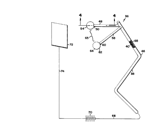
Figures 3 and 4 illustrate a vaginal insert, generally shown as 36, that
has a main body or portion 38. The main body 38 is elongated and defines a main
lumen 40. Additionally, the main body 38 is bent at an intermediate section 46.
A first member or portion 48 is operably connected to and projects
outward from the main body 38. The first projecting member 48 has a projecting
end 50 and defines a first branch lumen 52 that is in fluid communication with a first
balloon 54. The first balloon 54 is operably connected to the projecting end 50 and
is formed from a porous membrane 56.
A second projecting member or portion 58 is substantially similar to
the first member 48. Specifically, the second member 58 is operably connected tothe main body 38, has a projecting end 60, and defines a second branch lumen (not
shown). Additionally, a second balloon 62 is formed from a porous membrane 64
and is operably conn~cte~ to the projecting end 60. The second balloon 62 is in
fluid communication with the second branch lumen.
~ First and second projecting members 48 and 58 are positioned so that
35 they form a gap or opening 65 therebetween. The gap 65 is sized to receive the
bladder neck 24 when the first and second projecting members 48 and 58 engage the
anterior vaginal wall 32. Additionally, the first and second projecting members 48
CA 022~0113 1998-09-29
WO 97/36641 PCTrUS97/04599
and 58 are positioned so that when they engage the anterior vaginal wall 32, theintennefli~te section 46 of the main body 38 engages the posterior vaginal wall 34.
A first electrode 66 iS positioned within the first balloon 54. The first
electrode 66 is connected to a first lead 68 that extends through the first branch
lumen 52 and the main lumen 40. The first lead 68 iS then connected to a power
source 70. In one embodiment, a second electrode 72 iS remotely located and is
connected to the power supply 70 by a second lead 74. In this configuration, thesecond electrode 72 is a patch-type electrode that can be placed against the patient's
skin. A third electrode (not shown) is substantially similar to the first electrode 66
and is located within the second balloon 62. A third lead (not shown) connects the
third electrode to the first lead 68 so that the first 66 and third electrodes have the
same polarity.
The power supply 70 can be a simple DC power source that provides
a direct current between the first and third electrodes and the second electrode.
Alternatively, the power supply 70 can provide a current having a predet~rmined
type of ~v~ro,.n. Additionally, the power supply can provide an electric current at
inte"~ le~lt intervals.
In use, the vaginal insert 36 iS placed within the vagina 26 and
oriented so that the first and second balloons 54 and 62 engage and press into the
anterior vaginal wall 20 SO that the bladder neck 24 iS positioned within the gap 65.
In this position, the intP.rmediate section engages the posterior vaginal wall 34
providing frictional engagement to secure the vaginal insert 36 in a stationary
position. One skilled in the art will realize that the first and second balloons 54 and
62 can be positioned along other portions of the urinary tract such as the urethra 20.
After the vaginal insert 36 is secured in position, a fluid cont~ining an
agent is injected into the main lumen 40 so that the fluid flows through the first 52
and second branch lumens and inflates the first and second balloons 54 and 62. The
second electrode 72 is placed against the patient's body at a position such as the
patient's abdomen or thigh. The power supply 70 iS then activated, which causes a
current to flow between the first 66 and third electrodes and the second electrode 72.
Electrons that form the current will flow from the first 66 and third electrodes,
through the anterior vaginal wall 32, through the tissue proximate to the bladder
neck 24, and to the second electrode 72. The electrons carry the agent from the first
and second balloons 54 and 62 to the tissue proximate to the bladder neck 24.
One skilled in the art will realize that the vaginal insert 36 shown in
Figure 3 can have many alternative embodiments. For example, the first and second
balloons 54 and 62 can be replaced with hollow spheres (not shown) that define aplurality of delivery ports. In this embodiment, the spheres may be covered with a
CA 022~0113 1998-09-29
W O 97/36641 PCTnUS97/04~99
porous membrane (not shown) to diffuse the current and prevent a hot spot at tissue
that is ~ G~nt to the delivery ports.
In another alternative embodiment, the first and second balloons 54
and 62 can be replaced with solid spheres (not shown) that have surface mounted
5 electrodes (not shown) and are covered with a m~teri~l (not shown) that can beimpregn~tecl with and release the agent. The material might be configured to
n~hlr~lly release the agent or to release the agent only if subjected to some type of
active delivery merll~ni~m such as iontophoresis or phonopholesis. Examples of
suitable materials include a polymer matrix such as a hydrogel, a foam such as an
10 open cell foam or a hydrophilic foam, and or any other m~tçri~l that can contain and
release the agent.
In yet another alternative embodiment, the main body 38 defines first
and second main lumens (not shown). The first main lumen is in fluid
communication with the first balloon 54 via the first branch lumen 52. The second
15 main lumen is in fluid communication with the second balloon 62 via the second
branch lumen (not shown). In this embodiment, the first electrode 66 is positioned
in the first balloon 54 and the second electrode 70 positioned in the second balloon
62. The first lead 68 extends through the first branch lumen 52 and the first main
lumen. The second lead 74 extends through the second branch lumen and the second20 main lumen. Finally, the first and second leads 68 and 74 are c~nn~cte~l to the
power supply 70 in a manner that creates a bipolar electrode configuration.
An advantage of this design is that two agents can be simlllt~neously
delivered. The first fluid having an agent and charged ions of one polarity are
injected into the first balloon 54 and a second fluid cont~ining an agent and ions of
25 an opposite polarity are injected into the second balloon 62. As a result, two
dirr~nt agents can be simultaneously delivered, which can minimi7e the overall
length of time required to deliver the prescribed dose of agents.
Additionally, the same agent can be delivered from both the first and
second balloons 54 and 62 using a bipolar configuration. Delivery in this manner is
30 accomplished by linking the agent that is injected into the first balloon 54 to ions
having one polarity and the agent that is injected into the second balloon 62 to ions
having an opposite polarity. An advantage of this type of delivery is that the current
density can be decreased, while still delivering the agent in an acceptable amount of
time. Reducing the current density will help to alleviate the discomfort experienced
35 by the patient. An alt~ tive advantage is that delivery can be performed twice as
fast, thereby minimi7inE the length of time the patient is in ~ comfort
Figure 5 illustrates an ~ltçrn~tive vaginal insert, generally shown as
76, in which a first member or portion 78 is operably connected to the main portion
CA 022~0ll3 l998-09-29
W O97/36641 PCTAUS97/04599
38, has a projecting end 80, and has a curved segment 82 that is shaped to conform
to the bladder neck 24. The curved segment 82 can have a C-shaped configuration.Additionally, the first projecting member 78 defines a first branch lurnen 84 that is
in fluid col.llllullication with the main lumen 40. Delivery ports 86 are defined in
5 the curved segment 82 and are in fluid communication with the first branch lumen
84.
The second projecting member 88iS subst~nti~lly similar to the first
projecting member 78 and has a projecting end 90, a curved segment 92, a second
branch lumen (not shown) that is in fluid communication with the main lumen 40,
10 and delivery ports 94 that are in fluid con~lunication with the second branch lumen.
Additionally, the second projecting member 88 has a flexible segm~nt 97 that
allows the first and second projecting members 78 and 88 to move between an openstate and a closed state.
When in a closed state, the projecting ends 80 and 90 of the first and
15 second projecting members 78 and 88 are adjacent to one another. In the closed
state, the curved segment~ 82 and 92 form a gap or opening 96 that is sized to allow
the bladder neck and pro~ l,al tissue to pass tht;l~ ough. When in the open state,
the opening 96is configured to receive the bladder neck 24 and proximal tissue.
Referring to Figure 6, a first electrode 98is positioned in the first
20 branch lumen 84 and is conn~cted to the first lead 68 that extends through the main
lumen 40. The second electrode 72iS remotely located and conn~cte~l to the second
lead 74. A third electrode (not shown) is positioned in the second branch lumen and
is also connected to the first lead 68. The power supply 70iS connected to the first
and second leads 68 and 74so that the polarity of the first 98 and third electrodes is
opposite to the polarity of the second electrode 72.
In use, the vaginal insert 76iS inserted into the vagina 26so that the
first and second projecting members 78 and 88 are in the open state and engage the
anterior vaginal wall 32, thereby projecting along opposite sides of the bladder neck
24. The first and second projecting members 78 and 88 are then shifted to the closed
state so that the delivery ports 86 and 94 subsl~llially circumscribe the bladder neck
24. Fluid is then injected through the main luInen 40so that it seeps through the
delivery ports 86 and 94. Simultaneously, current is passed between the first 98 and
third electrodes and the second electrode 72. The iontophoretic current helps tooll the fluid through the tissue ofthe anterior vaginal wall 32 and to the tissue
proximal to the bladder neck 24. If the vaginal insert 76 has a bipolar configuration,
fluids Co~ g agents with oppositely charged ions can be injected into the first 84
and second branch lurnens, respectively.
CA 022~0113 1998-09-29
WO 97/36641 PCT/US97/04599
Similar to the embodiment shown in Figure 3, one skilled in the art
will realize that the vaginal insert 76 also can be configured to have a bipolarconfiguration in which the first electrode 98 having one polarity is positioned in the
first branch lumen and the second electrode 72 having an opposite polarity is
S positioned in the second branch lumen. The main body 38 has first and second main
lumens (not shown). The first lead 68 extends through the first main lumen and the
second lead 74 extends through the second main lumen.
Additionally, the first and second projecting members 78 and 88 can
be covered with a polymer matrix to help diffuse the current density and prevent hot
spots at tissue ~ cent to the delivery ports 86 and 94. One skilled in the art will
also realize that the first and second projecting m~mhers 78 and 88 can be formed
from a solid m~teri~l; can have surface mounted electrodes; and can be coated with a
material such as polymer matrix, hydrogel, or foam that is hn~le~ l with an
agent. ~ltern~tively, the first and second projecting members 78 and 88 can be
formed from the agent-ret~ining material itsel~
Figure 7 illustrates yet another ~lt~rn~tive embodiment. In this
embollim~nt a vaginal insert, generally shown as 100, has a U-shape and defines a
main portion 102 that is central between first and second portions 104 and 106. First
and second portions 104 and 106 have a circumference that is smaller than the
circumference of the main portion 102. Additionally, a first electrode 108 is wound
around the first portion 104 and a second electrode 110 is wound around the second
portion 106. The first and second electrodes 108 and 110 are connected to a power
supply 112 via first and second leads 114 and 116.
The first portion 104 and first electrode 108 are covered by a first cap
118 formed from an absorbent material such as a polymer matrix, a hydrogel, or afoam. Any of these sub~L~ces can be h~le~ ted with an agent. The second
portion 106 and second electrode 110 are similarly covered with an absorbent
material that can be impregn~ted with an agent.
One skilled in the art will realize that the power supply 112 can have
several possible configurations. In one configuration, the power supply 112 can be a
battery that provides a simple direct current. In this situation, the patient would
activate the power supply 112 before the vaginal insert 100 is placed in the vagina
26. In an ~ e embodiment, the power supply 70 contains an inductive coil,
similar to that of a l-~-sru-l--er. In this embodiment, the vaginal insert 100 is
positioned within the vagina 26 and the patient or caregiver can place a magnetic
source against the body and proximal to the power supply 70. The m~gn~tic sourcewill generate a field that induces an electrical current in the m~,~n~tic coil.
CA 022~0113 1998-09-29
WO 97/36641 PCT/US97/04599
In yet another embodiment the inductive circuit is a resonant circuit
that is tuned to a frequency in the radio frequency (RF) range (i.e., greater than about
9 KHz). In this embodiment, the resident circuit in~ ces an electrical current when
subject to a m~gnetic field that is oscillating at the resident frequency. One skilled
5 in the art will realize that the power supply 112 can include an AC/DC converter so
that a direct current is passed between the first and second electrodes 108 and 110.
Alternatively, the power supply 112 can include additional ChCUiLI~ to generate a
current having a particular waveform. An advantage of using a resonant circuit is
that i~llelrelcllce from stray m~gn~tjc fields is minimi7P.~l
In use, the first cap 118 is hll~le~ l with an agent having a pre-
determined polarity. Additionally, the second cap 120 can be hllple~,llated with an
agent having an opposite polarity. The vaginal insert 100 is positioned within the
vagina 26 so that the first and second portions 104 and 106 engage the anterior
vaginal wall 32 and are positioned along opposite sides of the urinary tract. The
main portion then engages the posterior vaginal wall 34, thereby securing the
vaginal insert 100 in a stationary position. One skilled in the art will realize that the
vaginal insert 100 can be positioned p,o~illlal to any portion of the urinary tract such
as the bladder neck 24 or the urethra 20. The electric current then passes between
the first and second electrodes 108 and 110 and transports the agent from the first
and second caps 118 and 120 to tissue proximal to the urinary tract that is located
and between the first and second portion 104 and 106.
Figure 8 illu~ es another alternative embodiment that has a
configuration s~lbst~nti~lly similar to the vaginal insert 100 and uses
magnetophoresis to transport the agent. In particular, the vaginal insert, generally
shown as 122, has a main portion 124, a first portion 126, and a second portion 128.
First and second caps 130 and 132 formed of an agent-ret~ining material such as a
polymer matrix cover the first and second portions 126 and 128, respectively. The
first and second caps 130 and 132 are impregnated with m~gn~tic particles in
addition to an agent. In use, the vaginal insert 122 is positioned in the vagina 26 so
that a portion of the urinary tract is positioned between the first and second portion
126 and 128. A user can then place a magnetic source against their body. The
magnetic source generates a field that causes the magnetic particles to vibrate. This
vibrating action drives the agent from the caps 130 and 132, through the anterior
vaginal wall 32, and into tissue proximal to the pa ient's urinary tract.
Referring to Figure 9, another vaginal insert, generally shown as 134,
has a base member 136 having a delivery portion 138 and a handle portion 140. The
delivery portion 138 defines a chamber 142 and plurality of ports 144. A first
electrode 146 is wound around the delivery portion 138. A first lead 149 extends
CA 022~0113 1998-09-29
WO 97/36641 PCT/US97/04599
11
from the first electrode 146 and through the handle portion 140 so that it may be
connected to the power source 70.
A membrane 148 is operably connected to the base member 136 and
defines a reservoir lS0. The membrane 148 has a porous section for eng~ging the
anterior vaginal wall 32. The ports 144 provide fluid communication between the
chamber 142 and the reservoir 150. Additionally, the porous section of the
membrane 148 can be located in only that portion of the membrane 148 that is
placed against the anterior vaginal wall 32, thereby minimi7in~ the amount of tissue
outside of the urinary tract that is exposed to the agent. However, one skilled in this
art will realize that the entire m~mhr~n~ 148 could be porous.
The handle portion 140 defines a supply port 152 that is covered by a
septum 154, which seals if punctured by a needle. The supply port 152 provides apassage for supplying fluid into the chamber 142 and the reservoir 150. A secondelectrode 156 is a patchtype electrode and configured to be placed against the
patient's body. The second electrode 156 has a second lead 158 for connecting to the
power source 70.
In use, a patient or caregiver will inject a fluid co~ g an agent
through the supply port 152 and fill the reservoir 150 and chamber 142. The fluid is
supplied in an amount that fills the reservoir l S0 with a ~l~s~u~e that p~ the
membrane 148 to remain pliable. The vaginal insert 134 is then placed in the vagina
26 so that the pores in the membrane 148 engage the anterior vaginal wall 32. The
second electrode 156 is then placed against the patient's body at a position such as
the patient's abdomen or thigh. The power supply 70 is activated so that an
iontophoretic current passes between the first and second electrodes 146 and 156 and
transports the agent from the reservoir l S0 to tissues surrounding the urinary tract.
An advantage of vaginal insert 134 is that upon insertion, the vaginal
walls will exert pres:iule against the membrane 148 and cause the fluid and agent to
pass through the pores and through the anterior vaginal wall 32 where the agent can
be absorbed by tissue surrounding the urinary tract.
Figures 10 and 11 A illustrate another vaginal insert, generally shown
as 160, having a base member 162 and a first electrode 164 mounted on a surface
166 of the base member 162. The first electrode 164 is connectçd to the power
source 70 via a first lead 167. A second, patch-type electrode 168 is configured to
~ be placed against the skin and is attached to the power source 70 via a lead 170. An
agent-ret~ining member 172 is operably connectçd to the base member 162 and has
an outer surface 174. The base m~mber 162 and agent-retaining member 172 are
curved, thereby defining a channel 176 that is sized to engage the anterior vaginal
wall 32 and receive tissue proxirnal the urethra 20. In this configuration, the agent-
CA 022~0113 1998-09-29
WO 97/36641 PCT/US97/04599
12
lGl~ini~-g member 172 can be formed from a material such as polymer matrix,
hydrogel, or hydrophilic foam that can be impregnated with the agent. The agent-re~ining member 172 is hllple~ ated with an agent.
In use, the vaginal insert 160 is placed in the vagina 26 so that the
S curved outer surface 174 engages the anterior vaginal wall 32 and the channel 176
receives the tissue surrounding the urethra 20. The second electrode 168 is thenplaced against the patient's abdomen or thigh and the power source 70 is activated.
The iontophoretic current passes bc;lw~xll the first and second e}ectrodes 164 and
168 and transports the agent from the agent-ret~ining member 172 to the tissue
proximal to the urethra 20.
In an alternative embodiment illustrated in Figure 1 lB, the base
member 162 can define a chamber 163 and a plurality of delivery ports 165. The
delivery ports provide fluid col-~ ication between the charnber 163 and the
surface of base member 162. An electrode 169 is positioned within the chamber
163. The electrode is operably comlecled to the first lead 167. The base member
162 also defines a hole (not shown) that is covered by a septum (not shown). In
order to fill the charnber, the user can simply inject the agent or a solution cont~ining
the agent through the septum.
This alternative embodiment may contain the agent-retaining member
172. If the agent-~ g member 172 is used, it may contain a second theldpeulic
agent or a penetration enh~n~er. Alternatively, the agent-ret~ining member could be
loaded only with water in order to provide a path for the iontophoretic current that
passes between the electrodes 169 and 168. One skilled in the art will realize that
the alternative embodiment shown in Figure 1 lB is used in a sllhst~ti~lly similar
manner to the embodiment shown in Figure 11 A.
Referring to Figures 12 and 13, another vaginal insert, generally
shown as 178, has a base member 180 on which a first electrode 182 is mounted.
The first electrode 182 is co~ e~l~d to a power source 70 via a first lead 184. A
second, patch-type electrode 186 is configured to be placed against the patient's
body at a point such as the abdomen or thigh. The second electrode 186 is then is
connected to the power source 70 via a second lead 188. A cotton-fiber matrix 190
is mounted on the base member. The cotton-fiber matrix is absorbent and swells
when it becomes wetted. One skilled in the art will realize that other materials that
can be in.pl~e~ rd with an agent and that swells when wetted can be ~ubsliLu~ed for
the cotton-fiber matrix 190.
The base member 180 and cotton-fiber matrix 190 are sized to fit
within a tubular tampon-like applicator 192 when the cotton-fiber matrix 190 is not
CA 022~0113 1998-09-29
W O 97/36641 PCTrUS97/04599
13
wetted. The tampon-like applicator 192 has a leading end 194 that is open and a
trailing end 196 that is open.
In use, a user will position the base unit 180 and unwetted cotton-
fiber matrix 190 in the tampon-like applicator 192 so that the first lead 184 extends
S through the trailing end 196. The cotton-fiber matrix 190 is then wetted with a fluid
that contains an agent. The user uses the tampon-like applicator 192 to insert the
base member 180 and cotton-fiber matrix 190 combination into the vagina 26 much
like a tampon. During this process, the cotton-fiber matrix 190 exr~n-l~ as it is
pushed out of the leading end 194 of the tampon-like applicator 192. After the
tampon-like applicator 192 is removed from the vagina 26, the swelled cotton-fiber
matrix 190 will conform to the con~oul~ of the interior vaginal wall. The secondelectrode 186 is placed against the patient's body at a point such as the abdomen or
thigh. When an electric current passes between the first and second electrodes 182
and 186, the agent will be t,a~s~nJ~led from the cotton-fiber matrix 190 to tissue
surrounding the urinary tract, including the urethra 20.
One skilled in the art will realize that any of the embodiments
described above can use ~ltern~tive methods to lla~ls~oll the agent. ~or example, the
active ~ ll meçh~ni~m can be phonophoresis in which the electrodes are be
replaced with an ultrasonic tr~n~d~lcers. Phonophoresis uses ultrasonic waves totransport the agent from the vaginal insert to the target tissue that is proximal to the
urinary tract. Another alternative transport mech~ni~m is magnetophoresis. a
magnetophoresis transport mech~ni~m has both an agent and magnetic particles
impregnated in an agent-ret~ining m~teri~l that forms part of the vaginal insert. The
magnetic particles will vibrate when subjected to an oscillating m~gn~ tic field,
thereby driving the agent toward the target tissue. Additionally, all of the
embo~iment~ described above can be used with passive delivery, in which case
electrodes, ultrasonic trAn~lucers, and magnetic particles are not nece~ry. One
skilled in the art will also realize that any agent-ret~ining m~t~ri~l may be
substituted for a polymer matrix, hydrogel, or foam.
Although the description of vaginal inserts and methods has been
quite specific, it is contemplated that various modifications could be made.
Accordingly, it is intentletl that the scope and spirit of the present invention can be
dictated by the appended claims, rather than by the foregoing description.
.