Note: Descriptions are shown in the official language in which they were submitted.
CA 022~1048 1998-10-07
W097/46868 PCT~S97/09920
ELECTRONIC ASSAY DEVICE AND METHOD
Related Applications
The present application repeats a substantial portion of
prior application no. 08~455,236, filed May 31, 1995 which is
a continuation of application serial no. 08/111,347, filed
August 24, 1993 and now abandoned. The present application
adds and claims additional disclosure not presented in the
prior applications. Since the present application names an
inventor named in the prior application, it may constitute a
continuation-in-part of the prior applications.
The subject matter of this application is related to a
disposable single-use digital electronic instrument that is
entirely self-contained, including all chemistry reagents, as
disclosed in U.S. Application Serial No. 08/512,844 entitled
"Dry Reagent Particle Assay And Device Having Multiple Test
Zones And Method Therefor" filed August 9, 1995 by Joel M.
Blatt and Michael P. Allen, U.S. Application Serial No.
08/642,228 entitled "Method And Device For Measuring Reflected
Optical Radiation" filed April 30, 1996 by Raymond T. Hebert
et al., and U.S. Application Serial No. 642,228 entitled
"Method And Device Producing A Predetermined Distribution Of
Detectable Change In Assays" filed May 11, 1996 by Joel M.
Blatt et al. The above applications have the same assignee as
~ the present invention and is incorporated herein by reference
in its entirety.
CA 022~1048 1998-10-07
W 097/46868 PCTrUS97/09920
Field of the Invention
This invention relates to a disposable self-contained,
electronic assay device for use in determining the amount of
one or more analytes in a body fluid such as blood or urine.
In particular, this invention relates to a small disposable,
electronic credit-card sized device which, upon application of
a body fluid to a sample receptor, automatically performs an
analysis and presents the concentration of the analyte and/or
another message output in readable form.
Background of the Invention
Qualitative and quantitative self-tests have developed
gradually over the last half century. An advancement in non-
instrumented tests came with the application of lmmunochemical
reagents on a solid support. This led to a number of
commercially useful diagnostic tests including those for HCG
(pregnancy), LH, FSH, CKMB, Staphylococcus, and Rubella.
Measurement of the hormone HCG to detect pregnancy was among
the first of these tests to become commercially successful in
the home market. The first home pregnancy test, the e.p.t.TM
used a solution phase chemical reaction that formed a brown
ring on the surface of the urine solution in the presence of
HCG. The 2 hour long protocol associated with this test was
sensitive to vibration and timing, causing false results.
Two additional test systems that appeared in the late
l9~0s were the ~ipoScanTM by Home Diagnostics Inc. and the
ChemcardTM by Chematics Inc. Both tests measure cholesterol in
whole-blood using visual color comparison. Since visual color
matching is subjective, these tests do not achieve the
CA 022~1048 1998-10-07
W097/46868 PCT~S97109920
quantitative performance necessary for cholesterol testing
(Pradella et al, Clin. Chem. 36:1999-1995 (1990)).
For many analytes such as the markers for pregnancy and
ovulation, qualitative or semi-quantitative tests are
appropriate. There are, however, a variety of analytes that
require accurate quantification. These include glucose,
cholesterol, HDL cholesterol, triglyceride, a variety of
therapeutic drugs such as theophylline, vitamin levels, and
other health indicators. Generally, their quantification has
been achieved through the use of an instrument. Although
suitable for clinical analysis, these methods are generally
desirable for point-of-care testing in physicians offices
rather than the home due to the expense of the instrument.
Recently, a number of non-instrumented methods for
measurement of analytes have started to emerge. The key to
achieving instrument-free quantification is through the use of
migration distance, rather than color matching, as the visual
signal. In migration distance assays, chemical/biochemical
reactions occur as the analyte is wicked through a solid
support. During wicking, the analyte reacts with a signal-
producing reagent and forms a visible signal along the
support. The migration distance or the distance of signal
border is related to analyte concentration. The operator
reads the height of the color bar, much the same way one reads
a thermometer, and finds the concentration from a calibrated
scale.
There are a few migration-type assays commercially
available. These include Environmental Test Systems' QuantabTM
, which measures chloride in swimming pools and during the
CA 022~1048 1998-10-07
W097/46868 PCT~S97/09920
mixing of concrete, Syva's AccuLevel~ for the measurement of
therapeutic drugs, and ChemTrak's AccuMeter~ for measurement
of cholesterol in whole blood. Other companies such as
Enzymatics and Crystal Diagnostics have more recently
announced the introduction of their Q.E.D. TM and ClinimeterTM
technologies to measure, respectively, alcohol in saliva and
cholesterol in blood. ActiMedTM Laboratories disclose a
thermometer-type cholesterol assay device in Ertinghausen,
J.S. Patent No. 5,087,556 (1992).
Although these single use, thermometer-type, non-
instrumented quantitative devices and non-instrumented color
comparison devices for qualitative measurement have shown
adequate performance, they have several problems associated
with reliability and convenience. First, the colors generated
on these devices are not always uniform and sharp. In the
case of migration type assays the border is often light in
color, unclear and difficult to read. This translates
directly into user errors since the user must make a judgment
related to the position of the color band border. In the case
of non-instrumented pregnancy tests it is sometimes difficult
to visually interpret the intensity of the colored spot
(especially at HCG concentrations close to the cut-off
sensitivity), and interpretation of the result is sometimes a
problem. Anytime a non-technical operator is required to make
a visual judgment or interpretation, an error is possible and,
sometimes, unavoidable.
Second, the assay protocol for these tests is sometimes
difficult and lengthy, taking 15 minutes to 1 hour to obtain a
result. Third, these tests often do not have sufficient
CA 022~1048 1998-10-07
W 097/46868 PCTAUS97/09920
procedural and reagent references to assure ade~uate test
performance. Fourth, non-instrumented devices can only
measure single endpoint type tests since enzyme rates or
ratiometric analysis of two analytes cannot be measured.
Therefore, the test menu of potential analytes is limited.
As an example of the significance of the problems, a
recent article in Clinical Chemistry (Daviaud et al, Clin.
Chem. 39:53-59 (1993)) evaluated all 27 home use pregnancy
tests sold in France. The authors state, "among the 478
positive urine samples distributed, 230 were falsely
interpreted as negative".
Thus, a need exists in the field of diagnostics for a
single-use assay which is sufficiently accurate and reliable
to permit point-of-care use by untrained individuals in
locations such as the home, sites of medical emergencies, or
locations other than a clinic.
SUMMARY OF T~E INVENTION
The present invention provides an assay device for
determining the presence of one or more selected analytes in a
sample. The device includes a housing having an exterior
surface and defining an interior area. A sample receptor
means receives the sample and is located on the exterior
surface of the housing. The sample treatment means reacts the
sample with a reagent to yield a physically detectable change
which correlates with the amount of selected analyte in the
sample. The sample treatment means is located within the
housing and is in fluid communication with the sample receptor
means. The device also includes a detector means which
CA 022~1048 1998-10-07
W O 97/46868 PCT~US97/09920
responds to the physically detectable change and produces an
electrical signal which correlates to the amount of the
selected analyte in the sample. The detector means is located
within the housing and is in electrical or optical
communication with the sample treatment means. A processing
means converts the electrical signal to a digital output and
is located within the housing and connected to the detector
means. A starting means automatically activates the
processing means and detector means upon the application of
the sample to the device. The starting means is located
within the housing and connected to the processing means. A
display means visually displays the digital output external to
the housing and is connected to the processing means.
One preferred embodiment of the present invention
provides a multi-assay device for determining the presence of
a plurality of selected analytes in a sample. The multi-assay
device includes a housing having an exterior surface and
defining an interior area. A sample receptor means receives
the sample and is located on the exterior surface of the
housing. A sample treatment means reacts the sample with a
plurality of reagents corresponding to the plurality of
selected analytes to yield physically detectable changes which
each correlate with the amount of one of the selected analytes
in the sample. The sample treatment means is located within
the housing and is in fluid communication with the sample
receptor means. A detector means responds to the physically
detectable change and produces electrical signals which each
correlate to the amount of one of the selected analytes in the
sample. The detector means is located within the housing and
CA 022~1048 1998-10-07
W 097146868 PCT~US97/09920
is in electrical or optical communlcation with the sample
treatment means. A processing means converts each electrical
signal to a digital output corresponding to one of the
selected analytes and is located within the housing and
connected to the detector means. The device includes a
display means which externally displays the digital output
corresponding to one of the selected analytes. Each digital
output corresponding to one of the selected analytes includes
at least a first component providing the assay results such as
the identity of the selected analyte and a second component
providing the amount of the selected analyte. The display
means is connected to the processing means.
Another preferred embodiment of the present invention
provides an assay device which provides quantitative
measurement of one or more selected analytes in a sample using
reflected optical radiation. The device includes a housing
having an exterior surface and sealing an interior area. A
receptor is configured to receive the sample containing the
analyte selected for determining its presence and is located
on the exterior surface of the housing. At least one assay
strip reacts the sample with a self-contained reagent to yield
a physically detectable change in at least one sampling area
on each assay strip which correlates with the amount of
selected analyte in the sample. Each assay strip is in fluid
communication with the receptor. A reflectometer is included
in the device which has an optical radiation source, a
detector configured to quantitatively detect optical
radiation, and an optics assembly configured to direct the
illumination from the optical radiation source to each
CA 022~1048 1998-10-07
W097/46868 PCT~S97/09920
sampling area and to direct the radiation diffusely reflected
from each sampling area to the detector. The detector
produces electrical signals which correlates to the amount of
one of the selected analytes in the sample. A processor is
configured to store assay calibration information which is
uniquely characteristic to each specific self-contained
reagent and physically detectable change within each sampling
area and to the specific reflectometer of the individual assay
device. The processor is further configured to calibrate each
sampling area and the reflectometer using the stored assay
calibration information. The processor is further configured
to convert the electrical signal to a digital output. The
processor is sealed within the housing and connected to the
reflectometer. A starter is configured to automatically
activate the processor and reflectometer upon the application
of the sample to the device. The starter is located within
the housing and connected to the processor. A display is
configured to visually display the digital output external to
the housing and is connected to the processor.
The present invention also provides a method for
determining the presence of one or more selected analytes in a
sample within a disposable housing. The method includes the
steps of: reacting the sample within the housing with a
reagent corresponding to the selected analyte to yield a
physically detectable change which correlates with the amount
of the selected analyte in the sample; calibrating the
physicalIy detectable change using assay calibration
information uniquely characteristic to each specific reagent
in the housing and to the physically detectable change for
CA 022~1048 1998-10-07
W 097/4686~ PCTrUS97/09920
each selected analyte to determine the amount of the selected
analyte; and, displaying the amount of each selected analyte.
Another method provided by the present invention
automatically starts a diagnostic device to analyze a sample.
The method includes the steps of: sensing the introduction of
a sample to the device and generating a signal to activate the
device. Preferably, the sensing step includes creating a
potential between a plurality of electrodes and changing the
electrical potential between the electrodes upon contacting
the electrodes with the sample.
The present invention also provides a method of
displaying quantitative assay results for a plurality of
selected analytes in a sample on a diagnostic device. The
method includes the steps of: simultaneously displaying the
identity and amount of one of the selected analytes for a
predetermined period of time; and, repeating the prior step
for another one of the plurality of selected analytes.
The advantages, embodiments, variations and the like of
the present invention will be apparent to those skilled-in-
the-art from the present specification taken with the
accompanying drawings and appended claims.
BRIEF DESCRIPTION OF THE DR~WINGS
In the drawings, which comprise a portion of this
disclosure:
Fig. 1 is an isometric view of an embodiment of the
disposable device of this invention;
CA 022~1048 1998-10-07
W097/46868 PCT~S97/09920
Fig. 2 is a schematic view of the device of this
invention, showing one configuration of the electronic and
sample processing components for single analyte testing;
Fig. 3 is an exploded cross-sectional side view of one
configuration of the sample processing components for single
analyte testing;
Fig. 4 is an exploded perspective view of a preferred
embodiment of a diagnostic device of the present invention;
Fig. 5 is an isolated perspective view of the underside
of the cover of the device in Fig. 4;
Fig. 6 is an isolated perspective view of the top face of
the optics assembly of the device in Fig. 4;
Fig. 7 is an isolated perspective view of the bottom face
of the optics assembly of the device in Fig. g;
Fig. 8 is an isolated perspective view of the top side of
the shield of the device in Fig. 4;
Fig. 9 is a schematic circuit configuration for one
embodiment of the automatic start utilized by the present
invention;
Fig. 10 is a side view of one embodiment of an assay
strip suitable for use in an NTx assay;
Fig. 11 is a top plan view of the assay strip in Fig. 10;
Fig. 12 is a side view of one embodiment of an assay
strip suitable for use in a general chemistry assay; and
Fig. 13 is a top plan view of the assay strip in Fig. 12.
DESCRIPTION OF THE ~K~Kk~ EMBODIM~NTS
The present invention represents a substantial
improvement in the art by providing assay methods and devices
CA 022~1048 1998-10-07
W 097/46868 PCT~US97/09920
that can produce qualitative or quantitative results. The
assay chemistry is self-contained within the instrument and
dry formulated on a solid matrix (i.e. membrane) where
reflectance is used, or lyophilized and deposited or spotted
and dried in a reaction compartment where transmission is
used, or present on an electrode where the chemistry produces
a change in electrical current or pH. The chemistry operates
in response to the analyte to produce a color change within
the chemistry matrix or in a fluid defined by the sample (i.e.
plasma) and reconstituted reagents. Alternately, the
chemistry will produce a change in electric current (i.e.
produce or consume electrons, or cause changes in electrical
conductivity) or cause a pH change that can easily be
detected. This type of chemistry is common in home glucose
instruments that contain chemistry reagents impregnated in a
reagent strip.
Substantially all types of assays can be carried out with
the present invention for a wide variety of analytes. Assays
that can be performed include, but are not limited to, general
chemistry assays and immunoassays. Both endpoint and reaction
rate type assays can be accomplished with the present
invention.
Analyte, as used herein, is the substance to be detected
which may be present in the test sample. For example, general
chemistry assays can be performed for analytes such as, but
not limited to, glucose, cholesterol, HDL cholesterol, LDL
cholesterol, triglycerides, and BUN. For immunoassays, the
analyte can be any substance for which there exists a
naturally occurring specific binding member (such as, an
CA 02251048 1998-10-07
W097/46868 PCT~S97tO9920
antibody), or for which a speci-fic binding member can be
prepared. Thus, an analyte is a substance that can bind to
one or more specific binding members in an assay. Analyte
also includes any antigenic substances, haptens, antibodies,
macromolecules, and combinations thereof. As a member of a
specific binding pair, the analyte can be detected by means of
naturally occurring specific binding partners (pairs) such as
the use of intrinsic factor protein as a member of a specific
binding pair for the determination of Vitamin B12, or the use
of lectin as a member of a specific binding pair for the
determination of a carbohydrate. The analyte can include a
protein, a peptide, an amino acid, a hormone, a steroid, a
vitamin, a drug including those administered for therapeutic
purposes as well as those administered for illicit purposes, a
bacterium, a virus, and metabolites of or antibodies to any of
the above substances. In particular, such analytes include,
but are not intended to be limited to, ferritin; creatinine
kinase MB (CK-MB); digoxin; phenytoin; phenobarbital;
carbamazepinei vancomycin; gentamicin, theophylline; valproic
acid; quinidine; luteinizing hormone (LH); follicle
stimulating hormone (FSH); estradiol, progesterone; IgE
antibodies; vitamin B2 micro-globulin; glycated hemoglobin
(Gly Hb); cortisol; digitoxini N-acetylprocainamide (NAPA);
procainamide; antibodies to rubella, such as rubella-IgG and
rubella-IgM; antibodies to toxoplasma, such as toxoplasmosis
IgG (Toxo-IgG) and toxoplasmosis IgM (Toxo-IgM); testosterone;
salicylatés; acetaminophen; hepatitis B core antigen, such as
anti-hepatitis B core antigen IgG and IgM (Anti-HBC); human
immune deficiency virus 1 and 2 (HIV 1 and 2); human T-cell
CA 022~1048 1998-10-07
W O 97/46868 PCT~US97/09920
leukemia virus 1 and 2 (HTLV);-hepatitis B antigen (HBAg);
antibodies to hepatitis B antigen (Anti-HB); thyroid
stimulating hormone (TSH); thyroxine (T4); total
triiodothyronine (Total T3); free triiodothyronine (Free T3);
carcinoembryonic antigen (CEA); and alpha fetal protein (AFP).
~rugs of abuse and controlled substances include, but are not
intended to be limited to, amphetamine; methamphetamine;
barbiturates such as amobarbital, secobarbital, pentobarbital,
phenobarbital, and barbital; benzodiazepines such as librium
and valium; cannabinoids such as hashish and marijuana;
cocaine; fentanyl; LSD; methaqualone; opiates such as heroin,
morphine, codeine, hydromorphone, hydrocodone, methadone,
oxycodone, oxymorphone, and opium; phencyclidine; and
propoxyphene. The details for the preparation of such
antibodies and their suitability for use as specific binding
members are well known to those skilled in the art.
The present invention provides assays which preferably
use specific binding members. A specific binding partner or
member, as used herein, is a member of a specific binding
pair. That is, two different molecules where one of the
molecules through chemical or physical means specifically
binds to the second molecule. Therefore, in addition to
antigen and antibody specific binding pairs of common
immunoassays, other specific binding pairs can include biotin
and avidin, carbohydrates and lectins, complementary
nucleotide sequences, effector and receptor molecules,
cofactors and enzymes, enzyme inhibitors and enzymes, and the
like. Furthermore, specific binding pairs can include members
that are analogs of the original specific binding members, for
CA 022~1048 1998-10-07
W O 97/46868 PCTrUS97tO9920
example, an analyte-analog. Immunoreactive specific binding
members include antigens, antigen fragments, antibodies, and
antibody fragments, both monoclonal and polyclonal, and
complexes thereof, including those formed by recombinant DNA
molecules. The term hapten, as used herein, refers to a
partial antigen or non-protein binding member which is capable
of binding to an antibody, but which is not capable of
eliciting antibody formation unless coupled to a carrier
protein.
The analyte-analog can be any substance which cross-
reacts with the analyte-specific binding member, although it
may do so to a greater or lesser extent than does the analyte
itself. The analyte-analog can include a modified analyte as
well as a fragmented or synthetic portion of the analyte
molecule, so long as the analyte-analog has at least one
epitope site in common with the analyte of interest. An
example of an analyte-analog is a synthetic peptide sequence
which duplicates at least one epitope of the whole-molecule
analyte so that the analyte-analog can bind to an analyte-
specific binding member.
The sample to be tested by the present invention for thepresence of an analyte can be derived from any biological
source, such as a physiological fluid, including whole blood
or whole blood components including red blood cells, white
blood cells, platelets, serum and plasma; ascites; urine;
sweat; milki synovial fluidi peritoneal fluidi amniotic fluid
cerebrospinal fluidi and other constituents of the body which
may contain the analyte of interest. The test sample can be
pre-treated prior to use, such as preparing plasma from blood,
14
CA 022~1048 1998-10-07
WO 97/46868 PCTAUS97/09920
diluting viscous flulds, or the like; methods of treatment can
involve filtration, distillation, concentration, inactivation
of interfering compounds, and the addition of reagents.
Besides physiological fluids, other liquid samples can be used
such as water, food products and the like for the performance
of environmental or food production assays. In addition, a
solid material suspected of containing the analyte can be used
as the test sample. In some instances it may be beneficial to
modify a solid test sample to form a liquid medium or to
release the analyte. The analyte can be any compound or
composition to be detected or measured and which has at least
one epitope or binding site.
Single or multiple assays can be done at one time. For
example, a single assay can be performed measuring cholesterol
or one device can be set up to measure both total and HDL
cholesterol from a single sample. One test device can be set
up to measure one, two, three, or more analytes at one time.
The device of this invention is ideal for on-site testing
in remote locations throughout the world in health fairs,
occupational health settings, physician offices, and in the
home. The device can include automatic reagent handling
(sample filtration, component separation, blood separation or
the like), automatic sample measurement, automatic reagent
delivery, and on-board controls such that non-technical users
can operate the test easily without prior training. Also
since the device uses a digital display (like a calculator)
there is no need for visual interpretation of color quality or
intensity, or visual reading of a signal migration distance.
~, . . ., _ . . ,
CA 022~1048 1998-10-07
W O 97/46868 PCT~US97/09920
Thus, user errors will be significantly reduced using this
disposable electronic device.
One of the key features of this invention is the
inexpensive cost of the device such that it becomes
economically practical for the device to be used as a single,
disposable unit. The device includes an electronic component,
a chemistry reagent component, and a housing which contains
the electronics and chemistry. It is desirable that the
electronics and housing are integrated into a single piece.
However, the reagent strip can be replaced once or several
times such that the electronics component is re-used.
Referring to the drawings, ~ig. 1 is an isometric view of
one embodiment of the disposable device of this invention.
The device 12 has a sample receptor 14 and a visual readout
display 16 such as a liquid crystal display. The thickness
"t", width "w" and depth "d" can be varied to provide the
desired overall dimensions. The device 12 can be of any
convenient size with the optimal dimensions determined by
several factors including, but not limited to: 1) the size of
the electronic components, 2) the size of the chemistry
components, and 3) marketing consumer studies. The device 12
may assume any convenient shape including square, rectangle,
triangle, oval, round, or any other desired shape as long as
the electronics and chemistry can be cost effectively
contained with acceptable performance.
The instrument is designed for a single use and can
measure, for example, transmission, reflectance, electrical
conductivity, electrical current or pH change. The instrument
is fabricated in a unitized integrated format to reduce the
16
CA 022~1048 1998-10-07
W097/46868 PCT~S97/09920
cost of manufacture. The instrument may have the following
generally described components: light source such as a light
emitting diode (LED); optics; a detector which senses
reflected or transmitted light; a processor with memory which
controls the assay start and stop, receives and processes
input from the detector, stores assay calibration information
and the like; an analog to digital converter or the like (a
current integrating comparator can be used); a power source
which can be a battery or solar cell or any convenient power
source; a temperature compensation mechanism (optional); and
a liquid crystal display (LCD) with 1 to 6 digits (preferably
3 ~ digits). The instrument described may contain one, all,
or none of the above-mentioned components or may contain other
components that are necessary for the diagnostic reflectance
or transmission instrument to operate.
Fig. 2 is a schematic view of the device 12 of this
invention designed for reflectance measurement of a detectable
signal, showing one configuration of the electronic and sample
processing components for single analyte testing. Mounted in
a housing 18 are all of the components, including a power
supply 38 required to conduct the assay. A reagent strip 20
has an electrode pair 22 mounted thereon between a sample
application zone 24 and a reagent zone 26, or at the site of
the sample application, to detect the presence and movement of
sample liquid on the reagent strip 20. Presence of sample
liquid bridging the electrode pair 22 reduces the resistance
across the electrodes, signaling the presence of a conductor
(sample liquid) therebetween. An LED 28 is positioned between
the detectors 30 and 32. The detectors 30 and 32 are
CA 022~1048 1998-10-07
W 097/46868 PCT~US97109920
conventional light detectors wh-ich detect light reflected at a
preselected wavelength corresponding to a property of the
physically detectable label. Temperature sensor 34 is mounted
on the reagent strip to detect the temperature of the system
and provide ambient temperature information for calibration
adjustment at temperature extremes. It is suitable to locate
the temperature sensor anywhere in or on the device. For
example, the temperature sensor may be located on the
microprocessor. The electrode pair 36 is positioned to detect
movement of sample liquid beyond the detection zone occupied
by the light sensors.
The power supply 38 has a lead from its negative pole
connected to one side of the electrode pairs 22 and 36, and a
lead from its positive pole being connected to an analog to
digital converter 40 and display 16. A processor and memory
component 42 is connected to the analog to digital converter
40 and the display 16. External calibration ports 44 are
connected to the analog to digital converter 40.
This embodiment of Fig. 2 includes two sets of electrodes
(22 and 36) which function to turn the instrument on in one of
two modes when the sample is present by using the conducting
properties of the sample to complete the circuit between the
electrodes. Electrode set 22 is the "reference-on" electrode
which is positioned immediately downstream from the sample
application port. The sample comes into contact with this
electrode set almost immediately after application onto the
device. The instrument calibrating-to-self-zero feature is
energized allowing the light source to warm up and the optical
system (~ED, detector, and optics) to zero by taking readings
18
CA 022~1048 1998-10-07
W O 97/46868 PCT~US97/09920
on the unreacted reagent area. Electrode set 36 is called the
"read-on" electrode which is positloned at the end of the
chemistry reagent, and is downstream from the electrode set 22
and downstream from the optical system. When the sample
reaches this electrode set the chemical reactions are well
underway and the instrument begins to read the reagent system.
The reading may begin immediately when the sample reaches
electrode set 36 or there may be some time delay of about less
than 1 second to 10 minutes (preferably from about 30 seconds
to 2 minutes). There may be single or multiple readings or
the readings may continue until the reagent system response
has stabilized either to an endpoint, maximum or minimum, or
to a constant reaction rate. These readings may be initiated
by either electrode set 22 or 36.
As described above, the electrode set 35 indicates that
sufficient sample was introduced to the device and has been
transported across the reagent strip. Vse of the electrode
set 35 is optional.
The instrument functions of "automatic zero" and "read"
are initiated in response to the presence of a sample. The
electrode method is described above; however, any convenient
and inexpensive method can be used. Another method uses a
solar cell which activates when the device is removed from the
light-impermeable foil storage pouch. When the user removes
the device from the pouch the ambient light turns the
instrument on, activates the self-reference and allows the LED
and optics to warm up. This also initiates a timer on the
processor which automatically activates the "read on" function
19
CA 022~1048 1998-10-07
W O 97/46868 PCT~US97/09920
after a specified time. This system eliminates the need for
the "reference on" and "read on" electrode pairs.
Although the auto start configuration of the present
invention may be used in an integrated assay device, the
present invention can be used in any other instrumented
reflectance or transmission meter, potentiometric,
amperometric, conductimetric or pH meter or the like, as part
of a replaceable reagent. Thus, the auto start embodiment of
the present invention also encompasses non-integrated assay
instruments and analytical assay instruments comprising the
present assay device.
The optics are optional for some embodiments of the
present invention. The optical system may include a light
source such as an LED, a detector and an optical surface. The
optical surface may be as simple as a clear or transparent
coating over the light source and detector. A simple aperture
can be used in lieu of a lens to focus and meter the light.
The coating can be any plastic, silicone, glass, or the like.
The optical system of the device may be set up to measure
single or multiple analytes. For single analytes only one
light source and detector are necessary; for two analytes, two
sets of light source and detector may be used and so on.
The processor 42 can be any common or custom integrated
circuit with memory. The processor 42 must have the capacity
to either store a set of pre-programmed calibration curves or
have the capability to be programmed during device
manufacturing. In the case of preprogrammed calibration, a
method of curve selection during manufacture is necessary.
This can be done by laser burning of a selection of circuit
CA 022~1048 1998-10-07
W O 97/46868 PCTAUS97/09920
pathways or any convenient means. In the case of post-
manufacture calibration, a method to load calibration data
onto the chip is necessary, for example external calibration
contacts 44. External calibration can be accomplished with
external electrical contacts or may be done with a non-contact
method using radio waves, magnetic fields, pulse light, laser
or the like. The non-contact method of calibration may be
more practical and efficient from a manufacturing viewpoint
The processor 42 will also control the entire operation
of the instrument including, but not limited to, turning the
instrument "re~erence-on" and "read-on" in response to
electrode power or time signals; timing, recording, and
processing the instrument zero function; controlling any time
delays or timed steps during reading; determining when the
reaction has stabilized; receiving and processing information
from the temperature senscr; and receiving input from the
optical system and converting it to output, based on
calibration information, to the display. The processor can
also calculate the time taken for the sample to travel from
electrode set 22 to electrode set 36 and if too much time is
taken an error code will show on the display. The processor
will also determine if the chemistry reaction has occurred
within the specified time, to a specified endpoint range or
within a specified reaction rate range to control for inactive
reagents. Any other electronic control checks can also be
included.
The~power supply 3~ can be any convenient device
including, but not limited to, a battery or a solar cell. The
shelf life of the final product will be about 6 months to
CA 022~1048 1998-10-07
W O 97146868 rCTAUS97/09920
about 24 months at room temperature. The power supply must
have stability consistent with this shelf life. Use of a
solar cell would have the advantage of allowing the instrument
to initiate and automatically zero itself immediately after
the assay device is taken out of the storage foil pouch. This
would eliminate the need for electrode set 22 ("reference-on"
electrodes).
The display 16 preferably is a liquid crystal device LCD
or any conventional, inexpensive display device. The number
size in the display should be sufficiently large to allow most
people to read the assay value, even if they have poor vision.
The display height may be from about 0.5 to about 2.0 cm and
most likely from about 0.75 cm to about 1.5 cm. The number of
digits in the display can be anywhere from 1 to about 10
digits, however, most assays require only 3 ~ digits and
therefore the display in this device will likely have a 3 to 5
digit display. In addition to showing the assay result, the
display may show messages such as "SAMPLE VOLUME OK" and
"RESULT OK".
In the case of measuring one analyte, only one display is
usually necessary. In the case where two or more analytes are
measured simultaneously, then at least two display
configuration options exist which include a single display
which alternates between results with only one result on the
display at one time, or two or more results being shown on the
display at one time. For example, if both total cholesterol
and HDL cholesterol are measured then the display can
alternate between the total and HDL values or show both values
at one time. The ideal situation would be having all values
22
CA 022~1048 1998-10-07
W O 97/46868 PCT~US97/09920
displayed simultaneously. However, manufacturing cost is a
consideration.
Fig. 3 is an exploded cross-sectional side view of one
configuration of the sample processing components for single
analyte testing. The reagent strip 20 rests on a lower plate
of housing 18 supporting the electrodes 22 and 36, LED 28 and
detectors 30 and 32. A separation device 46 rests on the
input end of strip 20. The strip 20 includes a plurality of
zones 48, 50 and 52, the functions of which will be described
in detail hereinafter.
The chemical reagents are dry formulated on the reagent
strip 20 which can be any convenient bibulous material
including, but not limited to, fabric or mesh made of cotton,
nylon, polyester, polypropylene, polyethylene or the like;
paper such as Whatman lC, 2C, 31ET or 3MM, or S&S 903C, 470,
604 or the like; glass fiber such as Whatman GFA or GFD, or
S&S 3362 or 32; plastic fiber, metal fiber, or any hydrophilic
synthetic membrane; synthetic membranes such as Millipore
IMMOBILON, Pall nylon, S&S nitrocellulose, cellulose acetate,
regenerated cellulose, Gelman VERSAPORE or the like. The
reagent strip 20 can also be made of any convenient bibulous
material including porous plastics such as polyethylene and
polypropylene, examples of which are made by Porex
Technologies Corp., or synthetic or natural mesh screens,
examples of which are made by Tetko. By "porous" is meant that
the material is one through which the test sample can easily
pass and~includes both bibulous and non-bibulous solid phase
materials.
.. . . .
CA 022~1048 1998-10-07
W 097/46868 PCTAJS97/09920
An assay device for the present invention can have many
configurations, several of which are dependent upon the
material chosen as the reagent strip 20. Other configurations
of the reagent strip include a fiberglass, cellulose, or nylon
pad for use in a pour and flow-through assay device having
multiple layers for multiple assay reagents; a test strip for
wicking or thin layer chromatographic capillary action (e.g.,
nitrocellulose) techniquesi or other porous or open pore
materials well known to those skilled in the art (e.g.,
polyethylene sheet material).
In a preferred embodiment, the present dry reagent assay
device uses a lateral flow bibulous materia~ with proximal and
distal ends, containing at least one central zone along its
length. The strip configuration may be of any dimensions
which provide the desired number of zones and which permit (a)
the desired binding or chemical reactions to be completed in a
reproducible manner and (b) detection of the reaction
indicator to occur. Preferably, the present strip is a total
of no more than about 100 mm in length and about 6 mm wide,
and more preferably, from about 10 mm to about 40 mm in length
and about 1 mm to about 5 mm wide.
The bibulous strip can comprise a plurality of zones
along its length. Each zone can be from about 0.1 mm to about
10 mm wide, more preferably from about 1 mm to about 5 mm
wide. There will be a minimum of two zones and a maximum of
about 10 or more zones, depending on the number of assays to
be conducted on one bibulous strip. The strip can be one
continuous section or composed of one, two, three or more
sections. Each zone may be a separate bibulous material, all
24
CA 022~1048 1998-10-07
W097/46868 PCTAJS97/09920
in fluid communication, or one or more zones can be a common
material with the other zones being made of separate
materials.
The assay devices include a bibulous substrate to which
members of specific binding pairs, which may be labeled, are
diffusively or non-diffusively immobilized. Non-diffusive
immobilization can be conducted by adsorbing, absorbing,
crosslinking or covalently attaching a reagent such as an
unlabeled member of a binding pair to the bibulous substrate.
Diffusive immobilization can be conducted by formulating
the assay reagent(s) to be immobilized (e.g., by dissolving in
a suitable solvent such as water, a Cl-C4 alcohol or mixture
thereof, along with any desired additives), applying the
resulting formulation to the bibulous material of the
membrane, filter or transport layer in the desired
location(s), and drying the material. Diffusive immobilization
allows rapid reconstitution and movement of reagents, whether
reacted or unreacted, through the bibulous substrate.
Suitable additives may include detergents, proteins, blocking
agents, polymers, sugars or the like. Alternatively, the
additive(s) and assay reagent(s) may be applied to the
membrane, filter or transport layer by precoating with a
"blocking agent", water soluble polymer, sugar or detergent,
followed by depositing the conjugate or conjugate formulation
and drying the material.
The separation device 46 for filtering the sample from
unwanted contaminants such as red cells in blood can be
constructed using synthetic membranes, fibrous depth filters
such as glass fiber, plastic fiber, metal fiber, cellulose
.
CA 022~1048 1998-10-07
W O 97/46868 PCT~US97/09920
fiber or the like or any combin-ation of filters and membranes.
For example, the separation device 46 can include micro-porous
synthetic membranes of pore size from about 0.2 ~m to about 12
~m (preferably about 0.4 ~m to about 7 ~m). Examples include:
Pall nylon, S&S nitrocellulose, cellulose acetate, regenerated
cellulose, nucleopore Poretics or the like. The separation
materials may be untreated or can be coated with proteins,
dextrans, sugars, or carbohydrates for red cell stabilization,
LDL precipitating reagents such as magnesium chloride and
dextran sulfate, antibodies, or red cell agglutinating agents
to facilitate red cell removal. The sample transport area can
be untreated or have various reagents diffusively or non-
diffusively immobilized, such as stabilizing proteins,
detergents, anticoagulants like heparin or EDTA, LDL
precipitating reagents, antibodies, or red cell agglutinating
agents like wheat germ lectin or anti-human RBC.
The housing 18 of the device can be made of any
conventional material including, but not limited to,
thermoplastics such as polyethylene, Delrin, ABS and
polystyrene.
For immunoassays, the present invention preferably uses
particle detection for a detectable response or signal in each
test zone related to the level of analyte in the sample.
Other means for providing a detectable response in the test
zones are suitable for use in the present invention. For
example, and not for limitation, the analyte may be labeled
with an indicator to measure electrical conductance or the
reflectance or absorption of a characteristic light
wavelength. The analyte may also be reacted with other
26
CA 022~1048 1998-10-07
W O 97/46868 PCTAUS97/09920
chemicals to convert a dye, chr-omogenic compound or the like
into a colored form detectable by means of transmission or
reflectance photometry. As used herein, "indicator" is meant
to include all compounds capable of labeling the analyte or
conjugate thereof and generating a detectable response or
signal indicative of the level of analyte in the sample.
The present device may be used on-site in the home and in
the physician's office, or in remote locations in emergency
medicine. Therefore, the device may advantageously include
sample pre-treatment as previously defined, as well as a
sample wlthdrawal device (e.g., a fingerstick) or any
combination thereof. Sample pretreatment can also adjust the
pH to within a specified range, reference salt concentration,
turbidity and/or viscosity, and/or reduce or remove
interfering substances such as immunochemical cross-reactants,
redox substances and the like.
A preferred embodiment of a single-use diagnostic device
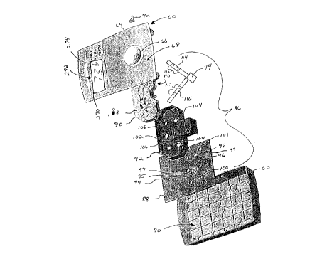
60 is illustrated in Fig. 4. The device 60 includes a housing
62 and cover 64 having a receptor such as inlet port 66 which
extends from the exterior surface 68 of the cover to the
interior 70 of the housing for receiving a sample 72
containing the one or more selected analytes to be determined.
The inlet port 66 allows the sample 72 to be introduced to
a sample receiving device 74 which is attached to the interior
surface 76 of the cover 64 as seen in Fig. 5. The sample
receiving device 74 includes a pad which is in fluid
communication with two assay strips and serves to distribute
the sample between the two strips. Optionally, the sample
receiving device 74 can also include a sample filter pad which
27
. ., ~ .
CA 022~1048 1998-10-07
W 097/46868 PCTAUS97/09920
removes undesired contaminants-from the sample. The sample
filter pad can be the same as the receiving pad with one pad
performing bother functions. The device 60 can include more
than one sample filter pad along the pathway of the sample
flow which remove different types of contaminants. The two
assay strips contain chemical reagents for determining the
presence of one or more selected analytes.
Referring to Fig. 4, the interior 70 of the housing
encloses a reflectometer 86 which includes a printed wiring
assembly having a printed circuit board (PCB) 88. The
reflectometer 86 also includes an optics assembly 90 and a
shield 92. The PCB 88 has one face 94 with a reference
detector 96 and zone detectors 98, 100 mounted directly
thereto. The face 94 of the PCB also has two LEDs 95, 97, one
for each pair of illumination channels, mounted directly to
the PCB. The LEDs 95, 97 are preferably in bare die form
without an integral lens, enclosure, or housing. As a result,
the LEDs 95, 97 provide illumination in all directions above
the face 94 and are directed only by the optics assembly 90.
Similarly, the zone detectors 98, 100 and reference detector
96 are bare die mounted directly to the face 94 of the PCB.
The LEDs 95, 97 and the detectors 96, 98, 100 are all
positioned in the same plane.
Fig. 4 also illustrates the position of the shield 92
relative to the PCB 88. Aperture 102 is provided through the
shield 92 to prevent obstructing the LEDs 95, 97 and the
referencé detector 96. Openings 104 are provided to prevent
obstructing zone detectors 98, 100. The shield 92 includes
upstanding walls 106 which prevent stray radiation from
28
.. . .
CA 022~1048 1998-10-07
W O 97/46868 PCTrUS97/09920
entering the zone detectors 98j 100. The upstanding walls 106
are positioned adjacent the reflecting and refracting elements
of the optics assembly 90 when the reflectometer 86 is fully
assembled.
The optics assembly 90 is a generally planar support
having at least a top face 108 and a bottom face 110. The
bottom face 110 is configured to receive illumination from the
LEDs 95, 97 and the optics assembly 90 directs the
illumination to one or more sampling areas 112 on a first 114
10 and second 116 assay strip. The top face 108 of the optics
assembly is also configured to transmit the diffusely
reflected optical radiation returning from the sampling areas
112 to one or more of the zone detectors 98, 100.
The top face 108 of the optics assembly is shown isolated
in Fig. 6 and is configured to transmit illumination directed
toward the sampling areas 112 on the first 114 and second 116
assay strips shown in phantom. The top face 108 also
transmits the optical radiation diffusely reflected from the
sampling areas 112 to one or more of the zone detectors 98,
100. The top face 108 also supports the underside of the
first 114 and second 116 assay strips and positions the assay
strips over the detectors 98, 100 between the upstanding
prongs 80 and by having pins 78 press-fit into corresponding
holes located at the distal ends 82 of the assay strips 114,
116.
The discrete light paths or channels are illustrated with
more clarity by referring to the detectors and the LEDs in
Fig. 4, and more specifically to Figs. 6 and 7 isolating the
optics assembly. The illumination from each LED 95, 97
29
CA 022~1048 1998-10-07
W 097/46868 PCTrUS97109920
located underneath the optics assembly 90 is partially
collimated by respective pairs of refracting elements 118,
120. Stray illumination off of the surface of reflecting
elements from each LED 95, 97 is directed to reference
detector 96. The partially collimated illumination is split
into two channels for each pair of refracting elements 118,
120 for a total of two pairs of channels or four individual
channels of illumination. Illumination in each pair of
channels is then deflected off a series of reflecting element
pairs in the following sequence: pairs of reflecting elements
122 and 124, pairs of reflecting elements 126 and 128, and
pairs of reflecting elements 130 and 132.
The illumination of each channel is then passed through
pairs of refracting elements 134 and 136 which spread the
illumination for each channel in a predetermined shape across
the sampling areas 112. More specifically, the pair of
refracting elements 134 spreads the illumination across first
detection zones 138 and 140 on assay strips 114 and 116
respectively. The pair of refracting elements 136 spreads the
illumination across second detection zones 142 and 144 on
assay strips 114 and 116 respectively.
The diffused optical radiation reflected downward by the
first detection zones 138 and 140 is partially collimated by a
pair of refracting elements 146. Similarly, the diffused
optical radiation reflected downward by the second detection
zones 142 and 144 is partially collimated by a pair of
refracting elements 148. Pairs of refracting elements 150 and
152 further direct the partially collimated diffuse optical
radiation from the refracting elements 146 and 148 to
CA 022~1048 1998-10-07
W 097/46868 PCT~US97/09920
detectors 98 and 100. More spe-cifically, detector 98 receives
the diffused optical radiation from the first and second
detection zones 138, 142 on the first assay strip 114.
Detector 100 receives the diffused optical radiation from the
first and second detection zones 140, 144 on the second assay
strip 116.
Each pair of refracting elements such as 146 and 150 used
for detection zone 138 constitutes an anamorphic lens system
which can differentially image the detection zone 138 onto the
detector 98 so that the boundaries of detector 98 clearly
define boundaries of detection zone 138 in each axis
independently. The leading edge 99 and the trailing edge 101
of the detector 98 define the leading edge 137 and the
trailing edge 139 of the detection zone 138 with regard to the
placement of the chemical reagents on the assay strip 114.
The anamorphic lens system is designed to accommodate
placement tolerance of the detector die 98 and the LED dies 95
and 97 by differentially magnifying the detection zone 138
onto the detector 98 through anamorphic refractive elements
146 and 150 such that the illumination zone overfills the
detection zone 138 in the direction of sample flow and
underfills perpendicularly to the direction of sample flow.
Furthermore, the present invention intends to provide
uniformity of sensitivity throughout the detection zone 138.
Fig. 8 is a view isolating the top side of the shield 92
and more specifically illustrates that the shield 92 is
integrally formed with a base 160 which partially supports the
assay strips 114, 116 at the ends proximal 162 to the inlet
port as seen in Fig. 5. When the shield and the optics
CA 022~1048 1998-10-07
W O 97/46868 PCTrUS97/09920
assembly 90 are assembled, the base 160 nests into the cut-
away portion 164 of the optics assembly 90 as seen in Fig. 6.
As seen in Figs. 6 and 8, the top surface 166 of the base is
approximately flush with the top face 108 of the optics
assembly, providing uniform support across the length of the
assay strips 114, 116.
As previously disclosed herein, the automatic start
feature of the present invention is also illustrated in Fig.
8. The base 160 includes two channels 168 and 170 spaced in
parallel across the top surface 166 of the base. The channels
168 and 170 are positioned to contact the sample upon its
delivery through the inlet port either instantaneously or
immediately downstream from the inlet port. Each channel 168,
170 is sized to accommodate an electrode 172, 174 respectively
therein. Preferably, the diameter of each electrode is about
0.5 mm. The electrodes 172, 174 connect to a control circuit
which causes a transition from a inactive or dormant state to
an active state.
In the inactive state, very little power is consumed by
the device 60. The microprocessor 42 is supplied only enough
power required to maintain the volatile random access memory.
The self-powering, automatic start feature allows the
microprocessor in the inactive state to consume preferably
less than about 5 ~AH, more preferably less than about 1 ~AH
to 2 ~AH, and most preferably about 0.1 ~AH. The automatic
start feature of the present invention changes the state of
the procéssor from "stop" to "idle", or directly to a fully
"active" state. The number of operational states of the
CA 022~1048 1998-10-07
WO 97/46868 PCT~US97/09920
particular microprocessor is easily accommodated by the
automatic start feature.
In the active state, the device 60 becomes operational as
the result of introducing the sample and wetting the pair of
electrodes 172, 174. Thus, the automatic start feature of the
present invention eliminates the need for a manual switch, or
an on/off control that consumes a significant amount of power
in the inactive state that will otherwise be needed later for
operation of the device. The automatic start feature also
provides a relatively precise starting time for the assay
which is uniform regardless of the individual device. Manual
switches or other means activated by the operator can not
provide this accuracy. The precise starting time prevents
wasting power caused by activating the microprocessor
prematurely.
With the present invention, one skilled in the art can
incorporate a battery in the device of the appropriate size
taking into account the power consumption of the particular
microprocessor, other than the examples herein, to provide the
desired shorter or longer shelf-life.
Fig. 9 illustrates the electrodes 172, 174 and a control
circuit 176 of the automatic start feature in more detail.
The electrodes 172, 174 are connected to the input of a
control circuit that will respond to a change in potential
between them. Electrode 174 is preferably a copper wire which
is connected to ground and exhibits a zero voltage potential.
Electrode 172 is preferably a zinc clad copper wire which
connects to a power supply 38 through a resistor 178 and also
to the processor 42. The power supply 38 also connects to the
33
CA 022~1048 1998-10-07
W O 97/46868 PCT~US97/09920
processor 92. When the device is in the dormant state, the
potential exhibited at the electrode 172 reflects the
potential of the battery which in this preferred embodiment is
about +3v. After the sample is introduced to the device, the
potential exhibited at the electrode 172 reverses polarity to
-0.7v. The value of the resistor 178 for the preferred
embodiment is about 2.2 Mohm.
The electrodes are made of dissimilar metals that create
an electric potential between them when exposed to the sample.
In the dormant or dry state, a potential of opposite polarity
is applied to the electrodes through a resistor. Preferab7y,
copper and zinc-clad copper wires are used as the electrodes.
However, tin and silver are suitable and other metals which
provide the needed potential and are easily incorporated into
the manufacturing process can be used with the present
invention.
In order to assure a shelf life of at least two years for
the preferred embodiment illustrated in ~igs. 4 and 9, two A76
batteries having a capacity of 150 mAH are used. The control
circuit consumes about 0.1 ~AH at room temperature. This
provides for adequate power for the device 60 in the
operational state which consumes about 100 ~a for about three
minutes to complete the test.
Referring to Fig. 4, another feature of the present
invention is illustrated in the manner that the quantitative
assay results are displayed. In this preferred embodiment, an
LCD 270 having a 3 ~ digit display capability is integrally
mounted through the exterior surface 68 of the cover and is
electrically connected to the processor. Each digit of the
34
CA 022~1048 1998-10-07
W 097/46868 PCT~US97/09920
LCD 270, or fraction thereof, can be considered a separate
screen for displaying pertinent information.
In the assay example illustrated, three different types
of assay result information are printed along side the LCD 270
on the exterior surface 68 of the cover for HDL cholesterol,
LDL cholesterol, and total cholesterol. When each assay
result is displayed, a first component of the corresponding
digital output displays a numerical output in a first screen
272. Simultaneously, a second component of the corresponding
digital output displays a character in a second screen 274
indicating the identity of the assay result by pointing to the
appropriate marking on the exterior surface of the cover. The
simultaneous display of the identity and amount of one of the
selected analytes, HDL cholesterol, remains for a
~5 predetermined period of time controlled by the processor.
Upon expiration of the predetermined time period, the identity
and amount of another one of the selected analytes, in this
example LDL cholesterol, is displayed. This procedure is
repeated for the total cholesterol result to complete a cycle.
The cycle begins again by displaying the information for the
first analyte, HDL cholesterol.
During the cycling of the display, the assay results can
be updated by the processor. As previously discussed, other
messages can be displayed by the LCD at various times during
the cycle of the assay results. It is suitable to provide
larger LCDs to display the assay results of all the analytes
simultaneously. However, the added cost is commercially
undesirable, particularly in a disposable device. It is also
CA 022~1048 1998-10-07
W O 97/46868 PCT~US97/09920
suitable to have the identity of the assay results displayed
by the LCD 270 instead of printing a mark on the cover 64.
As previously discussed, the diagnostic device 60 can be
of any convenient size with the optimal dimensions determined
by several factors including convenience of use to the
consumer. Preferably, the device 60 has a volume range of
about 5 cm3 to about 500 cm3. More preferably, the volume of
the device 60 is in the range of about 20 cm3 to about 50 cm3.
In the operation of one of the preferred embodiments of
the present invention, the presence of one or more selected
analytes in a sample is determined by reacting the sample,
within the housing of a disposable device, with a reagent
corresponding to the selected analyte to yield a physically
detectable change which correlates with the amount of the
selected analyte in the sample. Subsequently, the physically
detectable change is calibrated using the assay calibration
information previously described and transformed to a
numerical output. The assay calibration information is
uniquely characteristic to the specific reagent in the housing
and to the physically detectable change for each selected
analyte.
The term specific reagent refers to the reagent contained
in the individual device housing. The chemistry (i.e.
manufacturing lot number, etc.) of the specific reagent is
known when the housing, interior components, and reagent are
manufactured. As a result, the present invention can use
assay calibration information that is unique to the specific
reagent. Similarly, the assay calibration information can
include specific, individual information on each component
36
.
CA 022~1048 1998-10-07
W O 97/46868 PCTrUS97/09920
used in manufacturing the individual assay device.
Preferably, the device is manufactured with the assay
calibration information stored in the processor within the
housing and all of the components sealed in the housing.
The assay calibration information can be used to
determine the accuracy of the assay by measuring an electrical
signal produced in response to the physically detectable
change with a pre-determined range for the electrical signal.
The physically detectable change can also be calibrated to a
reference standard contained in or calculated using the assay
calibration information. The assay results can also be
adjusted to the ambient temperature of the device housing
using the calibration information. The assay calibration
information can be compared with the display output to
determine the accuracy of the assay by including a pre-
determined range for the display output in the information.
Another method of determining the accuracy of the assay is to
time the presence of the sample and compare the time required
to achieve the assay result to the calibration information
which can include a pre-determined range for that parameter.
Preferably, the quantitative assay results are displayed
for each selected analyte in the sample by simultaneously
displaying the identity and amount of one of the selected
analytes for a predetermined period of time. Upon expiration
of the predetermined time period the identity and amount of
another one of the selected analytes is displayed. This
procedure is repeated for each of the analytes to complete a
cycle. The cycle begins again by displaying the information
for the first analyte.
CA 022~1048 1998-10-07
W 097/46868 PCTAUS97/09920
Preferably, the operation of the device begins
automatically by sensing the introduction of the sample to the
housing and generating a signal to activate the device. One
of the preferred methods of sensing the introduction of the
sample to the device includes creating a potential between a
plurality of electrodes and changing the electrical potential
between the electrodes upon contacting the sample with the
electrodes. As discussed above, at least one electrode is
connected to a power supply and the device. The other
electrode is connected to a ground to create an electrical
potential therebetween which changes upon contact of the
electrodes with the sample. The voltage transition is then
signaled to the device.
Having generally described the present invention, a
further understanding can be obtained by reference to the
following specific examples, which are provided herein for
purposes of illustration only and are not intended to be
limiting of the present invention.
Example 1
Figs. 10 and 11 illustrate a laminated strip layout 200
for an NTx assay which is suitable for use in the preferred
embodiment of the diagnostic device 60 described above. The
strip layout 200 includes a sample distribution pad 202 for
receiving the sample through the inlet port (not shown) to the
top side 204 of the sample distribution pad 202 at the
proximal end 206 of the strip. The distribution pad 202 which
is made of material from CytoSep No. 1662 having approximately
square dimensions of about 7 mm with a thickness of about
38
CA 022~1048 1998-10-07
W O 97/46868 PCTrUS97/09920
0.023 mm. The sample distribution pad 202 attaches to and is
in fluid communication with two assay strips like 114 and 116
previously illustrated in Fig. 4. One of these strips is
represented in Figs. 10 and 11 as assay strip 208 which is
made of multiple components.
The sample flows to a sample treatment pad 210 and
subsequently to a conjugate pad 212. Both pads 210 and 212
are made of a material from Accuwik No. 14-20 and each is
about 4 mm long and 3 mm wide with a thickness of about
0.00945 inches. The conjugate pad 212 contains a diffusively
immobilized conjugate of blue polystyrene microparticles with
a mouse monoclonal antibody to NTx and is in fluid
communication with a reagent strip 214 made of nitrocellulose
material from Schleicher & Schuell P/N AE98 having a size of
about 12.4 mm long and about 3 mm wide with a thickness of
about 0.004685 inches. The reagent strip 214 contains the
chemical reagents for performing the assay to produce a
physically detectable change on the underside 218 of the strip
to be measured by the detector previously described. There
are two zones of non-diffusively immobilized materials on
reagent strip 214: the first zone containing NTx antigen and
the second zone containing goat antibody to mouse IgG. The
reagent strip 214 allows the treated sample to flow quickly
towards the distal end 216 of the strip where excess sample is
collected by an end pad 220. As seen in Fig. 6, the top face
108 of the optics assembly provides an indentation 84 for each
assay strip to accommodate the end pad 220. The end pad is
made of material from Schleicher & Schuell P/N G~ 002 having
39
... .. .. .. . . ...
CA 022~1048 1998-10-07
W 097/46868 PCTrUS97/09920
dimensions of about 3 mm wide and about 4 mm long with a
thickness of about 0.019 inches.
The pads 210, 212, and 214 are supported and attached to
a backing material 222 which is made of poly(ethylene
terephthalate) plastic from Adhesives Research with an
adhesive P/N 8565. The backing material is about 22.5 mm long
and about 3 mm wide with a thickness of about 0.01 mm. The
distal end 216 of the strip includes an index hole 224 in the
backing material 218 which engages the pin 78 for positioning
the strip 208 as seen in Fig. 6.
Example 2
Figs. 12 and 13 illustrate a laminated strip layout 230
for a general chemistry assay which is suitable for use in the
preferred embodiment of the diagnostic device 60 described
above. The strip layout 230 includes a sample distribution
pad 232 for receiving the sample through the inlet port (not
shown) on the topside 234 of the pad 232 at the proximal end
236 of the strip 238. The distribution pad 232 is made of
material from CytoSep No. 1662 having approximately square
dimensions of about 7 mm with a thickness of about 0.023
inches. The sample distribution pad 232 attaches to and is in
fluid communication with two assay strips like 114 and 116
previously illustrated in Fig. 4.
The sample flows from the distribution pad 232 to a
sample treatment pad 240 which is made of a material from Pall
Biosupport Accuwik No. 14-20, is about 7 mm long and 3 mm wide
with a thickness of about 0.00945 inches. The sample
treatment pad 240 is in fluid communication with a transport
.... . ... .
CA 022~1048 1998-10-07
W O 97/46868 PCTAUS97/09920
matrix 242 made of polyester substrate from Tetko P/N 7-2F777
BM having a size of about 11 mm long and about 3 mm wide with
a thickness of about 0.00846 inches. The transport matrix 242
allows the treated sample to flow quickly towards the distal
end 244 of the strip. Substantially overlapping the transport
matrix 242 is a spreading layer 246 which assists in spreading
the treated sample across the length of the strip. A reagent
layer 248 substantially overlaps the spreading layer 246 and
contains the chemical reagents for performing the assay to
produce a physically detectable change on the top surface 250
of the reagent layer which is measured by the detector
previously described. The reagent layer contains the dried
chemical components needed to measure creatinine in the
sample: the solution for dipping the indicator included 0.5%
w/v sucrose, 1.0% w/v polyvinyl-pyrrolidone (avg. mw. about
40,000), 5% v/v surfactant lOG (p-
isononylphenoxypoly(glycidol)) and 75 mg/ml bis(4-(N-(3'-
sulfo-n-propyl)-N-n-propyl)amino-2,6-dimethyl-phenyl)methane,
disodium salt; the enzyme solution used for dipping the
reagent layer included 1000 u/ml horse radish peroxidase (EC
1.11.17), 500 u/ml sarcosive oxidase (EC 1.5.3.1), 5000 u/ml
creatinine amidinohydrolase (EC 3.5.3.3), 1200 u/ml creatinine
amidohydrolase (EC 3.5.2.10) (all from the Toyobo Company), 1~
w/v poly(vinyl alcohol) (avg. mw. about 70,000), 1% v/v Triton
X-100 (t-octylphenoxypolyethoxyethanol), 1% w/v sucrose, 5
mg/ml Bovine Serum Albumin, and 50 mM buffer 3-(N-morpholino)-
2-hydroxypropanesulfonic acid, sodium salt, pH 7.5.
The sample treatment pad 240 and the transport matrix 242
are supported and attached to a backing material 252 which is
41
~ .. . . .
CA 022~1048 1998-10-07
W O 97/46868 PCTAUS97/09920
made of poly(ethylene terephthalate) plastic from Adhesives
Research with an adhesive P/N 8565. The backing material is
about 22.5 mm long and about 3 mm wide with a thickness of
about 0.01 mm. The distal end 244 of the strip includes an
index hole 254 in the backing material 252 which engages the
pin 78 for positioning the strip 238 as seen in Fig. 5.
Numerous modifications and variations of the present
invention are possible in light of the above teachings. It is
therefore to be understood that within the scope of the
appended claims, the invention may be practiced otherwise than
as specifically described herein.
42