Note: Descriptions are shown in the official language in which they were submitted.
CA 022~1216 1998-10-07
W097/38637 PCT~S97/05927
A MOISTURE TRANSPORT SYSTEM FOR CONTACT
ELECTROCOAGUhATION
Field of the Invention
The present invention relates generally to the
field of apparatuses and methods for ablating or
coagulating the interior surfaces of body organs.
Specifically, it relates to an apparatus and method
for ablating the interior linings of body organs such
as the uterus and gallbladder.
Backqround of the Invention
Ablation of the interior lining of a body organ
is a procedure which involves heating the organ
lining to temperatures which destroy the cells of the
lining or coagulate tissue proteins for hemostasis.
Such a procedure may be performed as a treatment to
one of many conditions, such as chronic bleeding of
- 20 the endometrial layer of the uterus or abnormalities
of the mucosal layer of the gallbladder. Existing
methods for effecting ablation include circulation
of heated fluid inside the organ (either directly or
inside a balloon), laser treatment of the organ
lining, and resistive heating using application of RF
energy to the tissue to be ablated.
U.S. Patent 5,084,044 describes an apparatus for
endometrial ablation in which a bladder is inserted
into the uterus. Heated fluid is then circulated
through the balloon to expand the balloon into
c~ntact with the endometrium and to ablate the
endometrium thermally. U.S. Patent 5,443,470
describes an apparatus for endometrial ablation in
which an expandable bladder is provided with
electrodes on its outer surface. After the apparatus
is positioned inside the uterus, a non-conductive gas
or liquid is used to fill the balloon, causing the
CA 022~l2l6 l998-l0-07
W097/38637 PCT~S97/05927
balloon to push the electrodes into contact with the
endometrial surface. RF energy is supplied to the
electrodes to ablate the endometrial tissue using
resistive heating.
These ablation devices are satisfactory for
carrying out ablation procedures. However, because
no data or feedback is available to guide the
physician as do how deep the tissue ablation has
progressed, controlling the ablation depth and
ablation profile with such devices can only
be done by assumption.
For example, heated fluid method is a very
passive and ineffective heating process which relies
on the heat conductivity of the tissue. This process
does not account for variations in factors such as
the amount of contact between the balloon and the
underlying tissue, or cooling effects such as those
of blood circulating through the organ. RF ablation
techniques can achieve more effective ablation since
it relies on active heating of the tissue using RF
energy, but presently the depth of ablation using RF
techniques can only be estimated by physician since
no feedback can be provided as to actual ablation
depth.
Both the heated fluid techniques and the latest
RF techniques must be performed using great care to
prevent overablation. Monitoring of tissue surface
temperature is normally carried out during these
ablation procedures to ensure the temperature does
not exceed 100~ C. If the temperature exceeds 100~
C, the fluid within the tissue begins to boil and to
thereby produce steam. Because ablation is carried
out within a closed cavity within the body, the steam
cannot escape and may instead force itself deeply
into the tissue, or it may pass into areas adjacent
CA 022~1216 1998-10-07
W097/38637 ~CT~S97/05927
to the area intended to be ablated, causing embolism
or unintended burning.
Moreover, in prior art RF devices the water
drawn from the tissue creates a path of conductivity
through which current traveling through the
electrodes will flow. This can prevent the current
from traveling into the tissue to be ablated.
Moreover, the presence of this current path around
the electrodes causes current to be continuously
drawn from the electrodes. The current heats the
liquid drawn from the tissue and thus turns the
ablation process into a passive heating method in
which the heated liquid around the electrodes causes
thermal ablation to continue well beyond the desired
ablation depths.
Another problem with prior art ablation devices
is that it is difficult for a physician to find out
when ablation has been carried out to a desired depth
- within the ti~sue. Thus, it is often the case that
too much or too little tissue may be ablated during
an ablation procedure.
It is therefore desirable to provide an ablation
device which eliminates the above-described problem
of steam and liquid buildup at the ablation site. It
is further desirable to provide an ablation method
and device which allows the depth of ablation to be
controlled and which automatically discontinues
ablation once the desired ablation depth has been
reached.
Summary Of The Invention
An apparatus and method for use in performing
ablation or coagulation of organs and other tissue
includes an electrode carrying member which is
substantially absorbent and/or permeable to moisture
and gases such as steam and conformable to the body
CA 022~1216 1998-10-07
W097/38637 PCT~S97/05927
cavity. Suctioning means may additionally be
positioned within the electrode carrying member to
aide the removal of moisture, and/or gas and/or
liquid, present or generated during the ablation
procedure. An array of electrodes is mounted to the
surface of the electrode carrying member and arranged
to produce ablation to a predetermined depth. The
electrodes may be provided with means for variably
controlling ablation depth by changing the electrode
density or center to center spacing.
Following placement of the ablation device into
contact with the tissue to be ablated, an RF
generator is used to deliver RF energy to the
electrodes and to thereby induce current flow from
the electrodes to tissue to be ablated. As the
current heats the tissue, moisture (such as steam or
liquid) leaves the tissue causing the tissue to
dehydrate. The moisture permeability and/or
absorbency of the electrode carrying member allows
the moisture to leave the ablation site so as to
prevent the moisture from providing a path of
conductivity for the current.
Brief DescriPtion Of The Drawinqs
Fig. 1 is a front elevation view of an ablation
device according to the present invention, with the
handle shown in cross-section and with the RF
applicator head in a closed condition.
' ~Fig. 2 is a front elevation view of an ablation
device according to the present invention, with the
handle shown in cross-section and with the RF
applicator head in an open condition.
Fig. 3 is a side elevation view of the ablation
device of Fig. 2.
CA 022~1216 1998-10-07
W097/38637 PCT~S97/05927
Fig. 4 is a top plan view of the ablation device
of Fig. 2.
Fig. 5A is a front elevation view of the
applicator head and a portion of the main body of the
ablation device of Fig. 2, with the main body shown
n cross-sectlon.
Fig. 5B is a cross-section view of the main body
taken along the plane designated 5B-SB in Fig. 5A.
Fig. 6 is a schematic representation of a uterus
showing the ablation device of Fig. 1 following
insertion of the device into the uterus but prior to
retraction of the introducer sheath and activation of
the spring members.
Fig. 7 is a schematic representation of a uterus
showing the ablation device of Fig. 1 following
insertion of the device into the uterus and following
the retraction of the introducer sheath and the
expansion of the RF applicator head.
Fig. 8 is a cross-section view of the RF
applicator head and the distal portion of the main
body of the apparatus of Fig. 1, showing the RF
applicator head in the closed condition.
Fig. 9 is a cross-section view of the RF
applicator head and the distal portion of the main
body of the apparatus of Fig. 1, showing the
configuration of RF applicator head after the sheath
has been retracted but before the spring members have
been released by proximal movement of the shaft.
Fig. 10 is a cross-section view of the RF applicator
head and the distal portion of the main body of the
apparatus of Fig. 1, showing the configuration of RF
applicator head after the sheath has been retracted
and after the spring members have been released into
the fully opened condition.
Fig. 11 is a cross-section view of an RF
applicator head according to the present invention
CA 022~1216 1998-10-07
W097/38637 PCT~S97/05927
which utilizes an alternative spring member
configuration.
Fig. 12 is a side elevation view of an alternate
embodiment of the distal end of an ablation device
according to the present invention.
Fig. 13 is a top plan view of the ablation
device of Fig. 12.
Fig. 14 is a representation of a bleeding vessel
illustrating use of the ablation device of Fig. 12
for general bleeding control.
Figs. 15 and 16 are representations of a uterus
illustrating use of the ablation device of Fig. 12
for endometrial ablation.
Fig. 17 is a representation of a prostate gland
illustrating use of the ablation device of Fig. 12
for prostate ablation.
Fig. 18 is a cross-section view of target tissue
for ablation, showing ablation electrodes in contact
- with the tissue surface and illustrating energy
fields generated during bi-polar ablation.
Figs. l9A - l9C are cross-section views of
target tissue for ablation, showing electrodes in
contact with the tissue surface and illustrating how
varying active electrode density may be used to vary
the ablation depth.
Fig. 20 is a side elevation view, similar to the
view of Fig. 2, showing an ablation device according
to the present invention in which the electrode
carrying means includes inflatable balloons. For
pùrposes of clarity, the electrodes on the electrode
carrying means are not shown.
Detailed DescriPtion
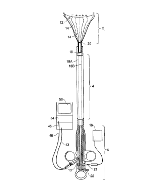
Referring to Figs. 1 and 2, an ablation device
according to the present invention is comprised
generally of three major components: RF applicator
CA 022~l2l6 l998-l0-07
W097/38637 PCT~S97/05927
head 2, main body 4, and handle 6. Main body 4
includes a shaft 10. The RF applicator head 2
includes an electrode carrying means 12 mounted to
the distal end of the shaft 10 and an array of
electrodes 14 formed on the surface of the electrode
carrying means 12. An RF generator 16 is
electrically connected to the electrodes 14 to
provide mono-polar or bipolar RF energy to them.
Shaft 10 is an elongate member having a hollow
interior. Shaft lO is preferably 12 inches long and
has a preferred cross-sectional diameter of
approximately 4 mm. A collar 13 is formed on the
exterior of the shaft 10 at the proximal end. As
best shown in Figs. 6 and 7, passive spring member 15
are attached to the distal end of the shaft 10.
Extending through the shaft 10 is a
suction/insufflation tube 17 (Figs. 6-9) having a
plurality of holes 17a formed in its distal end. An
arched active spring member 19 is connected between
the distal ends of the passive spring members 15 and
the distal end of the suction/insufflation tube 17.
Referring to Fig. 2, electrode leads 18a and 18b
extend through the shaft 10 from distal end 20 to
proximal end 22 of the shaft 10. At the distal end 20
of the shaft 10, each of the leads 18a, 18b is
coupled to a respective one of the electrodes 14. At
the proximal end 22 of the shaft 10, the leads 18a,
18b are electrically connected to RF generator 16 via
an electrical connector 21. During use, the leads
18'a,~18b carry RF energy from the RF generator 16 to
the electrodes. Each of the leads 18a, 18b is
insulated and carries energy of an opposite polarity
than the other lead.
Electrically insulated sensor leads 23a, 23b
(Figs. 5A and SB) also extend through the shaft 10.
Contact sensors 25a, 25b are attached to the distal
CA 022~1216 1998-10-07
W097~8637 PCT~S97/05927
ends of the sensor leads 23a, 23b, respectively and
are mounted to the electrode carrying means 12.
During use, the sensor leads 23a, 23b are coupled by
the connector 21 to a monitoring module in the RF
generator 16 which measures impedance between the
sensors 25a, 25b. Alternatively, a reference pad may
be positioned in contact with the patient and the
impedance between one of the sensors and the
reference pad.
Referring to Fig. 5B, electrode leads 18a, 18b
and sensor leads 23a, 23b extend through the shaft 10
between the external walls of the tube 17 and the
interior walls of the shaft 10 and they are coupled
to electrical connector 21 which is preferably
mounted to the collar 13 on the shaft 10. Connector
21, which is connectable to the RF generator 16,
includes at least four electrical contact rings 21a -
21d tFigs. 1 and 2) which correspond to each of the
- leads 18a, 18b, 23a, 23b. Rings 21a, 21b receive,
from the RF generator, RF energy of positive and
negative polarity, respectively. Rings 21c, 21d
deliver signals from the right and left sensors,
respectively, to a monitoring module within the RF
generator 16.
Referring to Fig. 5A, the electrode carrying
means 12 is attached to the distal end 20 of the
shaft 10. A plurality of holes 24 may be formed in
the portion of the di~tal end 20 of the shaft which
lies within the electrode carrying means 12.
' -The electrode carrying means 12 preferably has a
shape which approximates the shape of the body organ
which is to be ablated. For example, the apparatus
shown in Figs. 1 through 11 has a bicornual shape
which is desirable for intrauterine ablation. The
electrode carrying means 12 shown in these figures
includes horn regions 26 which during use are
CA 022~1216 1998-10-07
W097/38637 PCT~S97/05927
positioned within the cornual regions of the uterus
and which therefore extend towards the fallopian
tubes.
Electrode carrying means 12 is preferably a sack
formed of a material which is non-conductive, which
is permeable to moisture and/or which has a tendency
to absorb moisture, and which may be compressed to a
smaller volume and subsequently released to its
natural size upon elimination of compression.
Examples of preferred materials for the electrode
carrying means include open cell sponge, foam,
cotton, fabric, or cotton-like material, or any other
material having the desired characteristics.
Alternatively, the electrode carrying means may be
formed of a metallized fabric. For convenience, the
term "padn may be used interchangeably with the term
electrode carrying means to refer to an electrode
carrying means formed of any of the above materials
or having the listed properties.
Electrodes 14 are preferably attached to the
outer surface of the electrode carrying means 12,
such as by deposition or other attachment mechanism.
The electrodes are preferably made of lengths of
silver, gold, platinum, or any other conductive
material. The electrodes may be attached to the
electrode carrying means 12 by electron beam
deposition, or they may be formed into coiled wires
and bonded to the electrode carrying member using a
flexible adhesive. Naturally, other means of
attaching the electrodes, such as sewing them onto
the surface of the carrying member, may alternatively
be used. If the electrode carrying means 12 is
formed of a metallized fabric, an insulating layer
may be etched onto the fabric surface, leaving only
the electrode regions exposed.
CA 022~l2l6 l998-l0-07
W097/38637 PCT~S97/05927
The spacing between the electrodes (i.e. the
distance between the centers of ad~acent electrodes)
and the widths of the electrodes are selected so that
ablation will reach predetermined depths within the
tissue, particularly when maximum power is delivered
through the electrodes (where maximum power is the
level at which low impedance, low voltage ablation
can be achieved).
The depth of ablation is also effected by the
electrode density (i.e., the percentage of the target
tissue area which is in contact with active electrode
surfaces) and may be regulated by pre-selecting the
amount of this active electrode coverage. For
example, the depth of ablation is much greater when
the active electrode surface covers more than 10~ of
the target tissue than it is when the active
electrode surfaces covers 1~ of the target tissue.
For example, by using 3-6 mm spacing and an
- electrode width of approximately 0.5 - 2.5 mm,
delivery of approximately 20 - 40 watts over a 9-16
cm2 target tissue area will cause ablation to a depth
of approximately 5-7 millimeters when the active
electrode surface covers more than 10~ of the target
tissue area. After reaching this ablation depth, the
impedance of the tissue will become so great that
ablation will self-terminate as described with
respect to the operation of the invention.
By contrast, using the same power, spacing,
electrode width, and RF frequency will produce an
ab~lation depth of only 2 - 3 mm when the active
electrode surfaces covers less than 1 ~ of the target
tissue area. This can be better understood with
reference to Fig. l9A, in which high surface density
electrodes are designated 14a and low surface density
electrodes are designated 14b. For purposes of this
comparison between low and high surface density
........... .
CA 022~1216 1998-10-07
W097/38637 PCT~S97/05927
electrodes, each bracketed group of low density
electrodes is considered to be a single electrode.
Thus, the electrode widths W and spacings S extend as
shown in Fig. l9A.
As is apparent from Fig. l9A, the electrodes
14a, which have more active area in contact with the
underlying tissue T, produce a region of ablation A1
that extends more deeply into the tissue T than the
ablation region A2 produced by the low density
electrodes 14b, even though the electrode spacings
and widths are the same for the high and low density
electrodes.
Some examples of electrode widths, having
spacings with more than 10~ active electrode surface
coverage, and their resultant ablation depth, based
on an ablation area of 6 cm2 and a power of 20 - 40
watts, are given on the following table:
ELECTRODE WIDTH SPACING APPROX. DEPTH
1 mm 1 - 2 mm 1 - 3 mm
1 - 2.5 mm 3 - 6 mm 5 - 7 mm
1 - 4.5 mm 8 - 10 mm 8 - 10 mm
Examples of electrode widths, having spacings
with less than 1 ~ active electrode surface coverage,
and their resultant ablation depth, based on an
ablation area of 6 cm2 and a power of 20 - 40 watts,
are given on the following table:
ELECTRODE WIDTH SPACING APPROX. DEPTH
301 mm 1 - 2 mm 0.5 - 1 mm
1 - 2.5 mm 3 - 6 mm 2 - 3 mm
1 - 4.5 mm 8 - lO mm 2 - 3 mm
CA 022~1216 1998-10-07
W097/38637 PCT~S97/05927
Thus it can be seen that the depth of ablation
is significantly less when the active electrode
surface coverage is decreased.
In the preferred embodiment, the preferred
electrode spacing is approximately 8 - 10 mm in the
horn regions 26 with the active electrode surfaces
covering approximately 1~ of the target region.
Approximately 1 - 2 mm electrode spacing (with 10
active electrode coverage) is preferred in the
cervical region (designated 28) and approximately 3 -
6 mm (with greater than 10~ active electrode surface
coverage) is preferred in the main body region.
The RF generator 16 may be configured to include
a controller which gives the user a choice of which
electrodes should be energized during a particular
application in order to give the user control of
ablation depth. For example, during an application
for which deep ablation is desired, the user may
- elect to have the generator energize every other
electrode, to thereby optimize the effective spacing
of the electrodes and to decrease the percentage of
active electrode surface coverage, as will be
described below with respect to Fig. 18.
Although the electrodes shown in the drawings
are arranged in a particular pattern, it should be
appreciated that the electrodes may be arranged in
any pattern to provide ablation to desired depths.
Referring to Figs. 6 and 7, an introducer sheath
32 facilitates insertion of the apparatus into, and
rèmoval of the apparatus from, the body organ to be
ablated. The sheath 32 is a tubular member which is
telescopically slidable over the shaft 10. The
sheath 32 is slidable between a distal condition,
shown in Fig. 6, in which the electrode carrying
means 12 is compressed inside the sheath, and a
proximal condition in which the sheath 32 is moved
CA 022~1216 1998-10-07
W097/38637 PCT~S97/05927
proximally to release the electrode carrying means
from inside it (Fig. 7). By compressing the
electrode carrying means 12 to a small volume, the
electrode carrying means and electrodes can be easily
inserted into the body cavity (such as into the
uterus via the vaginal opening).
A handle 34 attached to the sheath 32 provides
finger holds to allow for manipulation of the sheath
32. Handle 34 is slidably mounted on a handle rail
35 which includes a sleeve 33, a finger cutout 37,
and a pair of spaced rails 35a, 35b extending between
the sleeve 33 and the finger cutout 37. The shaft 10
and sheath 32 slidably extend through the sleeve 33
and between the rails 35a, 35b. The tube 17 also
extends through the sleeve 33 and between the rails
35a, 35b, and its proximal end is fixed to the handle
rail 35 near the finger cutout 37.
A compression spring 39 is disposed around the
- proximal most portion of the suction/insufflation
tube 17 which lies between the rails 35a, 35b. One
end of the compression spring 39 rests against the
collar 13 on the shaft 10, while the opposite end of
the compression spring rests against the handle rail
35. During use, the sheath 32 is retracted from the
electrode carrying means 12 by squeezing the handle
34 towards the finger cutout 37 to slide the sheath
32 in the distal direction. When the handle 34
advances against the collar 13, the shaft 10 (which
is attached to the collar 13) is forced to slide in
the proximal direction, causing compression of the
spring 39 against the handle rail 35. The movement
of the shaft 10 relative to the suction/insufflation
tube 17 causes the shaft 10 to pull proximally on the
passive spring member 15. Proximal movement of the
passive spring member 15 in turn pulls against the
active spring member 19, causing it to move to the
CA 022~1216 1998-10-07
WO 97/38637 PCT/US97/05g27
opened condition shown in Fig. 7. Unless the shaft
is held in this retracted condition, the compression
spring 39 will push the collar and thus the shaft
distally, forcing the RF applicator head to close. A
locking mechanism (not shown) may be provided to hold
the shaft in the fully withdrawn condition to prevent
inadvertent closure of the spring members during the
ablation procedure.
The amount by which the springs 15, 19 are
spread may be controlled by manipulating the handle
34 to slide the shaft 10 (via collar 13), proximally
or distally. Such sliding movement of the shaft 10
causes forceps-like movement of the spring members
15, 19.
A flow pathway 36 is formed in the handle rail
35 and is fluidly coupled to a suction/insufflation
port 38. The proximal end of the suction/insufflation
- tube 17 is fluidly coupled to the flow pathway so
that gas fluid may be introduced into, or withdrawn
from the suction/insufflation tube 17 via the
suction/insufflation port 38. For example, suction
may be applied to the fluid port 38 using a
suction/insufflation unit 40. This causes water
vapor within the uterine cavity to pass through the
permeable electrode carrying means 12, into the
suction/insufflation tube 17 via holes 17a, through
the tube 17, and through the suction/insufflation
unit 40 via the port 38. If insufflation of the
ut~rine cavity is desired, insufflation gas, such as
carbon dioxide, may be introduced into the
suction/insufflation tube 17 via the port 38. The
insufflation gas travels through the tube 17, through
the holes 17a, and into the uterine cavity through
the permeable electrode carrying member 12.
CA 022~l2l6 l998-l0-07
W097l38637 PCT~S97/05927
If desirable, additional components may be
provided for endoscopic visualization purposes. For
example, lumen 42, 44, and 46 may be formed in the
walls of the introducer sheath 32 as shown in Fig.
5B. An imaging conduit, such as a fiberoptic cable
48, extends through lumen 42 and is coupled via a
camera cable 43 to a camera 45. Images taken from
the camera may be displayed on a monitor 56. An
illumination fiber 50 extends through lumen 44 and is
coupled to an illumination source 54. The third
lumen 46 is an instrument channel through which
surgical instruments may be introduced into the
uterine cavity, if necessary.
Because during use it is most desirable for the
electrodes 14 on the surface of the electrode
carrying means 12 to be held in contact with the
interior surface of the organ to be ablated, the
electrode carrying means 12 may be provide to have
- additional components inside it that add structural
integrity to the electrode carrying means when it is
deployed within the body.
For example, referring to Fig. 11, alternative
spring members 15a, 19a may be attached to the shaft
10 and biased such that, when in a resting state, the
spring members are positioned in the fully resting
condition shown in Fig. 11. Such spring members
would spring to the resting condition upon withdrawal
of the sheath 32 from the RF applicator head 2.
Alternatively, a pair of inflatable balloons 52
mày be arranged inside the electrode carrying means
12 as shown in Fig. 20 and connected to a tube (not
shown) extending through the shaft 10 and into the
balloons 52. After insertion of the apparatus into
the organ and following retraction of the sheath 32,
the balloons 52 would be inflated by introduction of
an inflation medium such as air into the balloons
CA 022~1216 1998-10-07
W097/38637 PCT~S97/05927
16
via a port similar to port 38 using an apparatus
similar to the suction/insufflation apparatus 40.
Structural integrity may also be added to the
electrode carrying means through the application of
suction to the proximal end 22 of the
suction/insufflation tube 17. Application of suction
using the suction/insufflation device 40 would draw
the organ tissue towards the electrode carrying means
12 and thus into better contact with the electrodes
14.
Figs. 12 and 13 show an alternative embodiment
of an ablation device according to the present
invention. In the alternative embodiment, an
electrode carrying means 12a is provided which has a
shape which is generally tubular and thus is not
specific to any particular organ shape. An ablation
device having a general shape such as this may be
used anywhere within the body where ablation or
- coagulation is needed. For example, the alternative
embodiment is useful for bleeding control during
laparoscopic surgery (Fig. 14), tissue ablation in
the prostate gland (Fig. 17), and also intrauterine
ablation (Figs. 15 and 16).
Operation
Operation of a preferred ablation device
according to the present invention will next be
described.
Referring to Fig. 1, the device is initially
cdnfigured for use by positioning the introducer
sheath 32 distally along the shaft 10, such that it
compresses the electrode carrying means 12 within its
walls.
At this time, the electrical connector 21 is
connected to the RF generator 16, and the fiberoptic
cable 48 and the illumination cable 50 are connected
CA 022~1216 1998-10-07
W097/38637 PCT~S97/05927
to the illumination source, monitor, and camera, 54,
56, 45. The suction/insufflation unit 40 is attached
to suction/insufflation port 38 on the handle rail
35. The suction/insufflation unit 40 is preferably
set to deliver carbon dioxide at an insufflation
pressure of 20 - 200 mmHg.
Next, the distal end of the apparatus is
inserted through the vaginal opening V and into the
uterus U as shown in Fig. 6, until the distal end of
the introducer sheath 32 contacts the fundus F of the
uterus. At this point, carbon dioxide gas is
introduced into the tube 17 via the port 38, and it
enters the uterine cavity, thereby expanding the
uterine cavity from a flat triangular shape to a 1-2
cm high triangular cavity. The physician may observe
(using the camera 45 and monitor 56) the internal
cavities using images detected by a fiberoptic cable
48 inserted through lumen 42. If, upon observation,
- the physician determines that a tissue biopsy or
other procedure is needed, the required instruments
may be inserted into the uterine cavity via the
instrument channel 46.
Following insertion, the handle 34 is withdrawn
until it abuts the collar 13. At this point, the
sheath 32 exposes the electrode carrying member 12
but the electrode carrying member 12 is not yet fully
expanded (see Fig 9), because the spring members 15,
19 have not yet been moved to their open condition.
The handle 34 is withdrawn further, causing the shaft
10' to move proximally relative to the
suction/insufflation tube 17, causing the passive
spring members 15 to pull the active spring members
19, causing them to open into the opened condition
shown in Fig. 10.
The physician may confirm proper positioning of
the electrode carrying member 12 using the monitor
CA 022~l2l6 l998-l0-07
W097/38637 PCT~S97/05927
18
56, which displays images from the fiberoptic cable
48.
Proper positioning of the device and sufficient
contact between the electrode carrying member 12 and
the endometrium may further be confirmed using the
contact sensors 25a, 25b. The monitoring module of
the RF generator measures the impedance between these
sensors using conventional means. If there is good
contact between the sensors and the endometrium, the
measured impedance will be approximately 20 - 180
ohm, depending on the water content of the
endometrial lining.
The sensors are positioned on the distal
portions of the bicornual shaped electrode carrying
member 12, which during use are positioned in the
regions within the uterus in which it is most
difficult to achieve good contact with the
endometrium. Thus, an indication from the sensors
- 25a, 25b that there is sound contact between the
sensors and the endometrial surface indicates that
good electrode contact has been made with the
endometrium.
Next, insufflation is terminated. Approximately
l - 5 cc of saline may be introduced via
suction/insufflation tube 17 to initially wet the
electrodes and to improve electrode electrical
contact with the tissue. After introduction of
saline, the suction/insufflation device 40 is
switched to a suctioning mode. As described above,
the application of suction to the RF applicator head
2 via the suction/insufflation tube 17 collapses the
uterine cavity onto the RF applicator head 2 and thus
assures better contact between the electrodes and the
endometrial tissue.
If the generally tubular apparatus of Figs. 12
and 13 is used, the device is angled into contact
CA 022~l2l6 l998-l0-07
W097l38637 PCT~S97/05927
with one side of the uterus during the ablation
procedure. Once ablation is completed, the device
tor a new device) is repositioned in contact with the
opposite side and the procedure is repeated. See.
Figs. 15 and 16.
Next, RF energy at preferably about 500 kHz and
at a constant power of approximately 30 W is applied
to the electrodes. As shown in Fig. 5a, it is
preferable that each electrode be energized at a
polarity opposite from that of its neighboring
electrodes. By doing so, energy field patterns,
designated 100, 102 and 104 in Fig. 18, are generated
between the electrode sites and thus help to direct
the flow of current through the tissue T to form a
region of ablation A. As can be seen in Fig. 18, if
electrode spacing is increased such by energizing,
for example every third or fifth electrode rather
than all electrodes, the energy patterns will extend
- more deeply into the tissue. (See, for example,
pattern 102 which results from energization of
electrodes having a non-energized electrode between
them, or pattern 104 which results from energization
of electrodes having two non-energized electrodes
between them).
Moreover, ablation depth may be controlled as
described above by providing low surface density
electrodes on areas of the electrode carrying member
which will contact tissue areas at which a smaller
ablation depth is required (see Fig. 19A).
~ -Referring to Fig. l9B, if multiple, closely
spaced, electrodes 14 are provided on the electrode
carrying member, a user may set the RF generator to
energize electrodes which will produce a desired
electrode spacing and active electrode area. For
example, alternate electrodes may be energized as
shown in Fig. l9B, with the first three energized
CA 022~1216 1998-10-07
W097l38637 PCT~S97/05927
electrodes having positive polarity, the second three
having negative polarity, etc.
As another example, shown in Fig. l9C, if
greater ablation depth is desired the first five
electrodes may be positively energized, and the
seventh through eleventh electrodes negatively
energized, with the sixth electrode remaining
inactivated to provide adequate electrode spacing.
As the endometrial tissue heats, moisture begins
to be released from the tissue. The moisture
permeates the electrode carrying member 12 and is
thereby drawn away from the electrodes. The moisture
may pass through the holes 17a in the
suction/insufflation tube 17 and leave the
suction/insufflation tube 17 at its proximal end via
port 38 as shown in Fig. 7. Moisture removal from
the ablation site may be further facilitated by the
application of suction to the shaft 10 using the
- suction/insufflation unit 40.
Removal of the moisture from the ablation site
prevents formation of a liquid layer around the
electrodes. As described above, liquid build-up at
the ablation site is detrimental in that provides a
conductive layer that carries current from the
electrodes even when ablation has reached the desired
depth. This continued current flow heats the liquid
and surrounding tissue, and thus causes ablation to
continue by unpredictable thermal conduction means.
Tissue which has been ablated becomes dehydrated
a~d -thus decreases in conductivity. By shunting
moisture away from the ablation site and thus
preventing liquid build-up, there is no liquid
conductor at the ablation area during use of the
ablation device of the present invention. Thus, when
ablation has reached the desired depth, the impedance
at the tissue surface becomes sufficiently high to
CA 022~1216 1998-10-07
W097~8637 PCT~S97105927
stop or nearly stop the flow of current into the
tissue. RF ablation thereby stops and thermal
ablation does not occur in significant amounts. If
the RF generator is equipped with an impedance
monitor, a physician utilizing the ablation device
can monitor the impedance at the electrodes and will
know that ablation has self-terminated once the
impedance rises to a certain level and then re~; n.~
fairly constant. By contrast, if a prior art bipolar
RF ablation device was used together with an
impedance monitor, the presence of liquid around the
electrodes would cause the impedance monitor to give
a low impedance reading regardless of the depth of
ablation which had already been carried out, since
current would continue to travel through the low-
impedance liquid layer.
Other means for monitoring and terminating
ablation may also be provided. For example, a
- thermocouple or other temperature sensor may be
inserted to a predetermined depth in the tissue to
monitor the temperature of the tissue and terminate
the delivery of RF energy or otherwise signal the
user when the tissue has reached a desired ablation
temperature.
Once the process has self terminated, 1 - 5 cc
of saline can be introduced via suction/insufflation
tube 17 and allowed to sit for a short time to aid
separation of the electrode from the tissue surface.
The suction/insufflation device 40 is then switched
t~ provide insufflation of carbon dioxide at a
pressure of 20 - 200 mmHg. The insufflation pressure
helps to lift the ablated tissue away from the RF
applicator head 2 and to thus ease the closing of the
RF applicator head. The RF applicator head 2 is
moved to the closed position by sliding the handle 34
in a distal direction to fold the spring members 15,
CA 022~1216 1998-10-07
W097/38637 PCT~S97/05927
19 along the axis of the device and to cause the
introducer sheath 32 to slide over the folded RF
applicator head. The physician may visually confirm
the sufficiency of the ablation using the monitor 56.
Finally, the apparatus is removed from the uterine
cavity.