Note: Descriptions are shown in the official language in which they were submitted.
CA 022~1248 1998-10-07
W O97t38621 PCTrUS97/06265
PORTABLE SCANNING LASER OPHTHALMOSCOPE
FIELD OF INVENTION
The present invention relates to a sc~nning
laser ophthalmoscope and more particularly to a
portable scanning laser ophthalmoscope with a wide
field of view that allows a clinician to directly
view the interior of the patient's eye without the
use of an external monitor.
BACKGROUND OF T~E INVENTION
Scanning laser ophthalmoscopes such as shown
in U.S. Patents Webb 4,765,730; Webb 4,764,006 and
Webb 4,768,873 are known to include a turning
mirror to direct a laser beam to a multi-faceted
rotating polygonal reflector scanner that scans a
laser beam in a first direction to form a line of
light. A second scanner is employed in the form
of a galvanometer reflector scanner to scan the
line of light generated by the first scanner in a
.~
second direction perpendicular to the first
direction of scanning. The scanned light is
directed to a patient's eye by a series of
focusing mirrors. Light reflected from the
patient's eye follows the same path via the
scanners and focusing mirrors back to the turning
mirror. The turning mirror is small so that the
CA 022~1248 1998-10-07
W O 97/38621 PCTrUS97/06265
light reflected from the eye passes around it to
an optical detector in the form of an avalanche
diode. The output of the optical detector is
coupled to the display to provide a two
dimensional picture of the patient's retina.
Although this type of scanning laser
ophthalmoscope is capable of producing an image of
the patient's retina without requiring the
patient's pupil to be dilated with drugs and
without requiring contact with the patient's eye,
it has several drawbacks. First, the scanned
laser light source employed in Webb's sc~nn; ng
laser ophthalmoscope is very bright and leaves the
patient dazzled for some time following the
diagnostic procedure implemented with the
ophthalmoscope. Further, the Webb system is
large, complex and very costly. The Webb system
also suffers from a small field of view that is on
the order of only 30~.
Another type of scanning laser ophthalmoscope
is shown in U.S. Patent Kobayashi 4,781,453 that
utilizes a first acousto optical modulator for
modulating the intensity of a laser beam to
project a fixation target. The frequency of the
drive signal for the first acousto optical
modulator is also varied so as to select, with the
use of a lens and device having a slit therein, a
single wavelength of a laser beam having a number
of wavelengths therein. The single selected
wavelength of the laser is then passed to a
scanning system. The scanning system includes a
second acousto optical modulator that is driven so
as to scan the selected wavelength of the laser in
CA 022~1248 1998-10-07
W O97/38621 PCT~US97/06265
a first direction. Prior to scanning, however,
the range of the second acousto optical modulator
must be changed to accommodate the selected
wavelength of the laser. The scanned laser is
guided by relay lenses from the second acousto
optical modulator to a mirror that is mounted on
a galvanometer for scanning the laser in a second
direction perpendicular to the scanning direction
of the second acousto optical modulator. A small
mirror then reflects the scanned light to a
patient's eye. The light reflected from the eye
passes around the small mirror and is captured by
a lens and focused on a photosensor. A filter
corresponding to the selected wavelength of the
laser is disposed in front of the photosensor to
allow passage of the selected light to the sensor.
An image of the eye at a depth corresponding to
the selected wavelength is stored in a frame
memory associated with the selected wavelength,
wherein the system includes different frame
memories for the different wavelengths that can be
selected. The different images stored in the
frame memories can be selected via the electronics
of the system for individual display in different
colors on a color monitor. The Kobayashi
ophthalmoscope is an extremely complex device in
which the scanning range of the second acousto
optical modulator must be changed to accommodate
a selected wavelength of the laser light each time
a new wavelength is selected via the first acousto
optical modulator. Further, the filter disposed
in front of the photo sensor must also be changed
in accordance with the selected wavelength. The
CA 022~l248 l998-l0-07
W O97/38621 PCTrUS97/06265
field of view of this scanning laser
ophthalmoscope is also small, being on the same
order as that described above for the Webb
scanning laser ophthalmoscope.
In both the Webb and Kobayashi systems, a
mirror is disposed in the optical path of the
light reflected from the patient's eye to the
detector which causes a shading off effect. This
shading off effect is realized as a darkening of
the edges of an image feature with a gradual
lightening of the image feature towards the center
thereof. For example, this effect causes the
displayed image of a blood vessel to appear as
dark parallel lines with a lighter center
therebetween. This effect is further exacerbated
by the small aperture diameter employed in the
image detection portion of the these systems.
This small aperture although eliminating unwanted
reflections from detection, brings substantially
all of a given scene into focus at the same focal
plane. The result is that the image of the
patient's fundus appears similar regardless of the
wavelength of the laser beam and the portion of
the patient's eye at a particular depth therein
reflecting the selected wavelength of the light.
Further, the known scanning laser
ophthalmoscopes such as described above are large
and nonportable. As a result patients must be
taken to the instrument for the eye examination
which can be difficult with a sic~ patient that is
bedridden. These ophthalmoscopes are also
extremely complex and costly due to their optical
CA 022~1248 1998-10-07
W O97/38621 ~CT~US97/06265
arrangements and the necessity of image detectors
and monitors for displaying an image of the eye.
SUMMARY OF THE INVENTION
In accordance with the present invention, the
disadvantages of prior scanning laser
ophthalmoscopes have been overcome. The scanning
laser ophthalmoscope of the present invention is
portable; provides a wide field of view; and
allows a clinician to directly view the interior
of a patient's eye without the use of an external
monitor. The scanning laser ophthalmoscope of the
present invention is extremely simplified compared
to prior devices and eliminates the shading off
effects found in the displayed eye images produced
by prior scanning laser ophthalmoscopes.
More particularly, the portable scanning
laser ophthalmoscope of the present invention
includes a housing that is sufficiently small to
be carried and held by a clinician. The housing
contains a source of laser light that is scanned
by a scanning system for generating a two
dimensional area of illumination. The housing
also includes a battery for providing power to the
scanning system. An optical system contained in
the housing directs illumination from the scanning
system to the patient's eye to illuminate the
fundus and also intercepts light reflected by the
patient's eye to generate a magnified image of the
interior of the patient's eye. The optical system
also includes an eyepiece lens through which a
CA 022~1248 1998-10-07
W O97t38621 PCT~US97106265
clinician looks to directly view the magnified
image of the interior of the patient's eye.
In accordance with another feature of the
present invention, shading off in the image
captured by the scanning laser ophthalmoscope is
prevented by separating the optical path between
the patient's eye and the scanning system from the
optical path between the patient's eye and the
eyepiece lens with a beam splitter that does not
bloc~ the chief ray reflected at any given
position in the patient's eye on its path to the
eyepiece lens.
In accordance with another feature of the
present invention, the scanning system includes
only one scanner with a moveable reflective
surface and a passive, stationary optical element.
The passive optical element is positioned in a
path of the laser light such that the light
impinges on the optical element at a point and the
optical element generates a line of light from
that point. The single scanner with moveable
reflective surface is then used to scan the line
of light generated by the passive optical element
in a direction perpendicular to the line so as to
generate the two dimensional area of illumination.
Because only a single scanner with a moveable
reflective surface is employed as opposed to two
such scanners, the scanning laser ophthalmoscope
is more rugged than prior devices and more
compact, enabling the ophthalmoscope to be
portable. Further, because a passive, stationary
optical element is employed as opposed to an
active optical device, such as an acousto optical
CA 022~l248 l998-l0-07
W O 97/38621 PCTrUS97/06265
modulator that requires a drive signal to scan,
the electronics of the present scanning laser
ophthalmoscope are again greatly simplified.
In accordance with a further feature of the
present invention, a nonsymmetric aspheric lens is
employed to focus the illumination light from the
illumination system on an area generally proximate
to the patient's pupil and to capture light
reflected from the patient's eye and to focus that
reflected light onto an image plane. This
aspheric objective lens which is positioned
between the patient's eye and the eyepiece lens
greatly simplifies the optical system of the
scanning laser ophthalmoscope of the present
invention and greatly reduces the optical
components thereof.
Further, in accordance with another feature
of the present invention, the laser light from the
source is polarized in a first direction and a
polarizer disposed between the aspheric objective
lens and the eyepiece lens is polarized in a
second direction that is different from the first
direction to pass only desired light to the
eyepiece lens. Thus, unwanted reflections are
eliminated from the magnified image viewed by the
clinician.
These and other advantages and novel features
of the present invention, as well as details of an
illustrated embodiment thereof, will be more fully
understood from the following description and
drawings.
CA 022~1248 1998-10-07
W O97/38621 PCTrUS97/06265
BRIEF DESCRIPTION OF THE DRAWING
Fig. 1 is a perspective view of a portable
scanning laser ophthalmoscope in accordance with
the present invention positioned with respect to
a patient's eye so that a clinician can view the
interior thereof;
Fig. 2 is a plan view of the portable
scanning laser ophthalmoscope of Fig. 1 shown in
relation to the patient's eye;
Fig. 3 is a prospective view of the
components of the portable scanning laser
ophthalmoscope of Fig. 2; and
Figs. 4A and 4B respectively illustrate an
image of an eye as viewed via the scanning laser
of ophthalmoscope of Figs. 1-3 and of an eye image
displayed with a prior device that produces a
shading off effect.
DESCRIPTION OF THE PREFERRED EMBODIMENT
A portable scAnning laser ophthalmoscope 10
in accordance with the present invention as shown
in Fig. 1 includes a housing 12 that is
sufficiently small to be carried by a clinician
and held in a clinician's hand during an
examination of a patient's eye 14. More particu-
larly, during an eye examination, the clinician
holds the housing 12 of the scanning laser
ophthalmoscope 10 so that a housing portion 16
containing an objective lens is positioned near
CA 022~1248 1998-10-07
Wo97/38621 PCT~S97/~265
the patient's eye 14. The clinician then presses
an on-off button 18 so as to provide power to a
scanning system of the ophthalmoscope 10 from a
battery contained within the housing 12. The
battery is easily accessible to a user via an
access panel 20. When the scanning laser
ophthalmoscope is turned on, the scanning system
thereof illuminates a two-dimensional area of the
interior of the patient's eye 14. Light reflected
from the patient's eye due to this illumination is
captured by the optical system of the ophthalmo-
scope 10 so that a magnified image of an interior
portion of the patient's eye 14 can be viewed
directly by the clinician through an eyepiece lens
22.
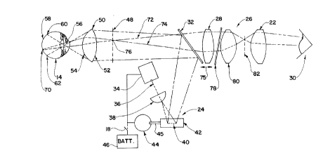
The portable scanning laser ophthalmoscope 10
as shown in detail in Figs. 2 and 3 includes a
scanning system 24 for scanning the two-
dimensional area of illumination that illuminates
the interior of the patient's eye 14. The
scanning laser ophthalmoscope 10 also includes an
optical system 26 with a movable field lens 28 to
capture light reflected from the patient's eye 14
so that a clinician 30 can view an interior
portion of the patient's eye 14 through the
eyepiece lens 22. The optical path from the
scanning system 24 to the patient's eye is
separated from the optical path from the patient's
eye to the field lens 28 and eyepiece lens 22 so
that in the portable scanning laser ophthalmoscope
10 of the present invention there is no scanner,
mirror or other optical element that totally
blocks light reflected from the patient's eye 14
CA 022~1248 1998-10-07
W O 97/38621 PCTrUS97/06265
in a given region of the optical path to the
eyepiece lens 22. This feature illuminates
shading off problems of prior scanning laser
ophthalmoscopes.
In order to accomplish the separation of the
scanning system 24 from the optical path between
the patient's eye and the eyepiece lens 22, the
scanning laser ophthalmoscope 10 includes a beam
splitter 32. The beam splitter 32 is a partially
reflecting illumination mirror that reflects at
least 25% of the illumination light from the
scanning system 24 to the patient's eye 14 while
passing therethrough light reflected from the
patient's eye 14 so that the interior portion of
the patient's eye can be viewed by the clinician
through the eyepiece lens 22. It has been found
that the shading off effects plaguing prior
scanning laser ophthalmoscopes were caused by an
optical element such as a scanner or mirror placed
in the optical path from the patient's eye to the
eye image capturing system. These optical
elements block the chief ray from any given image
position on its route from the patient's eye to
the image capturing optics thereby causing shading
off. The present invention eliminates this
problem by separating the path to the eyepiece
lens 22 from the scanning system 24 and by
employing optical elements within the optical path
from the patient's eye to the lens 22 that do not
block the chief rays from any given image position
on their route to the lens 22. As a result, the
image of the eye viewed by a clinician with the
ophthalmoscope 10 is as shown in Fig. 4A as
CA 022~1248 1998-10-07
W O 97/38621 PCTAUS97/0626
opposed to an image of the eye as shown in Fig. 4B
that has the shading off effect.
The shading off effect can be seen in Fig.
4B as a darkening of the edges of a feature of the
image and as a lightening of the center of the
feature. For example, in Fig. 4B the imaged
features of blood vessels are shown as dark
parallel lines with a gradual lightening towards
the center of the blood vessel. This effect is
not present in the image displayed by the scanning
laser ophthalmoscope 10 of the present invention
as depicted in Fig. 4A because the chief ray from
any given position in the eye is not blocked on
its route to the eyepiece lens 22.
As shown in Figs. 2 and 3, the scanning
system 24 of the ophthalmoscope 10 includes a
laser source 34. The laser source 34 generates a
laser beam 36 that impinges on a passive,
stationary optical element 38 at a point. The
passive, stationary optical element 38, which may
be a cylindrical lens as shown, generates a line
40 of light from the point of light impinging on
the lens 38. The line 40 of laser light is
scanned in a direction perpendicular to the
direction of the line 40 by a scanner mirror 42 on
which the line of light impinges. The scanner
mirror 42 is driven by a scanner motor 44 that is
coupled to the mirror 42 via a shaft 45. A
battery 46 provides power the scanner motor 44 and
to the laser source 34 via the on-off switch 18.
As the scanner mirror 42 vibrates, it scans the
line 40 horizontally across the face of the
partially reflective beam splitter 32 as shown in
CA 022~1248 1998-10-07
W O 97/38621 PCTrUS97/06265
Fig. 3 so that a rectangular shaped area of
illumination is generated on the face of the beam
splitter 32. The beam splitter 32 reflects the
rectangular area of illumination light towards the
eye 14 so that it is centered on a real image
plane 48 and on a nonsymmetric aspheric objective
lens 50. The illumination light as it travels
towards the patient's eye 14 is slightly
diverging. The weaker surface 52 of the aspheric
lens makes the slightly diverging illumination
light parallel and directs the illumination light
to the stronger surface 54 of the aspheric lens
50. The stronger surface 54 of the aspheric lens
focuses the illumination light to a point 56 that
is centered on the patient's pupil or generally
proximate thereto. The illumination light
continues its path until it strikes the retina 58
of the eye 14, thus illuminating an area of the
patient's eye within the boundaries of the rays 60
and 62. The use of the passive optical element 38
that converts a point of light impinging thereon
into a line of light without an external drive
signal applied thereto as required by acousto
optic modulators and without movement of an
element as in scanning mirrors, substantially
simplifies the optical system of the present
invention and reduces the size thereof so as to
enable the scanning laser ophthalmoscope 10 to be
packaged in a portable housing 12.
In order to focus the eye image capturing
system 26 onto different areas of a patient's eye
14, the optical system 26 includes the moveable
field lens 34. More particularly, as shown in
CA 022~1248 1998-10-07
W O 97/38621 PCT~US97/06265
Figs. 2 and 3, an illuminated point 70 on the
fundus 58 of the patient's eye 14 reflects light
shown by the rays 72 and 74 wherein the reflected
light is captured and focused by the aspheric
objective lens 50 to a point 76 on the image plane
48. The light reflected from the patient's eye 14
passes through the beam splitter 32 to the field
lens 28. The field lens 28 is moveable in the
direction of the arrow 75 so as to change the
position of the image plane 48 closer to or
farther from the lens 50, thus changing the
location of the point 70. The light reflected
from the patient's eye passes through the field
lens 28 and from there through a polarizer film 78
to an image lens 80. The image lens 80 and field
lens 28 form a magnified image 82 of the interior
of the patient's eye which is observed by the
clinician 30 as he looks into the eyepiece lens
22.
In order to pass only desired light to the
eyepiece lens 22, the laser light from the source
34 is polarized in a first direction and the
polarizer film 78 of the optical system 26 is
polarized in a second direction that is different
from the first direction. In particular, the
polarizer film 78 is preferably polarized in a
direction perpendicular to the polarization of the
laser light from the source 34. This polarization
of the polarizer film 112 blocks unwanted
reflections from the patient's cornea, the
aspheric lens 50 and other elements of the system
from reaching the image lens 80 and eyepiece lens
22 so that only the randomized reflected image
CA 022~l248 l998-l0-07
W O97/38621 PCTrUS97/06265
from the interior of the patient's eye passes
through the optical system into the eyepiece lens
22.
The aspheric lens 50 of the present invention
focuses the illumination light from the
illumination system 24 on an area of the patient's
eye that is generally proximate to the pupil and
the aspheric lens 50 also intercepts light
reflected from the patient's eye 14 and focuses
the intercepted light onto the image plane 48 that
is disposed between the aspheric lens and the
eyepiece lens 22. In order to provide such an
aspheric lens, each surface 52 and 54 of the lens
is preferably described by the polynomial
function:
f(Y,A2,A4,A6, C, CC) = A2y2+A4Y4 'A6y6+cy2/ (1+~ C2CC
where A2, A4 and A6 are constants; C represents the
curvature of the surface; and cc represents the
conic constant. For the stronger surface 54 of
the lens 50, these values should be within the
following ranges:
~ 0.0 ~ A2 ~ 0.003
-0.02 ~ A4 < 0.02
-0.01 < A6 < 0.01
-0.1 < C < 0.0
-2.0 < cc < 1.0
For the weaker surface 52 of the lens 50 these
values should be within the following ranges:
CA 022~1248 1998-10-07
W O97/38621 PCTrUS97/06265
-0.003 < A2 < ~-~
0.0 < A4 < 0.001
-O. 001 A6 < O. 001
0.03 < C < 0.06
5 -2.0 < CC < 0.0
Further the curvature C of the weaker surface 52
is preferably greater than -1/2 times the
curvature C of the stronger surface 54.
In a preferred embodiment of the present
invention, the stronger surface 54 of the lens 50
has the values of: A2 = 0.000444, A4 = 0.000001,
A6 = ~-~~ C = -0. 092 and cc = -0. 933; whereas the
weaker surface 52 of the lens 50 has values of:
A2 = -0.00243, A4 = 0. 0000012, A6 = ~-~, C = 0. 045
and cc = -1. 213.
While the diameter d of the lens 50 may be
varied, the preferred diameter is 35 millimeters.
The aspheric lens 50 produces a 60~ field of view
for the scanning laser ophthalmoscope 10 which is
extremely wide compared to prior scanning laser
ophthalmoscopes and ophthalmoscopes in general.
Further, the real image produced by the aspheric
lens 50 is substantially free from distortions.
Many modifications and variations of the
present invention are possible in light of the
above teachings. Thus, it is to be understood
that, within the scope of the appended claims, the
invention may be practiced otherwise than as
described hereinabove.
What is claimed and desired to be secured by
Letters Patent is: