Note: Descriptions are shown in the official language in which they were submitted.
CA 022~1336 1998-10-13
W O 97/39684 PCT~US97/04674
METHOD AND SYSTEM FOR 3-D ACOUSTIC
MICROSCOPY USING SHORT PULSE EXCITATION
AND 3-D ACOUSTIC MICROSCOPE FOR USE lH~EIN
Technical Field
This invention relates to the non-destructive
evaluation of objects and materials and, in particular,
to method and systems for 3-D acoustic microscopy using
short pulse excitation and 3-D acoustic microscopes for
use therein.
Back round Art
The practice of clinical pathology centers
around the microscopic analysis of biopsies obtained
from the body. Although tissue biopsies are fundamen-
tally three-dimensional, they must be sectioned for two-
dimensional analysis by light microscopy because of theopaqueness of most biological specimens. As a conse-
quence, multiple two-dimensional samples must be pre-
pared for each biopsy. Sample preparation can be very
costly for each section. Moreover, to accurately
characterize the properties of the entire 3-D sample, a
large number of sections must be prepared.
Acoustic microscopy is a well established
technique dating to the early 1970s. The most recog-
nized system was produced in the Applied Physics Depart-
ment at Stanford University by Calvin Quate. U.S.Patent Nos. 4,006,444; 4,028,933; 4,267,732; 4,430,897;
and 5,319,977 disc'ose various acoustic microscopes
wherein Mr. Quate is a named inventor.
CA 022~1336 1998-10-13
W O 97139684 PCTrUS97/04674
Several small commercial versions of this
microscope, and similar microscopes, have been produced
over the last decade. All of these microscopes are
inherently two-dimensional, where an image is commonly
obtained through some form of mechanical scanning.
Short pulse laser excitation of acoustic waves
is also a well established technique for ultrasonic
frequencies less than 100 MHz. A large body of work was
done on this at IBM by von Gutfeld in the early 1980s as
described in the U.S. patent to von Gutfeld et al.
4,512,197.
Recent work by a group in the Physics Depart-
ment at Brown University led by Tauc and Maris has shown
that laser excitation can be extended to produce ultra-
sonic pulses at frequencies greater than 1 GHz. U.S.
Patent No. 4,710,030 in the name of Tauc et al. disclos-
es some of this work.
Synthetic Aperture techniques are common in
ultrasonic and RADAR systems as disclosed in the U.S.
patents to Fort et al. 5,269,309 and 5,465,722. For
example, Synthetic Aperture Radar (SAR), pioneered by
ERIM over two decades ago, is now routinely used in many
forms of surveillance.
However, all work to date on laser-generated,
high frequency, acoustic waves uses weakly focused
optical sources, resulting in spatially extended excita-
tion (i.e., equivalent aperture many ultrasonic wave-
lengths across). Such excitation produces nearly plane
wave propagation of the resultant ultrasonic pulse.
CA 022~1336 1998-10-13
W O 97/39684 PCTAJS97/04674
S-lmm~ry Of The Invention
An object of the present invention is to
provide a method and system for examining an object
internally using the principles of acoustic microscopy,
optical excitation of acoustic pulses and synthetic
aperture reconstruction for the production of true 3-D
acoustic microscope images. These techniques can be
applied to human pathology and ultrasonic non-destruc-
tive testing.
Another object of the present invention is to
provide a method and system for 3-D acoustic microscopy
using short pulse excitation wherein living tissue can
be investigated three dimensionally with the same
diagnostic accuracy as current 2-D methods.
It is still another object of the present
invention to provide a 3-D acoustic microscope which can
be integrated into an optical needle probe such as the
tip of a conventional biopsy needle so that in si tu
imaging of internal organs can be performed in real-
time. Such an instrument will not remove any tissue
from the organ under investigation.
Yet still another object of the present
invention is to provide a method and system for 3-D
acoustic microscopy using short pulse excitation for 3-D
imaging of optically opaque small tissue samples with
high frequency ultrasound.
It is still another object of the present
invention to provide a method and system for 3 D acous-
tic microscopy using short pulse laser excitation and
. . .
CA 022~1336 1998-10-13
WO 97/39684 PCTrUS97/04674
synthetic aperture reconstruction to produce dynamically
focused 3-D images of small tissue samples with a
resolution approaching the finest of optical micro-
scopes.
Yet still a further object of the present
invention is to provide a method and system for 3-D
acoustic microscopy using short pulse excitation in a
simple and effective manner for obtaining true 3-D
acoustic microscopic images with near optimal resolution
over a wide depth of field.
In carrying out the above objects and other
objects of the present invention, a method is provided
for examining an object internally. The method includes
the steps of positioning an opto-acoustic transducer
having an optically reflecting surface and an absorbing
layer capable of converting a burst of electromagnetic
energy into a thermal pulse relative to the object so
that the thermal pulse propagates as an acoustic wave in
the object and is internally reflected within the
object. The method also includes the steps of scanning
a plurality of bursts of the electromagnetic energy over
the surface of the absorbing layer to obtain acoustic
waves in the object, detecting acoustic waves reflected
within the object at a plurality of positions at the
optically reflecting surface of the opto-acoustic trans-
ducer to generate resultant signals, and calculating a
3-D representation of the object from the resultant
signals. Finally, the method includes the step of
displaying the 3-D representation as an image.
Still further in carrying out the above
objects and other objects of the present invention, a
CA 022~1336 1998-10-13
W O 97/39684 PCTrUS97/04674
method is provided for examining an object internally.
The object is capable of converting a burst of electro-
magnetic energy into a thermal pulse which propagates as
an acoustic wave in the cbject and is internally re-
flected within the object. The method includes thesteps of scanning a plurality of bursts of the electro-
magnetic energy over a surface of the object to generate
acoustic waves in the object, detecting the acoustic
waves reflected within the object at a plurality of
positions on the surface of the object to generate
resultant signals, and calculating a 3-D representation
of the object from the resultant signals. Finally, the
method includes the step of displaying the 3-D represen-
tation as an image.
In carrying out the above objects and other
objects of the present invention, systems are provided
for carrying out the above method steps.
Yet still further in carrying out the above
objects and other objects of the present invention, a
3-D acoustic microscope is provided and is adapted to be
used in a system for 3-D acoustic microscopy to examine
an object internally. The microscope includes an opto-
acoustic transducer adapted to be coupled to the object.
The transducer has an optically reflecting surface and
an absorbing layer capable of converting a burst of
electromagnetic energy into a thermal pulse which
propagates as an acoustic wave in the object and is
internally reflected within the object. The microscope
also includes a first acoustooptic scanning device for
scanning a plurality of bursts of the electromagnetic
energy over a surface of the absorbing layer to generate
acoustic waves in the object. A second acoustooptic
CA 022~1336 1998-10-13
W O 97t39684 PCT~US97/04674
scanning device is provided for scanning a probe beam at
a plurality of positions on the reflecting surface of
the opto-acoustic transducer. A housing houses the
opto-acoustic transducer and the first and second
acoustooptic scanning devices to define an optical
needle probe.
Further in carrying out the above objects and
other objects of the present invention, a 3-D acoustic
microscope adapted to be used in a system for 3-D
acoustic microscopy to examine an object internally is
provided. The object is capable of converting a burst
of electromagnetic energy into a thermal pulse so that
the thermal pulse propagates as an acoustic wave in the
object and is internally reflected within the object.
The microscope includes a first acoustooptic scanning
device for scanning a plurality of bursts of the elec-
tromagnetic energy over a surface of the object to
generate acoustic waves in the object and a second
acoustooptic scanning device for scanning a probe beam
at a plurality of positions on the surface of the
object. The microscope also includes a housing for
housing the first and second acoustooptic scanning
devices to define an optical needle probe.
The above objects and other objects, features,
and advantages of the present invention are readily
apparent from the following detailed description of the
best mode for carrying out the invention when taken in
connection with the accompanying drawings.
CA 022~1336 1998-10-13
W O 97/39684 PCT~S97104674
Brief Description Of The Drawin~c
FIGURE 1 is a schematic view of a 3-D optical-
ly excited acoustic microscope with optical detection;
FIGURE 2a is a view similar to the view of
Figure 1 wherein optical detection is accomplished with
an unfocused probe beam source and an array of coherent
optical detectors;
FIGURE 2b is a schematic view of an opto-
acoustic detector with a single optical fiber for use in
the method and system of the present invention;
FIGURE 3 is a schematic view of a 3-D optical-
ly excited acoustic microscope with acoustic detection;
and
FIGURE 4 is a block diagram of a 3-D optically
excited acoustic microscope.
Best Mode For Carryin~ Out The Invention
In contrast to the prior art, the method and
system of the present invention provides 3-D acoustic
microscopy using short optical pulses which are highly
focused so that the excitation aperture is comparable to
an acoustic wavelength. If confined to an aperture
comparable to a wavelength, then the excitation will
launch spherical waves into the specimen under study.
One possible embodiment of a 3-D microscope
system exploiting this principle is generally illustrat-
ed at 10 in Figure I. A sample or object under investi-
CA 022~1336 1998-10-13
W097/39684 PCT~S97/04674
gation 12 is opto-acoustically coupled to a transducer,
generally indicated at 14, which includes optically
absorbing and reflecting materials. For pathological
samples, the object 12 is an internal organ in intimate
contact with the transducer 14 insuring coupling of
ultrasonic pulses from the transducer 14 to the tissue
sample.
The transducer 14 includes a material layer 16
semi-transparent at the optical excitation wavelength
and having a thickness preferably equal to one quarter
of an ultrasonic wavelength, A, at the primary ultrason-
ic frequency of operation. A thin metal film or absorb-
ing layer 18 (e.g. aluminum) is deposited onto one
surface of the semi-transparent layer 16. The thin film
18 is an optical absorber, converting a laser pulse from
a source 19 into a thermal pulse which then propagates
as a sound wave in the object under investigation 12.
The semi-transparent layer or substrate 16 is
typically chosen to be A/4 thick so that the absorber-
object interface is mechanically clamped at the primaryoperating frequency, A. The absorbing layer 18 may be
on either surface of the substrate layer 16, where the
absorbing layer 18 also acts as a partial optical
reflector if it is placed on the outer surface of the
substrate 16.
Each laser excitation pulse is highly focused
onto the absorbing layer 18 so that a thermal wave is
generated in an area comparable to (A) 2, where the
duration of the optical pulse is chosen to maximize
acoustic generation at the primary ultrasonic frequency
CA 022~1336 1998-10-13
W O 97/39684 PCT~US97/04674
of operation, ~. The resultant ultrasonic pulse propa-
gates as a spherical wave, interacting with the tissue.
Reflected ultrasonic waves propagate back to
the free surface at the substrate-air boundary. This
boundary acts as the optical reflector in the opto-
acoustic transducer. The modulated surface may be
probed or scanned by a focused, continuous wave, laser
beam generated by a source 21 and operating at a wave-
length different than that of the excitation beam. The
spot size diameter of the receive beam, controlled by
focusing optics, is on the order of A/2, where ~ is the
acoustic wavelength. The beam is reflected or scattered
at the substrate-air interface, and collected by light
gathering optics. The surface displacement may be
measured from the reflected beam by optical filtering,
such as with a Fabry-Perot etalon and an intensity
detector.
The excitation beam generated by the source 19
is preferably scanned over the transducer 14 acousto-
optically by a first acoustooptic scanning device suchas a Bragg cell 23.
In like fashion, the probe beam generated by
the source 21 is scanned over the transducer 14 either
mechanically, by motor-controlled mirrors, or acousto-
optically by a second acoustooptic scanning device suchas a Bragg cell 25.
For each scan position, a generating laser
pulse propagates ultrasonic radiation into the object
under investigation 12. The scattered or reflected
sound is detected by the probe beam and the procedure is
CA 022~1336 1998-10-13
W~ 97/39684 PCTrUS97/04674
-10 -
repeated for other positicns. Obviously, the probe-beam
must be scanned over the surface of the transducer 14
independent of excitation beam scanning if a single
optical detector such as a photodetector 20 is used for
detection.
Alternatively, the receive beam can be
unfocused as generated by unfocused probe beam source
21' with detection by an array of (coherent~ optical
detectors 20' as shown in ~igure 2a. The system of
Figure 2a is generally indicated at 10'. Some optical
magnification may be needed between the transducer 14
and the detecting array 20' to ensure that each element
corresponds to the light reflected from an area of the
transducer 14 comparable to the size of the excitation
area (i.e., one optical detector element corresponds to
an equivalent acoustic element at the surface of the
transducer 14). A preferred optical detecting element
is disclosed in the above-noted patent application and
is also discussed now with reference to Figure 2b.
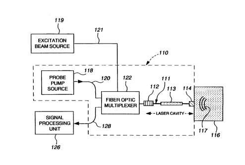
In general, an opto-acoustic detector, gener-
ally indicated at 110 in Figure 2b, is particularly
useful in high frequency ultrasound array imaging. The
opto-acoustic detector 110 includes a fiber laser for
generating optical frequencies related to ultrasound
pressure waves incident on the detector. The optical
cavity of the fiber laser is modulated by incident
acoustic pressure such that changes in optical path
length and phase are induce~, modulating the optical
frequency. The optical cavity is disposed such that its
cross-sectional area perpendicular to the optical path
is less than or comparable to A2, where A is the wave-
length of the incident ultrasound.
CA 022~1336 1998-10-13
W O 97/39684 PCTrUS97/04674
The laser cavity has an optical fiber, gener-
ally indicated at 111, bounded by first and seeond
reflectors 112 and 114, respeetively, on opposite sides
of the laser cavity. The reflectors 112 and 114 are
disposed such that a single longitudinal mode is sup-
ported by the laser cavity.
The detector 110 ineludes an integrated
narrowband reflector 112 which can be placed directly in
the optical fiber 111 to limit lasing to a single longi-
tudinal mode in the absence of acoustic modulation.
The second reflector 114 is acousticallycoupled to a medium 116 supporting the ultrasound radia-
tion or waves 117.
Either reflector 112 and/or reflector 114 have
a frequency response which limits the linewidth of the
laser output, thereby enabling single mode operation.
The opto-acoustic detector also includes an
active medium 113 which is a section or portion of the
optical fiber 111 doped with a gain material that
converts incident pump to laser power.
Excitation beam power is delivered through the
laser cavity from an external excitation beam source 119
via an optical fiber 121 and coupled to the detector~s
laser cavity by a fiber-optic multiplexer 122.
Pump power is delivered to the laser cavity
from an external probe pump source 118 via an optical
fiber 120 and coupled to the detector's laser cavity by
the fiber-optic multiplexer 122. The signal power is
CA 022~1336 1998-10-13
W097/39684 PCT~S97/04674
recovered from the detector 110 through the multiplexer
122, or another output coupling device, and transmitted
to a signal processing unit 126 via another optical
fiber 128. The unit 126 is responsible for generating
an output signal corresponding to the received acoustic
pressure incident on the detector 110.
An enhancement to the ultrasound detector 110
is an acoustic signal enhancing feature. This feature
allows for greater surface displacements involving the
aforementioned broadband reflector 114. The enhanced
displacements cause greater fluctuations in the optical
path length of the fiber laser cavity, thereby increas-
ing the sensitivity of the detectors 110 as described in
detail in the above-noted application.
A further extension of the present invention
is the use of the fiber-optic ultrasound detectors 110
in high density, high frequency arrays in direct contact
with the specimen under investigation. In arrays of
this type, the equivalent ultrasound element size is
determined by the sensing area of the optical detector
llO. For the fiber-optic device, the sensing area is
essentially the cross-sectional area of the fiber core,
typically comparable to or less than ~2, even at ultra-
sound operating frequencies greater than 500 MHz. The
reduced element size permits closely spaced optical
detectors llO, enabling high density arrays for high
frequency imaging. In addition, high density fiber
arrays can deliver optical excitation pulses to create
an acoustic transmitting array. The wavelength of the
excitation pulse is selected to be different than the
lasing wavelength of the fiber cavity. Either the
mirror absorbs the excitation pulse at this wavelength,
CA 022~1336 1998-10-13
W 097/39684 PCT~US97/04674
or the mirror is transparent to this wavelength and
passes the excitation pulse to an appropriate absorber.
Referring again to Figure 2a, the basic
recording is repeated as the focused excitation pulse is
scanned over precisely the same surface probed or
scanned by the unfocused probe beam. This means that if
there are N elements in the optical detector array, then
there will be N7 recordings over a two-dimensional
aperture. In general, the number of firing positions
can be arbitrary. Acoustooptic devices, such as the
Bragg cells 23 and 25, have been omitted from Figure 2a
for purposes of simplicity.
A slight variation of the system of Figure 2a
is illustrated by the system in Figure 3, which utilizes
acoustic detection. Optical detection of acoustic
pulses is inherently insensitive. Although it is
anticipated that complete optical detection will have
sufficient signal to noise ratio to permit high frequen-
cy microscopic imaging, for applications needing high
sensitivity, direct piezoelectric detection can be used.
A highly focused, single element conventional ultrasonic
microscope transducer 22 is used in the system of Figure
3 to detect the reflected ultrasonic wave. The trans-
ducer 22 is focused onto the surface of the opto-acous-
tic transducer 14, where the focal spot is confined toa region comparable to (A)2 (i.e., f/number of about 1
for the ultrasonic lens). The transducer 22 is scanned
in a plane parallel to the face of the opto-acoustic
transducer 14 recording signals equivalent to the
scanned optical probe beam system of Figure 1.
CA 022~l336 l998-l0-l3
W O 97/39684 PCTrUS97/04674
-14-
A schematic view of the three-dimensional
acoustic microscope system using the optical excitation
technique described with reference to Figures 1, 2a, 2b
and 3 is presented in Figure 4. A radio-frequency
waveform (100-1000 MHz, depending on the microscope
application) output from either the optical detector 20
or the ultrasonic transducer 22 iS digitized using a
waveform recorder or digitizer 24. The output of this
recorder 24 for each firing is stored in a memory 26
until the entire surface of the opto-acoustic transducer
14 has been scanned. These data are then reconstructed
using 3-D synthetic aperture beam forming equations
within an appropriately programmed computer as indicated
at block 28 to permit complete spherical focus on both
transmit and receive. Such reconstruction routines are
currently used in low-frequency ultrasonic imaging with
1-D arrays. These standard methods can be easily
extended for 2-D arrays, resulting in full 3-D recon-
structions.
The output of the reconstruction hardware is
then displayed using a conventional 3-D display system
30 currently employed in medical imaging. The entire
data acquisition, reconstruction and display system
should be synchronized by a master scan controller 32.
The systems shown in Figures 1-3 are conceptu-
ally simple. Nevertheless, they produce truly three-
dimensional images, with high resolution maintained over
a large depth of field. Such systems can have a dramat-
ic impact on the current practice of clinical pathology,
especially if they can both provide detailed information
equivalent to the highest performance optical micro-
scopes currently in routine use and be incorporated into
CA 022~1336 1998-10-13
W 097/39684 PCT~US97/04674
the tip of a conventional biopsy needle for in situ,
real-time imaging. The first and second acoustooptic
devices, Bragg cells 23 and 25, together with the opto-
acoustic transducer 14 define a 3 -D acoustic microscope
when placed in a housing, indicated in phantom at 27 in
Figure 1. The lines leading from the sources 19 and 21
and to the photodetector 20 may comprise a single
optical fiber and may have the same general configura-
tion as shown in Figure 2b, wherein a single optical
fiber 111 serves multiple purposes.
In addition to medical applications, there may
be several uses of this technology in non-destructive
evaluation (NDE) of materials. In such uses, there may
not be a need for an opto-acoustic transducer. The
disclosed microscopes can replace mechanically scanned
systems in all NDE applications. Moreover, in applica-
tions permitting needle insertion, such as NDE of soft
plastics, the electronic scanning capability of this
system is vastly superior to traditional mechanical
scanning.
While the best mode for carrying out the
invention has been described in detail, those familiar
with the art to which this invention relates will
recognize various alternative designs and embodiments
for practicing the invention as defined by the following
claims.
.