Note: Descriptions are shown in the official language in which they were submitted.
CA 02251674 1998-10-14
WO 97/41425 PCT/CA97/00276
BIOSENSOR DEVICE AND METHOD
Field of the Invention
The present invention relates to biosensors, in particular, to a biosensor for
measuring a
binding event between a ligand and a ligand-binding receptor, and to methods
employing such
biosensor.
Background of the Invention
To a great extent, diagnostic tools used for detecting or quantitating
biological analytes are
based on ligand-specific binding between a ligand and a receptor. Ligand-
receptor binding
pairs used commonly in diagnostics include antigen-antibody, hormone-receptor,
drug-receptor,
cell surface antigen-lectin, biotin-avidin, and complementary nucleic acid
strands, wherein said
ligand is typically the smaller of the two binding pair members. The analyte
to be detected
may be either member of the binding pair; alternatively, the analyte may be a
ligand analog
that competes with the ligand for binding to the complement receptor.
A variety of methods for detecting ligand/receptor interactions have been
developed. The
simplest of these is a solid-phase format employing a reporter-labeled ligand
whose binding to
or release from a solid surface is triggered by the presence of analyte ligand
or receptor. In
a typical solid-phase sandwich type assay, for example, the analyte to be
measured is a ligand
with two or more binding sites, allowing ligand binding both to a receptor, e.
g. , antibody,
carried on a solid surface, and to a reporter-labeled second receptor. The
presence of analyte
is detected (or quantitated) by the presence (or amount) of reporter bound to
solid surface.
In a typical solid-phase competitive binding assay, an analyte ligand {or
receptor) competes
with a reporter-labeled analyte analog for binding to a receptor (or Iigand)
carried on a solid
support. The amount of reporter signal associated with the solid support is
inversely
proportional to the amount of sample analyte to be detected or determined.
The reporter label used in both solid-phase formats is typically a visibly
detectable particle
or an enzyme capable of converting a substrate to an easily detectable
product. Simple
spectrophotometric devices allow for the quantitation of the amount of
reporter label, for
quantifying amount of analyte.
Detecting or quantitating ligand-specific binding events is also important in
high-
throughput methods being developed for combinatorial library screening. In a
typical method,
a large library of possible effector molecules (ligands) is synthesized. The
library members
are then screened for effector activity by their ability to bind to a selected
receptor. The
approach has the potential to identify, for example, new oligopeptide antigens
capable of high-
specificity binding to disease related antibodies, or small-molecule compounds
capable of
CA 02251674 1998-10-14
WO 97/41425 PCT/CA97/00276
2
interacting with a selected pharmacological target, such as a membrane bound
receptor or
cellular enzyme.
High-throughput screening methods typically employ simple ligand displacement
assays
to detect and quantitate ligand binding to a receptor. Displacement assays
have the advantage
of high sensitivity, e.g., where the displaced ligand is radiolabeled, and
also allow for the
determination of ligand-receptor binding affinity, based on competitive
displacement of a
binding agent whose binding affinity to the target receptor is known.
In both diagnostics and high-throughput screening, there is increasing
interest in
developing electrochemical biosensors capable of detecting and quantifying
ligand-receptor
binding events. Such biosensors are designed to produce electrical signals in
response to a
selected analyte-specific event, such as a ligand-receptor binding event. The
interest in
biosensors is spurred by a number of potential advantages over strictly
biochemical assay
formats, such as those discussed above.
First, biosensors may be produced, using conventional microchip technology, in
highly
reproducible and miniaturized form, with the capability of placing a large
number of biosensor
elements on a single substrate.
Secondly, because small electrochemical signals can be readily amplified (and
subjected
to various types of signal processing if desired), biosensors have the
potential for measuring
minute quantities of analyte, and proportionately small changes in analyte
levels.
A consequence of the features above is that a large number of different
analytes can be
detected or quantitated by applying a small sample volume, e.g., 10-50 ~.1, to
a single multi-
sensor chip.
Heretofore, electrochemical biosensors have been more successfully applied to
detecting
analytes that are themselves electrochemical species, or can be participate in
catalytic reactions
that generate electrochemical species, than to detecting ligand-receptor
binding events. This
is not surprising, given the more difficult challenge of converting a
biochemical binding event
to an electrochemical signal. One approach to this problem is to provide two
separate reaction
elements in the biosensor: a first element contains a receptor and bound
enzyme-linked ligand,
and the second element, components for enzymatically generating and then
measuring an
electrochemical species. In operation, analyte ligand displaces the ligand-
enzyme conjugate
from the first element, releasing the enzyme into the second element region,
thus generating
an electrochemical species which is measured in the second element.
Two-element biosensors of this type are relatively complicated to produce,
particularly by
conventional silicon-wafer methods, since one or more biological layers and
permselective
CA 02251674 1998-10-14
WO 97/41425 PCT/CA97/00276
3
layers must be deposited as part of the manufacturing process. Further,
enzymes or receptors
in the biosensor can denature on storage, and the device may have variable
"wetting" periods
after a sample is applied.
Biosensors that attempt to couple electrochemical activity directly to a
ligand-receptor
binding event, by means of gated membrane electrodes, have been proposed. For
example,
U.S. Patent Nos. 5,204,239 and 5,368,712 disclose gated membrane electrodes
formed of a
lipid bilayer membrane containing an ion-channel receptor that is either
opened or closed by
ligand binding to the receptor. Electrodes of this type are difficult to make
and store, and are
limited at present to a rather small group of receptor proteins.
Alternatively, direct ligand/receptor binding may be measured electrically by
embedding
the receptor in a thin polymer film, and measuring changes in the film's
electrical properties,
e.g., impedance, due to ligand binding to the receptors. U.S. Patent No.
5,192,507 is
exemplary. Since ligand binding to the receptor will have a rather small
effect on film
properties, and since no amplification effect is achieved, the approach is
expected to have
limited sensitivity.
It would thus be desirable to provide a biosensor capable of detecting and
quantifying
ligand-binding events and characterized by: (i) direct electrochemical
conversion of the binding
event to electrical signal; (ii) a high electron flow "turnover" from each
binding event; (iii)
adaptable to substantially any ligand, and (iv) good storage characteristics
and rapid wetting
with sample application. In addition, the device should be easily produced,
and preferably
amenable to manufacture using standard microchip technologies.
summary of the Invention
One aspect of the invention is a biosensor apparatus for detecting a binding
event between
a ligand and ligand-binding receptor. An electrode in the apparatus includes
an electrode
substrate with a detection surface covered by a monolayer of hydrocarbon
chains. The chains
are anchored at their proximal ends to the detection surface, and are
sufficiently close-packed
and ordered to form an effective barrier to electron flow across the monolayer
mediated by a
redox ion species in an aqueous solution in contact with the monolayer.
The ligand whose binding to a receptor is to be detected is attached to the
distal ends of
a portion of the monolayer chains, such that binding of a ligand-binding
receptor to ligand
perturbs the monolayer sufficiently to measurably increase electron flow
across the monolayer
mediated by such redox ion species.
CA 02251674 1998-10-14
WO 97/41425 PCT/CA97100276
4
The aqueous solution of redox species in contact with the monolayer is held in
a chamber
that is also designed to receive sample receptor, to bring the receptor into
contact with ligand
on the monolayer. Ion-mediated electron flow across said monolayer, in
response to binding
events occurring between said receptor and ligand, is measured in an
electrical circuit in the
apparatus.
In a preferred embodiment, the monolayer is composed of 8-22 carbon atom
chains
attached at their proximal ends to the detection surface, e.g., a gold
surface, by a thiolate
linkage. The chains have a preferred molecular density of about 3 to 5
chains/nm2.
The dielectric constant of the monolayer in the presence of the solution of
redox species,
but in the absence of the binding receptor, is preferably less than about 2,
with a change in the
dielectric constant of 10% or more, by receptor binding to the ligand, being
readily detectable.
Exemplary ligand-receptor pairs include antigen-antibody, hormone-receptor,
drug-
receptor, cell-surface antigen-lectin, biotin-avidin, substrate/antibody and
complementary
nucleic acid strands, where the ligand is typically the first-named of these
pairs. Where the
apparatus is used to detect a ligand or analog of the ligand, the apparatus
may further include
a receptor which competes with the analyte ligand or analog for binding to the
ligand on the
monolayer. One exemplary ligand is an oligosaccharide ligand, and one
exemplary receptor,
the Verotoxin receptor, also known "Shiga-like toxin".
The electrode employed in the biosensor may be prepared, in accordance with
another
aspect of the invention, by (i) subjecting the conductive metal surface of the
electrode substrate
to mild oxidation conditions, (ii) adding to the substrate, a solution of
hydrocarbon chains
having lengths between 8-22 carbon atoms and derivatized at one chain end with
a thiol group,
and (iii) applying a positive potential to the electrode. The potential placed
on the electrode
is preferably at least 250 mV vs NHE (normal hydrogen electrode), in a
soiution containing
the alkyl thiol to be deposited, and electrolytes including lithium ion and
perchlorate anions.
A selected portion of the hydrocarbon chains are derivatized at their ends
opposite the thiol
group, with the ligand of interest. The oxidative conditions applied to the
electrode surface are
such as to produce deposition of a monolayer of close-packed, oriented chains
on the substrate,
as .evidenced by the ability of the electrode to form an effective barrier to
electron ion flow
across the monolayer mediated by a redox ion species in an aqueous solution in
contact with
the monolayer.
In another general embodiment of the biosensvr apparatus, ligand molecules are
attached
to the hydrocarbon chains forming the monolayer in the electrode through a
heterodimer-
subunit complex composed of first and second peptides that together form a-
helical coiled-coil
CA 02251674 1998-10-14
WO 97/41425 PCT/CA97/00276
heterodimer, where: {i) the first peptide is covalently bound to the electrode
surface through
a spacer, such as an oligopeptide or hydrocarbon chain; (ii) the ligand is
covalently attached
to the second peptide; (iii) binding of the second peptide to the first
peptide, to form such
complex, is effective to measurably reduce the electron flow across the
monolayer mediated
5 by such redox ion species, relative to electron flow observed in the
presence of the first peptide
alone; and (iv) binding of a ligand-binding receptor to the ligand, with such
forming part of
said complex, is effective to measurably increase the electron flow across of
the monolayer
mediated by such redox species.
Also contemplated is an electrode for use in a biosensor apparatus of this
type, composed
of a substrate having a detection surface and ligand molecules attached to
surface through an
a-helical coiled-coil heterodimer of the type detailed above.
1fie electrode just described can be produced, in accordance with another
aspect of the
invention, by contacting together: (a) a detection surface having attached
thereto, a first
heterodimer-subunit peptide, and (b) a second heterodimer subunit capable of
binding to the
first subunit to form an a-helical heterodimer, and having a covalently
attached ligand capable
of binding specifically to such ligand-specific receptor.
These and other objects and features of the invention will become more fully
apparent
when the following detailed description of the invention is read in
conjunction with the
accompanying drawings.
Brief Description of the Drawings
Fig. 1 is a simplified, partly schematic view of the a biosensor apparatus
constructed in
accordance with the invention;
Fig. 2 is an enlarged view of a region the electrode in the biosensor shown in
Fig. 1;
Figs. 3A-3C illustrate three methods for forming a biosensor electrode having
a lipid
monolayer and attached ligand molecules, in accordance with the invention;
Fig. 4. is a plot of monolayer thickness as a function of applied voltage in
an electrode
monolayer formed in accordance with the method illustrated in Fig. 3B;
Fig. 5 illustrates the triggering of conductance by receptor-ligand
interaction on a
biosensor electrode, in accordance with the invention;
Figs. 6A and 6B illustrate the perturbation of lipid monolayer structure with
binding of
PAK peptide to disaccharide ligands on a monolayer;
CA 02251674 1998-10-14
WO 97/41425 PCT/CA97/00276
6
Fig. 7 shows plots of changes in oxidation (solid circles) and reduction (open
squares)
current of Fe(CN)63/°' as a function of time after addition of PAK
peptide to the monolayer
illustrated in Figs. 6A and 6B;
Figs. 8A-8C illustrate the perturbation of lipid monolayer structure with
binding of
Verotoxin to trisaccharide ligands on a monolayer;
Fig. 9 shows plots of changes in oxidation (solid circles) and reduction {open
squares)
current of Fe(CN)6~h' as a function of time after addition of Verotoxin to the
monolayer
illustrated in Figs. 8A and 8B;
Fig. 10 is a plot of electrode current of Fe(CN)6''/'' as a function of
temperature in a
monolayer electrode constructed in accordance with the invention;
Figs. 11A and 11B demonstrate ion gating effects with a negatively charged
ligand in an
electrode monolayer;
Figs. 12A and 12B demonstrate ion gating effects with a positively charged
ligand in an
electrode monolayer;
Figs. 13A and 13B illustrate the structure of an electrode monolayer having an
embedded
K coil peptide subunit (13A), and an embedded K coil/E coil heteroduplex;
Fig. 14 shows the change in oxidation (solid circles) and reduction (open
squares) current
as a function of time after addition of E coil peptide subunit to an electrode
of the type
illustrated in Fig. 13A containing an embedded K coil peptide subunit;
Fig. 15 shows changes in oxidation of Fe(CN)6''/' (open circles) and reduction
(open
squares) as a function of time after addition of PAK peptide to an electrode
containing di-
saccharide ligands on a K coil/E coil lipid monolayer;
Fig. 16 shows changes in oxidation of Fe(CN)'~/'' (open circles) and reduction
(open
squares) as a function of time after addition of Verotoxin peptide to an
electrode containing
trisaccharide ligands on a K coil/E coil lipid monolayer;
Figs. 17A-17E show a synthetic pathway used for producing a trisaccharide-
hydrocarbon
conjugate employed in the monolayer shown in Figs. 8A-8C; and
Fig. 18 shows a synthetic pathway used in producing a disaccharide-hydrocarbon
conjugate
employed in the monolayer shown in Figs. 6A and 6B.
Detailed Description of the Invention
A. Biosensor Apparatus
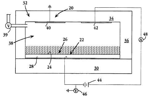
Fig. 1 is a simplified schematic view of a biosensor apparatus 20 for
detecting a binding
event between a ligand and a ligand-binding receptor or agent, in accordance
with the
CA 02251674 2004-03-30
-_
WO 97/41d25 PCT/CA97/00276
7
invention. The apparatus includes a working electrode 22 having a conductive
detection surface
24, and a hydrocarbon-chain monolayer 26 formed on the detection surface. In
the
embodiment shown, the detection surface is the upper surface of a conductive
film 28 deposited
on an electrode substrate 30, which may be non-conductive material. Details of
the monolayer
formed on the detection surface, and the method of forming the monolayer on
the surface, are
discussed below.
A cover 32 in the apparatus has an upper wall 34, and side walls, such as wall
36, which
are joined to edge regions of the electrode substrate to form a closed chamber
38 therewith.
The chamber serves to hold an aqueous electrolyte solution required for
biosensor operation,
as will be described. Liquid may be introduced into or withdrawn from the
chamber through
a valued port 39 as shown. Although not shown, the chamber may include a
second port or
vent to facilitate liquid flow through the port.
A reference electrode 4(? and a counter electrode 42 in the apparatus are
carried on the
chamber-facing surface of wall 34, as shown, and are thus both in conductive
contact with
electrode 22 when the chamber is filled with electrolyte solution. The
reference electrode,
which is held at ground, serves as the voltage potential reference of the
working electrode,
when a selected potential is placed on the working electrode by a voltage
source 44. This
potential is measured by a voltage meaning device 46 which may additionally
include
conventional circuitry for maintaining the potential at a selected voltage,
typically between
about -500 to + 800 mV .
Voltage source 44 is connected to counter electrode 42 through a current
measuring device
48 as show, for measuring current flow between the two electrodes during
biosensor operation.
The reference and counter electrodes are Pt, Ag, Ag/AgCI, or other suitable
electrodes. The
reference and working electrodes, and the circuitry connecting them to the
working electrode,
are also referred to herein, collectively, as means for measuring ion-mediated
electron flow
across the working-electrode monolayer, in response to ligand-receptor binding
events
occurring at the monolayer surface.
Fig. 2 is an enlarged view of a portion of the working electrode, including
the electrode
monolayer, showing individual hydrocarbon chains, such as chains 50, forming
the monolayer,
and ligand molecules, such as molecules 52, covalently attached to distal ends
of the
hydrocarbon chains. The ligand employed in the biosensor is a selected binding
partner in a
ligand/receptor binding pair, where the analyte to be detected is related to
one of the two
binding partners. Ligand-receptor binding pairs used commonly in diagnostics
include antigen-
antibody; hormone-receptor, drug-receptor, cell surface antigen-lectin, biotin-
avidin, and
CA 02251674 1998-10-14
WO 9?/41425 PCT/CA97/00276
8
complementary nucleic acid strands, where the ligand is typically the smaller
of the two binding
pair members. The analyte to be detected may be either member of the binding
pair, or
alternatively, a ligand analog that competes with the ligand for binding to
the complement
receptor.
The ligand molecules are attached to distal ends of the chains through
conventional
derivatization reactions, e.g., ester, ether, amide, or sulfhydryl linkages,
according to standard
methods. The number of chains in the monolayer carrying distal-end ligands is
preferably
about 1 to 10 mole percent of the total chains, but may range from 0.01 to
100%.
The chains forming the monolayer are typically 8-22 carbon, saturated
hydrocarbon chains,
although longer chains, chains with some unsaturation, chains with non-carbon
chain atoms,
such as lipid ethers, and/or chains with minor branching, such as by non-chain
methyl groups,
may be employed, within the constraint that the chains, at a sufficient
packing density, form
a sufficiently close packed and ordered monolayer to be effective as a barrier
to electron flow,
under biosensor operating conditions, as discussed below. This density is
calculated to be
between 3-5 chains/nm2.
As an example of the variation in chain composition allowed, the embodiment of
the
invention shown in Fig. 13B has a hydrocarbon-chain monolayer that includes
coil-coil peptide
heterodimers embedded in the planar chain matrix, while still retaining a low
dielectric barrier
to ion flow through the monolayer.
In the embodiment shown, the chains are coupled to the electrode detecting
surface
through sulfhydryl linkages, although other suitable coupling groups may be
employed.
Methods for producing monolayers having suitable hydrocarbon chain densities
will now be
discussed.
B. Electrode Monola~rer Production
Figs. 3A-3C illustrate three methods for forming hydrocarbon chain monolayers
suitable
for use in the biosensor electrodes.
One approach, illustrated in Fig. 3A, involves passive diffusion of chains,
such as
hydrocarbon chains 54 and ligand-derivatized chains, such as chains 56, onto
the surface an
electrode 58, under conditions effective to couple the diffused chains to the
electrode detection
surface. The diffusion method illustrated in 3A is a two-step process. In the
first step,
hydrocarbon chains alone (in the absence of ligand-derivatized chains) are
allowed to react with
the detected surface over an extended period, e. g. , 24-48 hours, until a
selected packing density
less than full packing density is achieved.
CA 02251674 1998-10-14
WO 97/41425 PCT/CA97/00276
9
The diffusion reaction is carried out under conditions suitable for coupling
the derivatized
chains to the detection surface. Where the chains have thiol coupling groups,
and the electrode
surface is gold, the surface is subjected to mild electro-chemically oxidizing
conditions, with
a perchlorate salt present in solution, then reacted with the chains under
mildly oxidizing
conditions.
The extent of packing can be monitored, for example, by ellipsometry
measurements to
determine the thickness of the layer on the detection surface. At maximum
density, i.e.,
saturation, a given chain length will produce a given monolayer thickness. As
a guide, C~
chains produce a maximum monolayer thickness of about 30A, and shorter length
chains,
proportionately thinner monolayers. Thus, in the case of a monolayer formed of
C~ chains,
the passive buildup of the monolayer may be stopped when a 25A monolayer
thickness is
observed.
The second diffusion step involves the passive diffusion of ligand-derivatized
thiol-chains
56 onto the partially formed monolayer, indicated at 60, again under suitable
thiolate coupling
conditions, until a high-density monolayer 62 is achieved, as evidenced, for
example, by the
measured thickness of the monolayer andlor a plateauing of the thickness/time
curve.
Although this approach has been applied successfully to monolayer production
in the
invention, it suffers from two limitations. First, rather long diffusion times-
- on the order of
one to several days-- are required to reach maximum packing density. Secondly,
the percent
chains containing attached ligands is difficult to control reproducibly, so
that the final
monolayers will have variable mole percentages of ligands, and thus, different
performance
characteristics.
These limitations are substantially overcome in the method illustrated in Fig.
3B, in
accordance with another novel aspect of the invention. In this approach, a
mixture of free and
ligand-carrying hydrocarbon chains, such as chains 66, 68, respectively, at a
desired mole
ratio, are actively driven to the surface by applying a positive voltage
potential to the substrate,
here indicated at 64. In practice, the hydrocarbon chain mixture (about 1 mM
hydrocarbon
chains) in an ethanolic solution of 100 mM Li perchlorate, neutral pH, is
added placed over
the electrode, and a selected potential is applied to the electrode. The
buildup of the monolayer
can be monitored by increase in layer thickness, as above. Preferably,
however, monolayer
formation is monitored by measuring electron flow across the monolayer, e.g.,
employing the
circuit configuration shown in Fig. 1. In this case, formation of the
monolayer, indicated at
70, will be characterized by a steady drop in electrode current, until a
stable low current flow
is reached, at which point maximum chain packing has been achieved.
CA 02251674 2004-03-30
WO 97!41425 PCT/CA97/00276
'The time required to achieve saturation packing density will vary with
applied voltage, and
can be a short as 10 seconds -- that is, about 4 orders of magnitude faster
than monolayer
formation by diffusion. Fig. 4 is a plot of monolayer thickness formed using a
thiol-group C~
hydrocarbon chain under coupling conditions like those above, after 10 minutes
at the electrode
5 voltage indicated. As seen, complete or nearly complete monolayer formation
(30 A thickness)
occurs within 10 minutes at about 1 V {vs. NHE) potential and above. At lower
positive
voltages, additional reaction time is required. Preferably the voltage applied
to the electrode
is at least voltage between about +250 mV relative to a normal hydrogen
electrode (+250 vs.
NHE) and I.2V (vs. NHE),
10 Not only are rapid monolayer formation times achieved, but the percentages
of ligand- and
non-ligand chains present in the reaction mixture are precisely represented in
the monolayers,
giving highly reproducible electrode characteristics.
Fig. 3C shows a modification of the Fig. 3B method, where the hydrocarbon-
chain
mixture reacted with the electrode (indicated at 71) includes non-Iigand
chains, such as chains
72, and peptide subunit conjugates, such as indicated at 74, containing a
peptide subunit 76 that
is capable of forming a stabilized, alpha-helical peptide heterodimer with an
oppositely charged,
complementary subunit. Such heterodimer subunits are described in PCT patent
application
WO CA 95/00293, for "Heterodimer Polypeptide Immunogen Carrier Composition and
Method", publication date 23 November 1995.
Exemplary subunits are referred to herein as K coils, referring to a
positively charged subunits
whose charge is provided dominantly by lysine residues, and E coils, referring
to negatively
subunits whose charge is provided dominantly by glutamic acid residues.
In the embodiment shown, subunit 76 is attached to the distal end of a
hydrocarbon chain
78 (end opposite the chain's thiol group) by suitable Lipid-to-peptide
conjugation, e.g., by ester
linkage to a hydrocarbon fatty acid. Alternatively, and as described below,
the peptide subunit
may be linked to the electrode surface through a peptide spacer, e.g.,
tripeptide spacer that
extends from one end of the subunit and includes cysteine as a terminal
residue, for sulfhydryl
attachment to the electrode surface. In both cases, the peptide subunit
conjugate is mixed with
the hydrocarbon chains, at a selected mole ratio, then driven into a monolayer
formation by
applying a positive voltage to the electrode, as above, until a densely packed
monolayer 80 is
formed.
A suitable ligand is then attached to the monolayer by contacting the
monolayer with a
ligand-coil conjugate 82 composed of the appositely charged complement of the
monolayer coil,
indicated at 84, coupled to a selected ligand 86. The two oppositely charged
subunits
CA 02251674 1998-10-14
WO 97/41425 PCT/CA97/00276
11
spontaneously self assemble into heterodimers, effectively coupling the ligand
to the monolayers
with tie high affinity constant of the two heterodimers.
The method provides, in addition to the advantages mentioned above with
respect to Fig.
3B, a "universal" biosensor substrate which can be modified to include one of
a large number
of different ligands in the substrate monolayer, simply by contacting the
universal substrate
with a conjugate of the oppositely charged peptide subunit and the selected
ligand. In the
example shown in Fig. 3C, a universal substrate monolayer 80 is converted to a
ligand-specific
monolayer 88 by addition of the ligand-specific conjugate 82.
C. Biosensor Characteristics: Directly Attached Ligand
This section examines the dielectric properties of the biosensors of the
invention, as
evidenced by the conductance properties of the biosensor monolayer membranes
in the presence
and absence of ligand-receptor binding. The present section considers
membranes having
directly attached ligands of the type described with respect to Figs. 3A and
3B. The next
section examines similar electrical properties in biosensor membranes in which
the ligand is
attached through heterodimer peptide subunits, as described with respect to
Fig. 3C.
The basic operational features of the biosensor are illustrated in Fig. 5. The
figure shows
a biosensor electrode 90 in a biosensor apparatus of the type described in
Fig. 1, where an
electrode monolayer 92 is formed, as above, of a densely ordered array of
hydrocarbon chains
containing ligand molecules, such as molecule 94, attached to the distal ends
of some of the
chains.
The electrode is in contact with a solution of ionic species, indicated at 98,
capable of
undergoing a redox reaction, i.e., losing or gaining an electron, at a
suitably charged electrode.
Exemplary redox species are Fe(CN)6'~'~, as a negatively charged species, and
Ru(NH3)62+r~+
as a positively charged species. Other probes which can be used include
Mo(CN)6~ (Fp =
+800 mV), W(CN)6'~ (Eo=+580 mV), Fe(CN)4 (Eo=+580 mV), Ce'+'3+, (Eo=+1.4V),
and
Fe+"z+ (~_ +~6mV). Typical redox ion concentrations are between 0.01 and 10
mM. The
redox solution is contained in chamber, like chamber 38 in Fig. 1, and is in
contact with
reference and counter electrodes.
The voltage potential placed on the electrode, i. e. , between the electrode
and reference
electrode, is typically at least 90 mV above the electrochemical potential
(en) value of the redox
species, for oxidation, and at least 90 mV below the electrochemical
potential, for reduction
of the species. Consider, for example, Fe(CN)6''", with an Eo of 450 mV (vs.
NHE). Above
about 550 mV electrode potential, any Fe2+ species is oxidized to Fe3+, and at
an electrode
CA 02251674 1998-10-14
WO 97/41425 PCT/CA97/002'76
12
potential below about 350 mV, and Fe+3 is reduced to Fe+2. Similarly,
Ru(NH3)6z+rs+ h~
an Eo of +50 mV (vs. NHE), so oxidation is achieved at an electrode potential
above about
+150 mV, and reduction, below about -50 mV.
In the absence of receptor binding to the ligand, the monolayer retains its
dense ordered
packing, forming an effective barrier to electron flow across the monolayer
mediated by the
redox ion species, when a suitable oxidizing or reducing potential is placed
across the
monolayer. This is reflected by a low or zero measured current across the
membrane. The
dielectric constant of the monolayer in this condition is typically about 1-2.
With binding of a receptor 96 to a ligand on a monolayer, as shown at the
right in the
figure, the ordered structure of the monolayer is perturbed sufficiently to
allow the movement
of redox species through the monolayer, producing electron flow through the
electrode.
Measurements performed in support of the invention indicate that one
triggering event leads
to 102 to 106 ionic and electron transfer events per second, and thus is
highly multiplicative.
The biosensor records this binding event as an increase in current across the
electrode, i.e.,
between the working and counter electrodes.
By analogy to a transistor, the redox solution serves as the "source", the
monolayer as the
"gate", and the underlying electrode as the "drain". Current flow in a
transistor is initiated by
applying a threshold voltage to the gate. In the biosensor of the invention,
current flow is
initiated by a stimulus-- in this case, a ligand-receptor binding event-- to
the monolayer "gate" .
A biosensor electrode 100 constructed in accordance with the invention, and
having a
disaccharide ligand indicated at 102 is shown before and after receptor
binding in Figs. 6A and
6B, respectively. Synthesis of the disaccharide-hydrocarbon chain used in the
membrane is
described in Examples iD and lE. The electrode was prepared as described with
reference to
Fig. 3B, employing a ratio of non-ligand to ligand-chains of about 4 to 1. The
disaccharide
is specifically reactive with a Pseudomonas PAK peptide, indicated at 104,
forming a ligand-
receptor pair with the peptide.
The increase in biosensor electrode current, when PAK peptide receptor is
added to the
biosensor chamber, is seen in Fig. 7 for both oxidation (solid circles) and
reduction (open
squares) current from Fe(CN)''I~'. The increase over time presumably reflects
the kinetics of
binding, demonstrating that the biosensor is useful as well in measuring the
rate ligand-receptor
binding events. Fig. 6B illustrates the perturbation of the hydrocarbon chain
structure with
receptor binding.
CA 02251674 1998-10-14
WO 97/41425 PCT/CA97/00276
13
As another example, the biosensor electrode illustrated in Figs. 8A-8C
(electrode 22 from
Fig. 2) has a trisaccharide ligand 52 which is shown before and after receptor
binding in Fig.
6A and Figs. 6B and 6C, respectively. Synthesis of the trisaccharide-
hydrocarbon chain used
in the membrane is described in Examples 1B and 1C. The electrode was prepared
as
described with reference to Fig. 3B, employing a ratio of non-ligand to ligand-
chains of about
4 to 1. The disaccharide is specifically reactive with a Verotoxin, indicated
at 106, forming
a ligand-receptor pair. Verotoxin was prepared as described in Example 2.
Figs, 8B and 8C illustrate two possible binding configurations. The
configuration in Fig,
8B has little effect on the monolayer structure, and hence on biosensor
current, because binding
is "remote" from the membrane surface; the configuration illustrated in Fig.
8C, by contrast,
produces significant perturbation of the monolayer structure, and thus would
be expected to
significantly enhance biosensor current.
The oxidation and reduction current plots shown in Fig. 9 demonstrate that
Verotoxin
binding to the membrane does in fact produce a major change in monolayer
structure. As seen,
both oxidation and reduction current increase from near-zero levels, in the
absence of
Verotoxin, to a level in the p.Amp range an hour after Verotoxin is introduced
into the
biosensor.
In the examples above, the stimulation of biosensor current by receptor
binding may be
the result of (i) steric perturbation of the monolayer chains, as indicated in
Figs. 6B and 8C,
{ii) charge effects on the monolayer surface due to charged groups on the
receptor, or (iii) a
combination of the both effects. Studies conducted in support of the invention
indicate that
both effects can be operative.
The effect of hydrocarbon-chain disruption in the biosensor monolayer, was
examined by
plotting biosensor current as a function of electrode temperature. If lipid-
chain disruption leads
to greater electron flow in the biosensor, raising the temperature of the
monolayer, and thus
the motion of the lipid chains, should increase measured electron flow
mediated by redox
carriers. This was in fact observed, as seen in Fig. 10. The
current/temperature plot has a
peak corresponding to the phase transition temperature of the monolayer chains
(about 55°C),
consistent with the idea that maximum lipid disruption occurs at the point of
maximum extent
of phase boundaries in the hydrocarbon chains.
The effect on conductance of charge on the monolayer surface can be seen from
Figs. 11
and 12. In the study represented in Fig. 11A, a negatively charged ligand was
attached to the
distal ends of a portion of the chains forming the monolayer. In the figure,
the electrode is
indicated at 108, the monolayer, at 110, chains forming the monolayer, at 112,
and chain-
CA 02251674 1998-10-14
WO 97141425 PCT/CA97/00276
14
attached ligands, at 114. Electrode current was measured for the negatively
charged redox
species Fe(CN)6'''°', and independently, with the positively charged
species Ru(NH,)62+~'+, at
oxidation potentials indicated above.
As seen in Fig. 11B, the oxidation current for the positively charged species
shows the
ion-dependent behavior expected for ion migration through the monolayer,
indicating that the
monolayer is conductive to positively charged redox species. In this figure,
the electrode is
indicated at 116, the monolayer, at 118, chains forming the monolayer, at 120,
and chain-
attached ligands, at 122. Conversely, no significant electron flow was
observed with the
negatively charged redox species.
Similar results were obtained with a monolayer designed to contain a
positively charged
surface ligand, as illustrated in Fig. 12A. In this case, ion-dependent
current was observed for
oxidation of the negatively charged iron redox species, but not the positively
charged ruthenium
species.
D. Biosensor Characteristics: Heterodimer Attached Li,Qand
In another embodiment, the ligand in the biosensor is anchored to biosensor
surface, i.e.,
embedded within the hydrocarbon-chain monolayer, by a coiled-coil heterodimer
complex
formed of two subunit peptides.The heterodimer-subunit peptides employed in
the biosensor
invention are two non-identical, preferably oppositely charged polypeptide
chains, typically
each about 21 to about 70 residues in length, having an amino acid sequence
compatible with
their formation into two-stranded a-helical heterodimeric coiled-coils. They
are designated
herein as HSPI (heterodimer-subunit peptide 1), and HSP2 (heterodimer-subunit
peptide 2).
In the discussion below, HSP1 will refer to the peptide attached to the
biosensor surface in the
biosensor, and HSP2, to the peptide having an attached ligand. It will be
understood that these
designations refer to the functional role played by the subunit peptide,
not~the actual peptide
sequence.
In aqueous medium, the isolated heterodimer-subunit peptides are typically
random coils.
When HSP1 and HSP2 are mixed together under conditions favoring the formation
of a-helical
coiled-coil heterodimers, they interact to form a two-subunit a-helical coiled-
coil heterodimeric
complex.
Peptides in an a-helical coiled-coil conformation interact with one another in
a
characteristic manner that is determined by the primary sequence of each
peptide: The tertiary
structure of an a-helix is such that 7 amino acid residues in the primary
sequence correspond
to approximately 2 turns of the a-helix. Accordingly, a primary amino acid
sequence giving
SUBSTITUTE SHEET (RULE 26)
CA 02251674 2004-03-30
WO 97141425 PCTlCA9710027.6
rise to an a-helical conformation may be broken down into units of 7 residues
each, termed
heptads. The heterodimer-suhunit peptides are composed of a series of heptads
in tandem.
When the sequence 'of a heptad is repeated in a particular heterodimer-subunit
peptide, the
heptad may be referred to as a "heptad repeat", or simply "repeat".
5 Specific types of amino acid residues at deftned positions in each heptad
act to stabilize
the two-stranded a-helical coiled-coil heterodimeric structure or complex. The
heterodimer
peptides may also contain_ residues. that can be reacted (either intro- or
inter-helically) to
stabilize the a-helical or coiled-coil nature of the polypeptides. One example
of a stabilizing
modification is the incorporation of lactam bridges in the first and last
(terminal) repeats of
10 heterodimer-suhunit . peptides, as detailed in PCT application WO
CA95/00293 for
"Heterodimer Polypeptide Immunogen Carrier Composition and Method",
publication date 23
November 1995.
The dimerization of HSP1 and HSP2 is due to the presence of a repeated heptad
motif of
conserved amino acid residues in each peptide's primary amino acid sequence.
Repeating
15 heptad motifs having appropriate amino acid sequences direct the HSP 1 and
HSP2 palypeptides
to assemble into a heterodimeric a-helical coiled-coil structure under
permissible conditions.
The individual a-helical peptides contact one another along their respective
hydrophobic faces.
HSP1 and HSP2 may assemble into a heterodimer coiled-coil helix (coiled-coil
heterodimer) in either parallel or antiparallel configurations. In a parallel
configuration, the
two heterodimer-subunit peptide helixes are aligned such that they have the
same orientation
(amino-terminal to carboxyl-terminal). In an antiparallel configuration, the
helixes are arranged
such that the amino-terminal end of one helix is aligned with the carboxyl-
terminal end of the
other helix, and vice versa.
Heterodimer-subunit peptides designed in accord with the guidance presented in
the above
PCT application typically show a preference for assembling in a parallel
orientation vs. an
antiparallel orientation. Por example, the exemplary peptides identified by
SEQ ID NO:1 and
SEQ ID N0:2 in the above CA95/00293 PCT patent application, form parallel-
configuration
heterodimers as do other peptide sequences discussed in the PCT application.
When attaching
a ligand to HSP2; it is generally desirable to attach the ligand at or near
the end of the peptide
that will forrii the distal end of the heterodimer. In particular, where the
heterodimer forms
a parallel configuration, the HSP1 peptide is preferably anchored to the
biosensor surface at
its C terminus, and the ligand attached to the HSP2 peptiiie at its N
terminus.
As just noted, one of the two subunit peptides (HSP1) in the heterodimer is
attached to the
biosensor surface, and the second peptide (HSP2) contains a ligand intended to
participate in
CA 02251674 1998-10-14
WO 97/41425 PCT/CA97/00276
16
an analyte-dependent ligand/anti-ligand binding reaction. In both cases, the
peptide is
synthesized, or derivatized after synthesis, to provide the requisite
attachment function and
ligand, respectively.
Considering the modification of HSP1, the peptide may be synthesized, at
either its N or
C terminus, to carry additional terminal peptides that can function as a
spacer between the
biosensor surface and the helical-forming part of the peptide. Alternatively,
the HSP1 peptide
can be attached to the biosensor surface thorough a high-affinity binding
reaction, such as
between a biotin moiety carried on the peptide and an avidin molecule
covalently attached to
the surface.
Where the heterodimer is embedded in a hydrocarbon-chain monolayer, as
described
below, the spacer anchoring the HSP1 peptide to the biosensor surface may be a
hydrocarbon
chain. The chain is preferably a fractional length of the chains making up the
bilayer, such that
the distal ends of the heterodimer peptides in the assembled monolayer are at
or near the
exposed surface of the monolayer. Thus, for example, if the monolayer is made
up of 18-
carbon chains, the spacer is preferably 2-10 carbons in length, depending on
the length of the
assembled heterodimer.
The hydrocarbon-chain spacer, in the form of a omega-thio fatty acid, may be
coupled to
a terminal hydroxyl or amine coupling during solid-phase synthesis, as
outlined above. The
derivatized peptide, in turn, can be attached to a metal surface by standard
thiolate coupling
(Dakkouri, et al., Langmuir (1996) 12:2849-2852). supra).
Considering the ligand-attachment to HSP2, the ligand selected will be
determined by the
analyte to be tested. Ligand-receptor binding pairs, i.e., ligand/ligand-
binding agent pairs used
commonly in diagnostics include antigen-antibody, hormone-receptor, drug-
receptor, cell
surface antigen-lectin, biotin-avidin, substrate/enzyme, and complementary
nucleic acid strands.
The ligand is typically the smaller of the two binding pair members,
particularly where the
ligand is attached to a hydrocarbon-chain monolayer, as described below.
However, attachment
of either binding pair is contemplated herein.
Where the ligand is a polypeptide, e.g., peptide antigen, the antigen can be
synthesized
by either solid-state or recombinant methods, to include the peptide antigen
at the end of the
HSP2 peptide that will orient distally in the assembled heterodimer. Where the
ligand is a non-
peptide moiety, e.g., a non-peptide hormone, drug, or nucleic acid, the HSP2
peptide can be
synthesized to include one or more residues that can be specifically
derivatized with the ligand.
The ligand is preferably covalently attached to the N-terminal amino acid
residue, or to one or
the residues facing the exposed face of the heterodimer. Preferred coupling
groups are the thiol
SUBSTITUTE SHEET (RULE 26)
CA 02251674 2004-03-30
_-
WO 97141425 PCTlCA97/00276
17
groups of cysteine residues, which are easily modified by standard methods.
Other useful
coupling groups include the thioester of methionine, the imidazolyl group of
histidine, the
guanidinyl group of arginine, the phenolic group of tyrosine and the indolyl
group of
tryptophan. These coupling groups can be derivatized using reaction conditions
known to those
skilled in the art.
'fo attach the ligand-derivatized HSP2 peptide to the surface-immobilized HSP1
peptide,
the two peptides are contacted under conditions that favor heterodimer
formation. A medium
favoring coiled-coil heterodimer formation is a physiologically-compatible
aqueous solution
typically having a pH of between about 6 and about 8 and a salt concentration
of between about
50 mM and about 500 mM. Preferably, the salt concentration is between about
100 mM and
about 200 mM. An exemplary benign medium has the following composition: 50 mM
potassium phosphate, 100 mM KCI, pH 7. Equally effective media may be made by
substituting, for example, sodium phosphate for potassium phosphate and/or
NaCI for KCI.
Heterodimers may form under conditions outside the above pH and salt range,
medium, but
some of the molecular interactions and relative stability of heterodimers vs.
homodimers may
differ from characteristics detailed above. For example, ionic interactions
between the ionic
groups that tend to stabilize heterodimers may break down at low or high pH
values due to the
protonation of, for example, Glu side chains at acidic pH, or the
deprotonation of, for example,
Lys side chains at basic pH. Such effects of low and high pH values on coiled-
coil heterodimer
formation may be overcome, however, by increasing salt concentration.
Increasing the salt concentration can neutralize the stabilizing ionic
attractions or suppress
the destabilizing ionic repulsions. Certain salts have greater efficacy at
neutralizing the ionic
interactions. For example, in the case of the K-coil peptide in Fig. 2A, a IM
or greater
concentration of CIO' anions is required to induce maximal a-helical
structure, whereas a 3M
or greater concentration of Cl- ions is required for the same effect: The
effects of high salt on
coiled-coil formation at low and high pH also show that interhelical ionic
attractions are not
essential for helix formation, but rather, control whether a coiled-coil tends
to form as a
heterodimer vs. a homodimer.
Fig. 13A shows a biosensor electrode 124 in which the hydrocarbon chain
monolayer,
indicated at 126 includes a K coil peptide subunits, such as subunit 128, as
described above. In the
embodiment shown, each peptide subunit is coupled to the electrode surface via
a tripeptide
spacer, such as spacer 130 in subunit 128, which is itself attached to the
electrode surface
through a sulfhydryl linkage, as shown. The peptide, including the peptide
spacer, is formed
conventionally, e.g. , by solid phase synthesis. The amount of peptide subunit
in the monolayer
CA 02251674 1998-10-14
WO 97/41425 PCT/CA97/00276
L8
is about 20 mole percent. The monolayer was formed according to the method
described above
with respect to Fig. 3C. As indicated above, the peptide subunit may
alternatively be coupled
to the distal ends of a portion of the hydrocarbon chains in the monolayer,
placing the subunit
more on the monolayer surface. A hydrocarbon chain-peptide conjugate suitable
for this
application may be made, for example, by attaching an activated-end
hydrocarbon chain to the
terminal amino acid of the peptide, as the terminal step in solid phase
synthesis.
Presumably because of the positive charge imparted to the monolayer by the K
coil
subunits, the monolayer shows relatively high conductance to negatively
charged redox species,
such as Fe(CN)6~, as evidenced by a relatively high oxidation or reduction
current with the
redox species.
Fig. 13B shows the same monolayer, but after addition of complementary,
negatively
charged E coil subunits, such as indicated at I30. As shown, oppositely
charged subunits pair
to form charge-neutral heterodimers in the monolayer. This pairing is
effective to reduce
monolayer conductance substantially, as evidenced by the time-dependent fall
in measured
oxidation or reduction current in the presence of Fe(CN)6~ ions (Fig. 14).
As shown in Fig. 3C, the second peptide subunit, e. g., the E coil subunit,
added to the
monolayer may be derivatized with a ligand, producing a monolayer having
charge-neutral
heterodimers embedded therein (or attached to the monolayer surface), and a
ligand exposed
on the monolayer surface. The resulting electrode is effective to measure
ligand-specific
receptor binding events in a biosensor operated in accordance with the
invention.
The operating characteristics of such a biosensor are illustrated in Fig. 15.
The electrode
in this biosensor includes (i) a monolayer with embedded K coils covalently
attached to the
electrode surface, (ii) complementary E coils forming heterodimers with the K
coils in the
mono~ayer, and (iii) surface disaccharide ligands of the type shown in Fig. 6
attached
covalently to the E coils and disposed therefore at the monolayer surface. As
seen in Fig. 15,
addition of the PAK protein receptor (see Fig. 6B) produces a increase in both
oxidation and
reduction currents, with the current increase over time presumably reflecting
additional binding
events after receptor addition to the biosensor electrode.
A similar biosensor having a trisaccharide, rather than disaccharide, ligand
attached to the
E coil subunit in the electrode monolayer was tested with the Verotoxin
receptor described
above with respect to Figs. 8A-8C, with the results seen in Fig. 16. The solid
lines in the
figure show the increase in oxidation and reduction current observed, as a
function of time,
after addition of Verotoxin.
The following examples are intended to illustrate, but in no way limit the
invention.
SUBSTITUTE SHEET (RULE 26)
CA 02251674 2004-03-30
WO 97/41425 PCT/CA97/00276
l9
Example 1
synthesis of Receptors in a Form Suitable For Immobilization on a Gold
Electrode
Refering to Figures 17A-E and 18, selective tosylation of 1,16-dihydroxyhexane
provided
the monotosylated alcohol 1 in 42% yield.
Trisaccharide~ 2, obtained as described in the literature (Janson, et al. , J.
Org. Chem.
X3,:5629 (1988)), was converted into an anomeric mixture of
trichloroacetamidates 3.
Glycosylation of alcohol 1 with glycosyl donor 3 in CH~CI= in the presence of
a catalytic
amount of trimethylsilyl trifluoromethanesulfonate gave
trisaccharide.glycoside c~-tosylate 4;
which was used in the next step without purification. The tosyloxy group of
compound 4 was
displaced by thiocyanate to provide the trisaccharide glycoside 5, terminated
at the reducing
end by spacer-arm containing the masked thiol function. Reduction of
thiocyanate by the action
of sodium borohydride (Olsen, R.K., and Snyder, H.R., J. Org. Chem. ~:I84
(1965).)
followed by saponification of acetate groups gave trisaccharide receptor 6.
The disaccharide imidate 7 was reacted with alcohol 1 in a similar fashion to
that described
for the trisaccharide 4. Synthesis of the disaccharide glycosyl donor 7 is not
described here
but follows established methods that are considered a general art.
Nucleophilic substitution of
the tosyloxy group by thiocyanate was carried out as described for preparation
of 5 to give
compound 8. Reduction of thiocyanate accompanies by deacetylation afforded
synthetic
disaccharide receptor 9.
A. 16-~p-Toluensulfonxloxvlhexadecanol (Structure 17
To a solution of 1.1 g of 1;16-dihydroxyhexadecane in 10 ml of dry pyridine
0.8 g of
tosyl chloride was added. After 2 h mixture was concentrated diluted with ZO
ml of acetone,
5 g of SiO~ was added and acetone was removed in vacuum. The solid was applied
on Si02
and eluted with pentane-ethyl acetate (2:1) to yield 748 mg (42~%) of.C-101.
B. ~,3 4.6-tetra-O-acet-yl-D- al~actopyranasvl(al--~4 -6-O-acet3rl-2.3-di-O-
benzovl-D-
g_alactopyranosy1f61-i41-2.3.6-tri-O-~enzoyl-D- IQ ucopvranosyl(61-~Ol-(16-
thiocyanolhexadecanol (Structure 5)
A mixture of 277 mg of imidate, 100 mg of C-101 and 0.5 g of mol. sieves (4A)
was
stirred for 1 h. Then 8 ~d of TMSOTf was added. After 2 h 1 ml of EA was
added, solid was
removed by filtratioil. Filtrate was concentrated and dried in vacuum. A
solution of the
residue and 200 mg of KSCN in 6 ml of DMF was stirred at-80°C for 2
hours. Mixture was
concentrated, dissolved in 30 ml of CHZCI:, washed with water and concentrated
again.
Chromatography of the residue on Si0= with pentane-ethyl acetate (3:2) gave
225 ml (739'0) of
C-105.
CA 02251674 2004-03-30
WO 97/41425 PCT/CA97/00276
C. D-Galacton~rano~yt(al-~4)-D-~alactop r~anosvl(fii--41-D-glucop ranosytlal-
~O)-(16-
thio)hexadecanol (Structure 6)
To a solution of 60 mg of C-I05 in 4 ml of dry MeOH -40 mg of NaHH, was added
Ar.
After stirring for 2 h at 45°C mixture was concentrated and .dissolved
under gentle reflux in
5 a solution of 50 mg of NaOH in 10 ml of water. After stirring overnight at
45°C, the mixture
was neutralized with cationite (H'-form) and applied on "Seppak" (C-18). The
cartridge was
washed with 20, 40, 50, 80, and 100 % solution of MeOH in water. Fractions of
100 % MeOH
was concentrated to give 24.4 mg (81 %) of C-106.
10 D. l16-thiocyanolhexadecanyl 4-O-(2-acetamido-3.4.6-tri-O-acetyl-2-D
galactop ry anosyl)-
2.3.6-O-acetyl-B-D-galactopyranoside C-108 (Structure 8)
Mixture of 100 mg of imidate 7, 68 mg of C-I01 and 100 mg of mol, sieves (4A)
in 5 ml
of CH2Clz was stirred for 1 h. Then 5 ~,1 of TMSOTf was added. After 2 h 1 ml
of EA was
added, solid was removed by filtration. Filtrate was concentrated and dried in
vacuum. A
15 solution of the residue and 70 mg of KSCN in 3 ml of DMF was stirred at
80°C for 2 h.
Mixture was concentrated, dissolved in 30 ml of CHZCI=, washed with water and
concentrated
again. Chromatography of the residue on Si02 with pentane-ethyl acetate (2:1)
gave 82 mg
(70% ) of C-108.
20 E. X16-thiohydroxy)hexadecanyl 4-O-(2-aeetamido-2-deaxy-B-D-
galactoBvranosyl) ~-D-
galactop3rranoside (Structure 91
A solution of 54.2 mg of 8 (C-108) and 40 mg of NaBH' in 3 ml of dry MeOH was
refluxed for 2 h then neutralized with Dowex (H+), concentrated and
chromatographed on C-18
in H20 (50:50):100) to yield 19.7 mg (52%) of C-111.
. Example 2
Isolation of Verotoxin Receptor
Shiga-like (Vero) toxin I (SLT-n was purified from Escherichia coli JM101
(pJB128) in
a simple, one step procedure using CHROMOSORB-P containing covalently coupled
synthetic
analogs of the toxin's aGal(1,4),BGaI (digalactoside) host- cell receptors
(SYNSORB-P~.
Bacteria were grown in baffled Fernbach flasks at 37°C in carbenicillin-
(50 ~,g/ml)
.x.
supplemented tryptic soy broth (TSB) containing SYNSORB-P1 (IS g/L) to bind
toxin released
from growing cells: _ Late log phase cultures were treated for 30 minutes at
37°C with
Polymyxin B sulfate (0.1 mglmL) to release intracellular SLT-I and also allow
this to bind to
the SYNSORB-P1. Next, the SYNSORB-PI was collected and washed thoroughly with
250
mM NaCI (pH 3.8) to remove cells and cellular debris. THe SLT-I was eluted
from the
~,
washed SYNSORB-P1 using 50 mM Tris base (pH 10) containing 250 mM NaCI (TN)
and
* Trade-mark
CA 02251674 2004-03-30
~ j.
WO 97!41425 PCT/CA97/00276
21
concentrated using an Amicon ultrafiltration unit. The concentrated SLT-I was
stable for weeks
at 4°C and could be frozen for extended periods of time without
appreciable loss of activity.
On average, 61 %a (n = 10, SD mean = 8, range 48~'o to 76%) of the SLT
activity in the
original Potymyxin-treated TSB cultures was recovered in the TN fraction
eluted from the
SYNSOItB-P1. SDS-polyacrylamide gel electrophoretic analysis of the SLT-I
preparation
revealed two prominent Coomassie blue-stained bands. The molecular weight of
these two
bands was calculated to be 35,000 and 7,500, respectively. The 7.5 KDz band
reacted in
western immunoblots with SLT-I but not SLT-II B subunit-specific monoclonal
antibody.
Amino terminal microsequence analysis of both bands confirmed their identity
as the A and B
subunits of SLT-I. Average yield of SLT-I was 0.32 mglL (n = 8, SD mean = 0.3,
range
0.1 to 0.8) of TSB culture and its specific activity in the Vero cytotoxicity
assay was 4.4
~.
pg/mL/CD~. The results demonstrate the utility of SYNSORB in the facile and .
rapid
purification of carbohydrate binding toxins or lectins:
Although the invention has been described with respect to various specific
embodiments
and methods, it will be appreciated that various modifications and changes can
be made without
departing from the invention.
* Trade-mark