Note: Descriptions are shown in the official language in which they were submitted.
CA 02252913 2001-08-28
-1-
PERCUTANEOUS CATHETER DIRECTED INTRAVASCULAR
OCCLUSION DEVICES
The present application is related to co-pending application Serial No.
2,194,669 filed
on July 10, 1995, and entitled "METHOD OF FORMING MEDICAL DEVICES;
INTRAVASCULAR OCCLUSION DEVICES".
BACKGROUND OF THE INVENTION
I. FIELD OF THE INVENTION
The present invention generally relates to intravascular devices for treating
certain
medical conditions and, more particularly, relates to intravascular occlusion
devices for Atrial
Septal Defects (ASD) and Patent Ductus Arteriosus (PDA) treatment. The devices
made in
accordance with the invention are particularly well suited for delivery
through a catheter or
the like to a remote location in a patient's vascular system or in analogous
vessels within a
patient's body.
II. DESCRIPTION OF THE RELATED ART
A wide variety of intravascular devices are used in various medical
procedures.
Certain intravascular devices, such as catheters and guidewires, are generally
used simply to
deliver fluids or other medical devices to specific locations within a
patient's body, such as a
selective site within the vascular system. Other, frequently more complex,
devices are used in
treating specific conditions, such as devices used in removing vascular
occlusions or for
treating septal defects and the like.
In certain circumstances, it may be necessary to occlude a patient's vessel,
such as to
stop blood flow through an artery to a tumor or other lesion. Presently, this
is commonly
accomplished simply by inserting, for example, Ivalon particles (a trade name
for vascular
occlusion particles) and short sections of coil springs into a vessel at a
desired location.
These "embolization agents" will eventually become lodged in the vessel,
frequently floating
downstream of the site at which they are released before blocking the vessel.
This procedure
is often limited in its utility, in part, due to the inability to precisely
position the embolization
agents..
Balloon catheters similar to that disclosed by Landymore et al. in U.S. Patent
No.
4,836,204 have been used by physicians to temporarily occlude a septal defect
until the
patient stabilizes enough for open heart surgical techniques. Detachable
balloon catheters
CA 02252913 1998-10-29
WO 97/42878 PCT/US97/06194
-2-
are also used to block patients' vessels. When using such a catheter, an
expandable balloon
is carried on a distal end of a catheter. When the catheter is guided to the
desired location,
the balloon is filled with a fluid until it substantially fills the vessel and
becomes lodged
therein. Resins which will harden inside the balloon, such as an
acrylonitrile, can be
S employed to permanently fix the size and shape of the balloon. The balloon
can then be
detached from the end of the catheter and left in place.
Such balloon emboIization is also prone to certain safety problems, though.
For
example, if the balloon is not filled enough, it will not be firmly fixed in
the vessel and may
rotate or drift downstream within the vessel to another location, much like
the loose
embolization agents noted above. In order to avoid this problem, physicians
may overfill the
balloons; it is not uncommon for balloons to rupture and release the resin
into the patient's
bloodstream.
Mechanical embolization devices, filters and traps have been proposed in the
past,
some of which are disclosed in King et al., U.S. Pat. No. 3,874,388; Das, U.S.
Pat. No.
5,334,217; and Marks, U.S. Pat. No. 5,108,420. The devices disclosed are pre-
loaded into
the introduces or delivery catheter and are not easily loadable by the
physician. Further,
during deployment of these devices, recapture into the delivery catheter is
difFlcult if not
impossible, thereby limiting the effectiveness of these devices.
Also, even if some of these devices prove to be effective occluders, they also
tend to
be rather expensive and time-consuming to manufacture. For example, some
intravascular
blood filters are formed of a plurality of specially-shaped legs which are
adapted to fill the
vessel and dig into the vessel walls. In making most such filters, the legs
must be individually
formed and then painstakingly attached to one another, frequently requiring
attachment by
hand, to assemble the final filter. Not only does this take significant
skilled manpower, and
hence increase the costs of such devices, the fact that each item must be made
by hand tends
to make quality control more difficult. This same difficulty and expense of
manufacturing is
not limited to such filters, but is experienced in many other intravascular
devices as well.
When using these devices to occlude an ASD, the pressure and therefore the
chance
of dislodgment of the device increases with the square of the size of the
communication.
30' Consequently, these devices have to have a very large retention skirt.
Often times, the
position of the ASD dictates the size of the retention skirt. Hence, there is
a need for an
CA 02252913 1998-10-29
WO 97!42878 PCTIUS97/06194
-3-
ASD occluder which may be made with a relatively small retention skirt. Also,
the shape of
the prior devices (for example squares, triangles, pentagons, hexagons and
octagons) require
a larger contact area, having corners which extend to the free wall of the
atria. Each time the
atria contracts (approximately 100,000 times per day), internal wires within
the prior art
devices are bent creating structural fatigue fractures in approximately 30
percent of all cases.
Furthermore, the previous devices require a French 14-16 introducing catheter,
making it
impossible to treat children affected with cogentital defects with these
devices.
Accordingly, it would be advantageous to provide a reliable embolization
device
which is both easy to deploy through a 6-7 French catheter and which can be
accurately
placed in a vessel. It would also be desirable to provide a recoverable device
for deployment
in a vessel in a patient's body which is both economical and yields
consistent, reproducible
results.
SUMMARY OF THE INVENTION
The present invention provides a reliable intravascular occlusion device which
may be
1 S formed to treat, for example, Atria! Septa! Defects (hereinafter ASD) and
Patent Ductus
Arteriosus (hereinafter PDA). When forming these intravascular devices from a
resilient
metal fabric a plurality of resilient strands is provided, with the wires
being formed by
braiding to create a resilient material which can be heat treated to
substantially set a desired
shape. This braided fabric is then deformed to generally conform to a molding
surface of a
molding element and the braided fabric is heat treated in contact with the
surface of the
molding element at an elevated temperature. The time and temperature of the
heat treatment
is selected to substantially set the braided fabric in its deformed state.
After the heat
treatment, the fabric is removed from contact with the molding element and
will substantially
retain its shape in the deformed state. The braided fabric so treated defines
an expanded
state of a medical device which can be deployed through a catheter into a
channel ir1 a
patient's body.
Further embodiments of the present invention also provide specific shapes for
medical devices which may be made in accordance with the present invention to
address
predetermined medical procedures. Such devices of the invention are formed of
a braided
metal fabric and have an expanded configuration and a collapsed configuration.
In use, a
guide catheter can be positioned in a channel in a patient's body and advanced
to position the
CA 02252913 1998-10-29
WO 97/42878 PCT/US97/06194
-4-
distal end of the catheter adjacent a treatment site for treating a
physiological condition. A
medical device, formed in a predetermined shape, and made in accordance with
the process
outlined above, can be collapsed and inserted into the lumen of the catheter.
The device is
urged through the catheter and out the distal end, whereupon, due to its
memory property it
will tend to substantially return to its expanded state adjacent the treatment
site. In
accordance with a first of these embodiments, a generally elongate medical
device has a
generally tubular middle portion and a pair of expanded diameter portions,
with one
expanded diameter portion positioned at either end of the middle portion. In
another
embodiment, the medical device is generally bell-shaped, having an elongate
body having a
IO tapered first end and a larger second end, the second end presenting a
fabric disc which will
be oriented generally perpendicular to an axis of a channel when deployed
therein.
BRIEF DESCRIPTION OF THE DRAWINGS
Figures 1A and 1B each depict a metal fabric suitable for use with the
invention;
Figure 2A is exploded side view of a molding element having inserted a length
of a
metal fabric suitable for use in forming a medical device in accordance with
the invention;
Figure 2B is an exploded perspective view of the molding element shown in
Figure
2A;
Figure 3A is a perspective view showing the molding element of Figures 2A and
2B
in a partially assembled state;
Figure 3B is a close-up view of a portion of the highlighted area of Figure 3A
showing the compression of the metal fabric in one of the molding element's
cavities;
Figure 4 is a cross-sectional view showing the molding element of Figures 2A
and 2B
in an assembled state, and having the metal fabric formed within the molding
elements
cavities;
Figure SA is a side view of a medical device in accordance with the invention;
Figure SB is an end view of a medical device in accordance with the invention;
Figures 6A-6C are a side view, an end view and a perspective view,
respectively, of a
medical device in accordance with another embodiment of the invention;
Figure 7 is a side, cross sectional view of a molding element suitable for
forming the
medical device shown in Figures 6A-6C;
CA 02252913 1998-10-29
WO 97/42878 PCT/US97/06194
-5-
Figure 8 is a schematic illustration showing the device of Figures 6A-6C
deployed in
a central shunt of a patient's vascular system;
Figure 9A is a side view of a medical device in accordance with another
alternate
preferred embodiment;
Figure 9B is an end view of the medical device shown in Figure 9A;
Figure 10A is a side view of one molding element suitable for forming the
embodiment of Figures 9A and 9B;
Figure 10B is a cross-sectional view of another molding element suitable for
forming
the embodiment of Figures 9A and 9B;
Figure I OC is a cross-sectional view of still another molding element
suitable for
forming the embodiment of Figures 9A and 9B;
Figure 11 is an enlarged, partial sectional view of an ASD device shown
stretched
and partially extending out from the lumen of a delivery catheter;
Figure 12 is a partial sectional view of a PDA device of the type shown in
Figures 6a-
6c, wherein the PDA device is shown stretched and partially extending out from
the lumen of
a delivery catheter;
Figure 13 is an enlarged side elevational view of an ASD device, shown in its
pre-
shaped configuration;
Figure 14 is a side elevational view of the ASD device of Figure 13, shown
slightly
stretched and filled with polyester fibers;
Figure 15 is a side elevational view of the ASD device of Figure 13, shown
stretched
and filled with polyester fibers;
Figure 16 is a partial sectional side elevational view of the ASD device of
Figure 13
shown positioned within an ASD of a patient's heart;
Figure 17 is an enlarged side elevational view of an alternate ASD device,
shown in
its pre-shaped configuration; and
Figure 18 is a side elevational view of the ASD device of Figure 16, shown
stretched
and filled with polyester fibers.
DETAILED DESCRIPTION OF THE PREFERRED E11~IBODIMENTS
The present invention provides a percutaneous catheter directed intravascular
occlusion device for use in shunts in patients' bodies, such as vascular
channels, urinary
CA 02252913 1998-10-29
WO 97/42878 PCT/ITS97I06194
-6-
tracts, biliary ducts and the like. In forming a medical device via the method
of the
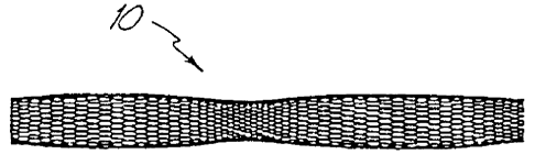
invention, a metal fabric 10 is provided. The fabric is formed of a plurality
of wire strands
having a predetermined relative orientation between the strands. Figures 1A
and 1B
illustrate two examples of metal fabrics which are suitable for use in the
method of the
invention.
In the fabric of Figure 1A, the metal strands define two sets of essentially
parallel
generally helical strands, with the strands of one set having a "hand", i.e. a
direction of
rotation, opposite that of the other set. This defines a generally tubular
fabric, known in the
fabric industry as a tubular braid. Such tubular braids are well known in the
fabric arts and
find some applications in the medical device field as tubular fabrics, such as
in reinforcing the
wall of a guiding or diagnostic catheter. As such braids are well known, they
need not be
discussed at length here.
The pitch of the wire strands (i.e. the angle defined between the turns of the
wire and
the axis of the braid) and the pick of the fabric (i.e. the number of turns
per unit length) may
be adjusted as desired for a particular application. For example, if the
medical device to be
formed is to be used to occlude the channel in which it is placed, the pitch
and pick of the
fabric will tend to be higher than if the device is simply intended to filter
bodily fluid passing
therethrough.
For example, in using a tubular braid such as that shown in Figure 1 A to form
a
device such as that illustrated in Figures SA and SB, a tubular braid of about
4 mm in
diameter with a pitch of about 50° and a pick of about 74 (per linear
inch) would seem
suitable for fabricating devices used in occluding channels on the order of
about 2 mm to
about 4mm in inner diameter, as detailed below in connection with the
embodiment of
Figures SA and SB.
Figure 1B illustrates another type of fabric which is suitable for use in the
method of
the invention. This fabric is a more conventional fabric and may take the form
of a flat
woven sheet, knitted sheet or the like. In the woven fabric shown in Figure
1B, there are also
two sets 14 and 14' of generally parallel strands, with one set of strands
being oriented at an
angle, e.g. generally perpendicular (having a pick of about 90°), with
respect to the other set.
As noted above, the pitch and pick of this fabric (or, in the case of a knit
fabric, the pick and
CA 02252913 1998-10-29
WO 97/42878 PCT/US97/06194
the pattern of the knit, e.g. Jersey or double knits) may be selected to
optimize the desired
properties of the final medical device.
The wire strands of the metal fabric used in the present method should be
formed of a
material which is both resilient and which can be heat treated to
substantially set a desired
shape. Materials which are suitable for this purpose include a cobalt-based
low thermal
expansion alloy referred to in the field as Elgeloy, nickel-based high
temperature high-
strength "superalloys" commercially available from Haynes International under
the trade
name Hastelloy, nickel-based heat treatable alloys sold under the name Incoloy
by
International Nickel, and a number of different grades of stainless steel. The
important
factor in choosing a suitable material for the wires is that the wires retain
a suitable amount
of the deformation induced by the molding surface (as described below) when
subjected to a
predetermined heat treatment.
One class of materials which meet these qualifications are so-called shape
memory
alloys. Such alloys tend to have a temperature induced phase change which will
cause the
material to have a preferred configuration which can be fixed by heating the
material above a
certain transition temperature to induce a change in the phase of the
material. When the
alloy is cooled back down, the alloy will "remember" the shape it was in
during the heat
treatment and will tend to assume that configuration unless constrained from
so doing.
One particularly preferred shape memory alloy for use in the present method is
nitinol, an approximately stoichiometric alloy of nickel and titanium, which
may also include
other minor amounts of other metals to achieve desired properties. NiTi alloys
such as
nitinol, including appropriate compositions and handling requirements, are
well known in the
art and such alloys need not be discussed in detail here. For example, U.S.
Patents
5,067,489 (bind) and 4,991,602 (Amplatz et al.), the teachings of which are
incorporated
herein by reference, discuss the use of shape memory NiTi alloys in
guidewires. Such NiTi
alloys are preferred, at least in part, because they are commercially
available and more is
known about handling such alloys than other known shape memory alloys. NiTi
alloys are
also very elastic - they are said to be "superelastic" or "pseudoelastic".
This elasticity will
help a device of the invention return to a present expanded configuration for
deployment.
In forming a medical device in keeping with the invention, an appropriately
sized
piece of the metal fabric is cut from the larger piece of fabric which is
formed, for example,
CA 02252913 1998-10-29
WO 97/42878 PCT/I1S97/06194
_g_
by braiding wire strands to form a long tubular braid. The dimensions of the
piece of fabric
to be cut will depend, in large part, upon the size and shape of the medical
device to be
formed therefrom.
When cutting the fabric to the desired dimensions, care should be taken to
ensure that
the fabric will not unravel. In the case of tubular braids formed of NiTi
alloys, for example,
the individual wire strands will tend to return to their heat-set
configuration unless
constrained. If the braid is heat treated to set the strands in the braided
configuration, they
will tend to remain in the braided form and only the ends will become frayed.
However, it
may be more economical to simply form the braid without heat treating the
braid since the
fabric will be heat treated again in forming the medical device, as noted
below.
In such untreated NiTi fabrics, the strands will tend to return to their
unbraided
configuration and the braid can unravel fairly quickly unless the ends of the
length of braid
cut to form the device are constrained relative to one another. One method
which has
proven to be useful to prevent the braid from unraveling is to clamp the braid
at two
locations and cut the braid to leave a length of the braid having clamps (15
in Figure 2) at
either end, thereby effectively defining an empty space within a sealed length
of fabric.
These clamps 1 S will hold the ends of the cut braid together and prevent the
braid from
unraveling.
Alternatively, one can solder, braze, weld or otherwise affix the ends of the
desired
length together (e.g. with a biocompatible cementitious organic material)
before cutting the
braid. Although soldering and brazing of NiTi alloys has proven to be fairly
difficult, the
ends can be welded together, such as by spot welding with a laser welder.
The same problems present themselves when a flat sheet of fabric such as the
woven
fabric shown in Figure 1B is used. With such a fabric, the fabric can be
inverted upon itself
to form a recess or depression and the fabric can be clamped about this recess
to form an
empty pocket (not shown) before the fabric is cut. If it is desired to keep
the fabric in a
generally flat configuration, it may be necessary to weld the junctions of the
strands together
adjacent the periphery of the desired piece of fabric before that piece is cut
from the larger
sheet. So connecting the ends of the strands together will prevent fabrics
formed of
untreated shape memory alloys and the like from unraveling during the forming
process.
CA 02252913 1998-10-29
WO 97/42878 PCT/ITS97/06194
-9-
Once an appropriately sized piece of the metal fabric is obtained, the fabric
is
deformed to generally conform to a surface of a molding element. As will be
appreciated
more fully from the discussion below in connection with Figures 2-10, so
deforming the
fabric will reorient the relative positions of the strands of the metal fabric
from their initial
order to a second, reoriented configuration. The shape of the molding element
should be
selected to deform the fabric into substantially the shape of the desired
medical device.
The molding element can be a single piece, or it can be formed of a series of
mold
pieces which together define the surface to which the fabric will generally
conform. The
molding element can be positioned within a space enclosed by the fabric or can
be external of
such a space, or can even be both inside and outside such a space.
In order to illustrate one example of how such a mold may be configured and
how it
may be used in accordance with the method of the invention, reference will be
had to Figures
2-5. In Figures 2-4, the molding element 20 is formed of a number of separate
pieces which
can be attached to one another to complete the molding element 20. In using
such a multi-
piece molding element, the mold can be assembled about the cut length of
fabric 10, thereby
deforming the fabric to generally conform to the desired surface (or surfaces)
of the molding
element.
In the molding element illustrated in Figures 2-4, the metal fabric 10 is
deformed to
generally conform to a surface of the molding element 20, the molding element
comprising a
center section 30 and a pair of end plates 40. Turning first to the center
section 30, the
center section is desirably formed of opposed halves 32, 32 which can be moved
away from
one another in order to introduce the metal fabric 10 into the mold. Although
these two
halves 32, 32 are shown in the drawings as being completely separated from one
another, it
is to be understood that these halves could be interconnected, such as by
means of a hinge or
the like, if so desired. The opposed halves of the molding element 20 shown in
the drawings
of Figures 2 and 3 each include a pair of semi-circular recesses opposed on
either side of a
ridge defining a generally semi-circular opening. When the two halves are
assembled in
forming the device, as best seen in Figure 3, the semi-circular openings in
the opposed halves
32, 32 mate to define a generally circular forming port 36 passing through the
center section
30. Similarly, the semi-circular recesses in the two halves together form a
pair of generally
CA 02252913 1998-10-29
WO 97/42878 PCT/US97/06194
-10-
circular central recesses 34, with one such recess being disposed on either
face of the center
section.
The overall shape and dimensions of the center section can be varied as
desired; it is
generally the size of the central recesses 34 and the forming port 36 which
will define the
S size and shape of the middle of the finished device, as explained below. If
so desired, each
half 32 may be provided with a manually graspable projection 38. In the
embodiment shown
in the drawings, this projection 38 is provided at a location disposed away
from the abutting
faces of the respective halves. Such a manually graspable projection 38 will
simply enable an
operator to more easily join the two halves to define the recesses 34 and
forming port 36.
The center section is adapted to cooperatively engage a pair of end plates 40
for
forming the desired device. In the embodiment shown in Figures 2 and 3, the
center section
30 has a pair of flat outer faces 39 which are each adapted to be engaged by
an inner face 42
of one of the two end plates 40. Each end plate includes a compression disk 44
which
extends generally laterally inwardly from the inner face 42 of the end plate.
This
compression disk 44 should be sized to permit it to be received within one of
the central
recesses 34 on either face of the center section 30. For reasons explained
more fully below,
each compression disk 44 includes a cavity 46 for receiving an end of the 1
ength of the metal
fabric 10.
One or more channels 48 for receiving bolts and the like may also be provided
through each of the end plates and through the center section 30. By passing
bolts through
these channels 48, one can assemble the molding element 20 and retain the
metal fabric in the
desired shape during the heat treatment process, as outlined below.
In utilizing the molding element 20 shown in Figures 2-4, a length of the
metal fabric
10 can be positioned between the opposed halves 32 of the center section 30.
In the
drawings of the molding element 20 of Figures 2-4, the metal fabric 10 is a
tubular braid such
as that illustrated in Figure 1 A. A su~cient length of the tubular braid
should be provided to
permit the fabric to conform to the molding surface, as explained below. Also,
as noted
above, care should be taken to secure the ends of the wire strands defining
the tubular braid
in order to prevent the metal fabric from unraveling.
A central portion of the length of the metal braid may be positioned within
one of the
two halves of the forming port 36 and the opposed halves 32 of the center
section may be
CA 02252913 1998-10-29
WO 97/42878 PCT/tTS97/06194
-11-
joined to abut one another to restrain a central portion of the metal braid
within the central
forming port 36 through the center section.
The tubular braid will tend to have a natural, relaxed diameter which is
defined, in
large part, when the tubular braid is formed. Unless the tubular braid is
otherwise deformed,
when the wire strands are in their relaxed state they will tend to define a
generally hollow
tube having the predetermined diameter. The outer diameter of the relaxed
braid may be, for
example, about 4 mm. The relative size of the forming port 36 in the central
section 30 of
the molding element and the natural, relaxed outer diameter of the tubular
braid may be
varied as desired to achieve the desired shape of the medical device being
formed.
In the embodiment shown in Figures 2 and 3, the inner diameter of the forming
port
36 is optimally slightly less than the natural, relaxed outer diameter of the
tubular braid 10.
Hence, when the two halves 32, 32 are assembled to form the center section 30,
the tubular
braid 10 will be slightly compressed within the forming port 36. This will
help ensure that
the tubular braid conforms to the inner surface of the forming port 36, which
defines a
portion of the molding surface of the molding element 20.
If so desired, a generally cylindrical internal molding section (not shown)
may also be
provided. This internal molding section has a slightly smaller diameter than
the inner
diameter of the forming port 36. In use, the internal molding section is
placed within the
length of the metal fabric, such as by manually moving the wire strands of the
fabric apart to
form an opening through which the internal molding section can be passed. This
internal
molding section should be positioned within the tubular braid at a location
where it will be
disposed within the forming port 36 of the center section when the molding
element is
assembled. There should be a sufficient space between the outer surface of the
interior
molding section and the inner surface of the forming port 36 to permit the
wire strands of the
fabric 10 to be received therebetween.
By using such an internal molding section, the dimensions of the central
portion of
the finished medical device can be fairly accurately controlled. Such an
internal molding
section may be necessary in circumstances where the natural, relaxed outer
diameter of the
tubular braid 10 is less than the inner diameter of the forming port 36 to
ensure that the braid
conforms to the inner surface of that forming port. However, it is not
believed that such an
CA 02252913 1998-10-29
WO 97/42878 PCT/US97/06194
-12-
internal molding section would be necessary if the natural, relaxed outer
diameter of the
braid were larger than the inner diameter of the forming port 36.
As noted above, the ends of the tubular braid should be secured in order to
prevent
the braid from unraveling. Each end of the metal fabric 10 is desirably
received within a
S cavity 46 formed in one of the two end plates 40. If a clamp (15 in Figure
2) is used, the
clamp may be sized to be relatively snugly received within one of these
cavities 46 in order to
effectively attach the end of the fabric to the end plate 40. The end plates
can then be urged
toward the center section 30 and toward one another until the compression disk
44 of each
end plate is received within a central recess 34 of the center section 30. The
molding
element may then be clamped in position by passing bolts or the like through
the channels 48
in the molding element and locking the various components of the molding
element together
by tightening a nut down onto such a bolt (not shown).
As best seen in Figure 3A, when an end plate is urged toward the center
section 30,
this will compress the tubular braid 10 generally along its axis. When the
tubular braid is in
its relaxed configuration, as illustrated in Figure 1A, the wire strands
forming the tubular
braid will have a first, predetermined relative orientation with respect to
one another. As the
tubular braid is compressed along its axis, the fabric will tend to flare out
away from the axis,
as illustrated in Figure 4. When the fabric is so deformed, the relative
orientation of the wire
strands of the metal fabric will change. When the molding element is finally
assembled, the
metal fabric will generally conform to the molding surface of this element.
In the molding element 20 shown in Figures 2-4, the molding surface is defined
by
the inner surface of the forming port, the inner surfaces of the central
recess 34 and the faces
of the compression disks 44 which are received within the recesses 34. If an
internal molding
section is used, the cylindrical outer surface of that section may also be
considered a part of
the molding surface of the molding element 20. Accordingly, when the molding
element 20
is completely assembled the metal fabric will tend to assume a somewhat
"dumbbell"-shaped
configuration, with a relatively narrow center section disposed between a pair
of bulbous,
perhaps even disk-shaped end sections, as best seen in Figure 4.
It should be understood that the specific shape of the particular molding
element 20
shown in Figures 2-4 is intended to produce one useful medical device in
accordance with
the present method, but that other molding elements having different shape
configurations
CA 02252913 1998-10-29
WO 97/42878 PCT/US97/06194
-13-
could also be used. If a more complex shape is desired, the molding element
may have more
parts, but if a simpler shape is being formed, the molding element may have
even fewer parts.
The number of parts in a given molding element and the shapes of those parts
will be dictated
almost entirely by the shape of the desired medical device as the molding
element must define
S a molding surface to which the metal fabric will generally conform.
Accordingly, the specific molding element 20 shown in Figures 2-4 is simply
intended
as one specific example of a suitable molding element for forming one
particular useful
medical device. Additional molding elements having different designs for
producing different
medical devices are explained below in connection with, e.g., Figures 8 and
10. Depending
on the desired shape of the medical device being formed, the shape and
configuration of
other specific molding elements can be readily designed by those of ordinary
skill in the art.
Once the molding element 20 is assembled with the metal fabric generally
conforming
to a molding surface of that element, the fabric can be subjected to a heat
treatment while it
remains in contact with that molding surface. This heat treatment will depend
in large part
upon the material of which the wire strands of the metal fabric are formed,
but the time and
temperature of the heat treatment should be selected to substantially set the
fabric in its
deformed state, i.e., wherein the wire strands are in their reoriented
relative configuration
and the fabric generally conforms to the molding surface.
The time and temperature of the heat treatment can vary greatly depending upon
the
material used in forming the wire strands. As noted above, one preferred class
of materials
for forming the wire stands are shape memory alloys, with nitinol, a nickel
titanium alloy,
being particularly preferred. If nitinol is used in making the wire strands of
the fabric, the
wire strands will tend to be very elastic when the metal is in its austenitic
phase; this very
elastic phase is frequently referred to as a "superelastic" or "pseudoelastic"
phase. By
heating the nitinol above a certain phase transition temperature, the crystal
structure of the
nitinol metal when in its austenitic phase can be set. This will tend to "set"
the shape of the
fabric and the relative configuration of the wire strands in the positions in
which they are held
during the heat treatment.
Suitable heat treatments of nitinol wire to set a desired shape are well known
in the
art. Spirally wound nitinol coils, for example, are used in a number of
medical applications,
such as in forming the coils commonly carried around distal lengths of
guidewires. A wide
CA 02252913 1998-10-29
WO 97/42878 PCT/US97/06194
-14-
body of knowledge exists for forming nitinol in such medical devices, so there
is no need to
go into great detail here on the parameters of a heat treatment for the
nitinol fabric preferred
for use in the present invention.
Briefly, though, it has been found that holding a nitinol fabric at about
500°C to about
550°C for a period of about 1 to about 30 minutes, depending on the
softness or harness of
the device to be made, will tend to set the fabric in its deformed state, i.e.
wherein it
conforms to the molding surface of the molding element. At lower temperatures
the heat
treatment time will tend to be greater (e.g. about one hour at about
350°C) and at higher
temperatures the time will tend to be shorter {e.g. about 30 seconds at about
900°C). These
parameters can be varied as necessary to accommodate variations in the exact
composition
of the nitinol, prior heat treatment of the nitinol, the desired properties of
the nitinol in the
finished article, and other factors which will be well known to those skilled
in this field.
Instead of relying on convection heating or the like, it is also known in the
art to
apply an electrical current to the nitinol to heat it. In the present
invention, this can be
accomplished by, for example, hooking electrodes to the clamps 15 carried at
either end of
the metal fabric illustrated in Figure 5. The wire can then be heated by
resistance heating of
the wires in order to achieve the desired heat treatment, which will tend to
eliminate the need
to heat the entire molding element to the desired heat treating temperature in
order to heat
the metal fabric to the desired temperature.
After the heat treatment, the fabric is removed from contact with the molding
element
and will substantially retain its shape in a deformed state. When the molding
element 20
illustrated in Figures 2-4 is used, the bolts (not shown) may be removed and
the various parts
of the molding element may be disassembled in essentially the reverse of the
process of
assembling the molding element. If an internal molding section is used, this
molding section
can be removed in much the same fashion that it is placed within the generally
tubular metal
fabric in assembling the molding element 20, as detailed above.
Figures SA and SB illustrate one embodiment of a medical device 60 which may
be
made using the molding element 20 of Figures 2-4. As discussed below, the
device of Figure
5 is particularly well suited for use in occluding a channel within a
patient's body and these
designs have particular advantages in use as vascular occlusion devices.
CA 02252913 1998-10-29
WO 97/42878 PCT/US97/06194
-15-
The vascular occlusion device 60 of Figure SA includes a generally tubular
middle
portion 62 and a pair of expanded diameter portions 64. One expanded diameter
portion is
disposed at either end of the generally tubular middle portion 62. In the
embodiment shown
in Figures SA and SB, the expanded diameter portions 64 include a ridge 66
positioned about
midway along their lengths.
The relative sizes of the tubular middle section and the expanded diameter
portions
can be varied as desired. In this particular embodiment, the medical device is
intended to be
used as a vascular occlusion device to substantially stop the flow of blood
through a patient's
blood vessel. When the device 60 is deployed within a patient's blood vessel,
as detailed
below, it will be positioned within the vessel such that its axis generally
coincides with the
axis of the vessel. The dumbbell-shape of the present device is intended to
limit the ability of
the vascular occlusion device 60 to turn at an angle with respect to the axis
of the blood
vessel to ensure that it remains in substantially the same position in which
the operator
deploys it within the vessel.
In order to relatively strongly engage the lumen of the blood vessel, the
maximum
diameter of the expanded diameter portions 64 (which occurs along the middle
ridge 66 in
this embodiment) should be selected so that it is at least as great as the
diameter of the lumen
of the vessel in which it is to be deployed, and is optimally slightly greater
than that diameter.
When it is deployed within the patient's vessel, the vascular occlusion device
60 will engage
the lumen at two spaced-apart locations. The device 60 is desirably longer
along its axis
than the dimension of its greatest diameter. This will substantially prevent
the vascular
occlusion device 60 from turning within the lumen at an angle to its axis,
essentially
preventing the device from becoming dislodged and tumbling along the vessel
with blood
flowing through the vessel.
The relative sizes of the generally tubular middle portion 62 and expanded
diameter
portion 64 of the vascular occlusion device 60 can be varied as desired for
any particular
application. For example, the outer diameter of the middle portion 62 may
range between
about one quarter and about one third of the maximum diameter of the expanded
diameter
portions 64 and the length of the middle portion 62 may comprise about 20% to
about 50%
of the overall length of the device. Although these dimensions are suitable if
the device 60 is
to be used solely for occluding a vascular vessel, it is to be understood that
these dimensions
CA 02252913 1998-10-29
WO 97/42878 PCT/US97/06194
-16-
may be varied if the device is to be used in other applications, such as where
the device is
intended to be used simply as a vascular filter rather than to substantially
occlude the entire
vessel or where the device is deployed in a different channel in a patient's
body.
The aspect ratio (i.e., the ratio of the length of the device over its maximum
diameter
or width) of the device 60 illustrated in Figures 5A and SB is desirably at
least about 1.0,
with a range of about 1.0 to about 3.0 being preferred and an aspect ratio of
about 2.0 being
particularly preferred. Having a greater aspect ratio will tend to prevent the
device from
rotating generally perpendicularly to its axis, which may be referred to as an
end over end
roll. So long as the outer diameter of the expanded diameter portions 64 of
the device is
large enough to seat the device fairly securely against the lumen of the
channel in which the
device is deployed, the inability of the device to turn end over end will help
keep the device
deployed precisely where it is positioned within the patient's vascular system
or in any other
channel in the patient's body. Alternatively, having expanded diameter
portions which have
natural, relaxed diameters substantially larger than the lumen of the vessels
in which the
device is deployed should also since to wedge the device into place in the
vessel without
undue concern being placed on the aspect ratio of the device.
The pick and pitch of the metal fabric 10 used in forming the device 60, as
well as
some other factors such as the number of wires employed in a tubular braid,
are important in
determining a number of the properties of the device. For example, the greater
the pick and
pitch of the fabric, and hence the greater the density of the wire strands in
the fabric, the
stiffer the device will be. Having a greater wire density will also provide
the device with a
greater wire surface area, which will generally enhance the tendency of the
device to occlude
a blood vessel in which it is deployed. This thrombogenicity can be either
enhanced by, e.g.
a coating of a thrombolytic agent, or abated, e.g. by a coating of a
lubricious, anti-
thrombogenic compound.
When the device is deployed in a patient's vessel, thrombi will tend to
collect on the
surface of the wires. By having a greater wire density, the total surface area
of the wires will
be increased, increasing the thrombotic activity of the device and permitting
it to relatively
rapidly occlude the vessel in which it is deployed. It is believed that
forming the occlusion
device 60 from a 4 mm diameter tubular braid having a pick of at least about
40 and a pitch
of at least about 30° will provide sufficient surface area to
substantially completely occlude a
CA 02252913 1998-10-29
WO 97/42878 PCT/US97/06194
- I 7-
blood vessel of 2 mm to about 4 mm in inner diameter in a suitable period of
time. If it is
desired to increase the rate at which the device 60 occludes the vessel in
which it is
deployed, any of a wide variety of known thrombotic agents can be applied to
the device.
Figures 6A-6C illustrate an alternative embodiment of a medical device in
accordance
with the present invention. This device 80 has a generally bell-shaped body 82
and an
outwardly extending forward end 84. One application for which this device is
particularly
well suited is occluding defects known in the art as central shunts or patent
ductus arteriosus
(PDA). PDA is essentially a condition wherein two blood vessels, most commonly
the aorta
and pulmonary artery adjacent the heart, have a shunt between their lumens.
Blood can flow
directly between these two blood vessels through the shunt, compromising the
normal flow
of blood through the patient's vessels.
As explained more fully below in connection with Figure 8, the bell-shaped
body 82
is adapted to be deployed within the shunt between the vessels, while the
forward end 84 is
adapted to be positioned within the aorta to help seat the body in the shunt.
The sizes of the
body 82 and the end 84 can be varied as desired for differently sized shunts.
For example,
the body may have a diameter along its generally cylindrical middle 86 of
about 10 mm and a
length along its axis of about 25 mm. In such a device, the base 88 of the
body may flare
generally radially outward until it reaches an outer diameter equal to that of
the forward end
84, which may be on the order of about 20 mm in diameter.
The base 88 desirably flares out relatively rapidly to define a shoulder
tapering
radiaily outwardly from the middle 86 of the body. When the device is deployed
in a vessel,
this shoulder will abut the lumen of the vessels being treated with higher
pressure. The
forward end 84 is retained within the vessel and urges the base 88 of the body
open to ensure
that the shoulder engages the wall of the vessel to prevent the device 80 from
becoming
dislodged from within the shunt.
As detailed above, in making a device of the invention it is desirable to
attach the
ends of the wire strands forming the metal fabric 10 to one another to prevent
the fabric from
unraveling. In the illustrations of Figures 6A-6C, a clamp I 5 is used to tie
together the ends
of the wire strands adjacent the front end 84 of the device. It is to be
understood that this
clamp 15 is simply a schematic illustration, though, and that the ends could
be attached in
CA 02252913 1998-10-29
WO 97!42878 PCT1US97/06194
-18-
other ways, such as by welding, soldering, brazing, use of a biocompatible
cementitious
material or in any other suitable fashion.
The rearward ends of the wire strands are shown as being attached to one
another by
an alternative clamping means 90. This clamp 90 serves the same purpose as the
schematically illustrated clamp 15, namely to interconnect the ends of the
wires. However
the clamp 90 also serves to connect the device 80 to a delivery system (not
shown). In the
embodiment shown, the clamp 90 is generally cylindrical in shape and has a
recess for
receiving the ends of the wires to substantially prevent the wires from moving
relative to one
another, and a threaded outer surface. The threaded outer surface is adapted
to be received
within a cylindrical recess (not shown) on a distal end of a delivery device
and to engage the
threaded inner surface of the delivery device's recess.
The delivery device (not shown) can take any suitable shape, but desirably
comprises
an elongate, flexible metal shaft having such a recess at its distal end. The
delivery device
can be used to urge the PDA occlusion device 80 through the lumen of a
catheter for
deployment in a channel of the patient's body, as outlined below. When the
device is
deployed out the distal end of the catheter, the device will still be retained
by the delivery
device. Once the proper position of the device 80 in the shunt is confirmed,
the shaft of the
delivery device can be rotated about its axis to unscrew the clamp 90 from the
recess in the
delivery means.
By keeping the PDA device 80 attached to the delivery means, the operator
could
still retract the device for repositioning if it is determined that the device
is not properly
positioned in the first attempt. This threaded attachment will also allow the
operator to
control the manner in which the device 80 is deployed out of the distal end of
the catheter.
As explained below, when the device exits the catheter it will tend to
resiliently return to a
preferred expanded shape which is set when the fabric is heat treated. When
the device
springs back into this shape, it may tend to act against the distal end of the
catheter,
effectively urging itself forward beyond the end of the catheter. This spring
action could
conceivably result in improper positioning of the device if the location of
the device within a
channel is critical, such as where it is being positioned in a shunt between
two vessels. Since
the threaded clamp 90 can enable the operator to maintain a hold on the device
during
CA 02252913 1998-10-29
WO 97/42878 PCT/US97/06194
-19-
deployment, the spring action of the device can be controlled and the operator
can control
the deployment to ensure proper positioning.
A PDA occlusion device 80 of this embodiment of the invention can
advantageously
be made in accordance with the method outlined above, namely deforming a metal
fabric to
generally conform to a molding surface of a molding element and heat treating
the fabric to
substantially set the fabric in its deformed state. Figure 7 shows a molding
element 100
which may be suitable for forming a PDA occlusion device 80 such as that shown
in Figures
6A-6C.
The molding element 100 generally comprises a body portion 110 and an end
plate
120. The body portion 110 is adapted to receive and form the body 82 of the
device 80
while the end plate is adapted to compress against the metal fabric to form
the forward end
84. The body portion 110 includes an elongate, generally tubular central
segment 112 which
is sized to receive the elongate body 82 of the device. The central segment
112 of the
molding element 100 optimally has an internal diameter slightly less than the
natural, relaxed
outer diameter of the tubular braid of which the device is formed. This
compression of the
braid will help yield devices with reproducibly sized bodies 82. The forward
end of the body
portion 110 includes a back plate 114 which has a generally annular sidewall
116 depending
downwardly therefrom. The sidewall defines a recess 118 which is generally
circular in
shape.
The end plate 120 of the molding element 100 has a generally disc-shaped face
122,
which desirably has a clamp port 124 approximately centered therein for
receiving a clamp
15 attached to the metal fabric, as noted above. The end plate also has an
annular sidewall
126 which extends generally upwardly from the face 122 to define a generally
cylindrical
recess 128 in the end plate 120. The sidewall 116 of the body portion 110 is
sized to be
received within the recess 128 of the end plate.
In use, the metal fabric is placed in the molding element and the body portion
110
and the end plate 120 are brought toward one another. The inner face of the
back plate 114
will engage the fabric and tend to urge it under compression generally
radially outwardly.
The fabric will then be enclosed generally within the recess 118 of the body
portion and will
generally conform to the inner surface of that recess. If one prevents the
entire clamp 15
from passing through the clamp port 124, the fabric will be spaced slightly
away from the
CA 02252913 1998-10-29
WO 97/42878 PCT/US97/06194
-20-
inner surface of the face 122, yielding a slight dome shape in the forward end
84 of the
device, as illustrated in Figures 6. Although the illustrated embodiment
includes such a
dome-shaped forward end, it is to be understood that the forward end may be
substantially
flat (except for the clamp 15), which can be accomplished by allowing the
clamp to be
S received entirely within the clamp port 124 in the end plate.
Once the fabric is compressed in the molding element 100 so that it generally
conforms to the molding surface of the molding element, the fabric can be
subjected to a heat
treatment such as is outlined above. When the molding element is opened again
by moving
the body portion 110 and the end plate 120 away from one another again, the
fabric will
generally retain its deformed, compressed configuration. The device can then
be collapsed,
such as by urging the clamps 1 S, 90 generally axially away from one another,
which will tend
to collapse the device toward its axis. The collapsed device 80 can then be
passed through a
catheter for deployment in a channel in a patient's vascular system.
Figure 8 schematically illustrates how a medical device 80 generally as
outlined above
can be used to occlude a patent ductus arteriosus. In this case, there is a
shunt, referred to
as a PDA above, which extends between a patient's aorta A and the pulmonary
artery P. The
device 80 can be passed through the PDA, such as by keeping the device
collapsed within a
catheter (not shown), and the forward end 84 of the device can be allowed to
elastically
expand to substantially recover its thermally set, "remembered" shape from the
heat
treatment process, such as by urging the device distally to extend beyond the
distal end of the
catheter. This forward end 84 should be larger than the lumen of the shunt of
the PDA.
The device can then be retracted so that the forward end 84 engages the wall
of the
pulmonary artery P. If one continues to retract the catheter, the engagement
of the device
with the wall of the PDA will tend to naturally pull the body portion 82 of
the device from
the catheter, which will permit the body portion to return to its expanded
configuration. The
body portion should be sized so that it will frictionally engage the lumen of
the PDA's shunt.
The device 80 will then be held in place by the combination of the friction
between the body
portion and the lumen of the shunt and the aortic blood pressure against the
forward end 84
of the device. Over a relatively short period of time, thrombi will form in
and on the device
80 and the thrombi will occlude the PDA. Those skilled in the art will
appreciate that in
order to speed up the occiusion of the PDA or ASD device, the device may be
coated with a
CA 02252913 1998-10-29
WO 97!42878 PCT/LTS97/06194
-21-
suitable thrombogenic agent, filled with a polyester fiber or braided with an
increased number
of wire strands.
Figures 9A and 9B are a side view and an end view, respectively, of yet
another
embodiment of the present invention. This device 180 can be used for a variety
of
applications in a patient's blood vessels. For example, if a fabric having a
relatively high pick
(i.e. where the wire density is fairly great) is used in making the device,
the device can be
used to occlude blood vessels. In other applications, it may serve as a filter
within a channel
of a patient's body, either in a blood vessel or in another channel, such as
in a urinary tract or
biliary duct. In order to fi~rther enhance or reduce the device's tendency to
occlude the
vessel, depending on the application of the device a suitable known anti-
thrombogenic
coating may be applied to the device.
This filter 180 has a generally conical configuration, tapering generally
radially
outwardly from its rearward end 182 to its forward end 184. A length of the
device adjacent
its forward end is adapted to engage the walls of a lumen of a channel. The
maximum
diameter of the filter device 180 is therefore at least as large as the inner
diameter of the
channel in which it is to be positioned so that at least the forward end will
engage the wall of
the vessel to substantially lock the device in place.
Having a series of unsecured ends 185 of the wire strands adjacent the forward
end
of the device will assist in seating the device in the channel because the
ends of the wires will
tend to dig into the vessel wall slightly as the forward end of the device
urges itself toward
its fully expanded configuration within the vessel. The combination of the
friction between
the outwardly urging forward end of the device and the tendency of the wire
ends to dig into
the vessel walls will help ensure that the device remains in place where it is
deployed rather
than floating freely within a vessel to reach an undesired location.
The method in which the device 180 of the invention is deployed may vary
depending
on the nature of the physiological condition to be treated. For example, in
treating an
arterio-venous fistula, the device may be carefully positioned, as described
above, to occlude
the flow of blood at a fairly specific location. In treating other conditions
(e.g. an arterio-
venous malformation), however, it may be desired to simply release a number of
these
devices upstream of the malformation in a vessel having a larger lumen and
simply allow the
devices to drift from the treatment site to lodge in smaller vessels
downstream.
CA 02252913 1998-10-29
WO 97/42878 PCTlUS97/U6194
-22-
The decision as to whether the device 180 should be precisely positioned at an
exact
location within the channel in a patient's body or whether it is more
desirable to allow the
devices) to float to their final lodging site will depend on the size of the
channels involved
and the specific condition to be treated. This decision should be left to the
individual
operator to be made on a case-by-case basis as his or her experience dictates;
there is no one
right or wrong way to deploy the device 180 without regard to the conditions
at hand.
In the embodiment shown in Figures 9A and 9B, the wall of the device extends
generally linearly from a position adjacent the clamp 90 and the other end of
the device,
approximating a conical shape. Due to the presence of the clamp 90, though,
the end of the
device immediately adjacent the clamp may deviate slightly from the cone
shape, as indicated
in the drawings. Alternatively, the wall may be curved so that the diameter of
the device
chanl;es more rapidly adjacent the rearward end than it does adjacent its
forward end, having
an appearance more like a rotation of a parabola about its major axis than a
true cone.
Either of these embodiments should suffice in occluding a vessel with the
device 180, such as
to occlude a vessel.
The ends of the wire strands at the rearward end 182 of the device are secured
with
respect to one another, such as by means of a threaded clamp 90 such as that
described
above in connection with Figures GA-6C. Portions of the wire strands adjacent
the forward
end 184 may also be secured against relative movement, such as by spot welding
wires to
one another where they cross adjacent the forward end. Such a spot weld is
schematically
illustrated at 186 in Figures 9A and 9B.
In the embodiment illustrated in Figures 9, though, the ends of the wire
strands
adjacent the forward end 184 in the finished device need not be affixed to one
another in any
fashion. These strands are held in a fixed position during the forming process
to prevent the
metal fabric from unraveling before it is made into a finished device. While
the ends of the
wire strands adjacent the forward end remain fixed relative to one another,
they can be heat
treated, as outlined above. The heat treatment will tend to fix the shapes of
the wires in their
deformed configuration wherein the device generally conforms to a molding
surface of the
molding element. When the device is removed from contact with the molding
element, the
wires will retain their shape and tend to remain intertwined. Accordingly,
when the device is
CA 02252913 1998-10-29
WO 97/42878 PCT/US97/06194
-23-
released from contact with the molding element, even if the ends of the wires
are released
from any constraint the device should still substantially retain its shape.
Figures 10A-IOC illustrate three suitable molds for use in forming the filter
180 of
Figures 9A and 9B. In Figure IOA, the molding element 200 is a single piece
which defines a
pair of generally conical portions abutting one another. In another similar
embodiment (not
shown), the molding element 200 may be generally ovoid, shaped not unlike an
American
football or a rugby ball. In the embodiment illustrated in Figure 10A, though,
the molding
element is a little bit less rounded. This molding element comprises two
conical segments
202 which abut one another at their bases, defining a larger diameter at the
middle 204 of the
element which can taper relatively uniformly toward the ends 206 of the
element 200.
When the a tubular braid is used in forming this device, the tubular metal
fabric may
be applied to the molding element by placing the molding element within the
tubular braid
and clamping the ends of the braid about the molding element before cutting
the braid to the
desired length. In order to better facilitate the attachment of the clamps 90
to the ends of the
tubular braid, the ends 206 of the molding element may be rounded, as shown,
rather than
tapering to a sharper point at the ends of the molding element. In order to
ensure that the
braid more closely conforms to the outer surface of the molding element 200,
i.e. the
molding element's molding surface, the natural, relaxed diameter of the braid
should be less
than the maximum diameter of the element, which occurs at its middle 204. This
will place
the metal fabric in tension about the middle of the element and, in
combination with the
clamps at the ends of the braid, cause the braid to generally conform to the
molding surface.
Figure l OB illustrates an alternative molding element 210 for forming a
device
substantially as shown in Figures 9A and 9B. Whereas the molding element 200
is intended
to be received within a recess in the metal fabric, such as within the lumen
of a 1 ength of
tubular braid, the molding element 210 has an internal cavity 212 adapted to
receive the
fabric. In this embodiment, the molding element may comprise a pair of molding
sections
214, 216 and these mold sections may be substantially identical in shape. Each
of the
molding sections 214, 216 generally comprise a conical inner surface 220
defined by a wall
222. Each section also may be provided with a generally cylindrical axial
recess 224 for
receiving a clamp 15 (or 90) carried by an end of the metal fabric.
CA 02252913 1998-10-29
WO 97!42878 PCT/LTS97/06194
-24-
The two molding sections should be readily attached to one another with the
larger,
open ends 226 of the sections abutting one another. The mold sections can
simply be
clamped together, such as by providing a reusable jig (not shown) which can be
used to
properly position the sections 214, 216 with respect to one another. If so
desired, bolt holes
S 228 or the like may be provided to allow a nut and bolt, or any similar
attachment system, to
be passed through the holes and attach the sections 214, 216 together.
In use, a suitably sized piece of a metal fabric, optimally a length of a
tubular braid, is
placed in the recess 212 of the molding element and the two molding sections
214, 2I 6 are
urged toward one another. The fabric should have a relaxed axial length longer
than the
axial length of the recess 212 so that bringing the sections toward one
another will axially
compress the fabric. This axial compression will tend to urge the wire strands
of the braid
radially outwardly away from the axis of the braid and toward engagement with
the molding
surface of the element 210, which is defined by the surface of the recess 212.
Once the metal fabric is deformed to generally conform to the molding surface
of
either molding element 200 or 210, the fabric can be heat treated to
substantially set the
shape of the fabric in its deformed state. If molding element 200 is used, it
can then be
removed from the interior of the metal fabric. If there is sufficient room
between the resilient
wire strands, the molding element can simply be removed by opening the web of
wire strands
and pulling the molding element out of the interior of the metal fabric. If
molding element
210 is employed, the two molding sections 214, 216 can be moved away from one
another
and the molded fabric can be retrieved from the recess 212. Depending on the
shape of the
molding surface, the resulting formed shape may resemble either a pair of
abutting hollow
cones or, as noted above, a football, with clamps, welds or the like provided
at either end of
the shape.
This shape can then be cut into two halves by cutting the wires in a direction
generally perpendicular to the shared axis of the cones (or the major axis of
the ovoid shape)
at a location about midway along its length. This will produce two separate
filter devices
180 substantially as illustrated in Figures 9A and 9B. If the wires strands
are to be joined
adjacent the forward end of the device (such as by the weldments shown as 186
in Figures
9A and 9B), this can be done before the conical or ovoid shape is severed into
two halves.
Much the same net shape could be accomplished by cutting the metal fabric into
halves while
CA 02252913 1998-10-29
WO 97/42878 PCT/L1S97/06194
-25-
it is still carried about molding element 200. The separate halves having the
desired shape
could then be pulled apart from one another, leaving the molding element ready
for forming
additional devices.
In an alternative embodiment of this method, the molding element 200 is formed
of a
material selected to permit the molding element to be destroyed for removal
from the interior
of the metal fabric. For example, the molding element may be formed of a
brittle or friable
material, such as glass. Once the material has been heat treated in contact
with the molding
surface of the molding element, the molding element can be broken into smaller
pieces which
can be readily removed from within the metal fabric. If this material is
glass, for example,
the molding element and the metal fabric can be struck against a hard surface,
causing the
glass to shatter. The glass shards can then be removed from the enclosure of
the metal
fabric. The resultant shape can be used in its generally conical shape, or it
can be cut into
two separate halves to produce a device substantially as shown in Figures 9A
and 9B.
Alternatively, the molding element 200 can be formed of a material which can
be
chemically dissolved, or otherwise broken down, by a chemical agent which will
not
substantially adversely affect the properties of the metal wire strands. For
example, the
molding element can be formed of a temperature-resistant plastic resin which
is capable of
being dissolved with a suitable organic solvent. The fabric and the molding
element can be
subjected to a heat treatment to substantially set the shape of the fabric in
conformance with
the surface of the molding element, whereupon the molding element and the
metal fabric can
be immersed in the solvent. Once the molding element is substantially
dissolved, the metal
fabric can be removed and either used in its current shape or cut into
separate halves, as
outlined above.
Care should be taken to ensure that the material selected to form the molding
element
is capable of withstanding the heat treatment without losing its shape, at
least until the shape
of the fabric has been set. For example, the molding element could be formed
of a material
having a melting point above the temperature necessary to set the shape of the
wire strands,
but below the melting point of the metal forming the strands. The molding
element and
metal fabric can then be heat treated to set the shape of the metal fabric,
whereupon the
temperature can be increased to substantially completely melt the molding
element, thereby
removing the molding element from within the metal fabric.
CA 02252913 1998-10-29
WO 97/42878 PCTIL1S97/06194
-26-
It should be understood that the methods outlined immediately above for
removing
the metal fabric 10 from the molding element 200 can be used in connection
with other
shapes, as well. Although these methods may not be necessary or desirable if
the molding
element is carried about the exterior of the metal fabric (such as are
elements 30-40 of the
molding element 20 of Figures 2-4), if the molding element or some portion
thereof is
enclosed within the formed metal fabric (such as the internal molding section
of the molding
element 20), these methods can be used to effectively remove the molding
element without
adversely affecting the medical device being formed.
Figure IOC illustrates yet another molding element 230 which can be used in
forming
a medical device such as that illustrated in Figures 9A and 9B. This molding
element
comprises an outer molding section 232 defining a tapered inner surface 234
and an inner
molding section 236 having an outer surface 238 substantially the same shape
as the tapered
inner surface 234 of the outer molding section. The inner molding section 236
should be
sized to be received within the outer molding section, with a piece of the
metal fabric (not
I 5 shown} being disposed between the inner and outer molding sections. The
molding surface
of this molding element 230, to which the fabric will generally conform, can
be considered to
include both the inner surface 234 of the outer molding section and the outer
surface 238 of
the inner molding section.
This molding element 230 can be used with a metal fabric which is in the form
of a
tubular braid. If such a fabric is used and a clamp 15 (not shown in this
drawing) or the Like
is provided to connect the ends of the wire strands adjacent one end of the
device, a recess
(not shown) analogous to the cavity 46 in the face of the compression disk 44
of molding
element 20 (Figures 2-4) can be provided for receiving the clamp.
However, the present molding element 230 can be used quite readily with a flat
woven piece of metal fabric, such as is illustrated in Figure IB. In using
such a fabric, a
suitably sized and shaped piece of fabric is cut; in using the molding element
230 to produce
a device 180 analogous to that shown in Figures 9A and 9B, for example, a
generally disk-
shaped piece of the metal fabric 10' can be used. The metal fabric is then
placed between the
two sections 232, 23G of the molding element and the sections are moved
together to deform
the fabric therebetween. After heat treatment, the fabric can be removed and
will retain
CA 02252913 1998-10-29
WO 97/42878 PCT/US97/06194
-27-
substantially the same shape as it had when it was deformed between the two
molding
sections.
As can be seen by the discussion of the various molding elements 200, 210 and
230 in
Figures 10A-1 OC, it should be clear that a number of different molding
elements may achieve
essentially the same desired shape. These molding elements may be received
entirely within a
closed segment of fabric and rely on tension andlor compression of the fabric
to cause it to
generally conform to the molding surface of the molding element, as with the
element 200 of
Figure 10A. The molding element 210 of Figure l OB substantially encloses the
fabric within
a recess in the mold and relies on compression of the fabric (in this case
axial compression of
a tubular braid) to deform the fabric to the desired configuration. Finally,
the fabric may be
compressed between two coating parts of the molding element to deform the
fabric, such as
between the two sections 232, 236 of molding element 230 in Figure l OC. Any
one or more
of these techniques may be used in achieving a finished product having a
desired shape.
Figures 11 and 13-15 illustrate alternate preferred embodiment of a medical
device in
accordance with the present invention for correcting an atrial septa) defect
(ASD). With
reference to Figures 13 and 15, the device 300 in its relaxed, unstretched
state has two disks
302 and 304 aligned in spaced relation, linked tol;ether by a short cylinder
306. It is
proposed that this device 300 may also be well suited in occluding defects
known in the art
as patent foraman ovate (hereinafter PFO). ASD is a congenital abnormality of
the atrial
septum characterized by structural deficiency of the atrial septum. A shunt
may be present in
the atrial septum, allowing flow between the right and left atriums. In large
defects with
significant left to right shunts through the defect, the right atrium and
right ventricle are
volume overloaded and the augmented volume is ejected into a low-resistance
pulmonary
vascular bed.
Pulmonary vascular occlusive disease and pulmonary atrial hypertension
develops in
adulthood. Patients with secundum ASD with a significant shunt (defined as a
pulmonary
blood flow to systemic blood flow ratio of greater than 1.5) are operated upon
ideally at five
years of age or whenever a diagnosis is made in later years. With the advent
of two
dimensional echocardiagraphy and Doppler color flow mapping, the exact anatomy
of the
defect can be visualized. The size of the defect will correspond to the
selected size of the
ASD device to be used.
CA 02252913 1998-10-29
WO 97/42878 PCT/ITS97/06194
-28-
The device 300, shown in its unconfined or relaxed state in Figure I3, is
adapted to
be deployed within the shunt comprising an ASD or a PFO. For exemplary
purposes. use of
the device 300 in an ASD closure procedure will be described below. Turning
first to the
constructional features of the device 300, the ASD occluder 300 is sized in
proportion to the
shunt to be occluded. In the relaxed orientation, the metal fabric is shaped
such that two
disk like members 302 and 304 are axially aligned and linked together by a
short cylindrical
segment 306. The length of the cylindrical segment 306 preferably approximates
the
thickness of the atrial septum, and ranges between 2 to 20 mm. The proximal
302 and distal
304 disks preferably have an outer diameter suffciently larger than the shunt
to prevent
dislodging of the device. The proximal disk 302 has a relatively flat
configuration, whereas
the distal disk 304 is cupped towards the proximal end slightly overlapping
the proximal disk
3 02.
The ends of this braided metal fabric device 300 are welded or clamped
together with
clamps 308 and 310 as described above to avoid fraying. Of course the ends may
alternately
be held together by other means readily known to those skilled in the art. The
clamp 310
tying together the wire strands at the proximal end also serves to connect the
device to a
delivery system (see Fig. 11). In the embodiment shown, the clamp 310 is
generally
cylindrical in shape and has a recess for receiving the ends of the metal
fabric to substantially
prevent the wires comprising the woven fabric from moving relative to one
another. The
clamp 310 also has a threaded surface within the recess. The threaded recess
is adapted to
receive and engage the threaded distal end of a delivery device 312.
The ASD occlusion device 300 of this embodiment of the invention can
advantageously be made in accordance with the method outlined above. The
device 300 is
preferably made from a .005 inches nitinol wire mesh. The braiding of the wire
mesh may be
carried out with 28 picks per inch at a shield angle of about 64 degrees using
a Maypole
braider with 72 wire carriers. The stiffness of the ASD device 300 may be
increased or
decreased by altering the wire size, the shield angle, the pick size, the
number of wire carriers
or the heat treatment process.
Those skilled in the art will recognize from the preceding discussion that the
cavities
of the mold must be shaped consistent with the desired shape of the ASD
device. Also, it
will be recognized that certain desired configurations may require that
portions of the
CA 02252913 1998-10-29
WO 97/42878 PCT/ITS97/06194
-29-
cavities be cammed. Figures 17 and 18 illustrate an ASD device having a
modified
configuration. The proximal disk 302 is a mirror image of distal disk 304. The
distance
separating the proximal and distal disks 302 and 304 is less than the length
of the cylindrical
segment 306. The cup shape of the disk, as illustrated in Figures 13, 14, 16
and I7, ensures
complete contact between the occlusion device 300 and the atrial septum. As
such, a neo
endocardium layer of endothelial forms over the occlusion device 300, thereby
reducing the
chance of bacterial endocarditis.
Referring next to Figures 11, 14-16 and 18 the use of the device will now be
discussed in greater detail. The device may be delivered and property placed
using two
dimensional echocardiagraphy and Doppler color flow mapping. As indicated
above, the
delivery device 312 can take any suitable shape, preferably comprising an
elongated flexible
metal shaft similar to a conventional guidewire. The delivery device 312 is
used to advance
the ASD occlusion device 300 through the lumen of a small diameter cylindrical
tube 314,
such as a delivery catheter, for deployment. The ASD device 300 is loaded into
the small
diameter cylindrical tube 3I4 by stretching the same to put it in an elongated
condition. The
device may be inserted into the lumen of the tube 314 during the procedure or
preassembled
at a manufacturing facility, in that the devices of the present invention do
not take on a
permanent set when maintained in a compressed state.
From a femoral vein approach, the delivery catheter or tube 314 is passed
across the
ASD. The device 300 is advanced through the delivery catheter until the distal
end 304
becomes unconstrained on exiting the end of the catheter, whereupon it assumes
its disk-like
shape in the left atrium. The delivery catheter 314 is then pulled back in the
proximal
direction across the ASD and the delivery device 312 is likewise pulled in a
proximal
direction, urging the distal disk 304 against the septum 318. The delivery
catheter 314 is
then further pulled away from the septum 318, allowing the proximal disk 302
to extend out
of the delivery catheter 314, where it resiliently returns to its predefined
expanded disk-like
shape(see figure 15). In this manner, the ASD device 300 is positioned such
that the distal
disk 304 presses against one side of the septum 318 while the proximal disk
302 presses
against the other side of the septum 318. In order to increase its occluding
ability, the device
3'0 can contain polyester fibers 316 (see Figures I S and 18). In instances
where the device is
improperly deployed on a first try, the device 300 may be recovered by pulling
the delivery
CA 02252913 1998-10-29
WO 97/42878 PCT/US97/06194
-30-
device 312 proximally, thereby retracting the device 300 back into the
delivery catheter 314
prior to a second attempt at positioning the device 300 relative to the
defect.
When the ASD occluding device 300 is properly placed, the physician rotates
the
delivery device 312, unscrewing the delivery device 312 from the clamp 310 of
the occluding
device 300. The threads on the clamp 310 are such that the rotation of the
delivery device
312 unscrews the delivery device 312 from the clamp 310 of the occluding
device 300, rather
than merely rotating the occluding device 300. As noted above in alternate
embodiments,
the threaded clamp can enable the operator to maintain a hold on the device
during
deployment, or enables the operator to control the spring action during
deployment of the
device to ensure proper positioning.
Generally, the method in accordance with the present invention further
includes a
method of treating a physiological condition of a patient. In accordance with
this method, a
medical device suitable for treating the condition, which may be substantially
in accordance
with one of the embodiments outlined above, is selected. For example, if a
patent ductus
1 S arteriosus is to be treated, the PDA occlusion device 80 of Figures 6A-GC
can be selected.
Once the appropriate medical device is selected, a catheter may be positioned
within a
channel in patient's body to place the distal end of the catheter adjacent the
desired treatment
site, such as immediately adjacent (or even within) the shunt of the PDA.
Medical devices made in accordance with the method of the invention outlined
above
have a preset expanded configuration and a collapsed conf guration which
allows the device
to be passed through a catheter (see Figure 12). The expanded configuration is
generally
defined by the shape of the medics! fabric when it is deformed to generally
conform to the
molding surface of the molding element. Heat treating the metal fabric
substantially sets the
shapes of the wire strands in the reoriented relative positions when the
fabric conforms to the
molding surface. When the metal fabric is then removed from the molding
element, the
fabric may define a medical device in its preset expanded configuration.
The medical device can be collapsed into its collapsed configuration and
inserted into
the lumen of the catheter. The collapsed configuration of the device may be of
any shape
suitable for easy passage through the lumen of a catheter and proper
deployment out the
distal end of the catheter. For example, the devices shown in Figures SA-SB,
GA-GC, and 13
may have a relatively elongated collapsed configuration wherein the devices
are stretched
CA 02252913 1998-10-29
WO 97/42878 PCT/LTS97/06194
-31-
along their axes (see Figures 11 and 12). This collapsed configuration can be
achieved
simply by stretching the device generally along its axis, e.g. by manually
grasping the clamps
15 and pulling them apart, which will tend to collapse the expanded diameter
portions 64 of
the device 60 inwardly toward the device's axis. The PDA occlusion device 80
of Figures 6
also operates in much the same fashion and can be collapsed into its collapsed
configuration
for insertion into the catheter by applying tension generally along the axis
of the device. In
this regard, these devices 60 and 80 are not unlike "Chinese handcuffs", which
tend to
constrict in diameter under axial tension.
Once the medical device is collapsed and inserted into the catheter, it may be
urged
along the lumen of the catheter toward the distal end of the catheter. This
may be
accomplished by using a guidewire or the like to abut against the device and
urge it along the
catheter. When the device begins to exit the distal end of the catheter, which
is positioned
adjacent the desired treatment site, it will tend to resiliently return
substantially entirely to its
preset expanded configuration. Superelastic alloys, such as nitinol, are
particularly useful in
1 S this application because of their ability to readily return to a
particular configuration after
being elastically deformed to a great extent. Hence, simply urging the medical
device out of
the distal end of the catheter tend to properly deploy the device at the
treatment site.
Although the device will tend to resiliently return to its initial expanded
configuration
(i.e. its shape prior to being collapsed for passage through the catheter), it
should be
understood that it may not always return entirely to that shape. For example,
the device 60
of Figure 5 is intended to have a maximum outer diameter in its expanded
configuration at
least as large as and preferably larger than, the inner diameter of the lumen
in which it is to
be deployed. If such a device is deployed in a vessel having a small lumen,
the lumen will
prevent the device from completely returning to its expanded configuration.
Nonetheless,
the device would be properly deployed because it would engage the inner wall
of the lumen
to seat the device therein, as detailed above.
If the device is to be used to permanently occlude a channel in the patient's
body,
such as the devices 60 and 80 described above may be, one can simply retract
the catheter
and remove it from the patient's body. This will leave the medical device
deployed in the
patient's vascular system so that it may occlude the blood vessel or other
channel in the
patient's body. In some circumstances, the medical device may be attached to a
delivery
CA 02252913 1998-10-29
WO 97/42878 PCT/US97/06194
-32-
system in such a manner as to secure the device to the end of the delivery
means, such as
when the threaded clamp 90 shown in Figures 6 and 9 are attached to a distal
end of the
delivery means, as explained above. Before removing the catheter in such a
system, it may be
necessary to detach the medical device from the delivery means before removing
the catheter
and the delivery means.
While a preferred embodiment of the present invention has been described, it
should
be understood that various changes, adaptations and modifications may be made
therein
without departing from the spirit of the invention and the scope of the
appended claims.
What is claimed is:
i0