Note: Descriptions are shown in the official language in which they were submitted.
CA 02253362 2002-06-18
INTRACORONARY STENTS
CONTAINING QUINAZOLINONE DERIVATIVES
Field of the Invention
The present invention relates to intracoronary stents coated with
compositions containing quinazolinones. More particularly, the present
invention
relates to a stent coated with a composition for the inhibition of restenosis,
comprising a quinazolinone derivative as herein defined as active ingredient
therein.
The present invention is a modification of and improvement on the
invention described in Israel Specif canon No. 110,831.
Background of the Invention
In U.S. Patent 3,320,124, issued in 1967, there is described and claimed a
method for treating coccidiosis with quinazolinone derivatives.
Halogufinone, otherwise known as 7-bromo-6-chloro-3-[3- (3-hydroxy-2-
piperidinyl)-2-oxopropyl]-4(3H)-quinazolinone, was frst described and claimed
i.n said patent by American Cyanamid Company, and was the preferred
compound taught by said patent and the one commercialized from among the
derivatives described and claimed therein.
Subsequently, U.S. Reissue Patent ?6,833 and U.S. Patents 4.824,847;
4,855.299: 4.861.758 and 5.215,993 all relate to the coccidiocidai properties
of
CA 02253362 1998-09-14 ~~~~~ ,.~ ~ ~,
~ ~,J~
2
halofuginone, while U.S. Patent 4,340,596 teaches that it can also be used for
combatting theileriosis.
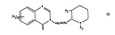
In U.S. Patent No. 5,449,678, there is described and claimed an anti-
fibrotic composition, comprising an amount of a compound of formula I:
N
RZ
~R1 I
N
\ ~L ~2~ N
I
R3
wherein:
n is 1 or 2;
R1 is a member of the group consisting of hydrogen, halogen, nitro, benzo,
lower
alkyl, phenyl and lower alkoxy;
R2 is a member of the group consisting of hydroxy, acetoxy, and lower alkoxy,
and
R3 is a member of the group consisting of hydrogen and lower alkenoxy-
carbonyl;
and physiologically acceptable salts thereof,
effective to inhibit collagen type I synthesis, as active ingredient therein.
After further research and development, it was discovered that the above-
identified compounds of formula I are effective in the inhibition of
restenosis,
which formally is not a fibrotic condition.
CA 02253362 1998-09-14
3
The pathogenesis of atherosclerosis involves abnormal migration and
proliferation of smooth muscle cells (SMCs) infiltrated with macrophages and
embedded in extracellular matrix (ECM) of adhesive glycoproteins,
proteoglycans and collagens (V. Fuster, et al., "The Pathogenesis of Coronary
Artery Disease and the Acute Coronary Syndromes," New En~. J. Med., Vol.
326, pp. 242-250 ( 1992); R. Ross, "The Pathogenesis of Atherosclerosis: A
Perspective for the 1990's," Nature, Vol. 362, pp. 801-809 (1993)]. Under
physiological conditions, the majority of arterial SMCs remains in the Go
phase
and cell growth is controlled by a balance between endogenous proliferation-
stimulating and proliferation-inhibiting factors. Following endothelial cell
perturbation due to atherogenic risk factors (i.e., hypertension,
hyperlipoproteinemia, diabetes mellitus), platelets and non-platelet-derived
growth factors and cytokines are released and stimulate monocyte and SMC
migration as well as SMC proliferation (V. Fuster, et al., ibid.; R. Ross,
ibid.).
Among these growth factors are platelet-derived growth factor (PDGF) [G.A.A.
Ferns, et al., "Inhibition of Neoinitmal Smooth Muscle Accumulation after
Angioplasty by an Antibody to PDGF," Science, Vol. 253, pp. 1129-1132
(1991)], basic fibroblast growth factor (bFGF) [V. Lindner, et al., "Role of
Basic
Fibroblast Growth Factor in Vascular Lesion Formation," Circ. Res., Vol. 68,
pp. 106-113 ( 1991 )], and interleukin-1 (IL-1 ) [H. Loppnow and P. Libby,
"Proliferating or Interleukin-1 Activated Human Vascular Smooth Muscle Cells
Secrete Copious Interleukin 6," J. Clin. Invest.. Vol. 85, pp. 731-738
(1990)].
Macrophages and platelets also release enzymes, i.e., elastase, collagenase,
heparanase) that digest various constituents of the ECM and release bFGF and
possibly other growth factors (TGFb) that are stored in basement membranes and
ECM [I. Vlodavsky, et al., "Extracellylar Matrix-bound Growth Factors,
CA 02253362 1998-09-14 ~~~ "~
J
4
Enzymes and Plasma Proteins," in: Molecular and Cellular Aspects of Basement
Membranes, Mono~aphs in Cell Biolo~y, D.H. Rohrbach and R. Timpl, Eds.,
Academic Press, New York, New York, U.S.A., pp. 327-346 (1993)]. A potent
growth-promoting activity towards SMCs is also exerted by thrombin, which,
under certain conditions, may be present within the vessel wall [R. Bar-
Shavit, et
al., "Thrombin Immobilized to Extracellular Matrix Is a Mitogen for Vascular
Smooth Muscle Cells: Non-Enzymatic Mode of Action," Cell Rep., Vol. 1, pp.
453-463 ( 1990); S.M. Schwartz, "Serum-Derived Growth Factor is Thrombin?"
J. Clin. Invest., Vol 91, p. 4 ( 1993)]. Molecules that interfere with the-
growth-
promoting activity of these growth factors may attenuate the progression of
the
atherogenic process.
Proliferation of arterial smooth muscle cells (SMC) in response to
endothelial injury is a basic event in the process of restenosis of coronary
arteries
after percutaneous transluminal coronary angioplasty (PTCA) [V. Fuster, et
al.,
ibid.J. Coronary bypass surgery or angioplasty are applied to reopen coronary
arteries that have been narrowed by heart disease. A major problem with both
procedures is that arteries rapidly reclog in about 30% of patients undergoing
antioplasty and about 10% of bypass surgery patients. Vascular SMC are
ordinarily protected by the smooth inner lining of the arteries, composed of
vascular endothelial cells. However, following bypass surgery or angioplasty,
SMC are often left exposed. In a futile effort to repair the wound, the cells
proliferate and clog the artery.
According to the invention claimed in Israel Specification No. 110,831,
there is provided a pharmaceutical composition comprising a compound of
formula I as hereinbefore defined, in a pharmaceutically effective amount for
CA 02253362 1998-09-14 PCT ~~ g 7 ~ 15 2 5 4
preventing restenosis by the inhibition of vascular smooth cell proliferation
and
in combination with a pharmaceutically acceptable carrier.
In preferred compositions of said invention, said compound is
halofuginone.
As is known, conventional balloon angioplasty, introduced over 15 years
ago, remains hampered by the persistence of two vexing problems: abrupt vessel
closure during intervention and restenosis during follow-up. Mechanical
intervention with intracoronary stems was introduced for human clinical
investigation already in 1986, and following FDA approval of the first
coronary
stmt for prevention of restenosis following balloon angioplasty, the market
for
the devices has grown from $220 million in 1994 to one that is expected to
capture as much as $1 billion in world-wide revenues in 1996 [W. Diner,
"Technology Strategies - Coronary Stents: Breaking J&J's Lock on the Market,"
Start-Up, pp. 20-26 (May 1996)].
According to said article, while angioplasty alone may result in restenosis
rates of 40% or more, some studies indicate that angioplasty, followed by
stent
deployment, reduces the rate to 20-30%, depending on the kind and location of
disease.
Obviously, a restenosis rate of 20-30% is also undesirable, and therefore it
has been suggested in the literature, and there are now manufactured and sold,
stems which are coated with materials designed to reduce restenosis. Thus,
e.g.,
Johnson & Johnson International Systems markets a heparin-coated form of the
Palmaz-Schartz stent and Medtronic Interventional Vascular markets a fibrin-
CA 02253362 1998-09-14 ~~~~ i~
J.J ~"c ~~
6
coated Wiktor stmt. Stents coated with an antithrombogenic silicon-carbide
material have been sold by Biotronik GmbH, and other suggestions include the
coating of metallic stems with polymers to diminish their thrombogenic
properties, with a nylon mesh, and with a medical grade silicon polymer. Drug-
eluting polymer coatings have. also been reported [see, e.g., Tao Peng, et
al.,
"Role of Polymers in Improving the Results of Stenting in Coronary Arteries,"
Biomaterials 1996, Vol. 17, No. 7, pp. 685-694 ( 1996)]. Thus, said article
teaches that polymer stents can incorporate or bind drugs for later local
controlled delivery at the target site that would inhibit thrombus formation
and
neointimal proliferation and that local administration of various drugs,
including
urokinase, heparin, taxol, hirudin and peptide, is being investigated to
prevent
thrombosis and restenosis.
Summary of the Invention
With the above state of the art in mind, it has now been realized that the
compositions taught in Israel Specification 110,831 can be utilized in an
especially effective manner by incorporating the same as a coating on
intracoronary stents, by methods known per se.
Thus, according to the present invention, there is provided an intracoronary
stent coated with a quinazolinone derivative of formula I:
CA 02253362 2002-06-18
7
wherein:
R1 is a member of the group consisting of hydrogen, halogen, nitro, benzo,
lower
alkyl, phenyl and lower alkoxy;
RZ is a member of the group consisting of hydroxy, acetoxy, and lower alkoxy,
and
R3 is a member of the group consisting of hydrogen and Iower alkenoxy-
carbonyi,
for preventing restenosis after angioplasty.
As indicated above, while described, exemplified and illustrated herein.
the subject matter of Israel Specification No. 110,831 does not constitute a
part
of the present invention, and is specifically disclaimed.
Preferably, said lower alkyl has 1 to 6 carbon atoms; said lower alkoxy has 1
to 6 carbon atoms; and said lower alkenoxy-carbonyl has 1 to 6 carbon atoms.
Preferably, said quinazolinone derivative is combined with a polymer carrier
that is non-degradable and insoluble in biological mediums.
With specific reference now to the examples in detail, it is stressed that
the particulars described are by way of example and for purposes of
illustrative
discussion of the preferred embodiments of the present invention only, and are
presented in the cause of providing what is believed to be the most useful and
readily understood description of the principles and conceptual aspects of the
invention. In this context, it is to be noted that only subject matter
embraced in
the scope of the claims appended hereto, whether in the manner defined in the
claims or in a manner similar thereto and involving the main features as
defined
in the claims, is intended to be included in the scope of the present
invention,
while subject matter of Israel Specifcation 110,831. although described and
exemplified to provide background and better understanding of the invention,
is
not intended for inclusion as part of the present invention.
CA 02253362 1998-09-14 ~~~~~~e ~ ~~ !~ ,~
g
Brief Description of the Drawings
In the drawings:
Fig. 1 is a characteristic curve showing the inhibitory effect of halofuginone
on
SMC proliferation;
Fig. 2 is a characteristic curve showing reversion of the antiproliferative
effect
of halofuginone on SMC;
Figs. 3a and 3b respectively are a bar graph and a characteristic curve,
showing
the effect of halofuginone on 3H-thymidine incorporation into vascular
SMCs;
Fig. 4 is a characteristic curve showing the effect of halofuginone on
vascular
endothelial cell proliferation;
Figs. 5a and Sb respectively are a bar graph and a characteristic curve,
showing
the effect of halofuginone on 3H-thymidine incorporation into vascular
endothelial cells;
Fig. 6 is a bar graph showing antiproliferative effect of halofuginone on 3T3
fibroblasts;
Fig. 7 is a bar graph showing the inhibitory effect of halofuginone on the
mitogenic activity of bFGF;
Figs. 8a and 8b are color light micrographs of the central artery of a rabbit
ear
after being subjected to crush injury, respectively showing an untreated
artery and an artery treated according to the present invention; and
Fig. 9 is a graph showing the effect of halofuginone on injury-induced artery
stenosis.
CA 02253362 1998-09-14 ~C ~i ~ 9 7 ~ I 5 2 5 4
9
EXAMPLES
1) Exuerimental Procedures
Cells
SMC were isolated from the bovine aortic media as previously described
[see, e.g., J.J. Castellot, et al., "Structural Determinants of the Capacity
of
Heparin to Inhibit the Proliferation of Vascular Smooth Muscle Cells: Evidence
for a Pentasaccharide Sequence that Contains a 3-0-Sulfate Group," J. Cell
Biol.,
Vol. 102, pp. 1979-1984 (1986); and A. Schmidt, et al., "The Antiproliferative
Activity of Arterial Heparan Sulfate Resides in Domains Enriched with 2-0-
Sulfated Uronic Acid Residues," J. Biol. Chem., Vol. 267, pp. 19242-19247
( 1992)].
Briefly, the abdominal segment of the aorta was removed and the fascia
cleaned away under a dissecting microscope. The aorta was cut longitudinally,
and small pieces of the media were carefully stripped from the vessel wall.
Two
or three such strips, with average dimensions of 2-3 mm, were placed in 100 mm
tissue culture dishes containing DMEM (4.5 g glucose/liter), supplemented with
10% FCS, 100 U/ml penicillin and 100 mg/ml streptomycin. Within 7-14 days,
large patches of multilayered cells migrated from the explnts. Approximately 1
week later, the cells were subcultured into 100-mm tissue culture plates (4-
6x10⁵
cells/plate). The cultures (passage 3-8) exhibited typical morphological
characteristics of vascular SMC and the cells were specifically stained with
monoclonal antibodies that selectively recognize the muscle form of actin (HF-
35). This antibody does not recognize endothelial cells or fibroblasts.
CA 02253362 1998-09-14 ~~~/~ 3~ ~ '~
...
Cultures of vascular endothelial cells were established from bovine aorta,
as previously described by D. Gospodarowicz, et al. ["Clonal Growth of Bovine
Endothelial Cells: Fibroblast Growth Factor as a Survival Agent," Proc. Natl.
Acad. Sci. U.S.A., Vol. 73, p. 4120 (1979)]. Stock cultures were maintained in
DMEM (1 g glucose/liter) supplemented with 10% calf serum, 50 U/ml
penicillin, and 50 mg/ml streptomycin at 37dC in 10% C02 humidified
incubators. Partially purified brain-derived bFGF (100 ng/ml) was added every
other day during the phase of active cell growth (D. Gospodarowicz, et al.,
ibid.,
and J. Vlodavsky, et al., "Vascular Endothelial Cells Maintained in the
Absence
of Fibroblast Growth Factor Undergo Structural and Functional Alterations That
Are Incompatible with Their In Vivo Differentiated Properties," J. Cell Biol.,
Vol 83, pp. 468-486 (1979)].
Cell Proliferation; 3H-Thymidine Incorporation
SMCs were plated (4x10⁴ cells/16 mm well) in DMEM supplemented
with 10% FCS. 24 hours after seeding, the medium was replaced with medium
containing 0.2% FCS, and 48 hours later, the cells were exposed to growth
stimulants and 3H-thymidine ( 1 mCi/well) for an additional 24-48 hours. DNA
synthesis was assayed by measuring the radioactivity incorporated into
trichloroacetic acid insoluble material [M. Benezra, et al., "Reversal of bFGF
Autocrine Cell Transformation by Aromatic Anionic Compounds," Cancer Res.,
Vol. 52, pp. 5656-5662 ( 1992)].
Growth Rate
SMCs (1.5x10⁴ cells/well) were seeded into 24 well culture plates and
exposed to growth stimulants as described above. 1 to 6 days after seeding,
the
CA 02253362 1998-09-14 PCT 9 ~ ~ 1 5 z 5 4
n
cells were fixed with 2.5% formaldehyde in PBS. The plates were immersed in a
bath of 0.1 M borate buffer (pH 8.5), stained ( 1 h, 24dC) with methylene blue
( 1 % in 0.1 M borate buffer, pH 8.5) andwashed four times in water. This
procedure removed practically all non-cell-bound dye. Specific cell
incorporated
methylene blue was dissolved with 0.5 ml of 0.1 N HC1 (1 h, 25dC) and
determined by measuring the absorbency at 620 nm (Bar-Shavit, et al., ibid.).
The initial cell plating density was chosen to ensure a linear relationship
between
cell number and absorbance at the end of the experiment. In each experiment, 3
wells were fixed before adding the test compound to determine the initial
average absorbance. This value was used to calculate doubling times (DT) of
control and drug-treated cells, using the following equation:
DT = In 2/IN [(ODt/ODc)/hJ
wherein:
DT - doubling time in hours;
Odt - optical density of a test well at the end of the experiment;
Odc = optical density of a control well at the beginning of the experiment;
h - duration of incubation in hours.
The growth rate was calculated by dividing the doubling time of drug-
treated cells by that of control cells [A. Horowitz, et al., "In Vitro
Cytotoxicity of
Liposome- Encapsulated Doxorubicin: Dependence on Liposome Composition
and Drug Release," Biochim. Biophys. Acta, Vol. 1109, pp. 203-209 ( 1992)].
Cell Number
SMCs were seeded (2.5x10³ cells/well) into 24-well plates in DMEM
(4.5 g glucose/liter), supplemented with 10% FCS and allowed to attach for 6
CA 02253362 1998-09-14 PCTA ~~
12
hours [A. Schmidt, et al., "The Antiproliferative Activity of Arterial Heparan
Sulfate Resides in Domains Enriched with 2-0-Sulfated Uronic Acid Residues,"
J. Biol. Chem., Vol. 267, pp. 19242-19247 (1992)]. The medium was removed
and experimental medium (with or without halofuginone) containing 10% FCS
was added to quadruplicate wells. After 4 days of incubation, the cell number
was determined, using a Coulter counter (Schmidt, et al., ibid.). The degree
of
inhibition was calculated from the following formula:
Inhibition = 1-net growth in presence of halofuginone/net growth in control
x 100
The net growth was determined by subtracting the initial cell number from
the final cell number.
Z) Experimental Results
i) Antiproliferative Effect of Halofu~inone toward Vascular SMC
Growth Rate
Sparsely seeded vascular SMC were exposed to 10% FCS in the absence
and presence of increasing concentrations of halofuginone. The cells were
dissociated with STV and counted daily. As shown in Fig. 1, 80-90% inhibition
of SMC proliferation was obtained in the presence of 75 ng/ml halofuginone,
with an almost complete inhibition at 125 ng/ml.
In another experiment, the SMCs were exposed to halofuginone for 48
hours, followed by removal of the drug and subsequent growth in regular growth
medium. As demonstrated in Fig. 2, removal of the drug resulted in a gain of
an
accelerated growth rate similar to that of the untreated SMCs.
CA 02253362 1998-09-14 ~~T~V ~ ~~
13
3H-Thymidine Incorporation
Subconfluent vascular SMCs maintained in a medium containing 10% FCS
were exposed (48 hours, 37dC) to 3H-thymidine in the absence and presence of
increasing concentrations of halofuginone. As demonstrated in Fig. 3a,
complete
inhibition of DNA synthesis was observed at 0.15 mg/ml halofuginone, while
65% inhibition was obtained at a concentration as low as 0.05 mg/ml (Fig. 3b).
ii) Antiproliferative Effect toward Vascular Endothelial Cells and 3T3
Fibroblasts
Vascular Endothelial Cells
Sparsely seeded bovine aortic endothelial cells were cultured in medium
containing 10% CS in the absence and presence of increasing concentrations of
halofuginone. The cells were dissociated with STV and counted daily.
Inhibition of endothelial cell proliferation was observed primarily during the
first
4 days, in cells treated with relatively high concentrations (0.1-0.125 mg/ml)
of
the drug (Fig. 4). Unlike the results with SMCs, the endothelial cells
regained an
almost normal growth rate (doubling time), starting on day 5 (Fig. 4),
indicating
that vascular EC are less susceptible than vascular SMCs to the inhibitory
effect
of halofuginone. Thymidine incorporation studies revealed a 50% inhibition of
DNA synthesis at 0.05 mg/ml halofuginone (Fig. 5).
3T3 Fibroblasts
Fig. 6 demonstrates that 3H-thymidine incorporation by actively growing
3T3 fibroblasts maintained in medium containing 10% FCS was almost
CA 02253362 1998-09-14
~C~'1' ~ '~ ~' ~ ~ ~. '
14
completely inhibited in the presence of 0.025 mg/ml halofuginone, suggesting
that fibroblasts are even more sensitive to the drug as compared to SMCs.
Effect on bFGF-Induced Cell Proliferation
Quiescent, growth arrested 3T3 fibroblasts maintained (48 hours) in
medium containing 0.5% FCS are readily stimulated to proliferate by low
concentrations at basic fibroblast growth factor (bFGF). Exposure to
halofuginone (0.025 mg/ml) resulted in an almost complete inhibition of bFGF-
stimulated thymidine incorporation in growth-arrested 3T3 fibroblasts (Fig.
7).
This result suggests that halofuginone efficiently antagonizes the growth-
promoting activity of bFGF.
iii) Arterial Stenosis Caused by Physical Iniury
Adult New Zealand rabbits were anesthetized by intramuscular injection of
ketamine (50 mg/kg). Physical injury was applied for 30 min. externally to the
central artery of each ear [Banai, et al., Circulation Res., Vol. 69, pp. 748-
756
( 1992)). After the operation, the rabbits were housed in accordance with
Animal
Welfare Act specifications. Halofuginone (0.2 ml of 0.09 mg/ml) was
introduced subcutaneously around the physical crush area 1 hour after the
crush
and once every 24 hours during the first 4 days. On day 14, the animals were
sacrificed and the ears fixed in 10% buffered formaldehyde for 72 hours. The
crush sites were further trimmed at 1 mm intervals, dehydrated in ethanol and
xylene, and embedded in paraffin. Serial (Smm) sections were stained by Movat
pentachrome method. Computerized planimetry was performed at the site
of the lesion and at an adjacent normal arterial segment displaced 2 mm from
the
location of the injury. Selection of the normal site was random; approximately
one-half were proximal and one-half distal to the injury site. The lumen, the
area
CA 02253362 1998-09-14 ~~~ ~ 9 7 / 1 5 2 5 ~
IS
cricumscribed by the internal elastic lamina ("original lumen") and the area
circumscribed by the external border of the media (total vessel area) were
traced,
and the ratio between neointima and media was calculated. In all cases, the
single
section demonstrating the greatest extent of neointimal proliferation was
selected
for planimetry.
Referring now to Figs. 8a and 8b, there are seen light micrographs of the
central artery of a rabbit ear 14 days after external crush injury (Movat
staining
of representative cross-sections).
In Fig. 8a, the SMCs are migrating from the media into the neointima
through the disrupted internal elastic lamina and the artery lumen is narrowed
by
the protruding neointima in the untreated artery. As can be seen, there is
striking
neointimal formation and an almost complete obliteration of the arterial
lumen.
In contradistinction, in Fig. 8b there is seen a rabbit ear artery subjected
to
crush injury and treatment with halofuginone. An almost complete inhibition of
neointimal formation is observed.
Fig. 9 shows a quantitative analysis of the ratio between enointima to
media performed in control rabbits and rabbits treated with halofuginone (H)
or a
synthetic heparin-mimicking compound (M). Each point represents one rabbit.
As stated hereinbefore, quinazolinone derivatives of formula I, and
preferably halofuginone, are incorporated in a polymeric matrix coating of the
metal stent. The drug is dispersed or dissolved in a polymeric solution or
melt
and applied onto a metal stent by dripping or spray process. The following
examples demonstrate the release of halofuginone from a polymeric coating. It
CA 02253362 1998-09-14
1,~
,i .-
16
is to be noted that the polymeric coating is chosen not to affect the normal
performance of the stmt, such as the expansion of the stmt in application or
deformation of the polymeric coating during its presence within the blood
vessel.
Example 1
Coating of a Metal Stent with a Polymeric Matrix
A solution of polyethylene vinyl acetate (EVA, 1 % w/w in
dichloromethane) and 0.1 wt% of halofuginone free base is prepared. The
desired stmt is dipped once into the EVA-drug solution and the stent is
allowed
to dry at room air to yield a smooth uniform coating of about 10 microns
thickness. If thicker coating is desired, the dipping process is repeated
several
times. To improve the adherence of the EVA coating on the stmt, the stmt is
pre-treated with a prime polymer coating that allows adhesion of the EVA
coating. The coating is flexible and does not affect the expandability of the
stmt.
The concentration of the polymer and drug may vary from 0.1 % to about 10% of
polymer concentration and the drug content may vary from 1 to about 20% per
polymer weight. The thickness of the coating can be varied by either the
number
of coating or the polymer concentration in the dipping solution.
The polymer carrier can be any pharmaceutically acceptable biopolymer
that is non-degradable and insoluble in biological mediums, has good stability
in
a biological environment, has a good adherence to the selected stent, is
flexible,
and that can be applied as coating to the surface of a stent, either from an
organic
solvent, or by a melt process. The hydrophilicity or hydrophobicity of the
polymer carrier will determine the release rate of halofuginone from the stent
surface. Hydrophilic polymers, such as copolymers of hydroxyethyl
CA 02253362 1998-09-14 ~ ~S 9 7 l 1 5 2 5 ~.
17
methacrylate-methyl methacrylate and segmented polyurethane (Hypol), may be
used. Hydrophobic coatings such as copolymers of ethylene vinyl acetate,
silicone colloidal solutions, and polyurethanes, may be used. The preferred
polymers would be those that are rated as medical grade, having good
compatibility in contact with blood. The coating may include other
antiproliferative agents, such as heparin, steroids and non-steroidal anti-
inflammatory agents. To improve the blood compatibility of the coated stent, a
hydrophilic coating such as hydromer-hydrophilic polyurethane can be applied.
Examule 2
Halofuginone Release from Polymer Coating
The releasee of drug from the stmt coating is determined in vitro by
placing the device into a large volume of physiologic solution (0.1 M
phosphate
buffer, pH 7.4) at 37°C. The drug release to the solution is
periodically
determined by HPLC. For the coating described in Example 1 (single dipping of
a 1 % polymer solution and 10% of halofuginone per polymer weight in EVA),
there was a release of about 5 to 10 mcg/cm2/day for a period of 3 weeks. When
a rate-limiting coating is applied, an additional dipping of the stent in a
solution
of 0.5% EVA containing between 0 and 5% of halofuginone is applied. The
drug release rate is decreased to about 20% of the release without the rate-
controlling membrane, then coated with a drug-free coating. In addition, there
is
no burst release and the drug is released at a constant rate for a longer time
period.
The drug release profile and the duration of drug releasee can be altered by
altering the coating thickness, the polymer carrier, the drug content in the
CA 02253362 1998-09-14
Ig
polymer, composition of the coating (hydrophilic or hydrophobic additives;
blends of polymers), the drug content within the various layers of coating,
the
configuration of the stem, and the properties of the rate-controlling
membrane. It
is possible to gain an adequate effect by coating the stent at specific sites
such as
on the outer surface only (can be chieved by either selected spray coating or
by
shielding the inner side of the stent) or coating certain parts of the stmt,
such as
the edges.
Conclusions
Current approaches to inhibit the proliferation of vascular SMC utilize
heparin, suramin, antibodies to various growth-promoting factors, anti-
thrombin
agents, and, most recently, antisense DNA technique. Heparin is a potent
anticoagulant and its anti-proliferative activity is relatively small and
subjected to
major variations depending on the source and manufacturing company. Suramin
is highly toxic at the effective dose, while antibodies are expensive, have a
short
half life and may elicit an immune response. Information on the antisense
approach is new, and at present very limited.
The present invention, in its most preferred embodiment, utilizes a highly
potent, inexpensive and non-toxic compound which inhibits the activity of
various growth factors, including bFGF, and inhibits autocrine growth of
vascular SMC and fibroblasts. Moreover, halofuginone is a low molecular
weight compound which can be administered orally. The compound has been
approved by the F.D.A. for use in farm animals. These characteristics make
halofuginone a most promising clinically useful drug to inhibit restenosis.
CA 02253362 1998-09-14
' ~ ,~ r' r
~ ~ ..d ;~, ~o .
19
Thus, the present invention provides for the use of halofuginone as a non-
toxic compound that efficiently inhibits SMC proliferation, to provide an
effective strategy for inhibiting the pathophysiology of arteriosclerosis,
restenosis after coronary angioplasty, and neointimal proliferation in
saphenous
vein grafts.
It will be evident to those skilled in the art that the invention is not
limited
to the details of the foregoing illustrative examples and that the present
invention
may be embodied in other specific forms without departing from the essential
attributes thereof, and it is therefore desired that the present embodiments
and
examples be considered in all respects as illustrative and not restrictive,
reference
being made to the appended claims, rather than to the foregoing description,
and
all changes which come within the meaning and range of equivalency of the
claims are therefore intended to be embraced therein.