Note: Descriptions are shown in the official language in which they were submitted.
CA 02253646 1998-11-05
WO 97141777 PCT/US97I07784
OPTICAL BIOPSY FORCEPS AND
METHOD OF DIAGNOSING TISSUE
Field of the Invention
This invention pertains to the field of medical diagnosis and
treatment. More specifically, the invention pertains to a forceps device
having
integrated optical fiber and remotely controllable biopsy forceps functions,
and
to the use i:hereof in medical diagnosis. The catheter is adapted for in vivo
tissue
identification of tissue types through optical techniques using the optical
fiber,
and biopsy sampling of identified tissue areas for withdrawal from the body
for
conventional examination and analysis.
>f3ac ground of the Prior Art
Numerous type of biopsy forceps devices have been developed
for in vivo medical diagnosis and treatment of various conditions. Such
devices
are designed for sampling tissue within the body, for example in endoscopic,
laparoscopic and vascular procedures to retrieve biopsy samples for analysis
and
identification of tissue types. These biopsy forceps devices generally include
small cutting jaws at the distal end, operated remotely from the proximal end
after the distal end of the device has been positioned or navigated to the
site of
interest.
One difficulty in using prior art biopsy forceps devices is in
knowing for certain the cx:act positioning of the distal tip, in relation to
the
suspected disease area, especially when the area of interest is very small.
Various types of optical catheters or probes have been developed for use in
locating or identifying sitca within the body. A method of diagnosing and
treating tissue in vivo using an optical guidewire is disclosed in U. S.
Patent
5,439,000, assigned to SpectraScience, Inc. One type of prior art system for
internal biopsy uses an optical catheter to locate the site, followed by
replacement of the optical catheter with a biopsy forceps for taking a sample.
However, this can result in errors and uncertainties in the final placement of
the
biopsy jaws with respect 1:o a previously identified small structure or area.
CA 02253646 2002-09-30
2
Other prior art systems have been proposed which use optical
viewing or imaging and a cutting device in the same device, to visually locate
and Lhel1 blVpstl a J4speVtel.J ~4i.A~~r4. ~~\/r N dal Lr 14,
by Laserscope, Inc. relates to a surgical device for internal
operations. A rigid tissue parting means is provided to enlarge a cavity which
can be viewed by a viewing system. Tissue collecting means are provided
adjacent to the viewing system. However, such devices have been hampered by
their thickness which is needed to accommodate the imaging system and the
cutting actuation system, and which precludes their use in very small areas.
Another shortcoming of such prior art systems is the offset or'parallax'
between
the viewing axis of the imaging system and the cutting position of the biopsy
l
sampling apparatus, such that the biopsy sample actually is taken from a zone
slightly displaced from the zone being viewed by the optics. This can result
in a
loss of accuracy in the case of v~rv small structures of interest.
Summar"v of the Invention
To overcome these and other problems, the present invention
provides an integrated fiber optic biops.~ forceps device, which is very thin.
enabling it to be used in very small areas of interest, and which has accurate
alignment of the optic field of view and the biopsy zone of sampling.
The present invention provides an optical biopsy forceps which is
adapted for tissue identification both by optical techniques and biopsy
sampling.
The forceps device includes an elongated catheter body for introduction into
the
body and navigation iu an area of interest. The distal end of the forceps
device
has a pair of cutting jaws, and flue tip of an optical fiber which runs
through the
forceps device. The proximal end has a control handle for manipulating the
forceps device and actuating the jaws.
In accordance with one aspect of the invention, there is provided a
method of diagnosing tissue at a site within a body. The method comprises
introducing into the body an integrated optical biopsy forceps which includes
a
flexible catheter body with an optical fiber extending therethrough with the
distal
end of the optical fiber positioned with its optical view axis aligned for a
tissue
analysis zone adjacent the distal tip of the catheter body. The optical biopsy
AMENDED SHEET
CA 02253646 1998-11-06
2/a,.
forceps additionally including cutting jaws mounted at the distal end of the
catheter body for selective opening and closing in a biopsy cutting movement
in
the tissue analysis zone, acid an actuator mechanism operatively connected to
the
~n,,EN~EO SNP .
CA 02253646 1998-11-05
WO 97/41777 PCTIUS97/07784
3
jaws for se;lectively controlling the opening and closing of the cutting jaws.
Then, tissue in the tissue analysis zone adjacent the distal end of the
forceps is
spectroscopically analyzed through the use of an electro-optic tissue analysis
system connected to the proximal end of the optical fiber. The optical biopsy
S forceps is spectroscopically guided within the body to an area of interest
as
identified by the spectroscopic analysis of tissue type in the tissue analysis
zone
adjacent the distal tip of the catheter body. Then, a biopsy sample is cut
from the
location of the optical tissue analysis zone by actuating the actuator
mechanism,
and the biopsy sample is withdrawn from the body.
In one embodiment, the cutting jaws are mounted for pivoting or
other movement bringing them together for cutting tissue placed therebetween.
and coupled to and controlled by the optical fiber that extends through the
catheter body to the handle at the proximal end of the device. The optical
fiber
extends through the handle and the catheter body from its proximal end for
I S connection to eleetro-optical analysis equipment, to a distal tip for
transmitting
andlor receiving light energy from tissue at the location of the tip. The
fiber tip
is positioned coaxially with the jaws at their zone of contact and cutting, so
that
the biopsy sample is taken exactly at the spot in the field of view of the
optical
fiber.
In another embodiment, the cutting jaws are mounted for pivoting
or other cr~ovement bringing them together for cutting tissue placed
therebetween, and controlled by wires extending through the catheter body to
the
control handle. The optical fiber extends through the device, from its
proximal
end for connection to elec;tro-optical analysis equipment, to a distal tip for
transmitting and/or receiving light energy from tissue at the location of the
tip.
The fiber tip is positioned coaxially with the jaws at their zone of contact
and
cutting, so that the biopsy sample is taken exactly at the spot in the field
of view
of the optical fiber.
One example of the utility of the invention is in the diagnosis of
arterial or vascular obstc~~ctions, such as atherosclerotic lesions and
thrombi.
After identification, the appropriate therapeutic catheter, whether balloon
CA 02253646 1998-11-06
4
angioplasty, drug delivery ar laser ablation, can be advanced alon, a
~uidewire
and employed to treat the patient. The present invention is also useful in
many
other fields in~ludin', but not limited to: oncolo~~y, urolo~;v.
~'astroenterolo~Tv,
neurosurgery. general sur~~ery, obstetrics/gynecolo~~y, etc. It can also t-e
used in
laparoscopic procedures for additional dig<~nostic information. and/or
~~uidance
of a therapeutic modality (e.g.. laser or cutting/coagulation devices. such as
a
bipolar electrocautery device).
These and other features and advanta~es of the invention will
become apparent from the following description of the preferred embodiments of
the invention.
Brief Descrirltion of the Drawing,
Figure 1 is an overall view of the optical biopsy forceps accoc-din~~
to the present invention:
1 ~ Figure ? is a cross-sectional view at an enlarged scale of the distal
end of the forceps of Fie. 1, with the forceps jaws open:
Figure 3 is a view of the distal end of the forceps of Fi'T. 1. with
the forceps jaws closed:
Figure -l is a perspective view of the fiber tube assembl and
?0 related components, for the distal end of the device of Fia. ~:
Figure ~A is a top view, at an enlar~~ed scale. of a component of
the distal end of the device of Fi<~. ?'
Figure ~B i.s ~ side sectional view taken along the line ~B-sB of
F; gnre _; ~:
Figure ~C is an end view of the component of the distal end of the
device of Fig. ?;
Figure 6A and 6B are top and side views. respectively, of a
~~~~~f~ v~. ~Y ~.
CA 02253646 1998-11-05
WO 97/41777 PCTIUS97107784
S
cutting jav~r component of the distal end of the device of Fig. 2;
Figure 7 is an overall view of another embodiment of the optical
biopsy forceps according to the present invention; and
Figure 8 is a cross-sectional view of the distal end of an optical
biopsy forceps provided in accordance with a further embodiment of the
invention.
pescri ion o~~Jl~e Preferred Embodiments
One prefewed embodiment of an integrated optical biopsy forceps
of the present invention is generally indicated by reference number 10 in Fig.
I .
Forceps 10 is adapted for use internally of the body, for example in
connection
with endoscopic, laparoscopic or vascular procedures. Forceps 10 includes a
control handle portion 12 at the proximal end, a middle portion 14 which
extends
over the main length of the device, and a distal end 16 which includes opposed
forceps cutting jaws and distal end of the optical fiber, as is explained in
greater
1 S detail below.
As seen in the left portion of Fig. 2, the main body or length of
the forceps 10 consists of coaxial inner and an outer tubular members. In one
preferred embodiment, the: inner tubular member is a hollow plastic tube 20,
and
the outer tubular member or catheter body is coil 22. The coil 22 is a finely
wound spiral coil of stainless steel as is generally known and used in
catheters
and guidewires. Alternatively, the outer tubular member could be made using
another plastic tube, or a plastic/metal composite structure, in place of coil
22.
The plastic tube 20 is positioned within coil 22 and these components are
dimensioned with respect to each other so that tube 20 may be flee to move
2S axially within coil 22 during actuation of the jaws, as is explained below.
Positioned within inner tube 20 arc a pair of control wires 40, 41,
and the optical fiber 50. 'lChese components, together with outer coil 22 and
inner plastic tube 20 extend over the main length of the device, from the
distal
end 16 to the handle portion 12. At the handle, coil 22 and tube 20 pass
through
a plastic sleeve 24, which serves as a reinforcement and strain relief, into a
bore
2S in the tip 13 of the handle 12. The plastic sleeve 24 and the proximal end
of
CA 02253646 1998-11-05
WO 97!41777 PC'TlUS97107784
6
the coil 22 are received and secured, as by bonding, in the tip 13 of the
handle
12.
The inner plastic tube 20, control wires 40, 41 and fiber SO are not
secured at tip 13, hut pass through bore 25, through a stainless steel
reinforcing
tube 29 to slider 30, which is movably received in a slot 28 in handle 12.
Reinforcing tube 29, tube 20 and control wires 40, 41 are secured to slider 30
which together form an actuator mechanism for the forceps 10. Movement of
slider 30 causes axial moverr~ent of reinforcing tube 29, tube 20 and control
wires 40, 41 relative to coil 22, which is used to actuate the cutting jaws.
Loops
26 and 27 are: provided in ha~~dle 12 and slider 30, to form finger holes
useful in
grasping and manipulating tine forceps.
Optical fiber _'i0 extends through slider 30, and out of handle 12.
in a protective cable or sheath 32, for connection to electro-optical units
(not
shown) which provide the illumination light to the fiber, and which receive
and
analyze the rcaurned right from the target at distal the end of the forceps.
'The
optical biopsy forceps of the present invention may be used with any type of
electro-optical technique for guiding the forceps. This may include systems
which use vif~wing or imaging, systems which use illumination with white light
to excite dyer in the area of interest, and spectroscopic techniques to
identify
tissue types )~~y spectral analysis of light returned from tissue illuminated
with
light of certain wavelengths. Such spectroscopic techniques utilize the
property
of certain tissue types to reflect or fluoresce light having characteristic
wavelengths.
As seen in I~igs. 2, SA, SB and SC, the distal end 16 of the optical
forceps includes a yoke 60, which serves as a mounting member for the cutting
jaws. Yoke tiU may be machined from stainless steel or formed of other
suitable
material. It ~;enerally has a proximal portion or section indicated by
reference
number 61. a center section tie, and a distal section 63 having inwardly
curved
opposing distal end portions 63a and 63b. Yoke b0 has a bore 64 running
therethrough. Each of the opposing distal end portions 63a and 63b has an arc
shaped groove 65 (Figs. 5B and SC'.) formed therein which defines a guide slot
CA 02253646 1998-11-05
WO 97!41777 PCT/US97/07784
7
for the distal end of the fiber 50. The diameter of the bore defined by the
arcuate
grooves 65 can be stepped to a smaller size at distal end portions 63a and
63b.
Sections 61 and 62 are generally circular in section. Section 61 has a
diameter
corresponding to the inside dimension of coil 22, while section 62 has a
diameter
corresponding to the outside dimension of coil 22, so that the end of coil 22
may
be received and bonded to section 61. The proximal end surface 56 of the yoke
60 cooperates with the distal end 21 of the inner tube 20 to provide a limit
stop
for the fiber tube assembly 52 when it is being advanced within the outer tube
22
to open the jaws. Center ;section 62 has a pair of holes 68, 69 which receive
pins
72, 73 to hold the jaws in place.
Distal section 63 is stepped down relative to section 62, as seen in
side view in Figs. 2 and SB, to allow the jaws 80 and 81 to fold against it
when
the jaws are closed (Fig. ?.) so as to have a thin profile for ease of
introduction
and navigation. Distal section 63 also has a vertical slot 70 provided therein
which is dimensioned to the size of the mounting ends of the lever arms 85 of
the
jaws. The: inner wall 71 of distal section 63 is stepped outwardly relative to
the
slot 70 to provide clearance for the ends of control wires 40 and 41.
Because jaws 80 and 81 are similar only one is described in detail
here. The two jaws are mirror-image identical, but with their serrations
staggered so that they will mesh. As seen in Figs. 6A and 6B, jaw 80 has a
rearward lever or mounting portion 85, and a distal cup or sample receiving
portion 8~'., which has sharp serrations 83 used to cut the tissue sample. The
lever portiion 85 has a hole 84 formed to receive the pin 72 which thus serves
to
retain the ,jaws, and also to acts as the pivot point. A hole 86 is provided
at the
forward apex of the relieved section, to receive the end of control wire 40
(or 41 )
which is crimped or bent .at a right angle at its tip to be effectively
captured. The
control wires are formed of wire which is stiff enough to push against the
jaws to
open there, but ilexibIe enough to flex as the wires are retracted to pull the
jaws
together.
As seen in Fig. 2, the distal end 16 of the optical forceps also
includes a. fiber tube assembly 52. It includes a tube 54 which may be
machined
CA 02253646 1998-11-05
WO 97!41777 PC'T/US9710?784
8
from stainless steel, or formed of other suitable material. The end of plastic
tube
20 overlaps .end SS of the tube 54 and is bonded to tube 54. The control wires
40, 41 and tle optical fiber '.i0 pass into it from the plastic tube 20. The
optical
fiber and the control wires pass axially through the tube 54 and are bonded to
the
S tube 54 by epoxy or other suitable adhesive. The optical fiber 50 includes a
jacket 87 of polyamide or similar material and an outer protective tube 88
made
of stainless :steel, for example. 'The jacket 87 extends the length of the
optical
fiber from its proximal end to its proximal end. The protective tube 88
extends
from the distal end of the optical fiber to at least a point located within
the distal
end of tube >4. The distal end of the optical fiber 50 is flush with the end
of the
protective tube 88, and may have a lens or clear epoxy coating, depending on
the
optical properties desired. '!.'he protective tube 88 at the distal end of the
optical
fiber is desi~;ned to give strength to prevent damage to the fiber by tweezers
and
the like when tissue is removed from the biopsy jaws.
Referring to Figs. I and 2, in operation, the slider 30 is retracted
toward the back of handle 12 to close the jaws. 'This causes movement (to the
left in Fig. 2.) of plastic tube 20, the fiber tube assembly 52, the control
wires 40,
41, and the optical fiber 50. 'This retracts the optical fiber into the yoke
60 and
the pulling of the control wires closes the jaws. In this configuration, the
distal
end is of the: same narrow diameter as the main body of the forceps catheter,
and
the closed jaws have a smooth, rounded shape to facilitate introduction and
navigation in the vascular, e;ndoscopic or laproscopic systems. Also, the
cutting
jaws are coaxially positioned with respect to the distal end of the optical
fiber.
Once in place in the general area of interest, the forceps jaws can
be opened b~y pushing slider 30 of the control handle forward. This causes
movement (to the right in Fig. 2) of plastic tube 20, the fiber tube assembly
52,
the control wires 40, 41, and the optical fiber 50. ~hhe control wires push
against
the jaws, causing them to open. Simultaneously, the tip of the optical fiber
is
axially extended. The distal end or tip of the optical fiber is positioned at
the
distal end of the catheter body with its optical view axis or view axis
aligned for
a tissue analysis zone adjacent the distal tip of~the catheter body and
positioned
CA 02253646 1998-11-05
WO 97/41777 PCTlUS97/07784
9
at the area, of contact of the cutting jaws when the cutting jaws are operated
to
their close;d cutting position. The device may then be used for optical tissue
identification. When an area of disease is identified and a biopsy of it is
needed,
slider 30 is pulled, retracting the tip of the fiber and simultaneously
causing the
jaws to close and cut a biopsy sample at the exact place being viewed by the
fiber. Thc~ biopsy sample is cut from the exact tissue site identified by the
spectroscopic analysis step without requiring moving or repositioning of the
catheter body. The forceps may then be withdrawn from the patient to recover
the sample for analysis. The analysis of the withdrawn sample can be conducted
using known laboratory techniques to confirm the identification of the tissue
sample made by spectroscopic analysis.
The optical biopsy forceps of the invention is used for
spectroscopically analyzing tissue in the tissue analysis zone adjacent the
distal
end of they forceps througlh the use of an electro-optic tissue analysis
system
connected to the proximal end of the optical fiber. The optical biopsy forceps
are guided spectroscopically within the body to an area of interest as
identified
by the spt:ctroscopic analysis of tissue type in the tissue analysis zone
adjacent
the distal tip of the catheter body.
Referring to Fig. 7, another embodiment of an integrated optical
biopsy forceps of the present invention is generally indicated by reference
number 90. The optical forceps 90 is generally similar to the optical forceps
10
shown in Fig. 1, and accordingly, corresponding elements have been given the
same reference number. The optical biopsy forceps is adapted for use
internally
of the body, for example in connection with endoscopic, laparoscopic or
vascular
procedures. Forceps 90 includes a handle portion 91 and an operating lever 92
at
the proximal end, a middle portion 14 which extends over the main length of
the
device, and a distal end 16. The distal end 16 includes forceps cutting jaws
80
and 81 and the distal end of the optical fiber 50 which is contained within a
plastic tube, corresponding to plastic tube 20 of forceps 10. and pass through
a
sleeve 24~ in the manner illustrated in Figs. 1-6 for the forceps 10.
CA 02253646 1998-11-05
WO 97141777 PC'T/US97107784
The operating lever 92 has its upper end 93 pivoted to the handle
91 by a pivot pin 94. The forceps 90 includes a reinforcing tube,
corresponding
to reinforcing; tube 29 of farc:eps 10, which encloses the fiber optical tube,
and
control wires 40 and 41. The; control wires pass around a post 95 and are
secured
5 to the operating lever 92 near its upper end 93 located within the handle.
The
optical fiber tube extends out of the handle in a protective sheath 32 as
described
above with reference to optical biopsy forceps 10. Loops 97 are provided in
the
handle 91 and the operating lever 92, forming finger holes useful in grasping
and
manipulating, the forceps. The operating Lever also has curved regions 99
10 forming finger rests, which t~ogethcr with the depending operating lever
arrangement of the forceps 90, enhance the ergonomics of the instrument.
The jaws 80 a:nd 81 are open when the relative position between
the handle 91 and the operating lever 92 is as illustrated in Fig. 6. When the
operating lever 92 is moved rearwardly toward the handle, in the direction of
the
arrow 89, the' control wires 40 and 41 are drawn around the post 95,
retracting
the optical fiber and operating the jaws 80 and 81 closed in a manner similar
to
that described for the operation of forceps 10. When the operating lever is
moved in the opposite direction, the cantrol wires arc advanced within tube
20,
causing the jaws to open.
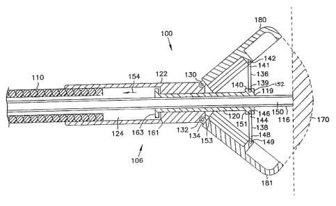
Referring to Fig. 8, there is illustrated the distal end 106 of an
integrated optical biopsy forceps provided in accordance with a further
embodiment of the invention. The optical biopsy forceps includes an optical
fiber 150 and opposed forceps cutting jaws t 80 and 181, which can be similar
to
the optical fiber and the jaws of forceps 10 shown in figs. 1-6. The optical
fiber
150 of the optical biopsy forceps includes an outer tubular, sheath-like
member
or catheter body 110, which corresponds to the outer sheath or coil 22 (Fig.
2),
and a reinforcement cover 1 16, which, for example, can be a metal coil or
cable,
a nylon sheath, or any other suitable cover. The reinforced optical f ber is
movable axially within the sheath 1 10. The optical biopsy forceps further
includes a tubular slide mernber 120 connected to the optical fiber and
movable
therewith, and coupled to the jaws 180 and 181 for actuating the jaws 180 and
CA 02253646 2001-06-1A
WO 97141777 PCTlUS97107784
11
181 as the optical fiber is rnovcd within the outer sheath 110.
'Ihe optical biopsy forceps includes a suitable handle (not shown)
for facilitating actuation of the tubular slide member 120. Preferably, the
handle
is similar to the handle 12 (Fig. 1 ) of the optical biopsyy forceps 10, but
the
handle can inehude any type of actuating mechanism capable of imparting
bidirectional axial movement to the optical fiber 150 of the optical biopsy
forceps. Referzing additionally to Fig. I, in such arrangement, the optical
fiber
150 positioned within the outer sheath, extends over the main length of the
device, from the distal end 106 to the handle. The proximal end of the sheath
110 passes through a sleeve, such as sleeve 24, and is secured to the tip of
the
handle. The sleeve provides reinforcement and strain relief where the sheath
110
is attached to the handle. The proximal end of the optical fiber 150 also
passes
through sleeve 24 and is secured to the slider 30 of the handle 12 distally of
the
proximal end of the optical fiber 150, the end portion of which passes through
I 5 the slider and out of the handle for connection to suitable electro-
optical units in
the manner that has been described for the optical fiber 50 of optical biopsy
forceps 10. The slider 30 of the handle is adapted to push the reinforced
optical
fiber 150, which in turn pushes the tubular slide member 120, to open the jaws
of
the optical biopsy forceps and to~ pull the reinforced optical fiber, pulling
the
tubular slide member 120, to close the jaws.
The optical biopsy forceps of the present invention can be used
with any type of electro-optical technique for guiding the forceps. This may
include systems which use viewing or imaging, systems which use illumination
with white light to excite dyes in the area of interest, and spectroscopic
techniques to identify tissue types by spectral analysis of light returned
from
tissue illuminated with light of certain wavelengths. Such spectroscopic
techniques utilize the property of certain tissue types to reflect or
fluoresce light
having characteristic wavelengdzs.
Considering the optical biopsy forceps in more detail, with
reference to Fig. 8, the sheath 110 is a flexible hollow catheter which can be
made a plastic tube, or a plastic~rmetal composite structure that defines an
CA 02253646 1998-11-05
WO 97141777 PCTIUS97I07784
opening or tore therethrough. By way of example, the outer sheath 110 can be
similar to those of disposable biopsy forceps commonly used with colonoscopes
used in the upper and lower gastrointestinal tracts, and broncoscopes used in
the
trachea and bronchus. Alternatively, the outer sheath 110 can be a rigid tube,
such as those of biopsy forceps commonly used with cystoscopes, colposcopes
and laproscopes.
At its distal e;nd, the optical fiber 150 extends through a central
bore 119 formed through a tubular slide member 120 which, in turn, is mounted
in a mounting member or jaw support block 122 which serves as a mounting
member for the cutting jaws. 180, 181. The jaw support block 122 can be
machined from stainless steel or formed of other suitable material. Thc.jaw
support block 122 has a bore 124 running therethrough which is generally
circular in section. The inner dimension of the jaw support block 122
corresponds to the outer dimension of the outer sheath 110 which is secured to
the support block in a suitable manner, such as with cement or by crimping.
'the
jaws 180, I 81 are hinged to the support block 122 which has a pair of holes
which receive pins 130, 132 which pass through ears 134 of the jaws to hold
the
jaws 180, 181 in place. The attachment of~the jaws to the support block by
ears
134, as seen in side view in Fig. 8, allows the jaws 180, 181 to fold against
the
front end of the support block when closed so as to have a thin profile for
the
distal end of the forceps for ease of introduction and navigation. 'Fhe jaw
support block 122 has a slot to control travel of the jaws 180 and 181.
The tubular slide member 120 is mounted in the bore 124 in the
jaw support block 122 and is free to move axially within support block 122
during actuation of the jaws. The fiber 150 is secured to the tubular slide
member 120 in a suitable manner such as with cement. The jaws 180, 181 are
connected to the tubular slide member 120 by a pair of control links 136, 138,
which are rigid members that function as a linkage mechanism connecting the
cutting jaw~~ to the tubular slide member. Control link 136 has one end 139
connected to tubular slide member 120 by a pin 140. The other end 141 of the
control link 136 is connected to jaw 180 by a pin 142. Similarly, control link
CA 02253646 1998-11-06
13
138 has one end 144 connected to tubular slide member 1?0 by a pin 146 and its
other end 148 connected to jaw 181 by a pin 149. Thus, axial movement of the
optical fiber in the direction of arrow 1 ~4, as the optical fiber is retracte
1. causes
axial movement of tubular slide member 1?0, pivoting the control lima 136..
138, about their ends 139 ar;d 144, respectively, drawing the jaws together to
actuate the cutting jaws 180, 181. The rearward surface 1 ~ 1 at the distal
end I ~~
of the tubular slide member 1?0 is adapted to engage the forward surface 1 >;
of
the jaw support block 1?~_. functioning.: as a travel limit stop surface to
limit the
axial movement of the tubular slide member 1?0 during retraction of the
optical
fiber 1 ~0. Similarly, when the optical fiber 1 ~0 is advanced into the sheath
1 1'_'.
the tubular slide member 120 is moved axially in the opposite direction.
causing
the control links 136. 138 to move the jaws apart. The forward surface 161 at
the proximal end 16~ of the tubular slide member 1?0 is adapted to enga~Te the
1 ~ rearward surface 16 3 of the jaw support block 1??. functioning as a tram.
e1 limit
stop surface to limit the axial movement of the tubular slide member 1?0
during
retraction of the optical fiber 1 ~0. Thus, both the proximal and distal ends
of the
tubular slide n_ember 1?0 include limit stops which prevent both over
distention
and over retraction of the optical fiber 1 ~0.
?0 Referring additionally to Fig. l, in operation of the optical biopsy
forceps. initially, the optical fiber 1 ~0 is ftrllv retracted (bv
retracting_> the slider
30 toward the back of the handle) to move the tubular slide member 1~0 in the
direction of the arrow 1 ~4 until its rearward surface 1 ~ 1 en_aUes forward
surface
1 ~~ Of the j2~.1' SLippOrt blOCl~:: ' ~'~. In this pOSitiOn, the COr1Lr01
lin~CS 1 36 arid 1 3Q
?~ have been drawn rearwardlv, drawing the jaws 180, 181 together so that
the,jaws
are closed. In this configuration. the distal end 106 of the forceps is
substantially
of the same narrow diameter as the outer sheath 116 which defines the main
body portion of the optical biopsy forceps. and the closed jaws have a smooth,
rounded shape to facilitate introduction and navi<~ation through the biopsy
30 channel of an endoscope, for example.
The endos;;opist advances the optical biopsy forceps through the
biopsy channel of the endosc:ope to the general area of interest. i.e., such
as a
CA 02253646 1998-11-05
WO 97/41777 PCTIUS97107784
tissue site or tissue analysis zone with a body, represented by the reference
numeral 17(1. Once in placf; in the general area of interest, the forceps jaws
can
be opened by advancing the slider 30, thereby advancing the optical fiber I 50
forwardly tl-trough the handle. This causes the tubular slide member 120 to
move
forwardly (to the right in Fig. 8), which in turn causes pivoting of the
control
links 136 anal 138. As the control links pivot, the control links push against
the
jaws, causing the jaws to o~>en. Simultaneously, the distal tip of the optical
fiber
150 is axially extended forvvardly beyond the jaws. The forceps may then be
used for optical tissue identification.
When an area of disease is identified and if a biopsy of it is
needed, the slider 30 is retracted. retracting the optical fiber 150 and thus
the
tubular slid; member 120, retracting the tip of the optical fiber and
simultaneously causing the jaws to close and cut a biopsy sample at the exact
place that h~~s been located by viewing through the optical fiber. To take the
i 5 tissue sample, the endoscopist holding the instrument by the handle,
gently pulls
back on the slider of the handle, retracting the optical fiber and tubular
slide
member 120, moving the optical fiber away from the tissue surface. As the
optical fiber is being retracted, the jaws begin to close as the tubular slide
member is moved in the direction of the arrow 154. While the jaws are being
closed, the endoscopist gently pushes on the instrument to urge the jaws
towards
the tissue surface so that a tissue sample will be captured by the jaws as
they
close. When the jaws are closed, the endoscopist pulls the entire assembly
away
from the tissue surface and then withdraws the optical biopsy forceps from the
endoscope so that the specimen tissue can be retrieved.
Thus, the present invention has provided an optical biopsy
forceps. An important feature of the invention is that the tip of the optical
fiber
50 (and optical fiber 150) is coaxial with, and perfectly aligned with, the
cone
where the two jaws 80, 81 (and jaws 180, 181 ) intersect and the sample is
taken.
Thus, there is no offset or'parallax' error between the spot where the optical
measurements were taken and the spot from which the biopsy sample will be
taken. This, together with the slim and compact profile of the device when the
CA 02253646 1998-11-05
WO 97141777 PCTIUS97I07784
~ J 5-
jaws are retracted, is a great improvement over prior art devices. In
accordance
with another feature, the fiber optic assembly, including the optical fiber
and the
tubular slide member of the biopsy forceps, can be produced as a disposable
assembly, with the rest of the biopsy forceps being produced as a non-
disposable
unit. 'The major advantage of forceps 100 as compared to forceps 10 is,
because
the biopsy jaw control wires 40, 41 are not required, larger diameter optical
fibers can be used to increase the detected signal relative to noise.
It will be appreciated from the foregoing that we have provided an
improved optical biopsy forceps which provides the physician a greater degree
of
accuracy and control ovex the diagnosis process than was previously possible.
While we have illustrated the invention with two illustrative embodiments of
the
invention, it will be appreciated that variations of shapes, materials and
assembly
are possible, within the scope of the invention.