Note: Descriptions are shown in the official language in which they were submitted.
CA 02254223 1998-11-16
1
DEVICE AND METHOD FOR ANALYZING A BIOLOGIC SAMPLE
Field of the Invention
This invention relates to a device for separating a fluid component,
such as plasma, from a biologic sample, such as blood, using microspheres and
analyte specific labeling . This invention also relates to a device and method
for
quantitative determination of an amount of analyte present in biologic fluids.
The
invention further relates to a quantitative assay method and device for
measuring one
or more analytes in a biologic fluid sample using a point-of care assay method
and
device. The test results can be analyzed using a suitable analyzer and,
optionally, the
assay test results are transmitted by way of digital transmission systems to
permit
further evaluation of the data.
Background of the Invention
There are presently many examples of one step assays for measuring
analytes in fluid. A common assay is the pregnancy test device which involves
contacting a urine sample with a test pad, which urine moves by capillary flow
along
the bibulous chromatography strips whereby the presence of human chorionic
gonadotropin (HCG) will be detected usually as shown by a coloured line
because of
the reaction between HCG and reagents in the bibulous chromatography strips.
This
is an example of a chromatographic assay.
U.S. Patent 5,766,961 issued June 16, 1998 and U.S. Patent 5,770,460
issued June 23, 1998 are both entitled "One-Step Lateral Flow Nonbibulous
Assay".
"Nonbibulous lateral flow" refers to liquid flow in which all of the dissolved
or
dispersed components of a liquid, which are not permanently entrapped or
filtered out,
are carried at substantially equal rates and with relatively unimpaired flow
laterally
through a stabilized membrane. This is distinguished from preferential
retention of
CA 02254223 1998-11-16
2
one or more components as would occur, for example, in materials capable of
absorbing or imbibing one or more components, as occurs in chromatographic
configurations. In this one-step assay, a sample (which may contain the
analyte of
interest) is collected on the "sample receiving zone" from which it flows to
the
"labelling zone" at which point it encounters a specific binding reagent for
the analyte
coupled to visible moieties (the "assay label"), then flows to a "capture
zone" where
the analyte bound to visible moieties is captured.
In U.S. Patent 5,540,888 issued July 30, 1996 and entitled "Liquid
Transfer Assay Devices", the invention described is a device for biochemical
diagnostic assays. It comprises two liquid flow channels of porous material
which
transfer liquid by capillary flow to a common site following simultaneous
application
of the liquid to the ends of the channels. The channels interconnect at a
certain point
and then both continue in an arrangement analogous to an electrical bridge
circuit. By
selecting the hydraulic resistances of the arms of this circuit, the flow can
be
controlled across the bridge.
U.S. Patent 5,300,779 issued April 5, 1994 entitled "Capillary Flow
Device" describes methods and devices for measuring an analyte in a sample
mixed
with reagents, the devices defining a flow path. The specific binding by
agglutination
may provide for changes in flow rate, light patterns of a flowing medium, or
light
absorption or scattering which permit measurement of the analyte of interest.
In U.S. Patent 5,110,724 issued May 5, 1992, entitled "Mufti-Analyte
Device", the invention described is an assay device for assaying multiple
analytes in a
drop-sized blood sample. A dispenser distributes a small volume blood sample
to
multiple transfer sites by capillary flow of the blood sample through sieving
and
distributing matrices which separate blood cells from plasma as the sample
fluid
migrates toward the transfer sites. A test plate in the device carries
multiple absorbent
pads, each containing reagent components for use in detection of a selected
analyte.
The test plate is mounted on the dispenser toward and away from a transfer
position at
CA 02254223 1998-11-16
3
which the exposed surface regions of the pads are in contact with associated
sample-
transfer sites, for simultaneous transfer of sample fluid from such sites to
the pads in
the support.
In U.S. Patent 5,039,617 entitled "Capillary Flow Device and Method
for Measuring Activated Partial Thromboplastin Time", the invention described
measures "activated partial thromboplastin time" (APTT) on a whole blood
sample by
applying the sample to a capillary tract with reagents capable of initiating
an APTT
analysis, wherein clotting time is measured by the cessation of blood flow in
the
capillary tract. This is an example of a risk evaluation based on coagulation.
In U.S. Patent 4,753,776 entitled "Blood Separation Device
Comprising a Filter and a Capillary Flow Pathway Exiting the Filter", the
invention
describes a method for separating plasma from red blood cells. The driving
force for
the movement of plasma from the filter to the reaction area of a device
utilizing the
method is capillary force provided by a tubular capillary. A filter is
selected from
glass microfiber filters of specified characteristics.
The U.S. Patent 5,135,719 issued August 4, 1992, entitled "Blood
Separation Device Comprising a Filter and Capillary Flow Pathway Exiting the
Filter", the similar invention is described and the glass fibre filters are
prepared from
fibers with diameters between 0.10 and 7.0 ~,m.
In U.S. Patent 4,447,546 issued May 8, 1984, entitled "Fluorescent
Immunoassay Employing Optical Fibre in Capillary Tube", a short length of
precise
diameter capillary tubing with an axially disposed optical fibre to which is
immobilized a monolayer of a component of the antibody antigen complex (eg. an
antibody) is described. The tubing is immersed in the sample.
U.S. Patent 5,610,077 issued March 11, 1997, entitled "Processes and
Apparatus for Carrying Out Specific Binding Assays", describes the well known
CA 02254223 1998-11-16
4
antibody binding to antigen assay. The sample which may contain the analyte
(a),
(the substance being tested for) is mixed with (b) an antibody which binds to
the
substance being tested for, which antibody is immobilized on a solid support,
and (c)
another antibody for the substance being tested for which is conjugated to a
detectable
marker, to thereby form a complex between (b), the substance being tested for
and (c)
and causes the marker to be immobilized and detected.
In U.S. Patent 4,943,522 issued July 24, 1990, entitled "Lateral Flow,
Non-Bibulous Membrane Assay Protocols", the described invention is a method
and
apparatus for conducting specific binding pair assays, such as immunoassays,
the test
substrate is a porous membrane on which a member of the binding pair is
affixed in an
"indicator zone". The sample is applied and is permitted to flow laterally
through the
indicator zone and any analyte in the sample is complexed with the affixed
specific
binding member, and detected. A novel method of detection employs entrapment
of
observable particle in the complex, for instance, red blood cells of blood can
be used
as the observable particles for detection of the complex.
An example of a method to separate red blood cells from whole blood
samples is found in U.S. Patent 5,118,428 issued June 2, 1992, entitled
"Method to
Remove Red Blood Cells from Whole Blood Samples". In the described invention,
red blood cells are removed from whole blood samples with a solution
containing an
acid. The agglutinated red blood cells are then removed from the resulting
suspension
by procedures of filtration, centrifugation or decantation, leaving an
essentially red
blood cell-free serum or plasma sample.
In U.S. Patent 5,073,484, entitled "Quantitative Analysis Apparatus
and Method", an analyte is measured along a liquid flow path which includes a
number of reaction-containing reaction zones spaced apart along the flow path.
Detector means are employed to detect analyte, reactant or predetermined
product in
the reaction zones, the number of zones in which detection occurs indicating
the
amount of analyte in the liquid.
CA 02254223 1998-11-16
In U.S. Patent 5,536,470 issued July 16, 1986, entitled "Test Carrier
for Determining an Analyte in Whole Blood", red blood cells cannot gain access
from
the blood sample application side, to the detection side and on the detection
side as a
result of an analysis reaction, an optically detectable change occurs.
A serious deficiency in current one-step assays for the measurement
and/or detection of an analyte is that they provide only qualitative results
rather than
quantitative results. That is to say that the presence or absence of the
analyte may be
determined but the actual amount or concentration of analyte present in the
sample
would still not be known. The assay of the present invention provides
quantitative
results as the test is performed in a determinable volume. In the prior art
methods it is
not possible to consistently identify the exact volume of the test sample in
repeated
testings since the fluids must wash through the test strips.
Prior art methods using chromatographic strips and fiberglass strips
require larger initial volumes of the biologic fluid in order to mobilize the
proteins
and labels in the strips. This is particularly true when the biologic fluid is
blood and
the plasma must first be separated from the blood sample. An advantage of the
device
and method of the present application is that very small fluid samples can be
used to
measure one or more analytes. The assay method and device of the present
invention
is also advantageous because the test volume can be made constant and
therefore
repeated testings will yield quantitative data which can be directly compared
between
samples and within a sample.
It is an advantage of the present invention that the assay device and
methodology allows for separation of the plasma from the whole blood during
the
assaying of a fluid sample. In other words it is not necessary to previously
separate
out the cellular component of the blood before assaying the sample. This is a
significant advantage as it allows that the assay can be used at the point of
patient
care, for example, by the patient themself, at the patient's bedside or in a
doctor's
office. In a preferred embodiment of the present invention there is provided
by the
CA 02254223 1998-11-16
6
device and assay methodology of the present invention a generic point-of care
platform suitable for use in one or more diagnostic or prognostic assays
performed on
one or more fluid samples.
Summary of the Invention
In accordance with an aspect of the present invention a method for
separating out the fluid component of a biologic sample using microspheres is
provided. In one embodiment, the biologic sample is placed in contact with a
group
of microspheres and the fluid component separates from the sample as the fluid
portion flows through the microspheres, by capillary action.
In accordance with an aspect of the present invention a quantitative
assay method and device are provided for measuring one or more analytes in a
fluid
sample using a point-of care assay method and device. The assay and device are
designed for use by a patient themself, at the bedside of a patient, or in a
doctor's
office. The test results are analyzed using a suitable analyzer and,
optionally, the
assay test results are transmitted by way of digital transmission systems to
permit
further evaluation of the data by an off site professional.
In accordance with an aspect of the present invention, an assay method
and portable assay device are provided for testing small volumes of biologic
fluids,
including blood, in a timely manner. In accordance with another aspect of the
present
invention, a method and device are provided for testing samples of biologic
fluids in
which a consistent volume of the biologic fluid sample is tested for one or
more
analytes and the data generated from the tests are used for collecting and
compiling in
a database pertaining, for example, to a particular disease condition.
Ultimately the
data collected can be used to train neural network algorithms and the
algorithms may
then be used to provide diagnostic and/or prognostic information based on the
individual test results of any given test subject.
CA 02254223 1998-11-16
In accordance with another aspect of the present invention in respect to
the analysis of blood, the cellular components of blood are separated from
plasma by
allowing the whole blood to be exposed to microsphere beads which permit the
plasma to pass in the spaces formed between the microspheres by capillary
action but
not the cellular component. The present invention is not limited to the
separation of
cells from plasma in blood but includes broader applications where microsphere
beads
may be used to separate a fluid component from a cellular component in a
biologic
fluid. The microsphere beads are effectively acting as a fluid filter.
According to another aspect of the present invention a device is
provided for separating plasma from blood in a sample. The device comprises a
plurality of microspheres disposed in abutting relation and forming
therebetween a
plurality of capillary channels, whereby when the microspheres are disposed in
fluid
communication with a blood sample cellular and plasma components of the
biologic
sample are separated by capillary flow of the plasma component through the
capillary
channels formed by the interstitial spacing between abutting microspheres.
According to another aspect of the present invention the device
comprises a plurality of groups of smaller microspheres each impregnated with
a
different label and interspersed with the larger microspheres in separate
zones of the
larger microspheres. The microspheres may be of substantially the same
diameter, or
the microspheres may be of differing diameters. The size of microsphere
selected
may be based on the viscosity of the sample or the size of the component one
wishes
to exclude or separate.
In accordance with yet another aspect of the present invention, the
microspheres are bundled in a fluid-permeable material or the microspheres are
maintained in abutting relation by a surface tension of the fluid which passes
through
them, for example plasma. In accordance with yet another aspect of the present
invention the microsphere beads, also known simply as microspheres, are dried
on a
surface of the device.
CA 02254223 1998-11-16
In accordance with another aspect of the present invention, the device
comprises a sample shelf adjacent to the fluid entrance and the microspheres
are
disposed on the sample shelf.
According to yet another aspect of the present invention the device
comprises a plurality of smaller microspheres which are impregnated with at
least one
label interspersed with a plurality of larger microspheres such that the
smaller
microspheres occupy the interstitial spacing between the larger microspheres
and
release a label into the fluid as it flows through the interstitial spacing
between the
larger microspheres. There may be a plurality of groups of smaller
microspheres each
impregnated with a different label and interspersed with the larger
microspheres in
separate zones of the larger microspheres. Alternatively, the smaller
microspheres
may be mobilized and carried forward by the fluid as it passes along the
capillary
channels formed by the larger microspheres.
In accordance with another aspect of the present invention, the device
comprises an indicator containing patient identification information to be
associated
with results of the assay, for example a bar code which can be read by a bar
code
reader.
According to another aspect of the present invention, a method of
separating fluid from a biologic sample is provided. The sample has a fluid
component and a non-fluid component and the method comprises the steps of,
(a) bringing the sample into fluid communication with a plurality
of microspheres disposed in abutting relation and forming therebetween a
plurality
interstitial spaces which connect to comprise capillary channels, and
(b) collecting the fluid component as it is separated by capillary
flow of the fluid component through the capillary channels. According to
another
aspect of the present invention there is provided, a method of conducting an
assay
utilizing a device comprising a capillary chamber defined by first and second
opposed
CA 02254223 1998-11-16
9
surfaces spaced a capillary distance apart having a fluid entrance and at
least one
reagent disposed within the capillary chamber, comprising the steps of,
(a) conveying a fluid sample into fluid communication with the
fluid entrance such that the fluid sample is drawn into the capillary chamber
by
capillary action and reacts with the reagent, and
(b) analyzing the reagent to determine whether the reagent binds to
an analyte in the fluid sample.
According to another aspect of the present invention the method further
comprise the step of analyzing the reagent to determine a proportion of the
reagent
which binds to the sample.
According to another aspect of the present invention, the method
further comprises a plurality of capillary chambers for conducting a plurality
of assays
on one or more fluid samples. According to another aspect of the present
invention
the results of the tests are recorded in a computer database and may be
further applied
in a trained neural network algorithm to generate a profile of one or more
selected
disorders. The assay further comprising the step of applying a receiver
operating
characteristic analysis to the data to determine a statistical significance of
the data.
In accordance with another aspect of the present invention a wick or a
capillary is brought into fluid communication with the fluid sample to remove
the
fluid sample from the capillary chamber.
In accordance with another aspect of the present invention
microspheres are used to separate a cellular component from a fluid component
in a
biologic fluid, for example plasma from whole blood, and the fluid component
can be
tested in chromatography test strips. Furthermore, the microsphere beads of
the
present invention may be used as a labeling device, in addition to a
filtration device,
in standard nitrocellulose chromatography assays.
CA 02254223 1998-11-16
Other and further details of this preferred embodiments are described
in the Detailed Description of the Preferred Embodiments together with the
drawings
described below.
Brief Description of the Drawings
5 For the purpose of illustrating the invention, there is shown in the
drawings a form which is presently preferred. It is not intended that this
invention be
limited to the precise arrangements and instrumentalities shown. The present
invention will be described in detail with reference to the accompanying
drawings, in
which like numerals denote like parts in the several views, and in which:
10 Figure 1 is an schematic, exploded, perspective view of an embodiment of
the device
of the present invention.
Figure 2 is a longitudinal cross section of the preferred embodiment
illustrated in
Figure 1 along line lA - lA.
Figure 2A is an end elevation view of the device illustrated in Figure 2 taken
from the
perspective of line 2A - 2A.
Figure 3 is a side view of an embodiment described in Example 1 illustrating
the
cover slip in relation to the beads when starting to form the curl.
Figure 4 is also a side view of an embodiment described in Example 1
illustrating the
curl after formation;
Figure 5 is another side view of an embodiment described in Example 1
illustrating
the position of the cover slip in relationship to the beads on the microscope
slide.
Figure 6 is a top plan view of an embodiment described in Example 1.
CA 02254223 1998-11-16
11
Figure 7 is a side view of an embodiment described in Example 2 illustrating
the label
pad variant.
Figure 7A is a side view of another embodiment described in Example 2
illustrating
the replacement of the label pad with microsphere beads.
Figure 8 illustrates an example ROC curve for the expected test results for a
neural
network risk analysis test.
Detailed Description of the Preferred Embodiments
The present invention relates to a method of separating a fluid
component from a biologic sample using microsphere beads. The present
invention
further relates to a device and a method for analyzing the presence or absence
of an
analyte in a biologic fluid sample. The invention also relates to quantifying
with
precision the amount of one ore more analytes present in a biologic fluid
sample. The
present invention further relates to an assay which can interpret test results
and be
used to further identify certain medical conditions from which a person or
animal may
be suffering or is likely to suffer from in the future. The present invention
further
relates to a prognostic assay technique in which the results of the test assay
defined in
the present invention may be used to predict the likelihood of a person or
animal
developing a certain condition or disease state at a future time. These
various
embodiments are described in detail herein.
Although the preferred embodiments described herein are described
with respect to the testing of human biologic samples it is well understood
that such
assays and methodologies could equally be used for assessing biologic samples
in
other animals. In particular the present invention would clearly have
applicability to
veterinary services.
CA 02254223 1998-11-16
12
In a biologic fluid sample having a fluid component and a non-fluid
component, the fluid component containing an analyte of interest the present
invention may be used to measure any of the following, alone or in
combination:
a) the presence of the analyte in the sample
b) the absence of the analyte in the sample
c) concentration of the analyte in the sample
d) total amount of analyte in the sample.
Suitable analytes which may be measured by the assay and device of the
present invention include soluble analytes: including but not limited to,
enzymes,
proteins, bacteria, viruses, antigens, antibodies, immunoglobulins, drugs, and
hormones. Other suitable analytes would be known to one skilled in the art.
The
assay and device of the present invention are useful for the detection and
measurement
of drugs of abuse in human biologic samples such as performance enhancing
drugs or
other street drugs.
Some biologic samples can be assayed without first separating out
cellular components; however, for example, in the case of blood, the cellular
component can interfere with the assay. In the case of biologic samples where
it is
necessary, or preferred, to remove the cellular component before assaying it
is
necessary to first separate the fluid component from any cellular components.
In the
case of blood, for example, it is necessary to separate the plasma from the
whole
blood so that the cellular components of the blood do not interfere with the
testing for
the analyte which is present in the plasma.
It is recognized in the present invention, surprisingly, the fluid
component of a biologic sample can be separated from its non-fluid component
by
applying the sample to a grouping of microsphere beads. When the sample is
applied
the fluid component will flow in between microsphere beads thereby separating
it
from the cellular components in a simple and effective way. The beads act as a
means
of separating the fluid component from the non-fluid component as the fluid
component moves by capillary action, through the spaces formed between the
beads,
CA 02254223 1998-11-16
13
when the beads are grouped together. So, in the case of blood, the plasma is
separated
from the cells in the blood sample. It has been surprisingly recognized in the
present
invention that microspheres have the ability to separate out the plasma from
whole
blood quickly and efficiently.
For the purposes of this patent application the spaces between the
beads are called "interstitial spaces" or "pores". It is believed that the
fluid flows by
capillary action from one interstitial space to the next.
In the present application the flow of the fluid passing through the
interstitial spaces between the beads is likened to flowing along channels
formed by
the spaces between the beads. The channels are referred to as "capillary"
channels
because it appears that the fluid flows between the beads by "capillary"
action.
When the microspheres are grouped together small spaces, interstitial
spaces, are formed between the microsphere beads. The size of the space formed
between the microspheres is a function of the radius of curvature of the
microspheres.
The radius of curvature is, for the purposes of the present invention, the
same as the
diameter of the microsphere. To understand the relationship between the
microsphere
bead size and the pore size which is formed between the beads, it is known
that the
ratio of the microsphere diameter to pore diameter is approximately 1 to 0.4.
In the
case of separating out the plasma from whole blood, a pore size of 4pm is
considered
optimal. Therefore, the bead size for this particular embodiment should be l
Oqm.
This permits an easy fluid flow (and therefore faster fluid flow) while still
preventing
cells from passing through the pores. The small spaces formed between the
beads
provide a certain capillarity when a fluid is present.
In the present invention the use of microspheres is an effective and
inexpensive means for separating plasma from whole blood as the erythrocytes
and
leukocytes in the blood will stay on one side of the beads while the plasma
portion of
the blood sample will pass through the beads, by capillary-like action along
the
interstitial spaces or pores, formed between the beads. It is considered that
the
CA 02254223 1998-11-16
14
capillary action observed in the present invention is related to the surface
tension
exerted by the microspheres on the fluid so as to draw the fluid forward. As
the fluid
is drawn forward between the microspheres it provides the additional advantage
of
mobilizing any reagents present in the region of the microspheres. For
example, the
microsphere layer could be impregnated with secondary antibodies or another
detection molecule.
The microsphere beads are effectively acting as a fluid filter and as
such can be used at any point in an assay where simple fluid filtration is
required.
Since it is believed that the microspheres act to filter the fluid component
from the
non-fluid component by capillary action, the microsphere filter may be termed
a
capillary filter and this term is used for that purpose herein.
The microspheres could have analyte specific antibodies bound to
them, for example, by adsorption or coupling. As the fluid containing the
plasma
passes through the capillary channels formed by the microspheres the analyte
will
mobilize the secondary antibodies contained on the microspheres and then react
with
the primary antibodies contained in the biochip. However, the microspheres may
act
solely to separate the cellular component from the fluid component and the
microspheres need not be labeled with antibodies.
Prior art technology has used chromatographic paper or other fibrous
material to wick the fluid component of a biologic sample away from the
cellular
component in order to perform tests on the fluid portion without interference
from the
cells or other substances present in the sample. The microspheres of the
present
invention provide an advantage over the prior art technology because it
provides
improved fluid flow without restriction by the fiber which is present in the
chromatographic paper. The microspheres provide a further advantage in that
they
provide an excellent surface for binding of proteins such as antibodies or
other
suitable labels.
CA 02254223 1998-11-16
The size of the microsphere beads used to separate the fluid component
can be varied based on the viscosity of the sample. Larger beads should be
used for
more viscous samples for faster fluid flow between the beads. Also, beads of
different
colours may be used to facilitate visualization of the beads when they are
used as
5 labels and bind to the analyte. The bound beads also serve to increase the
density of
any bound analyte for subsequent detection by a spectrometer. The regular
pattern of
the beads also means that diffraction difference could be used for detecting
and
measuring bound analyte.
The biologic sample may be applied to the top of the beads or at the
10 side of the beads.
In a preferred embodiment latex microsphere beads are used such as
those sold under the trademark Bang'sTM. The beads are supplied in a liquid
suspension. The beads can either be kept moist or dried when used for other
types of
beads could be used in the invention, including glass, so long as the beads
separating
15 out the fluid component.
The use of microsphere beads to quickly separate out a fluid
component from a biologic sample can be incorporated into assays for detecting
and
quantifying analytes present in the sample.
According to one aspect of the present invention this method of
separating out a fluid sample from a biologic sample using microsphere beads
is
incorporated into a one-step assay for analyzing one or more analytes which
may be
present in the fluid sample is provided. The assay is performed in association
with a
chamber of defined volume. In a preferred embodiment the chamber comprises
microsphere beads for separating out the fluid sample and detection means for
detecting and/or measuring an analyte in the sample. The detection means may
be
drawn from any of several known methods for detecting an analyte in a sample.
For
example the analyte may be recognized using detection protein, such as an
antibody or
antigen, which is specific to the analyte. When the analyte binds to the
detection
CA 02254223 1998-11-16
16
protein it changes density and may be measured. Alternatively, the detection
protein
may be bound to another label, which can be detected. For example the
detection
protein may be attached to a small bead so that when the detection molecule
binds to
the analyte the density will increase and this can be detected or measured.
Other
suitable labels would include metals such as gold, fluorescent labels,
chemical labels,
or colorimetric labels.
In accordance with an aspect of the present invention, this
invention pertains to a point-of care diagnostic or prognostic test in the
form of a
small chip or cassette for use in assaying biologic samples such as blood. The
present invention teaches a small, compact assay device referred to as a
"biochip" for
a simple assay taught in accordance with the present invention.
In the device of the present invention there is a pairing together of two
carrier surfaces in order to define a specific volume in which a quantitative
measurement of analyte(s) present in a drop of blood, urine, saliva or other
biologic
fluid may be measured. In a preferred embodiment the surfaces in question are
a
coverslip and a microscope slide but the present invention is not intended to
be
limited to only these specific embodiments. An important aspect of the present
invention is the fact that a fluid sample enters a space of defined volume by
capillary
action. The defined space is therefore referred to herein as a capillary
chamber. In the
case of a microscope slide and coverslip the capillary chamber is that volume
of space
between the bottom of the cover slip and the top of the slide.
In accordance with a preferred embodiment of the present invention,
the amount of fluid which is present between the plates or slides is
determined by the
volume of space between the slides. Therefore small test systems can be
designed
which allow for precision testing of very small volumes, in some cases, as
small as a
few microliters.
In order to quantitatively measure the concentration of an analyte in a
sample and to compare test results from one test to another it is advantageous
to have
CA 02254223 1998-11-16
17
a consistent test volume of the fluid sample each time the assay is performed.
In this
way the analyte measurement is assessed directly without having to adjust for
varying
volumes. The concentration or quantity of analyte can be assessed directly
without
difficulty and with consistency from test to test. The chamber of the biochip
of the
present invention provides that defined volume.
In accordance with one aspect of the present invention the fluid volume
in which the measurement of an analyte is performed is standardized. In
accordance
with another aspect of the present invention a method is provided for
separating the
plasma from the blood cells in a very small blood volume since it is most
practical to
be able to perform these tests with only a droplet of blood, for example from
a finger
prick, rather than requiring a larger volume only available by taking a tube
of blood
through a needle.
In one preferred embodiment the biochip test devices comprises a
chamber of a determinable volume. The chamber is defined by first and second
opposed carrier surfaces. The surfaces are positioned so that they are
separated by a
distance which is sufficiently narrow to permit fluid to flow between the two
surfaces
by capillary action. The chamber has a defined volume as it forms a defined
space.
The chamber has one or more points of fluid entrance which allow a fluid
sample to
enter. In this application, the chamber is also referred to as a capillary
chamber since
the fluid enters by capillary action.
For the purposes of the present invention this arrangement of the two
carrier surfaces joined together is referred to as a "biochip" but may also be
known as
a cassette or cartridge.
The intention is to provide a compact, portable test system which may
be standardized. In a particularly preferred embodiment the bottom surface is,
for
example, a microscope slide and the top surface is a microscope coverslip.
Microscope slides and coverslips are readily available and therefore are
useful carrier
surfaces. In another example two microscope slides could be mounted one on top
of
CA 02254223 1998-11-16
1g
the other, or any two plates, so long as there is a defined space between the
plates of a
determinable volume into which a fluid sample flows by capillary action.
Once in the capillary chamber, the fluid sample is retained by way of
surface tension at the ends and edges of the two surfaces. The device is of a
small size
which makes it portable and it can be inserted into an analyzer and reaction
products
between the analyte and detection molecules are measured using the analyzer.
For the
purposes of describing certain preferred embodiment the carrier surfaces will
be
referred to as plates; however, the invention is not to be limited only to
flat plates.
Similarly, all types of surfaces which are able to bind proteins, antigens and
other
detection molecules are contemplated with the scope of the present invention.
Specifically the composition of the carrier surface includes, but is not
limited to,
glass, plastic and metal.
In a preferred embodiment of the present invention a drop of biologic
sample is placed on the top surface of the microscope slide and, before
entering the
capillary chamber, the cellular component of the sample is removed by movement
of
the fluid component through a grouping of microsphere beads. For example, in
the
case of blood, the plasma is separated from the cellular component of blood by
movement through capillary channels formed by interstitial spaces between the
beads
and then the fluid enters the testing chamber in which the analyte reacts with
reagents
in the chamber and the reaction product is a measure of the analyte present in
the
sample.
Once the fluid has entered the defined space it is exposed to one or
more reagents present on an interior face of a carrier surface. The reagents
are
therefore exposed in the capillary chamber and available for reacting with one
or more
analytes which may be present in the fluid sample which ultimately fills the
capillary
chamber. The reagents are labelled and the quantity of analyte present in a
fluid
sample is measured based on a reaction product which results from the
interaction of
the analyte in the sample with the reagent in the chamber. The test results
are then
compared to standard calibrations to determine the quantity of analyte present
in the
CA 02254223 1998-11-16
19
sample. In a preferred embodiment of the present invention the reagent is one
or more
analyte specific antibodies which are adhered to the carrier surface,
preferably by
protein printing.
Alternatively, in another embodiment, an antigen is present on an
interior face of the carrier surface and the amount of antigen specific
antibody in the
sample is measured. When bound to the carrier surface the protein or other
detection
molecule will project into the defined space where it can react with the
analyte in the
sample. The detection molecule which is present on the interior face of the
carrier
surface may be bound to the surface by any one of several means known to a
person
skilled in the art.
Detection molecules are either coated, printed or otherwise bound to
one plate or the other using one of several techniques well known in the art.
Numerous techniques for immunoassays are known to persons in the art and are
described, for example, in "Principles and Practise of Immunology" (1997),
C.P. Price
and D.J. Newman eds. (Stockton Press) and this document is hereby incorporated
by
reference into the instant patent application and made a part hereof as if set
out in full
herein.
The distance between the two plates is limited only by the ability of the
plates to effectively draw a fluid such as plasma between the two plates by
capillary
action and to retain the fluid in the defined volume. The size of the plates
used would
also be dictated by practical considerations such as the desired volume for
testing.
Plates of larger surface areas would yield higher volumes.
In accordance with the present invention a fluid sample such as a drop
of blood is placed at one edge of the two plates and is drawn into the space
defined
between the plates. A fluid sample could be drawn against the edge of the two
plates
by any number of means which would be known to a person skilled in the art. In
its
simplest form the sample could be brought directly to touch the edges such
that a
portion of the fluid sample is drawn into the space so as to completely fill
the defined
CA 02254223 1998-11-16
volume of the space. For example by touching the patient's finger to the
plate. It is
important in the present assay that the sample always fill the defined volume
entirely
so that suitable quantitative analysis may be performed. In a standardized
model the
volume would be consistent from one biochip to another.
5 In another preferred embodiment the plates are joined together such
that the fluid sample may be readily removed. For example, at the end opposite
the
point of fluid entry.
In another example, the space between the two plates could be divided
into lanes and the volume of each lane would similarly be known. This approach
10 would allow multiple tests to be done on a single sample.
When dealing with a blood sample in which one wishes to measure a
plasma protein it is necessary to separate the plasma from the cells. In the
present
invention it is desirable that the test results be made available in a short
time frame,
preferably on the order of 1 to 30 minutes, from beginning to end. An
advantage of
15 the present invention is that the fluid sample enters the test chamber in a
shorter time
than prior art assays since the use of microsphere beads to separate the
plasma from
the blood sample, for example, eliminates the delay which would occur using
fiberglass or chromatographic strips. Cumbersome equipment such as a
centrifuge is
not required for cell separation. All of which facilitates the test being
performed at
20 the point-of care.
The present invention has further advantages over the prior art since
the biochip device of the present invention permits several assays to be
performed on
one sample. This facilitates the speed with which test results can be obtained
and
minimizes the amount of sample required for testing.
Analyte-specific antibodies themselves may be labeled with anyone of
several labels known to persons skilled in the art of such assays. Examples of
preferred labels include fluorescent labels, colorimetric labels, another
microsphere,
gold particles or any high contrast molecule. Other labels would be suitable
so long
CA 02254223 1998-11-16
21
as the presence of the label can be detected. Similarly microsphere beads
having a
diameter which is smaller than the test beads can be used so that the smaller
beads are
mobilized through the larger beads with the movement of the fluid sample (e.g.
plasma). The smaller beads can be labeled accordingly.
When the fluid sample containing the analyte enters into the defined
space between the two plates a further antibody-antigen reaction may occur. In
the
present invention the upper plate, for example a coverslip, has analyte-
specific
reagents bound on the surface which comes in contact with the fluid. In a
preferred
embodiment of the present invention the analyte-specific reagents are printed
on the
interior surface of the carrier plate using a protein printer. Suitable
protein printing
devices are well known in the marketplace. These include ink jet, spray, piezo-
electric and bubble jet protein printers. The piezo-electric printer is
preferred. The
analyte-specific reagent acts as a detection molecule, typically proteins.
These
molecules adhere to glass, metal and plastic surfaces. Preferred surfaces
include
polystyrene or polypropylene. The use of such printing devices is advantageous
in the
present invention to allow several different analyte-specific detection
molecules to be
printed onto the plate or coverslip such that different "lanes" are defined
and different
analytes may be assessed simultaneously using a single fluid sample.
Additional
background and calibration lanes can be provided in the same test chamber.
After the analyte reacts with the analyte-specific detection molecule a
measurable reaction product will be produced. It is preferred that the biochip
carrier
surfaces be colorless or transparent such that a colorimetric, or fluorescent
or other
reaction products can be read using a suitable spectrometer or other
appropriate
detection coupled to a reader. When the analyte and analyte-specific detection
molecule react together there is a change in density in the reaction lane. In
a preferred
embodiment of the present invention, the change in density is measured to
determine
the amount of analyte present in the sample. In order to reduce the background
noise
and therefore increase the sensitivity of the assay a mask is provided in
accordance
with a preferred embodiment of the present invention. Referring to Figure 1
the mask
CA 02254223 1998-11-16
22
32 is made of an opaque material except for the openings 36, 38 and 40 which
correspond to lanes 26, 28 and 30 on the plate. The mask is designed to fit
neatly over
the upper plate 10 so that only the lanes themselves are available to be read.
The use
of the mask has the advantage of reducing the amount of background noise and
setting
baseline values when reading the density change in the lanes.
In a preferred aspect of the present invention, the biochip is designed to
be read by a portable spectrometer which reads for example, the change in
color after
the analyte has reacted with the labeled antibody. The spectrometer could also
read
changes in density, film thickness, mass absorption or diffraction depending
on the
test reagents used. Once the analyzer, e.g. spectrometer, has performed the
necessary
data calculations the results are transmissible by digital transmission over
the
telephone lines or other computer network system.
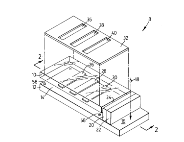
Turning to the figures, Figure 1, a preferred embodiment of the
biochip of the present application is illustrated in a schematic exploded
perspective
view. Two carrier plates 10 and 12 are provided. The two plates define a fixed
volume therebetween as indicated by reference number 14. Lower plate 12 may be
longer than upper plate 10 to provide a shelf which acts as an application
zone 16
upon which a biologic sample 18 may be applied. A shelf is not essential to
the
invention but provides a place to allow the sample to be separated by the
microsphere
beads. It is possible that the beads could be placed at the entrance of the
capillary
chamber 14 within the confines of the plates and the sample would be applied
to the
edge of the biochip where it would enter the chamber by capillary action.
Also affixed to application zone 16 is a collection of microsphere
beads 20 which may or may not also include a label zone 22. The microsphere
beads
20 may be grouped or bundled using a fluid-permeable material. For purposes of
the
schematic illustration, in Figures 1 and 2, the microsphere beads 20 and label
zone 22
are illustrated as separately defined regions; however the microsphere beads
may also
bear the label themselves and in this embodiment the two zones would converge
into
CA 02254223 1998-11-16
23
one with the microsphere beads playing two roles: separation of the fluid and
displaying a label to which the fluid is exposed.
More than one size of microsphere beads may be present. In one
embodiment, smaller microspheres could nestle in the interstitial spaces
formed by the
larger beads. The smaller beads could carry secondary labels which would bind
to the
analyte as it passes through the beads. Either the label would bind to the
analyte in
the fluid or the label attached to the small bead would attach to the analyte
in the fluid
and the small beads would then travel with the fluid into the capillary
chamber. At
the same time any cellular component in the fluid sample would not pass
through the
microsphere bead filter.
A patient ID may be affixed to either plate 10 or 12 so long as it does
not interfere with the test detection areas on the biochip or with reading the
biochip
after analyte has reacted with the substance bound to the carrier plate
surface. The
plates 10 and 12 are preferably colorless and/or transparent.
Three detection areas 26, 28, 30 are printed on the inner surface of
carrier plate 10: a calibration print zone 26, a detector print zone 28 and a
baseline
print zone 30. Three detection areas, or zones, are depicted for example only
to
illustrate how one test biochip may be set up; however, several lanes may be
present
and the number of lanes dedicated to calibration and/or background can vary
depending on what is being tested.
The test need not be limited to only three lanes. Several lanes could be
defined. In a preferred embodiment of the present invention three lanes are
printed on
the one plate to permit assessment of background readings as well as
calibration of the
biochip. It is understood that the background and calibration detection zones
need not
all be placed on the same biochip. It is advantageous to have the background
and
calibration readings made on the sample carrier plate in the same assay as the
test
analyte thereby reducing the variance in test results.
CA 02254223 1998-11-16
24
A background mask 32 is optionally provided. The mask is designed
to cover the outer surface of the carrier plate 10 without blocking the coated
or printed
detection zones/lanes. Therefore, openings 36, 38 and 40 are, for example,
present in
the mark to reduce background interference when reading test results. The
background mask is made of an opaque material with openings 36, 38 and 40
which
correspond to the detection zones 26, 28 and 30 identified on the inner
surface of the
upper plate. The opening 40 in the mask need not have a corresponding test
zone 30
as illustrated so long as the opening 40 is exposed to a part the plate 10
where
reagents are not present.
Although Figure 1 illustrates both an antibody/label zone 22 and a
microsphere zone 20, both of these zones are optional depending on the type of
test
one chooses to conduct. When fluid sample 18 is applied to application zone 16
it
flows through antibody/label zone 22 (if present) and microsphere bead zone 20
(if
present) before it reaches the edge 34 where the two plates 10 and 12 first
meet. In
the schematic illustration of Figure 1 there is a gap between the zone of
microsphere
beads and the fluid entry point identified by edge 34. Although this
arrangement of
the invention will work, it would be most preferred if the microsphere bead
zone 20
and/or label zone abutted against the edge 34 of the carrier plate 10. One
example of
such a configuration is illustrated in Figures 5 and 6. This configuration
provides the
least distance for the fluid sample to travel and this further minimizes the
amount of
fluid sample required for testing and is described in greater detail in
Example 1.
The fluid sample is drawn under edge 34 into the chamber 14 which
defines a known volume. The fluid sample should be of sufficient volume to
pass
along the application zone 16, through the microsphere and label zones) and to
completely fill the chamber 14. The biochip of the present invention can be
scaled to
a small size such that a single drop of blood could be a sufficient sample
size for
testing. Many dimensions are possible to construct based on the principles
taught
herein. Although dimensions of 1 cm x 3 cm make a device of convenient size,
the
nature of the testing to be done would dictate the optimum chip size. As
illustrated in
CA 02254223 1998-11-16
Figure 1 a shelf portion 16 extends on the bottom plate. On this shelf portion
the
biologic sample can be applied. In other embodiments, the portion of the test
which is
held, for example the microscope slide, may be large but the test assay itself
which
sits on the slide may be very small. The assay may be miniaturized to
accommodate
5 sample fluid volumes as small as about 1 microlitre.
Figure 2 is a sectional view taken along lines 2 - 2 illustrating the same
elements as referenced in Figure 1. Figure 2A is an end elevation view of
Figure 2
along lines 2A - 2A illustrating that the end of the device may be open, to
allow the
fluid to be removed from the chamber. One would want to remove fluid from the
10 chamber, for example, is you wanted to test the whole sample. A suitable
wicking
material would be applied to the open end and the fluid would be drawn through
thereby allowing additional fluid to enter the chamber. This could be either a
continuous or a discontinuous process.
Illustrated in all of Figures 1, 2 and 2A is a spot of glue 58 which is
15 one way to hold the plates 10 and 12 together. The glue 58 also illustrated
in Figure
6, another embodiment of the invention.
Figures 7 and 7A are illustrations of another use of the microsphere
method of separation in a one-step assay. In this embodiment the microspheres
are
used in conjunction with chromatography paper. The biologic sample 18 is
placed on
20 a surface such as a microscope slide 52'. It may be placed directly on the
microsphere
beads SO (as illustrated) or beside them. The fluid component of the sample
then
flows through the beads 50 separating from a non-fluid component present in
the
sample 18. The beads abut against or sit close to a fiberglass filter pad 60
which
abuts with a label pad 62. The label pad 62 is usually a fiberglass pad
impregnated
25 with the label of interest for labeling analyte in the fluid sample. The
fluid flows
through the filter 60 and label pad 62. Any analyte present in the fluid will
be labeled
as it flows through the label pad. The fluid then flows into the
nitrocellulose
chromatography strip 64 where the test results are read, usually as a color
change or
CA 02254223 1998-11-16
26
band on the nitrocellulose strip. Alternatively, since the microspheres 50 are
used as a
filter, the fiberglass filter 62 may be eliminated entirely (not illustrated).
Finally, as illustrated in Figure 7A, the fiberglass label pad 62 may be
replaced by microsphere beads 66. In this case the beads 66 are acting as a
source of
label, not as a filter and the fiberglass filter 60' serves as a spacer
between the two sets
of beads 50 and 66, respectively. For applications where filtration of a fluid
component is not required, the microspheres 66 can be used to label an analyte
present
in the fluid directly, without requiring the microsphere filter 50 or the
fiberglass
spacer 60'.
Figures 7 and 7A are illustrative of how current assay methodologies
may be modified using the microsphere bead technology of the present invention
as
taught herein.
The assay device and techniques of the present invention are very
useful in that they can be used for small volumes of many kinds of fluid
samples.
Although the description refers specifically to proteins any number of other
marker
would be suitable so long as a labeling system can be devised for the
detection and
measurement of the marker in the system. For example, the present invention
could
be used to measure and/or detect the presence of microorganisms such as
bacteria,
viruses, fungi or other infectious organisms. The biochip device of the
present
invention can be calibrated for the type of assay and the type of analyte so
that a table
of standard values may be constructed. The assay system or the present
invention can
detect the levels of a particular hormone or even the amount of a drug in a
patient's
system and this standardized data can be used to make diagnostic and/or
prognostic
determinations for a given individual.
Once the table of standard values is constructed data is collected on a
regular basis and databases constructed based on the patient's medical
history, current
health and the test results. Optionally, the data can be transmitted by
digital
transmission systems over a computer network via modem, the Internet, cable
lines,
CA 02254223 1998-11-16
27
telephone lines, satellite or other similar technology. These databases can be
used in
the development of neural network algorithms, for assessment of current
patient test
results and diagnoses as well as for predicting certain health outcomes for a
given
individual. One example of a neural network algorithm is found in Example 3
below
and a sample Receiver Operator Curve (ROC) is illustrated in Figure 8.
The development of the algorithms for the applied neural network will
be a function of the medical condition being assessed. Large amounts of
patient data
will first have to be accumulated in order to have reliable predictive
outcomes. The
neural network can be trained to recognize the concentration of analyte which
is
diagnostic or prognostic, using the standardized assays of the present
invention. The
data and algorithms are encoded in an electronic chip which is placed in the
reader,
for example a spectrometer, such that the printout from the reader will also
identify a
particular diagnosis or prognosis simultaneously with providing the test
result. In the
neural network algorithms, the diagnostic or prognostic test result will be
optimized as
the number of data points increases. With more patient data the predictive
and/or
diagnostic result will be made with greater certainty. The percent certainty
can be
calculated and provided to the physician or technician based on analysis of
the
measured data in comparison to a database contained in an electronic memory
chip
installed in the analyzer provided. Present technology makes it possible to
display
the actual standard curve on the reader itself at the time of printing out the
test results.
In the present invention, more than one test can be run simultaneously
on the same biochip and therefore the certainty of the diagnosis or prognosis
can be
improved. As the number of markers increases so does the certainty of
measurement.
One of the many examples of uses of the biochip/cassette of the present
invention is to measure blood proteins indicating peripheral vascular disease
using a
drop of the patient's blood.
CA 02254223 1998-11-16
28
Further details of the preferred embodiments of the invention are
illustrated in the following Examples which are understood to be non-limiting
with
respect to the appended claims.
Example 1 ~ Verification of Plasma Flow and Separation from Whole Human Blood
As illustrated schematically in Figures 3 to 6, approximately 15
microliters of 10 micrometer latex microsphere beads (Bang'sTM) 50 were
dropped
onto a glass slide 52 and allowed to dry. A glass coverslip 54 was placed on
the slide
and pushed, on edge, towards and along, the dried beads. The cover slip caused
the
dried beads to be separated from the glass slide and further caused the
collection of
dried beads to roll over thereby forming a curl 56. The cover slip was then
placed on
the slide with the "curl" touching the edge of the coverslip (illustrated in
Figures 5
and 6). The coverslip was fixed squarely in place on the slide with one edge
aligned
parallel to the edge of the curl of dried beads and this edge was left open to
allow fluid
to pass through the beads and into the capillary chamber formed between the
cover
slip and the glass slide. The coverslip was attached with nail polish at the
corners 58
of the coverslip to secure it to the microscope slide. The coverslip was
secured at a
spot where no capillary action was intended to take place to permit fluid to
flow freely
under the coverslip.
A 20 microliter drop of whole human blood 18 was placed on the
remaining 5 to 10 microliter microsphere beads. In other words, the sample of
whole
human blood was placed on the remaining portion of the beads which did not
form
part of the curl leaving the plasma component free to move by capillary action
through the curl portion of the microsphere beads and into the space defined
between
the coverslip and the slide (i.e. the capillary chamber). The effect was
observed under
a binocular light microscope. Upon application of the blood sample to the
beads the
plasma immediately began to separate from the whole blood. As the curl became
plasma soaked, capillary action between the coverslip and the slide drew the
pure,
clean, cell-free plasma under the coverslip into the chamber defined between
the
CA 02254223 1998-11-16
29
coverslip and the slide. This chamber defines a known space, the volume of
which
can be calculated and predetermined.
This demonstrated that the microsphere beads are able to readily and
effectively separate plasma from whole blood and to pass, via the capillary
channels
formed between the microsphere beads, into the capillary chamber.
Example 2' Micros~here Separation Combined With Chromato~raphy Strip
In an assay for an analyte in a human blood sample, this example
(schematically illustrated in Figure 7) demonstrated the use of microsphere
separation
of plasma from a blood sample of human whole blood. The plasma was separated
using latex microsphere beads (Bang'sTM) 50 and then drawn into a standard
nitrocellulose chromatography strip.
The fiberglass pads, which are usually used to retain red blood cells in
the prior art, were replaced with about 20 microliters of 10 micrometer latex
beads. A
drop of human blood (about 60 microliters) was placed on a surface 52', in
contact
with the latex microspheres. The fiberglass pad 60 effectively functions as a
spacer
between the beads 50 and the label pad 64 although it could also be used as a
second
filter. The fiberglass filter 60 may be eliminated entirely and the
microsphere beads
50 abut directly with the label pad 64 (not illustrated).
It was observed that the blood soaked the bead pile and within about 2
minutes clear plasma ran onto the nitrocellulose chromatography strip. This
was
observed with the visible eye and also under a microscope. This example
demonstrated that the microsphere method for separation of plasma from blood
can
also be used in conjunction with a standard nitrocellulose chromatography
strip. For
tests using such chromatography strips this is clearly an advantageous
methodology
for separating plasma from blood.
Illustrated in Figure 7A is another embodiment where, instead of a
fiberglass label pad 62, microsphere beads 66 are used as the label region of
the test
CA 02254223 1998-11-16
device. The fiberglass filter pad 60' is used as a spacer between the two sets
of beads,
50 and 66.
Example 3: Neural Network Marker Analysis
A neural network is a mathematical function N(W,a) which takes input
5 analyte vectors a=(al,a2...,an) and outputs numbers between 0 and 1. The
weight
parameters W are adjusted during the training period, using training patterns
{p=(bl,b2,...bn,T)} where bl,..,bn are training protein vectors, and T is the
target
output value. In the case of a coagulation test, T would be 1 for coagulation,
and 0 for
a non-coagulation.
10 The parameters W are adjusted to minimize the error E = E(N(W,a)-T)2
P
while maintaining good performance on new test data.
Once the Network is trained, a network cutoff C is chosen to classify
test data. Let TST(C,b,T) be the test result for a testing vector a, given
cutoff C, and
15 target output T.
{ 1 if N(a)>C
TST(C,b,T) _ {
{ 0 otherwise
Now, we can analyze the sensitivity and specificity of the test.
20 True Positive if T=1 and TST(C,b,T)=1
False Positive if T=0 and TST(C,b,T)=1
True Negative if T=0 and TST(C,b,T)=0
False Negative if T=1 and TST(C,b,T)=0
Sensitivity = TP/(TP+FN)
25 Specificity = TN/(TN+FP)
CA 02254223 1998-11-16
31
Plotting sensitivity versus 1-specificity for various cutoffs gives a ROC
(receiver operator characteristic) curve.
NEURAL NETWORKS
We start with a set of training patterns { p=( I1, I2 .... I1, TAR }, where
Ij is an input value, and TAR is the target value (TAR = 0 or TAR = 1 ). We
want to
train a neural network to give outputs which are close to the target values.
A neural network has 3 layers; the first INPUT layer, the second
HIDDEN layer, and the third OUTPUT layer:
INPUT HIDDEN OUTPUT
15
The neurons are connected by a set of weights { w(i,j,k) }. For
example, w(1,2,4) connects the second neuron of the first layer with the
fourth neuron
of the second layer.
For each pattern we assign a number called the activation to each
neuron, which measures the probability that it is firing. The activation is
defined
recursively as follows:
{Ij if i=1
a(i,j) _ {
{ 1/ (1 + exp (- sum(k) { w(i- l,k,j)a(i- l,k) } ))
The error is calculated as
ERMS=SQRT {sum{ (t- a(2,1))~2 } }
where the sum is over all patterns.
CA 02254223 1998-11-16
32
The weights are adjusted to minimize ERMS, while maintaining good
performance on new data.
CA 02254223 1998-11-16
33
QUANTITATIVE FLOW CHART
INPUT SERUM PROTEIN CONCENTRATIONS FOR
SPECIFIC DISORDERS OR CONDITIONS (FOR
EXAMPLE COAGULATION)
SCALE INPUTS TO VALUES BETWEEN 0 AND 1
RESULT: INPUT VECTOR (Q1, Q2, Q3, Q4)
FORWARD PASS THROUGH TRAINED NEUTRAL NETWORK,
WITH WEIGHTS { W(I,J,K) }
COMPARE OUTPUT OF NETWORK (OUT) TO CUTOFF (CUT)
{POSITIVE IF OUT>CUT
TEST RESULT = {
{NEGATIVE OTHERWISE
Those skilled in the art will recognize, or be able to ascertain using no more
than routine experimentation, many equivalents to the embodiments of the
invention
described specifically above. Such equivalents are intended to be encompassed
in the
scope of the following claims.