Note: Descriptions are shown in the official language in which they were submitted.
CA 022~68~ l998-ll-l6
W097/42975 PCT~S97/08487
DESCRIPTION
CHITOSAN RELATED COMPOSITIONS FOR DELIVERY OF NUCLEIC ACIDS INTO A CELL
Cross-Reference to Related Applications
This application claims the benefit of Mumper and
Rolland, ~.S. Provisional Application 60/018,342, entitled
"Chitosan Related Compositions and Methods for Delivery of
Nucleic Acids and Oligonucleotides into a Cell", filed May
17, 1996. This application is also related to Rolland and
Mumper, U.S. Patent Application Serial No. 08/372,213
entitled, "Formulated Nucleic Acid Compositions and
Methods of Administering the Same for Gene Therapy," filed
January 13, 1995. These applications are hereby
incorporated herein by reference in their entireties,
including any drawings and figures.
Field of Invention
This invention relates generally to the fields of
gene delivery and gene expression. In particular, it
relates to the delivery of nucleic acids and
oligonucleotides to cells using non-viral methods.
Backaround of the Invention
The following description of the background of the
invention is provided to aid in understanding the claimed
invention, but it is not admitted to constitute or
describe prior art to the claimed invention and should in
no way be construed as limiting the claimed invention.
Chitin, the chemical structure of which is shown in
Figure 1, is the main constituent in the shells of
crustaceans and is the most abundant naturally occurring
~ biopolymer other than cellulose. Chitosan, the chemical
structure of which is also shown in Figure 1, is derived
from chitin and can be formed by deacetylation of chitin.
Chitosan is commercially available in a wide variety of
CA 022~68~ l998-ll-l6
W097/42975 PCT~S97/08487
molecular weights (i.e., 10-1,000 kDa) and usually has a
degree of deacetylation ranging between 70% and 90%.
Chitosan has been reported to form compositions with
a variety of anionic drugs and polyanions such as
indomethacin, polyacrylate, pectin, acacia, alginate,
hyaluronate, and some polysaccharides (J. Kristl et al.,
Hydrocolloids and Gels of Chitosan as Drug Carriers. Int.
J. Pharm., 99; 13-19 (1993); S. Shiraishi et al.,
Controlled Release of Indomethacin by Chitosan-
Polvelectrolyte Complex: Optimization and In Vivo/In VitroEvaluation. J. Contr. ~el., 25; 217-225(1993); M.M.
Meshali and K.E. Gabr. Effect of Interpolymer Complex
Formation of Chitosan with Pectin or Acacia on the Release
Behavior of Chlorpromazine HCl. Int. J. Pharm., 89; 177-
181(1993); T. Nagai et al., Application of Chitin andChitosan to Pharmaceutical Preparations. In: "Chitin,
Chitosan, and Related Enzymes." Academic Press, New York,
1984, 21-39; H.E. Rios et al., Counterion Binding to
Cationic Polyelectrolytes in Aqueous Solution. J. Polym.
20 Sci., Polym. Phys. 29; 805-809(1991); T. Takahashi et
al., Characteristics of Polyion Complexes of Chitosan with
Sodium Alginate and Sodium Polyacrylate. Int. J. Pharm.,
61; 35-41(1990); K. Takayama et al., Effect of Inter-
polymer Complex Formation on Bioadhesive Propertv and Drug
Release Phenomenon of Compressed Tablets Consisting of
Chitosan and Sodium Hyaluronate. Chem. Pharm. Bull., 38;
1993-1997(1990); R. Srinivasan and R. Kamalam. Poly-
electrolyte Complexes of Glycol-Chitosan with Some Poly-
saccharides. I. Mixing Ratio and Dielectric Properties.
Biopolymers, 21; 251-263(1982).
These polyelectrolyte compositions with chitosan have
been well characterized in terms of optimal complexation
conditions (i. e., ionic strength, pH, temperature, and
~ ratios of components), composition morphology, and
composition stability. Chitosan has also been proposed
for use as a biomedical membrane, artificial skin, for
delivery of anti-cancer drugs to tumor cells, and as a
CA 022~68~ l998-ll-l6
W097/42975 PCT~S97/08487
pharmaceutical delivery system for prescription drugs. In
addition, chitosan has been shown to be biodegradable,
biocompatible, to have very low toxicity, and no
thrombogenic activity.
The use of chitosan as a component of a complex in a
non-viral gene delivery system in an in vitro use is
described in Mumper et al., Proceed . In tern . Symp .
Control . Rel . Bioact. Mater., 22:178-179, 1995, incor-
porated herein by reference in its entirety, including any
drawings and figures. Chitosan is described as effective
in condensing negatively charged plasmid DNA due to charge
interactions with the positively charged chitosan. Mumper
et al., report on the correlation between physicochemical
properties of the gene transfer complexes and their in-
vitro transfection efficiency. Specifically, they report
that the use of smaller molecular weight chitosan as a
component of the delivery system ( i . e ., chitosan in the
range of 2-4 kDa M.W.) results in the smallest particle of
the gene delivery system and also in an increased
transfection of cells with the condensed delivery system.
Chitosan has also been used with a pharmacologically
active compound such as insulin in the form of a solution
or as a coating on polystyrene microspheres. These
formulations involved the use of chitosan of molecular
weights of 10,000 or greater, preferably at least 100,000
or 200,000 and most preferably about 500,000. The
chitosan/insulin formulations were prepared by mixing
equal volumes of insulin and chitosan in solution. The
formulation was administered nasally to rats via micro-
syringe. These formulations have been reported asdisclosed in WO 90/09780.
The use of chitosan in microspheres containing naked
DNA has been reported by Alexakis et al., Applied
Biochemistry and Biotechnology, 50:93-106, 1995,
incorporated herein by reference in its entirety,
including any drawings and figures. The immobilized DNA
within chitosan-coated alginate microspheres was designed
CA 022~68~ 1998-ll-16
W097/42975 PCT~S97/08487
to test the role of metabolic byproducts of dlgestion in
promoting damage to DNA. The microspheres were designed
to pass through the digestive system without being taken
up by cells in the animal. Upon excretion, the intact
microsphere can be recovered and the DNA examined to asses
the role of metabolic byproducts of digestion in promoting
cancer through damage to nucleic acid. The microspheres
were designed to retain the DNA within their core during
transit through the animal. The microsphere prevented
access to DNA from hydrolytic enzymes but allowed
metabolic byproducts of digestion to cross or exit the
microsphere shell. The reported recovery rate of the
microspheres after administration was 97%. According to
the abstract, leakage of DNA from intact microspheres was
not observed.
Summary of the Invention
The invention features compositions of chitosan-
based compounds and nucleic acids or oligonucleotides.
The compositions are capable of non-viral gene delivery
(i.e., delivery of nucleic acid without the use of any
genomic viral components) via various routes of admin-
istration. The invention also features methods for the
preparation of chitosan-based compositions and methods for
the introduction of the compositions into a cell for
expression of nucleic acids, oligonucleotides or gene
products transported by the composition. The compositions
are useful for enhancing the administration to, and uptake
of, nucleic acids or oligonucleotides by an organism. The
compositions are also useful for in vitro transfections
and in vivo gene delivery, and among other things for the
administration of proteins, polypeptides, or peptides
encoded by the nucleic acid or oligonucleotide.
An efficient strategy for enhancing nucleic acid
delivery in vivo is to present, at the target site,
nucleic acid in composition of sufficient size to promote
its cellular uptake. The compositions of the present
CA 022~68~ 1998-11-16
W097/42975 PCT~S97/08487
invention, which are designed to administer nucleic acid
into a cell, comprise a substance which promotes condensa-
tion of nucleic acid when the substance is complexed with
the nucleic acid. The resulting composition is capable of
increasing the efficacy of transfecting cells in an
organism or cells in vitro cell culture.
Chitosan's properties are useful in complexing and
condensing nucleic acids or complexing oligonucletides.
DNA, which is a polyanionic nucleic acid has a high net
negative charge due to the presence of two phosphate
moieties on each base pair. Therefore, DNA is an excel-
lent candidate for complexation with chitosan and chitosan
oligomers for non-viral gene delivery. Neutralization of
the negative charge of DNA by the amine groups of chitosan
and chitosan oligomers results in condensation of DNA into
a compact particle which protects the DNA from nuclease
degradation and delivers the DNA, either specifically or
non-specifically, to target cells.
Chitosan has structural characteristics similar to
glycosamino-glycans (GAGs) and appears to mimic their
function (T. Chandy and C.P. Sharma. Chitosan As a
Biomaterial. Biomat ., Art . Cells, Art. Org., 18; 1-24
1990). GAGs are widely distributed among various tissues
and, like heparin sulphate proteoglycans (GAGS), may be a
component of cell membranes. Thus, chitosan may provide
natural targeting to cell surfaces (e.g., endothelial
cells). For example, chitin and chitosan have been
reported to selectively distribute to the surface of tumor
cells (T. Ouchi and T. Banba. Fixation of 5-Fluorouracil
to Chitosan and its Antitumor Activity. Trans. Soc.
Biomat. 11; 232(1988). A summary of the beneficial prop-
erties of chitin, chitosan, and chitosan oligomers for
gene and oligonucleotide delivery is shown in Table 1.
Thus, in one aspect, the invention features a
composition capable of delivering a nucleic acid or an
oligonucleotide to a cell. The composition includes a
CA 022~68~ l998-ll-l6
W097l4297S PCT~S97/08487
chitosan-based compound and a nucleic acid or an
oligonucleotlde.
By "composition" is meant any product resulting after
mixing a nucleic acid or an oligonucleotide with a
chitosan-based compound.
In preferred embodiments, the compositions are
suitable for in vivo delivery of a nucleic acid or
oligonucleotide, and are "pharmaceutical compositions".
Such compositions produce a physiological effect when
administered to an organisms, and preferably produce a
therapeutic effect. Also preferably, the compositions are
suitable for internal administration. Such pharmaceutical
compositions include a nucleic acid or oligonucleotide and
a chitosan-based compound, and preferably also include one
or more other pharmaceutically acceptable components.
Such components can, for example, include pharmaceutically
acceptable carriers and solutes.
By "mixing" is meant an intermingling or physical
mixture of substances. In a preferred embodiment the
nucleic acid or oligonucleotide is added by mixing to the
chitosan-based compound. In a more preferred embodiment
the chitosan-based compound is added by mixing to the
nucleic acid or oligonucleotide. In a most preferred
embodiment the pH of the chitosan-based compound is
adjusted before mixing with the nucleic acid or
oligonucleotide which has been separately adjusted for pH.
The chitosan, chitin, or chitosan oligomer is prefer-
ably bound to the nucleic acid or oligonucleotide
noncovalently. The composition preferably has a diameter
between 15 nm and 10,000 nm, more preferably between 15 nm
and 1,000 nm, and even more preferably between 15 and
500nm. The composition preferably has a net positive
charge ratio and a pH in the range of 4.0 to 8.0 ~more
preferably between 5.0 and 7.0, even more preferably
between 5.5 and 6.5). The composition preferably does not
contain any of the following: carbonyl iron powder,
hexamethylene diisocyanate or gluteraldehyde as described
CA 022~68~ l998-ll-l6
W097l42975 PCT~S97/08487
in Alexakis et al., Applied Biochemistry and Biotech-
nology, 50:93-106, 1995, incorporated herein by reference
in its entirety, including any drawings and figures.
The molecular weight of the composition preferably is
within the range of 5 kDA to 1,000 kDA, more preferably
between 5 kDA and 600 kDA, even more preferably between 5
kDA and 250 kDA. By "molecular weight" is meant, as is
commonly understood in the art, the relative mass of a
molecule or compound in relation to that of a Hydrogen
atom. In a preferred embodiment, the molecular weight of
compositions is determined by gel permeation
chromatography.
The composition is preferably capable of delivering
the nucleic acid or oligonucleotide into a cell. By
"delivering the nucleic acid or oligonucleotide into a
cell" is meant transporting a complexed and condensed
nucleic acid or a complexed oligonucleotide in a stable
and condensed state through the membrane of a cell ( in
vitro or in vivo), thereby transferring the nucleic acid
or oligonucleotide from the exterior side of the cell
membrane, passing through the lipid bilayer of the cell
membrane and subsequently into the interior of the cell on
the inner side (i.e., cytosol side) of the cell membrane
and releasing the nucleic acid or oligonucleotide once
within the cellular interior. The phrase "delivering the
nucleic acid or oligonucleotide into a cell" is also meant
to exclude the type of transport and/or diffusional loss
of DNA as described in Alexakis et al., Applied
Biochemistry and Biotec~nology, 50:93-106, 1995,
incorporated herein by reference in its entirety,
including any drawings and figures.
In a preferred embodiment at least 1% of the nucleic
acid or oligonucleotide in the composition is delivered
into the cell. In a more preferred embodiment, at least
10% of the nucleic acid or oligonucleotide is delivered
into the cell. In an even more preferred embodiment, at
least 50% of the nucleic acid or oligonucleotide is
CA 022~68~ l998-ll-l6
W097/42975 PCT~S97/08487
delivered into the cell. In a most preferred embodiment,
at least 90% of the nucleic acid or oligonucleotide is
delivered into the cell.
Furthermore, the composition may prevent lysosomal
degradation of the nucleic acid by endosomal lysis. In
addition, although not necessary, the composition may also
efficiently transport the nucleic acid through the nuclear
membrane into the nucleus of a cell.
By "chitosan-based compound" is meant any compound
having the polysaccharide chemical structure shown in
Figure 1 as common to chitosan and chitin. Chitosan is a
linear polysaccharide composed of two monosaccharides: N-
acetyl-D-glucosamine and D-glucosamine linked together by
B(1-4) glycosidic bonds (Figure 1). Chitosan is derived
from chitin (poly-N-acetyl-D-glucosamine). Chitin is
deacetylated to chitosan by the treatment of strong NaOH
at elevated temperatures with the material being kept in
the solid phase to gain the highest possible yield (O.
Skaugrud. Chitosan Makes the Grade. Manufacturing
20 Chemist, October (1989) 31-35). The term "chitosan based
compound" includes chitin, chitosan, chitosan oligomers,
as well as derivatives or analogues thereof that are
capable of forming suitable compositions in combination
with a nucleic acid or an oligonucleotide.
By "analogs" or "derivatives thereof" are meant
chitosan-based compounds having: (i) specific or non-
specific cell targeting moieties that can be covalently
attached to chitin, chitosan, and chitosan oligomers or
ionically or hydrophobically adhered to a chitosan-based
compound complexed with a nucleic acid or an oligo-
nucleotide, and (ii) various derivatives or modifications
of chitin, chitosan, and chitosan oligomers which serve to
alter their physical, chemical, or physiological proper-
~ ties. Examples of analogs include, but are not limited
to, chitosan-based compounds having specific or non-
specific targeting ligands, membrane permeabilization
agents, sub-cellular localization components, endosomo-
CA 022~68~ 1998-11-16
WO 97/42975 PCT/USg7/08487
lytic (lytic) agents, nuclear localization signals,
colloidal stabilization agents, agents to promote long
circulation half-lives in blood, and chemical derivatives
such as salts, O-acetylated and N-acetylated derivatives,
etc. These analogs can be formed by covalent attachment,
derivatization, or modification to the complexing agents
directly, adhered to complex particles by ionic or
hydrophobic interaction, or simply physically combined
with the complexing agents or their complex particles.
Examples of such analogs include, but are not limited to,
agents such as a lipophilic peptide binding molecule or
JTS-1 or a derivative as a lysis agent as described in
patent application no. 08/584,0~3, entitled "Lipophilic
Peptides For Macromolecule Delivery", filed on January 11,
1995, incorporated by reference herein in its entirety
including any drawings or figures. In a preferred
embodiment some sites for chemical modification of
chitosan include: C2 (NH-CO-CH3 or N~ (OH), or6 C
( CH20H ) .
By "nucleic acid" is meant both RNA and DNA
including: cDNA, genomic DNA, plasmid DNA, antisense
molecule, polynucleotides or olignucleotides, RNA or mRNA.
In a preferred embodiment, the nucleic acid administered
is plasmid DNA which comprises a "vector". By "vector"
is meant a nucleic acid molecule incorporating sequences
encoding polypeptide product(s~ as well as, various
regulatory elements for transcription, translation,
transcript stability, replication, and other functions as
are known in the art and as described herein. Vector can
include expression vector. An "expression vector" is a
vector which allows for production or expressing a product
encoded for by a nucleic acid sequence contained in the
vector. The product may be a protein or a nucleic acid
such as an mRNA which can function as an antisense
molecule. A "transcript stabilizer" is a sequence within
the vector which contributes to prolonging the half life
(slowing the elimination) of a transcript.
CA 022~68~ 1998-11-16
W097t42975 PCT~S97108487
A "DNA vector" is a vector whose native form is a DNA
molecule. By "non-viral" is meant any vector or
composition which does not contain genomic material of a
viral particle. An "antisense molecule" can be a mRNA or
an oligonucleotide which forms a duplex with a
complementary nucleic acid strand and can prevent the
complementary strand from participating in its normal
function within a cell. For example, expression of a
particular growth factor protein encoded by a particular
gene. A "gene product" means products encoded by the
vector. Examples of gene products include mRNA templates
for translation, ribozymes, antisense RNA, proteins,
glycoproteins, lipoproteins and phosphoproteins. "Post-
translational processing" means modifications made to the
expressed gene product. These may include addition of
side chains such as carbohydrates, lipids, inorganic or
organic compounds, the cleavage of targeting signals or
propeptide elements, as well as the positioning of the
gene product in a particular compartment of the cell such
as the mitochondria, nucleus, or membranes. The vector
may comprise one or more genes in a linear or circularized
configuration. The vector may also comprise a plasmid
backbone or other elements involved in the production,
manufacture, or analysis of a gene product. The nucleic
acid may be associated with a targeting ligand to effect
targeted delivery.
A "targeting ligand" is a component of the delivery
system or vehicle which binds to receptors, with an
affinity for the ligand, on the surface or within
compartments of a cell for the purpose of enhancing uptake
or intracellular trafficking of the vector. Glucans such
as Tris-galactosyl residues, carnitine derivatives,
mannose-6-phosphate, monoclonal antibodies, peptide
ligands, and DNA-binding proteins represent examples of
targeting ligands which can be used to enhance uptake.
"Intracellular trafficking" is the translocation of the
vector within the cell from the point of uptake to the
CA 022~68~ l998-ll-l6
W097/42975 PCT~S97/08487
nucleus where expression of a gene product takes place.
Alternatively, cytoplasmic expression of a nucleic acid
construct utilizing, for example, a T7 polymerase system
may be accomplished. Various steps in intracellular traf-
ficking include endosomal release and compartmentalizatlonof the vector within various extranuclear compartments,
and nuclear entry. "Endosomal release" is the egress of
the vector from the endosome after endocytosis. This is
an essential and potentially rate limiting step in the
trafficking of vectors to the nucleus. A lytic peptide
may be used to assist in this process. A "lytic peptide"
is a peptide which functions alone or in conjunction with
another compound to penetrate the membrane of a cellular
compartment, particularly a lysosomal or endosomal com-
partment, to allow the escape of the contents of thatcompartment to another cellular compartment such as the
cytosolic and/or nuclear compartment. "Compartmentali-
zation" is the partitioning of vectors in different
compartments within a defined extracellular or intracel-
lular space. Significant extracellular compartments mayinclude, for example, the vascular space, hair follicles,
interstitial fluid, synovial fluid, cerebral spinal fluid,
thyroid follicular fluid. Significant intracellular com-
partments may include endosome, potosome, lysosome,
secondary lysosome, cytoplasmic granule, mitochondria, and
the nucleus.
"Nuclear entry" is the translocation of the vector
across the nuclear membrane into the nucleus where the
gene may be transcribed.
"Elimination" is the removal or clearance of
materials (vectors, transcripts, gene products) from a
specific compartment over time. This term may be used to
reflect elimination from the body, the vascular
compartment, extracellular compartments, or intracellular
compartments. Elimination includes translocation
(excretion~ from a particular compartment or
biotransformation ~degradation).
,.. ... , ~, . . . . .
CA 022~68~ l998-ll-l6
W097/42975 PCT~S97/08487
12
The compounds which increase the efficacy of
transfection of a nucleic acid are suitable for internal
administration. By "suitable for internal administration"
is meant that the compounds are suitable to be
administered within the tissue of an organism, for example
within a muscle or within a joint space, intradermally or
subcutaneously. Other forms of administration which may
be utilized are topical, oral, pulmonary, nasal and
mucosal; for example, buccal, vaginal or rectal. These
substances may be prepared as solutions, suspensions,
gels, emulsions or microemulsions. Oil suspensions of
lyophilized nucleic acid, such as plasmid DNA may be
utilized. Delivery systems for these oil suspensions
include, but are not limited to, sesame oil, cottonseed
oil, soybean oil, lecithins, Tweens, Spans and Miglyols.
By "solutions" is meant water soluble substances
and/or surfactants in solution with nucleic acids. By
"suspensions" is meant water insoluble oils containing
suspended nucleic acids. By "gels" is meant high vis-
cosity substances containing nucleic acids. By "emulsion"is meant a dispersed system containing at least two im-
miscible liquid phases. Emulsions usually have dispersed
particles in the 0.1 to 100 micron range. They are
typically opaque and thermodynamically unstable. Nucleic
acids in the water phase can be dispersed in oil to make
a w/o emulsion. This w/o emulsion can be dispersed in a
separate aqueous phase to yield a w/o/w emulsion. Alter-
natively, a suitab~e oil could be dispersed in an aqueous
phase to form an o/w emulsion.
A "microemulsion" has properties intermediate to
micelles and emulsions and is characterized in that they
are homogenous, transparent and thermodynamically stable.
They form spontaneously when oil, water, surfactant and
co-surfactant are mixed together. Typically, the diameter
of the dispersed phase is 0.01 to 0.1 microns, usually of
the w/o and o/w type. The sustained-release compound
containing a nucleic acid is administered to the tissue of
T
CA 022~68~ 1998-ll-16
W097142975 PCT~S97108487
an organism, for example, by injection. In one embodiment
the tissue is preferably muscle tissue. In another
embodiment the tissue is preferably a joint space.
By "sustained-release compound" is meant a substance
with a viscosity above that of an isotonic saline solution
~150 mM NaCl) containing a nucleic acid; for example, DNA
in saline at l mg/ml has a viscosity of 3.0l mPa-sec, DNA
in saline at 2 mg/ml has a viscosity of 3.26 mPa-sec, DNA
in saline at 3 mg/ml has a viscosity of 5.85 mPa-sec
(Viscosity measurements were performed at 25~C in a
Brookfield DV-III Rheometer with a No. 40 Spindle at 75
rpm for 30 minutes). Preferably the sustained-release
compound has a viscosity in the range of about O.l-20,000
mPa sec above that of a complexation in which isotonic
saline is the delivery system for a nucleic acid. More
preferably the range is about O.l-5000 mPa-sec above that
of a complexation in which isotonic saline is the carrier
for a nucleic acid. Even more preferably the range is
about O.l-lO00 mPa-sec above that of a complexation in
which isotonic saline is the carrier for a nucleic acid.
"Targeted delivery" involves the use of targeting
ligands which specifically enhance translocation of a
nucleic acid to specific tissues or cells. A "target" is
a specific organ, tissue, or cell for which uptake of a
vector and expression of a gene product is intended.
"~ptake" means the translocation of the vector from the
extracellular to intracellular compartments. This can
involve receptor mediated processes, fusion with cell
membranes, endocytosis, potocytosis, pinocytosis or other
translocation mechanisms. The vector may be taken up by
itself or as part of a complex. "Binding" is an inter-
mediate step in uptake of some compositions involving a
high-affinity interaction between a targeting ligand and
~ a surface receptor on a target cell.
By "oligonucleotide" is meant a single-stranded
polynucleotide chain. In a preferred embodiment, the
oligonucleotide is less than lO0 residues in length. In
CA 022~68~ 1998-11-16
W097/4297S PCT~S97/08487
14
a more preferred embodiment, the oligonucleotide is less
than 50 residues in length. In a most preferred embodi-
ment, the oligonucleotide is less than 30 residues in
length.
In a preferred embodiment, the invention features a
composition capable of complexing and condenslng the
nucleic acid or oligonucleotide. These compositions
provide smaller, or condensed, and more stable nucleic
acid particles for delivery, thereby enhancing the
transfection rate of nucleic acid into the cell and the
subsequent expression therein.
By "complexing" is meant a high affinity inter-
action, based upon non-covalent binding, between the
chitosan-based substance and the nucleic acid or
oligonucleotide. By "affinity" is meant the selective
tendency of elements to combine with one, rather than
another element, when the physicochemical conditions are
appropriate. This interaction is most preferably an ionic
interaction but may be brought about wholly or in part by
hydrogen bonding, Van der Walls interactions or other
chemical attractions commonly recognized by those in the
art. The compounds which complex and condense a nucleic
acid may also interact or associate with the nucleic acid
by intermolecular forces and/or valence bonds such as:
Van der Waals forces, ion-dipole interactions, ion-induced
dipole interactions, hydrogen bonds, or ionic bonds.
These interactions may serve the following functions:
(1) Stereo selectively protect nucleic acids from
nucleases by shielding; (2) facilitate the cellular uptake
of nucleic acid by "piggyback endocytosis". By "piggyback
endocytosis" is meant the cellular uptake of a drug or
other molecule complexed to a delivery system that may be
taken up by endocytosis (C. V. Uglea and C. Dumitriu-
Medvichi, Medical Applications of Synthetic Oligomers.
In: "Polymeric Biomaterials." Edited by Severian
Dumitriu. Marcel Dekker, Inc. 1993) and incorporated
herein by reference including all drawings and figures.
CA 022~68~ l998-ll-l6
W097/42975 PCT~S97/08487
To achieve the desired effects set forth, it is desirable,
but not necessary, that the substances which condense and
complex nucleic acid have amphipathic properties; that is,
the substance has both hydrophilic and hydrophobic
regions. The hydrophilic region of the substance may
associate with the largely ionic and hydrophilic regions
of the nucleic acid, while the hydrophobic region of the
substance may act to retard diffusion of nucleic acid and
to protect nucleic acid from nucleases. Additionally, the
hydrophobic region may specifically interact with cell
membranes, possibly facilitating endocytosis of the
composition and thereby nucleic acid associated with the
compound. This chitosan-based composition may increase
the pericellular concentration of nucleic acid. Agents
which may have amphipathic properties and are generally
regarded as being pharmaceutically acceptable are chitin,
chitosan, and chitosan oligomers.
By "condensing" is meant charge neutralization,
exclusion of water and compactin~ into colloidal
particles. Compositions formed as a result of complexing
with chitosan-based compounds are smaller in size than the
naked nucleic acids which have not been so treated (e.g.,
See Example 7 infra). The composition which condense and
complex nucleic acid may also achieve one or more of the
following effects, due to their physical, chemical or
rheological properties: (1) Protect nucleic acid, for
example plasmid DNA, from nucleases; (2) increase the area
of contact between nucleic acid, such as plasmid DNA,
through extracellular matrices and over cellular
membranes, into which the nucleic acid is to be taken up;
(3) concentrate nucleic acid, such as plasmid DNA, at cell
surfaces due to water exclusion; (4) indirectly facilitate
uptake of nucleic acid, such as plasmid DNA, either
~ increasing interaction with cellular membranes and/or by
perturbing cellular membranes due to osmotic, hydrophobic
or lytic effects. The following may be suitable for use
CA 022~68~ l998-ll-l6
W097/42975 PCT~S97/08487
16
as compounds which condense and complex nucleic acid:
chitin; chitosan; chitosan oligomers.
By "increase the efficacy of transfection" is meant
that a nucleic acid or oligonucleotide when administered
to an organism in a composition comprising such a
substance will be more readily taken up into the interior
of a cell by translocating across the cellular membrane
than if administered in a composition without such a
substance, for example when administered in a formulation
such as a saline solution. The increased efficiency of
uptake of nucleic acid, or oligonucleotide into cells
could occur, for example, due to a better steric fit
between the composition containing the nucleic acid and a
pit on the surface of the cellular membrane or due to
protection of the nucleic acid from attack by nucleases.
In another preferred embodiment, the composition has
a net positive charge ratio. By "net charge" is meant the
resulting positive, negative or neutral character of a
compound which is determined after balancing the total
number of positive and negative charges possessed by a
molecule or compound. For example, the DNA molecule, has
a net negative charge due to the presence of two anionic
phosphate moieties on each base pair of the molecule. The
number of negatively charged phosphates exceed in number
the total number of positive charges on the DNA molecule.
Thus the surfeit of negative charges imparts a net
negative character or charge to DNA. The number of
negative charges to positive charges on compositions
determines the net charge ratio. The net charge ratio is
symbolized by (-/+) where a dash, "-", stands for a
negative charge and a plus sign, "+", stands for a
positive charge. A net charge ratio of 1:1(-/+) is
neutral; of 2:1(-/+) is negative and of 1:2(-/+) is
positive.
In another preferred embodiment, the composition is
suitable for use in vivo or in vitro. By "in vivo" is
meant in a living organism. By "in vitro" is meant any
CA 022~68~ 1998-11-16
W097/42975 PCT~S97/08487
method of maintaining cells in a living or potentially
living state while outside of a living organism. Various
methods of in vitro culture are well known to those
skilled in the art. In vitro encompass as within this
meaning techniques described as ex vivo. Ex vivo means
techniques in which cells can be co-transfected with a
composition containing nucleic acid and also containing a
selectable marker. This selectable marker is used to
select those cells which have become transformed. It is
well known to those skilled in the art the type of
selectable markers to be used with transfection studies.
Another embodiment features the composition addition-
ally mixed with a cryoprotectant. By "cryoprotectant" is
meant any chemical or compound which will serve to
protect nucleic acid and oligonucleotides and the
complexed particles during lyophilization, storage, and
subsequent rehydration. Examples of "cryoprotectants"
include, but are not limited to, such compounds as
lactose, sucrose, mannitol, and trehalose.
In another aspect, the nucleic acid or oligo-
nucleotide is delivered to a cell by the step of exposing
the composition to the cell. The method may be performed
in vitro, in vivo, or on a cell that has been removed from
a living organism. If the method is performed in vivo,
then the exposing step may be performed by administering
the composition to an organism.
By "administering" is meant the route of introduction
of the composition into a body. Administration can be
directly to a tarqet tissue or through systemic delivery.
In particular, administration may be by direct injection
to the cells. Routes of administration include, but are
not limited to, intramuscular, aerosol, oral, topical,
systemic, nasal, ocular, intraperitoneal and/or
~ intratracheal, buccal, sublingual, oral, intradermal,
subcutaneous, pulmonary, intra-artricular, and intra-
arterial. In a preferred embodiment administration is by
intravenous administration.
CA 022~68~ l998-ll-l6
W097/4297S PCT~S97/08487
18
By "organism" is meant a living entity capable of
replication. In a preferred embodiment the organism is an
animal, in a more preferred embodiment a mammal, and in a
most preferred embodiment a human.
In another aspect the invention provides a method of
making the compositions described above. The method
involves the steps of exposing the chitosan-based compound
to an acid, filtering the acid treated product and adding
the acid treated and filtered product to the nucleic acid
or oligonucleotide in an acceptable pharmaceutical
vehicle.
In an embodiment the molecular weight of the chitin,
chitosan, or chitosan oligomer used in the chitosan-based
composition is in the range of 5-1000 kDa, in a preferred
embodiment in the range of 5-600 kDa, in a more preferred
embodiment in the range of 5-250 kDa, in a most preferred
embodiment in the range of 5-100 kDa.
The chitin, chitosan, or chitosan oligomer preferably
is not used in a chitosan-based composition, as defined
herein, which includes a microsphere; either as a part of
a microsphere, a coating on a microsphere, or encapsulated
within a microsphere. The chitosan-based composition
herein preferably does not include in any way, shape, or
manner, a microsphere as part of itsl configuration as
described in WO 90/09780.
In WO 90/09780 pharmaceutical compositions can be
adsorbed to or encapsulated within pre-formed hollow
spheres made of cross-linked chitosan of a size measured
in microns (i.e., microspheres). The final size of the
particles described in WO 90/09780 can be controlled by
the initial size of the cross-linked chitosan microsphere
to which pharmaceutical compositions are either absorbed
or encapsulated. In WO 90/09780 the chitosan microsphere
composition cannot condense DNA. However, chitosan-based
compositions of the instant application preferably can
condense DNA (as the term condensed is defined herein).
CA 022~68~ 1998-11-16
WO97l42975 PCT~S97/08487
19
The spherically-shaped, chitosan-based compositions
described herein, are formed from a solution of chitosan,
chitin, or chitosan oligomer mixed with a solution of DNA.
These chitosan-based compositions are able to condense
DNA. The mixture of DNA and chitosan solution, as
described herein, is preferably capable of forming a final
composition that can be in the range of 0.015-10.0 mlcrons
in size. The size of the DNA/chitosan compositions of the
instant invention can be influenced by, but is not limited
to, the following: the concentration of DNA, the
concentration of chitosan, the method of mixing, the pH,
the temperature, and the order of mixing the components of
the composition.
The DNA/chitosan compositions of the instant
invention have different chemical and physical properties
than the microspheres compositions described in WO
90/09780.
In preferred embodiments the nucleic acid or
oligonucleotide is present in a concentration ranging from
10 to 4,000ug per ml of the acceptable pharmaceutical
carrier, more preferably in a concentration ranging from
100 to 400ug per ml of said acceptable pharmaceutical
carrler.
The composition preferably has a net positive charge
ratio and a pH in the range of 4.0 to 8.0 (more preferably
between 5.0 and 7.0, even more preferably between 5.5 and
6.5~.
In another embodiment of the invention, the compound
which complexes and condenses a nucleic acid is a
sustained-release compound which may be administered to an
organism or to cells in culture. By "sustained-release" is
meant that nucleic acid is made available for uptake by
surrounding tissue or cells in culture for a period of
time longer than would be achieved by administration of
the nucleic acid in a less viscous medium, for example, a
saline solution.
CA 022~68~ 1998-11-16
W097l42975 PCT~S97/08487
In yet another aspect, the composition is
administered to an organism. By "administering or
administration" is meant the route of introduction of the
composition into an organism. Administration can be
directly to a target tissue or through systemic delivery.
Administration can include but is not limited to: oral,
subcutaneous, intradermal, intramuscular, rectal, intra-
venous, intra tumoral, pulmonary, nasal, intra articular,
ocular, topical, and intra-osseous methods of delivery.
In particular, the present invention can be used for
administering nucleic acid for expression of specific
nucleic acid sequence in cells. Routes of administration
include intramuscular, aerosol, olfactory, oral, topical,
systemic, ocular, intraperitoneal and/or intratracheal.
A preferred method of administering is by oral delivery.
In addition, another means to administer the
chitosan-based compositions of the present invention is by
using a dry powder form for inhalation. Furthermore,
administration may also be by aerosolization with a
nebulizer mist and thereby inhaled. The specific delivery
route of any selected vector construct will depend on the
particular use for the nucleic acid associated with the
nucleic acid composition.
In general, a specific delivery approach for each
chitosan-based composition used will focus on uptake with
regard to the particular targeted tissue, followed by
demonstration of efficacy. Uptake studies will include
uptake assays to evaluate cellular uptake of the nucleic
acid and expression of the specific nucleic acid of
choice. Such assays will also determine the localization
of the target nucleic acid after uptake, and establishing
the requirements for maintenance of steady-state
concentrations of expressed protein. Efficacy and
cytotoxicity is then tested. Toxicity will not only
include cell viability but also cell function.
Incorporated DNA into compositions, as described herein,
which undergo endocytosis increases the range of cell
CA 022~68~ 1998-11-16
W097/42975 PCT~S97/08487
types that will take up foreign genes. The chosen method
of delivery should result in cytoplasmic accumulation and
optimal dosing. The dosage will depend on the route of
administration but should be between 0.1-1000 mg/kg of
body weight/day. This dose is readily determinable by
standard methods. It could be more or less depending on
the optimal dosing. The duration will extend through the
course of administration, possibly continuously.
Establishment of levels of expression of the nucleic acid
or oligonucleotide within the cell is dependent upon the
rate of uptake and degradation. Decreasing the degree of
degradation will prolong the intracellular half-life of
the nucleic acid or oligonucleotide to be delivered.
In another aspect, the composition is in a
pharmaceutically acceptable carrier. By "pharmaceutically
acceptable carrier" is meant, but not restricted to any
of the following: Methylcelluloses, hydroxypropylcellu-
loses, hydroxypropylmethylcelluloses; heteropolysacchar-
ides (pectins); poloxamers (Pluronics); poloxamines
(Tetronics); ethylene vinyl acetates; polyethylene gly-
cols; polyvinylpyrrolidones; saline; polyvinylalcohols;
polyvinylacetates; phosphatidylcholines (lecithins);
propylene glycol; miglyols; polylactic acid; polyhydroxy-
butyric acid; xanthan gum buffers. Also, copolymer
systems such as polyethylene glycol-polylactic acid (PEG-
PLA), polyethylene glycol-polyhydroxybutyric acid (PEG-
PHB), polyvinylpyrrolidone-polyvinylalcohol (PVP-PVA), and
derivatized copolymers such as copolymers of N-vinyl
purine (or pyrimidine) derivatives and N-vinylpyrrolidone.
Other and further objects, features, and advantages
will be apparent from the following description of the
drawings and the presently preferred embodiments of the
invention, as well as the examples provided herein.
~ .. . . . .
CA 022~68~ l998-ll-l6
W097/42975 PCT~S97/08487
Brief Description of the Drawings
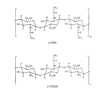
Figure 1 shows the chemical structures of Chitin
(poly-N-acetyl-D-glucosamine) and Chitosan (deacetylated
poly-N-acetyl-glucosamine). Some sites for chemical
modification of chitosan include: C2(NH-CO-CH3 or NH2), C3
(OH), or C6 (CH2OH).
Figure 2 is a graph of the effect of chitosan and
chitosan oligomer molecular weight on the resulting
particle size of chitosan-based compositions containing
DNA plasmids. The charge ratio of all DNA:Chitosan
compositions was 1:4 (-/+). The final pH of these
complexations ranged from pH 5 (for high molecular weight
chitosan-based compositions to pH 7.4 (for lower
molecular weight chitosan oligomer compositions).
Figure 3 is a transmission electron micrography (TEM)
of two complex compositions made with chitosan oligomer
(8 kDa). A) DNA:Chitosan:Lytic Peptide (1:6:1 -/+/-) made
in water with a DNA concentration of 50 ~g/ml. The
particle size of the composition as measured by light
scattering was 64 + 16 nm. B) DNA:Chitosan (1:12 -/+)
made in water with a DNA concentration of 50 ~g/ml. The
particle size of the complex as measured by light
scattering was 66 + 24 nm.
Figure 4 is a graph of the effect of adding a
negatively charged lytic peptide to preformed DNA:Chitosan
(8 kDa) compositions and the effect of making such
compositions in isotonic 10% lactose solutions with a DNA
concentration of 100 ~g/ml.
Figure 5 graphs the effect of lyophilization and
rehydration on the particle size of DNA:Chitosan (90
kDa):Lytic Peptide (1:6:1 -/+/-) compositions. The
composition was mixed in 10% lactose with a DNA
concentration of 100 ~g/ml, lyophilized, and rehydrated
~ with water to a final DNA concentration of 100 ~g/ml.
Figure 6 graphs the effect on compositlon of a
chitosan-based compound with and without the inclusion of
a lytic peptide. All compositions were made in water with
CA 022~68~ l998-ll-l6
W097/42975 PCT~S97/0~87
a DNA concentration of 200 ~g/ml. Panel (A) shows the
overall results of particle size, Panel (B) shows the
effect of chitosan molecular weight and complex charge
ratio on the size of the compositions, and Panel (C) shows
the effect of chitosan molecular weight, complex charge
ratio, and the inclusion of various amount of lytic
peptide on the size of the compositions.
Figure 7 shows the Zeta potential values of
compositions made with chitosan-based compounds with and
without the inclusion of a lytic peptide. The
compositions were made in water with a DNA concentration
of 2~0 ~g/ml.
Figure 8 shows the results of in vitro cell
transfection of HIG-82 (rabbit synovioctyes) in the
presence of 10% FBS. The transfection efficiency (in
RLU/~g protein) is shown as a function of chitosan
molecular weight and the inclusion of various amounts of
a lytic peptide. 10 ug of formulated CMV-Bgal was added
to the cells and the cells were harvested after 48 hours.
Description of the Preferred Embodiments
The following are preferred embodiments of the
present invention using compositions of chitosan-based
compounds for delivery of nucleic acid and
oligonucleotides to a cell. These embodiments are offered
by way of illustration and are not intended to limit the
invention in any manner.
I. Theory And Operation Of Invention
An important goal of the current invention is to
increase the efficacy of gene delivery and gene expression
in target cells. Gene delivery is the first step in the
process of ultimately obtaining expression of a product
~ encoded by a nucleic acid targeted for delivery to a cell.
One method of improving gene delivery is to effect the
uptake of nucleic acid by cells. ~ptake of nucleic acid
by cells is dependent on a number of factors, one of which
,,, ,, .. . ~ , ... . . .
CA 022~68~ l998-ll-l6
W097/4297S PCT~S97/08487
24
is the size of the composition carrying the nucleic acid
or oligonucleotide to be expressed in the target cell.
For instance, some investigators report a positive
correlation between the degree of condensation of DNA in
a complex to be delivered to a cell and the efficacy of
cellular DNA uptake (Wagner et al., PNAS, Vol 88, 1991).
Accordingly, it would be desirable to find a
substance able to complex and condense nucleic acids,
protect them from degradation by nucleases and enable
enhanced uptake by the target cell by either non-specific
adsorptive mechanisms or receptor mediated endocytosis and
also, permit attachment of additional moieties to the
composition to enhance the ability of the composition to
obtain expression of the product targeted to a cell.
Furthermore, these substances should be easily available,
biocompatible and capable of being modified to alter their
physical, chemical and physiological properties. Such
substances should be able to form compositions suitable
for administration to an organism by various means such
as, but not limited to, injection or oral delivery while
maintaining or regaining the physical characteristics
necessary to increase cellular uptake and expression of
nucleic acids or oligonucleotides.
Chitosan is such a substance. However, the majority
of pharmaceutically acceptable chitosan products have
molecular weights ranging from 100-1,000 kDa. They have
two distinct properties: i) higher molecular weight
chitosans are usually only soluble in dilute acid
solutions and are insoluble at pH > 6.5, and ii) aqueous
acidic solutions of these chitosans are quite viscous.
The present invention solves these problems by utilizing
techniques -or reducing the molecular weight of
conventional chitosan-based compounds, thereby providing
improved solubility and viscosity properties at
physiologically relevant pH's.
The embodiments and examples below demonstrate how
specific chitosan-based compositions stabilize and
CA 022~68~ l998-ll-l6
W097/42975 PCT~S97/08487
condense nucleic acid for cell delivery. Furthermore,
these embodiments and examples demonstrate how surface and
nuclear ligands can be used with a delivery peptide to
target nucleic acid into the cellular interior and/or the
cell nucleus. Such targeted delivery can be enhanced by
use of a lysis agent and lipophilic peptides. It was
found that though in vitro transfection results do not
necessarily predict effective in vivo delivery, the
chitosan-based compounds can be used in compositions which
enhance in vivo delivery, as well as in vitro transfection
of nucleic acids. Thus, the embodiments and examples
include in vivo and in vi tro techniques, various cellular
or animal models and methods for inserting nucleic acid
into cells.
Also supplied below are embodiments and examples of
specific chitosan-based compositions that can be used to
provide certain functionalities to the associated nucleic
acid in the composition, and thus within a transformed
cell or animal containing such a cell. Those in the art
will recognize that specific moieties of the chitosan-
based compounds can be identified as that containing the
functional region providing the desirable properties of
the composition. Such regions can be readily minimized
using routine deletion, mutation, or modification
techniques or their equivalent.
II. Utility Of The Invention
The compositions of the present invention enhance
delivery of nucleic acid into the cell preferably by
delivering stabilized and condensed nucleic acid into the
nucleus of the cell. These compositions can be used to
treat diseases by enhancing delivery of specific nucleic
acid to the appropriately targeted cells. These composi-
tions can also be used to create transformed cells, as
well as transgenic animals for assessing human disease in
an animal model.
CA 022~68~ 1998-11-16
WOg7/42975 PCT~S97/08487
26
The present invention also features the use of
compositions of chitosan-based compound with nucleic acid
noncovalently bound to the chitosan-based compound that is
capable of condensing the nucleic acid or oligonucleotide.
These chitosan-based compounds provide small, condensed
compositions, or reduced diameter compositions and more
stable nucleic acid particles for delivery, thereby
enhancing the transfection efficiency of the nucleic acid
into the cell and into the nucleus.
By taking advantage of the characteristics of these
compositions, the present invention enhances delivery of
nucleic acid to a cell. The components of the composi-
tions can be used alone, together or with other components
of a nucleic acid carrier as disclosed in PCT publication
WO 93/18759, Woo et al., entitled "A DNA Carrier System
and Method of Use," the whole of which (including
drawings) is hereby incorporated by reference in its
entirety. The chitosan composition, together with lysis
or lipophilic peptides can enhance the delivery of nucleic
acid to cells by enhancing the release of stable,
condensed nucleic acid from an endosome into the cellular
interior.
In addition a composition with a chitosan-based com-
pound, nucleic acid, and lysis agent, the present inven-
tion also features various compositions which can containa targeting ligand for a cell surface receptor and a
nuclear localization signal as well. The targeting
ligands are capable of binding to a cell surface receptor
and entering a cell through cytosis (e.g., endocytosis,
potocytosis, pinocytosis). By using targeting ligands
specific to certain cells, nucleic acid can be delivered
using the chitosan-based compositions directly to the
desired tissue. The nuclear localization signal are cap-
~ able of recognizing and transporting nucleic acid through
the nuclear membrane to the nucleus of the cell. Such
nuclear localization signals help enhance the compositions
ability to target nucleic acid to the cell nucleus.
CA 022~68~ 1998-11-16
W097/42975 PCT~S97/08487
The abilities of the above chitosan-based composi-
tions to deliver nucleic acid to specific cells and to the
nucleus also allows transgenic animal models to be used
for the dissection of molecular carcinogenesis and
disease, assessing potential chemical and physical car-
cinogens and tumor promoters, exploring model therapeutic
avenues as well as livestock agricultural purposes.
Furthermore, the above chitosan-based compositions per-
mit methods for administration and treatment of various
diseases. In addition, the above chitosan-based compo-
sitions can transform cells to produce particular pro-
teins, polypeptides, and/or RNA. Likewise, chitosan-
based compositions can be used in vitro with tissue
culture cells. In vitro uses allow the role of various
nucleic acids to be studied by targeting specific expres-
sion into specifically targeted tissue culture cells.
The present invention also encompasses transgenic
animals whose cells contain the nucleic acid referenced
above delivered via the chitosan-based compositions.
These cells include germ or somatic cells. Transgenic
animal models can be used for dissection of molecular
carcinogenesis and disease, assessing potential chemical
and physical carcinogens and tumor promoters, exploring
model therapeutic avenues and livestock agricultural
purposes.
The methods of use also include a method of treating
human disease, which is another aspect of the present
invention. The method of treatment includes the steps of
administering the chitosan-based compositions as
described herein so as to deliver a desired nucleic acid
to a cell or tissue for the purposes of expression of the
nucleic acid by the cell or tissue. Cell or tissue types
of interest can include, but are not limited to: liver,
muscle, lung, endothelium, joints, skin, bone, tumors and
blood.
The methods of treatment or use include methods for
delivering nucleic acid into a hepatocyte by contacting a
CA 022~68~ l998-ll-l6
W097/42975 PCT~S97/08487
28
hepatocyte with the above referenced chitosan-based
compositions. The surface ligand used with the chitosan-
based composition is one specific for recognition by
hepatocyte receptors. In particular, the asialooro-
somucoid protein is used as a cell surface ligand, apoE-3,
or a derivative as a lipophilic peptide binding molecule
and JTS-1 or a derivative as a lysis agent as described in
United States patent application no. 08/584,043, titled
"Lipophilic Peptides For Macromolecule Delivery", filed on
January 11, 1995, incorporated by reference herein in its
entirety including any drawings or figures. Furthermore,
these methods of use also include delivery of nucleic
acids using a chitosan-based composition with apoE-3 and
no surface or nuclear ligands. The term "hepatocyte" as
used herein refers to cells of the liver.
An embodiment of the methods of treatment or use
includes a method for delivering nucleic acid to muscle
cells by contacting the muscle cell with one of the above
referenced chitosan-based compositions. The surface
ligand used is specific for receptors contained on the
muscle cell. In particular, the surface ligand can be
insulin-like growth factor-I. In addition, the lipophilic
peptide binding molecule can be a apoE-3, or a derivative
and the lysis agent can be JTS-1 or a derivative.
Furthermore, these methods of treatment or use also
include delivery of nucleic acids using a chitosan-based
composition with apoE-3. The term "muscle cell" as used
herein refers to cells associated with skeletal muscle,
smooth muscle or cardiac muscle.
Another embodiment of the methods of treatment or use
includes a method for delivering nucleic acid to bone-
forming cells by contacting the bone-forming cell with the
above referenced chitosan-based composition. The surface
~ ligand used with the chitosan-based composition is
specific for receptors associated with bone-forming cells.
In particular, the surface ligands can include, but are
not limited to, bone morphogenetic protein or cartilage
CA 022~68~ l998-ll-l6
W097/42975 PCT~S97/08487
29
induction factor. In addition, the lipophilic peptide
binding molecule o~ a chitosan-based composition can be
apoE-3, or a derivative, and the lysis agent JTS-1 or a
derivative thereof. Furthermore, these methods of treat-
ment or use also include delivery of nucleic acids usinga chitosan-based composition with apoE-3. As used
herein the term "bone-forming cell" refers to those cells
which promote bone growth. Nonlimiting examples include
osteoblasts, stromal cells, inducible osteoprogenitor
cells, determined osteoprogenitor cells, chondrocytes, as
well as other cells capable of aiding bone formation.
Another related embodiment of the methods of treat-
ment or use includes a method for delivering nucleic acid
to a cell using the above referenced chitosan-based
compositions. The chitosan-based composition can use
folate as a ligand. In addition, the chitosan-based
compositions can use JTS-1 or a derivative as a lysis
agent, and apoE-3, or a derivative thereof as a lipophilic
peptide binding molecule. This method targets cells which
contain folate receptors, including, but not limited to,
hepatocytes.
Still another related embodiment of the methods of
treatment or use includes a method for delivering nucleic
acid to synoviocytes or macrophages using the above
referenced chitosan-based compositions. The chitosan-
based composition can use a ligand recognized by synovio-
cytes and/or macrophages. In addition, the chitosan-
based composition can use JTS-1 or a derivative as a
lysis agent, and apoE-3, or a derivative thereof as a
lipophilic peptide binding molecule. Furthermore, this
method of use also includes delivery of nucleic acids
using a chitosan-based composition with apoE-3 and no
surface or nuclear ligands. The term "synoviocytes"
refers to cells associated with the joints or with the
fluid space of the joints.
In addition to the above methods, the method of use
also includes delivery using a nuclear ligand binding
. . .
CA 022~68~ l998-ll-l6
W097/42975 PCT~S97/08487
complex with the above-referenced chitosan-based
compositions. Such nuclear carriers would help direct the
chitosan-based composition to the nucleus of the cell.
Furthermore, the above methods of use also include
chitosan-based compositions with the lipophilic peptide
binding molecule and the lysis agent, or any plurality of
confirmation thereof.
III. Administration
Administration as used herein refers to the route of
introduction of the chitosan-based composition into the
body. Administration includes but is not limited to
intravenous, intramuscular, systemic, subcutaneous, sub-
dermal, topical, or oral methods of delivery.
Administration can be directly to a target tissue or
through systemic delivery.
In particular, the present invention can be used for
administering nucleic acid for expression of specific
nucleic acid sequence in cells. Routes of administration
include intramuscular, aerosol, olfactory, oral, topical,
systemic, ocular, intraperitoneal and/or intratracheal.
A preferred method of administering chitosan-based
compositions is by oral delivery. Another preferred
method of administration is by direct injection into the
cells or by systemic intravenous injection.
Transfer of genes directly has been very effective.
Experiments show that administration by direct injection
of DNA into joints and thyroid tissue results in expres-
sion of the gene in the area of injection. Injection of
plasmids containing IL-1 into the spaces of the joints
results in expression of the gene for prolonged periods of
time. The injected DNA appears to persist in an uninte-
grated extrachromosomal state. This means of transfer is
one of the preferred embodiments.
In addition, another means to administer the
chitosan-based compositions of the present invention is by
using a dry powder form for inhalation. Furthermore,
, ,. .. _ ~, 1
CA 022 j j68 j 1998 -11-16
WO 97/42975 PCT/US97/08487
administration may also be through an aerosol composition
or liquid form into a nebulizer mist and thereby inhaled.
The special delivery route of any selected vector
construct will depend on the particular use for the
nucleic acid associated with the chitosan-based composi-
tion. In general, a specific delivery program for each
chitosan-based composition used will focus on uptake with
regard to the particular targeted tissue, followed by
demonstration of efficacy. ~ptake studies will include
uptake assays to evaluate cellular uptake of the nucleic
acid and expression of the specific nucleic acid of
choice. Such assays will also determine the localization
of the target nucleic acid after uptake, and establishing
the requirements for maintenance of steady-state concen-
trations of expressed protein. Efficacy and cytotoxicityis then tested. Toxicity will not only include cell
viability but also cell function.
Incorporated nucleic acid or oligonucleotide into
chitosan-based compositions, as described herein, which
undergo endocytosis increases the range of cell types that
will take up foreign genes from the extracellular space.
The chosen method of delivery should result in
cytoplasmic accumulation and optimal dosing. The dosage
will depend upon the disease and the route of administra-
tion but should be between 0.1-1000 mg/kg of body weight/
day. This level is readily determinable by standard
methods. It could be more or less depending on the
optimal dosing. The duration of treatment will extend
through the course of the disease symptoms, possibly
continuously. The number of doses will depend upon
disease delivery vehicle and efficacy data from clinical
trials.
Establishment of therapeutic levels of nucleic acid
or oligonucleotide within the cell is dependent upon the
rate of uptake and degradation. Decreasing the degree of
degradation will prolong the intracellular half-life of
the nucleic acid or oligonucleotide.
CA 022~68~ 1998-11-16
W097142975 PCT~S97/08487
IV. Cell Transfection
One embodiment of the present invention includes
cells transfected with nucleic acid associated with the
chitosan-based compositions described above. Once the
cells are transfected, the cells will express the protein,
polypeptide or RNA encoded for by the nucleic acid. Cells
include, but are not limited to, liver, muscle and skin.
This description is not intended to be limiting in any
manner.
The nucleic acid which contains the genetic material
of interest is positionally and sequentially oriented
within the host or vectors such that the nucleic acid can
be transcribed into RNA and, when necessary, be translated
into proteins or polypeptides in the transfected cells.
A variety of proteins and polypeptides can be expressed by
the sequence in the nucleic acid cassette in the
transfected ceLls. These products may function as intra-
cellular or extracellular structural elements, ligands,
hormones, neurotransmitters, growth regulating factors,
apolipoproteins, enzymes, serum proteins, receptors,
carriers for small molecular weight compounds, drugs,
immunomodulators, oncogenes, tumor suppressors, toxins,
tumor antigens, antigens, antisense inhibitors, triple
strand forming inhibitors, ribozymes, or as a ligand
recognizing specific structural determinants on cellular
structures for the purpose of modifying their activity.
Transfection can be done either by in vivo or ex vivo
techniques. One skilled in the art will be familiar with
such techniques for transfection. Transfection by ex vivo
techniques includes co-transfecting the cells with nucleic
acid containing a selectable marker. This selectable
marker is used to select those cells which have become
transfected. Selectable markers are well known to those
who are skilled in the art.
For example, one approach to nucleic acid delivery
for hepatic diseases is to remove hepatocytes from an
affected individual, genetically alter them in vitro, and
T -
CA 022~68~ l998-ll-l6
W097/42975 PCT~S97/08487
re-implant them into a receptive locus. The ex vivo
approach includes the steps of harvesting hepatocytes,
cultivating the hepatocytes, transducing or transfecting
the hepatocytes, and introducing the transfected
hepatocytes into the affected individual.
The hepatocytes may be obtained in a variety of ways.
They may be taken from the individual who is to be later
injected with the hepatocytes that have been transfected
or they can be collected from other sources, transfected
and then injected into the individual of interest.
Once the ex vivo hepatocyte is collected, it may be
transfected by contacting the hepatocytes with media con-
taining the chitosan-based composition and maintaining
the cultured hepatocytes in the media for sufficient time
and under conditions appropriate for uptake and
transfection of the hepatocytes. The hepatocytes may then
be introduced into an orthotopic location (the body of the
liver or the portal vasculature) or heterotopic locations
by injection of cell suspensions into tissues. One
skilled in the art will recognize that the cell suspension
may contain: salts, buffers or nutrients to maintain
viability of the cellsi proteins to ensure cell stability;
and factors to promote angiogenesis and growth of the
implanted cells.
In an alternative method, harvested hepatocytes may
be grown ex vivo on a matrix consisting of plastics,
fibers or gelatinous materials which may be surgically
implanted in an orthotopic or heterotopic location after
transduction. This matrix may be impregnated with factors
to promote angiogenesis and growth of the implanted cells.
Cells can then be re-implanted. The above are only
examples and are nonlimiting.
V. Direct Delivery to the Liver
Chitosan~based compositions of the present invention
can also be used in reversing or arresting the progression
of disease involving the liver, such as liver cancer. One
CA 022~68~ l998-ll-l6
W097/42975 PCT~S97/08487
3~
embodiment involves use of intravenous methods of adminis-
tration to delivery nucleic acid encoding for a necessary
molecule to treat disease in the liver. Chitosan-based
compositions which express a necessary protein or RNA can
be directly injected into the liver or blood supply so as
to travel directly to the liver.
VI. Direct DNA Delivery to Muscle
The muscular dystrophies are a group of diseases that
result in abnormal muscle development, due to many differ-
ent reasons. These diseases can be treated by using the
direct delivery of genes with the chitosan-based
compositions of the present invention resulting in the
production of normal gene product. Delivery to the muscle
using the present invention is done to present genes that
produce various antigens for vaccines against a multitude
of infections of both viral, bacterial, and parasitic
origin. The detrimental effects caused by aging can also
be treated using the chitosan-based compositions described
herein. Since the injection of the growth hormone protein
promotes growth and proliferation of muscle tissue, the
growth hormone gene can be delivered to muscle, resulting
in both muscle growth and development, which is decreased
during the later portions of the aging process. Genes
expressing other growth related factors can be delivered,
such as Insulin Like Growth Factor-1 (IGF-1).
Furthermore, any number of different genes may be
delivered by this method to the muscle tissue.
IGF-1 can be used to deliver DNA to muscle, since it
undergoes uptake into cells by receptor-mediated endocyto-
sis. This polypeptide is 70 amino acids in length and is
member of the growth promoting polypeptides structurally
related to insulin. It is involved in the regulation of
tissue growth and cellular differentiation affecting the
proliferation and metabolic activities of a wide variety
of cell types, since the polypeptide has receptors on many
types of tissue. As a result, the chitosan-based
CA 022~68~ l998-ll-l6
W097/42975 PCT~S97/08487
compositions of the present invention can utilize IGF-l as
a ligand for tissue-specific nucleic acid delivery to
muscle. The advantage of a IGF-1/nucleic acid delivery
system is that the specificity and the efficiency of the
delivery is greatly increased due to a great number of
cells coming into contact with the ligand/composition with
uptake through receptor-mediated endocytosis. Using the
nucleic acid described above in the chitosan-based
compositions of the present invention with the use of
specific ligands for the delivery of nucleic acid to
muscle cells provides treatment of diseases and
abnormalities that affect muscle tissues.
VII. Direct DNA Delivery to Osteogenic Cells
There are many other problems that occur during the
aging process, but one major problem is osteoporosis,
which is the decrease in overall bone mass and strength.
The direct delivery chitosan-based compositions of the
present invention can be used to deliver genes to cells
that promote bone growth. The osteoblasts are the main
bone forming cell in the body, but there are other cells
that are capable of aiding in bone formation. The stromal
cells of the bone marrow are the source of stem cells for
osteoblasts. The stromal cells differentiate into a
population of cells known as Inducible Osteoprogenitor
Cells (IOPC), which then under induction of growth
factors, differentiate into Determined Osteoprogenitor
Cells (DOPC). It is this population of cells that mature
directly into bone producing cells. The IOPCs are also
found in muscle and soft connective tissues. Another cell
involved in the bone formation process is the cartilage-
producing cell known as the chondrocyte.
A factor identified to be involved in stimulating the
IOPCs to differentiate is known as Bone Morphogenetic
Protein (BMP). This 19,000 MW protein was first
identified from demineralized bone. Another similar
factor is Cartilage Induction Factor (CIF), which also
CA 022~68~ 1998-11-16
W097/42975 ~CT~S97/08487
36
functions to stimulate IOPCs to differentiate thereby
initiating cartilage formation, cartilage calcification,
vascular invasion, resorption of calcified cartilage, and
finally induction of new bone formation. Cartilage
Induction Factor has been identified as being homologous
to Transfecting Growth Factor ~.
Since osteoblasts are involved in bone production,
genes that enhance osteoblast activity can be delivered
directly to these cells. Genes can also be delivered to
the IOPCs and the chondrocytes, which can differentiate
into osteoblasts, leading to bone formation. BMP and C~F
are the ligands that can be used to deliver genes to these
cells. Genes delivered to these cells promote bone forma-
tion or the proliferation of osteoblasts. The polypep-
tide, IGF-1 stimulates growth in hypophysectomized rats
which could be due to specific uptake of the polypeptide
by osteoblasts or by the interaction of the polypeptide
with chondrocytes, which result in the formation of
osteoblasts. Other specific bone cell and growth factors
can be used through the interaction with various cells
involved in bone formation to promote osteogenesis.
Nonlimiting examples of genes expressing the
following growth factors which can be delivered to these
cell types are Insulin, Insulin-Like Growth Factor-l,
Insulin-Like Growth Factor-2, Epidermal Growth Factor,
Transfecting Growth Factor-~, Transfecting Growth Factor-
~, Platelet Derived Growth Factor, Acidic Fibroblast
Growth Factor, Basic Fibroblast Growth Factor, Bone
Derived Growth Factors, Bone Morphogenetic Protein,
Cartilage Induction Factor, Estradiol, and Growth Hormone.
All of these factors have a positive effect on the
proliferation of osteoblasts, the related stem cells, and
chondrocytes. As a result, BMP or CIF can be used as
conjugates to deliver genes that express these growth
factors to the target cells by the intravenous injection
of the nucleic acid/chitosan compositions of the present
invention. Using the nucleic acid described above in the
CA 022~68~ l998-ll-l6
W097/42975 PCT~S97/08487
chitosan-based compositions of the present invention with
the use of specific ligands for the delivery of nucleic
acid to bone cells provides treatment of diseases and
abnormalities that affect bone tissues.
VIII. Direct DNA Delivery to the Synoviocytes
The inflammatory attack on joints in animal models
and human diseases may be mediated, in part, by secretion
of cytokines such as IL-1 and IL-6 which stimulate the
local inflammatory response. The inflammatory reaction
may be modified by local secretion of soluble fragments of
the receptors for these ligands. The complex between the
ligand and the soluble receptor prevents the ligand from
binding to the receptor is normally present on the surface
of cells, thus preventing the stimulation of the
inflammatory effect.
Therapy consists of the construction of a vector
containing the soluble form of receptors for appropriate
cytokines tfor example, IL-1), together with promoters
capable of inducing high level expression in structures of
the joint and composition which enables efficient uptake
of this vector. This composition is then used with the
nucleic acid carried by the chitosan-based compositions
of the present invention. This DNA is injected into
affected joints where the secretion of an inhibitor for
IL-1 such as a soluble IL-1 receptor or natural IL-I
inhibitor modifies the local inflammatory response and
resulting arthritis.
This method is useful in treating episodes of arth-
ritis which characterize many "autoimmune" or "collagenvascular" diseases. This method can also prevent dis-
abling injury of large joints by inflammatory arthritis.
In addition to the above, the present invention can
also be used with the following method. Current therapy
for severe arthritis involves the administration of
pharmacological agents including steroids to depress the
inflammatory response. Steroids can be administered
CA 022~68~ l998-ll-l6
W097/42975 PCT~S97/08487
38
systemically or locally by direct injection into the joint
space.
Steroids normally function by binding to receptors
within the cytoplasm of cells. Formation of the steroid-
receptor complex changes the structure of the receptor sothat it becomes capable of translocating to the nucleus
and binding to specific sequences within the genome of the
cell and altering the expression of specific genes.
Genetic modifications of the steroid receptor can be made
which enable this receptor to bind naturally occurring
steroids with higher affinity, or bind non-natural,
synthetic steroids, such as R~486. Other modifications
can be made to create steroid receptor which is
"constitutively active" meaning that it is capable of
binding to DNA and regulating gene expression in the
absence of steroid in the same way that the natural
steroid receptor regulates gene expression after treatment
with natural or synthetic steroids.
Of particular importance is the effect of gluco-
corticoid steroids such as cortisone, hydrocortisone,prednisone, or dexamethasone which are the most important
drugs available for the treatment of arthritis. One
approach to treating arthritis is to introduce a vector in
which the nucleic acid cassette expresses a genetically
modified steroid receptor into cells of the joint, e.g.,
a genetically modified steroid receptor which mimics the
effect of glucocorticoids but does not require the
presence of glucocorticoids for effect. This is termed
the glucocortico-mimetic receptor. This is achieved by
expression of a constitutively active steroid receptor
within cells of the joint which contains the DNA binding
domain of a glucocorticoid receptor. This induces the
therapeutic effects of steroids without the systemic
toxicity of these drugs.
Alternatively, steroid receptors which have a higher
affinity for natural or synthetic glucocorticoids, such as
R~486, can be introduced into the joint. These receptors
, ,,, . 1 1 .......
CA 022~68~ 1998-11-16
W097l42975 PCT~S97/08487
39
exert an increased anti-inflammatory effect when stimu-
lated by non-toxic concentrations of steroids or lower
doses of pharmacologically administered steroids.
Alternatively, constitution of a steroid receptor which is
activated by a novel, normally-inert steroid enables the
use of drugs which would affect only cells taking up this
receptor. These strategies obtain a therapeutic effect
from steroids on arthritis without the profound systemic
complications associated with these drugs. Of particular
importance is the ability to target these genes differen-
tially to specific cell types (for example synovial cells
versus lymphocytes) to affect the activity of these cells.
As described in U.S. Patent No. 5,364,791 to Vegeto,
et al., entitled "Progesterone Receptor Having C Terminal
Hormone Binding Domain Truncations," and U.S. Application,
Serial No. 07/939,246, entitled "Mutated Steroid Hormone
Receptors, Methods for Their Use and Molecular Switch for
Gene Therapy," Vegeto, et al., filed September 2, 1992,
both hereby incorporated by reference (including draw-
ings), genetically modified receptors, such as theglucocortico-mimetic receptor, can be used to create novel
steroid receptors including those with glucocortico-
mimetic activity. The steroid receptor family of gene
regulatory proteins is an ideal set of such molecules.
These proteins are ligand activated transcription factors
whose ligands can range from steroids to retinoids, fatty
acids, vitamins, thyroid hormones and other presently
unidentified small molecules. These compounds bind to
receptors and either up-regulate or down-regulate
transcription.
The preferred receptor of the present invention is
modification of the glucocorticoid receptor, i.e., the
glucocorticoid-mimetic receptor. These receptors can be
~ modified to allow them to bind various ligands whose
structure differs from naturally occurring ligands, e.g.,
RU486. For example, small C-terminal alterations in amino
acid sequence, including truncation, result in altered
.. ... . . . ..... .
CA 022~68~ l998-ll-l6
W097/42975 PCT~S97/08487
affinity and altered function of the ligand. By screening
receptor mutants, receptors can be customized to respond
to ligands which do not activate the host cells own
receptors.
A person having ordinary skill in the art will
recognize, however, that various mutations, for example,
a shorter deletion of carboxy terminal amino acids, will
be necessary to create useful mutants of certain steroid
hormone receptor proteins. Steroid hormone receptors
which may be mutated are any of those receptors which
comprise the steroid hormone receptor super family, such
as receptors including the estrogen, progesterone,
glucocorticoid-~, glucocorticoid-~, mineral corticoid,
androgen, thyroid hormone, retinoic acid, and Vitamin B3
receptors. Furthermore, DNA encoding for other mutated
steroids such as those which are capable of only
transrepression or of only transactivation are also within
the scope of the above embodiment. Such steroids could be
capable of responding to RV486 in order to activate
transrepression.
In addition to the above, the present invention can
also be used with the following method. Drugs which
inhibit the enzyme prostaglandin synthase are important
agents in the treatment of arthritis. This is due, in
part, to the important role of certain prostaglandin in
stimulating the local immune response. Salicylates are
widely used drugs but can be administered in limited doses
which are often inadequate for severe forms of arthritis.
Gene transfer using the present invention is used to
inhibit the action of prostaglandin synthase specifically
in affected joints by the expression of an antisense RNA
for prostaglandin synthase. The complex formed between
the antisense RNA and mRNA for prostaglandin synthase
interferes with the proper processing and translation of
this mRNA and lowers the levels of this enzyme in treated
cells. Alternatively RNA molecules are used for forming
a triple helix in regulatory regions of genes expressing
CA 022~68~ l998-ll-l6
WO 97142975 PCT/US97/08487
41
enzymes required for prostaglandin synthesls. Alterna-
tively, RNA molecules are identified which bind the active
site of enzymes required for prostaglandin synthesis and
inhibit this activity.
Alternatively, genes encoding enzymes which alter
prostaglandin metabolism can be transferred into the
joint. These have an important anti-inflammatory effect
by altering the chemical composition or concentration of
inflammatory prostaglandin.
Likewise, the present invention is useful for enhanc-
ing repair and regeneration of the joints. The regener-
ative capacity of the joint is limited by the fact that
chondrocytes are not capable of remodeling and repairing
cartilaginous tissues such as tendons and cartilage.
Further, collagen which is produced in response to injury
is of a different type lacking the tensile strength of
normal collagen. Further, the injury collagen is not
remodeled effectively by available collagenase. In addi-
tion, inappropriate expression of certain metalloprotein-
ases is a component in the destruction of the joint.
Gene transfer using promoters specific to chondro-
cytes (i.e., collagen promoters) is used to express
different collagens or appropriate collagenase for the
purpose of improving the restoration of function in the
joints and prevent scar formation.
Gene transfer for these purposes is affected by
direct introduction of nucleic acid into the joint space
where it comes into contact with chondrocytes and synovial
cells. Further, the genes permeate into the environment
of the joint where they are taken up by fibroblasts,
myoblasts, and other constituents of periarticular tissue.
IX. Direct Delivery to the Lungs
Chitosan-based compositions of the present invention
can also be used in reversing or arresting the progression
of disease involving the lungs, such as lung cancer. One
embodiment involves use of intravenous methods of adminis-
CA 022~68~ l998-ll-l6
W097t42975 PCT~S97/08487
42
tration to delivery nucleic acid encoding for a necessary
molecule to treat disease in the lung. Chitosan-based
compositions which express a necessary protein or RNA can
be directly injected into the lungs or blood supply so as
to travel directly to the lungs. Furthermore, the use of
an aerosol or a liquid in a nebulizer mist can also be
used to administer the desired nucleic acid to the lungs.
Finally, a dry powder form can be used to treat disease in
the lung. The dry powder form is delivered by inhalation.
These treatments can be used to control or suppress lung
cancer or other lung diseases by expression of a
particular protein encoded by the nucleic acid which is
chosen to be delivered.
Additional organs, tissues, cavities, cell or cells,
spaces for the administration of the molecules mentioned
herein may be found in "Nucleic Acid Transporters for
Delivery of Nucleic ~cids into a Cell"; Smith et al.,
.S. Patent Application Serial No. 08/484,777, filed
December 18, 1995, incorporated herein by reference in its
entirety including any drawings.
Examples
The following examples show methods of depolymerizing
chitosan and characterizing chitosan and chitosan oligo-
mers, methods of complexing chitosan and characterizingchitosan-based compositions, methods of preparation of
low weight molecular chitosans, methods of sodium nitrite
treatment of chitosans, the relation of the size of
chitosan-based compositions to the molecular weight of
chitosans used in the composition, modifications of
chitosan-based compositions, the net charge on chitosan-
based compositions, in vivo and in vitro studies of
chitosan-based compositions, and studies of the oral
administration of chitosan-based compositions. The
examples are solely for illustrative purposes and are not
meant to be limiting on the scope of the invention. The
invention is limited by the scope of the claims.
CA 022~68~ l998-ll-l6
W097/42975 PCT~S97/08487
43
Materials Used
SEACURE 143 (78.9% deacetylated), SEACURE 243 (80.1%
deacetylated), and SEACURE 340 (82.3% deacetylated) were
obtained from PRONOVA BIOPOLYMER (Raymond, WA). The
viscosities of 1% chitosan solutions (143, 243, and 340)
in 1% acetic acid were given as 14 cP, 90 cP, and 680 cP,
respectively. These viscosities corresponded to average
molecular weights of approximately 110 kDa, 230 kDa, and
540 kDa, respectively. SEACURE L210 (Poly-D-glucosamine
hydrolactate) was also obtained from PRONOVA BIOPOLYMER
(>80% deacetylated; 78 cP) for analysis. Glucosamine
hydrochloride was obtained from Fluka BioChemika (AG CH-
9470 Buchs). Sodium nitrite A.C.S. was from Fisher
Scientific (Fair Lawn, NJ). Lactose monohydrate powder
U.S.P. was from Penta Manufacturing (Livingston, NJ). A
endosomolytic (lytic) peptide (LP) was synthesized and
purified. DNA plasmids, containing a CMV promoter and
either chloramphenicol acetyltransferase (CMV-CAT) or B-
galactosidase reporter gene (CMV-B-gal), were prepared and
purified. A chemiluminescence detection system for B-
galactosidase (Galacto-LightTM) was from Tropix, Inc.
(Bedford, MA). A CAT ELISA kit was obtained from
Boehringer Mannheim (GmbH, Germany).
Example 1
De-polymerization of Chitosan
Chitosan (SEACURE 143) was de-polymerized according
to a modified method described by Peniston et al., United
States Patent 3,922,260; November 25, 1975 incorporated by
reference in its entirety including any drawings or
figures. Briefly, five-2 g (0.0124 moles) solutions of
SEACURE 143 in 100 ml of normal (6%) acetic acid were
treated at 25~C with five corresponding solutions of NaNO2
in 6% acetic acid. The five corresponding NaNO2 solutions
contained 1.24 x 10⁻⁴ moles NaNO2 , 2.48 x 10⁻⁴ moles NaNO2,
4.96 x 10⁻⁴ moles NaNO2 , 7.44 x 10⁻⁴ moles NaNO2 , and 1.24
x 10-3 moles NaNO2 , respectively. The five reactions
. ... -'- T - '
CA 022~68~ 1998-11-16
WO 97/42975 PCT/US97/08487
44
corresponded to 1%, 2%, 4%, 6%, and 10% of the theoretical
amount of NaNO2 needed for complete deamination of
chitosan. The NaNO2 was added over a 30 minute period
using vigorous magnetic stirring. The reactions were
allowed to continue for a total of 3 hours et which time
the five solutions were neutralized with dilute sodium
hydroxide to pH 7. 4 . The precipitated products were cen-
trifuged for 10 minutes at 4000 rpm, washed 2 times with
water, centrifuged two times as above, washed in ethanol
and centrifuged twice, again as above, washed in ether and
centrifuged again, as above, then vacuum dried at 25~C for
2 days. The weight yield (from 2 g SEACURE 143 original)
was 70%, 80%, 75%, 61%, and 32%, respectively.
Example 2
Characterization of Chitosan and Chitosan Oligomers
The molecular weights of SEACURE 143 and de-
polymerized SEACURE 143 products were determined using a
Beckman Ultraspherogel SEC2000 Column using a mobi~e phase
of 0.lM HOAc-NaOAc buffer (pH 5.5) with 0.05 M NaCl. A
flow rate of 1 ml/min was employed with both UV detection
at 280 nm and refractive index. Polyethylene glycol (PEG)
was used as the molecular weight standard. The percent
(%) Nitrogen was determined by Atlantic Microlab, Inc.
(Norcross, GA) using combustion analysis. Viscosities of
chitosan and chitosan oligomers at 4 ug/ml in 0.2% acetic
acid were determined using a Brookfield Model DV-III
Programmable Rheometer (Stoughton, MA). A Beckman DU640
Spectrophotometer was used to determine the absorbance of
1 ug/ml solutions of chitosan and chitosan oligomers in
0.2% acetic acid diluted with water.
The amine content of chitosan and chitosan oligomers
was determined using a modified ninhydrin assay as
described by Curotto et al., Analy. Biochem., 211 240-241
(~993). Briefly, chitosan and chitosan oligomers were
vacuum dried overnight and dissolved in 2% acetic acid at
a concentration of 1 mg/ml. Then to different volumes of
CA 022~68~ 1998-11-16
WO 97/42975 PCTtUS97tO8487
0.5 ml of 4 M acetic acid/acetate buffer (pH 5.5) was
added the sample (i.e., 100-500 ml). Each solution was
standardized to a volume of 1 ml with water. Then, 2 ml
ninhydrin reagent (J.L. Bailey. In: Techniques in Protein
Chemistry. (1967) 348-349. Elsevier, Amsterdam, The
Netherlands) was added to the samples which were vigor-
ously mixed using vortex mixing. The samples were
incubated at 100 ~C for 20 minutes and cooled on ice to
0~C. The absorbance of each sample was measured at 570 nm
against a ninhydrin reagent blank. All samples were
completed in triplicate and compared to a glucosamine
standard (100% amine content).
Example 3
Complexation of Chitosan and Chitosan Oligomers with DNA
Chitosan or chitosan oligomers were dissolved to 4
mg/ml in dilute acetic acid (either 0.2% or 1%) and
sterile filtered(0.2 mm). The resulting filtered higher
molecular weight chitosans (SEACURE 243 and SEACURE 340)
were dissolved in 1% acetic acid and sonicated with heat
to promote dissolution. The lower filtered molecular
weight chitosan and chitosan oligomers (8 kDa, 13 kDa, 22
kDa, 41 kDa, 70 kDa, and SEACURE 143) were dissolved in
0.2% acetic acid without sonication.
Chitosan or chitosan oligomers (4 mg/ml in either
O.2% or 1% acetic acid) were added to 100-400,ug DNA in
water or lactose, so that the final charge ratio (-/+) of
the compositions ranged from 1:0.8 (-/+) to 1:12 (-/+) in
per 1 ml of carrier solution for compositions made in
lactose. The final lactose concentration was 10%
(isotonic). An endosomolytic (lytic) peptide was added by
electrostatic interaction to selected compositions.
CA 022~68~ 1998-11-16
WO 97142975 PCT/USg7/08487
46
Example 4
Characterization of DNA:Chitosan and DNA:Chitosan Oligomer
Compositions
The unimodal size of the compositions was measured
using a Model N4MD Particle Sizer. For all samples,
measurements were made at 90~ for 120 seconds using a
viscosity setting of 1.005 cP and refractive index setting
of 1.333. To characterize the size and morphology of
compositions by transmission electron microscopy (TEM), a
JEOL Electron Microscope was uti~ized. Carbon coated 200
mesh copper specimen grids were glow-discharged for 1.5
minutes. One drop of complex complexation was deposited
on the grid and allowed to stand for 1.5 minutes. Excess
liquid was removed using filter paper. Next, 1 drop of 1%
uranyl acetate solution (0.2 ~m filtered) was deposited on
the grid and allowed to stain the sample for 10 minutes
prior to examination of the samples under the electron
microscope. Zeta potential values were measured using a
Coulter DELSA 440 (Amherst, MA).
In initial studies designed to show that chitosan
could complex and condense DNA into a compact particle, a
correlation was found between the molecular weight of the
commercially available chitosans and the resulting
particle size of the complex formed with DNA, irrespective
of the ratio of DNA to chitosan. The particle size of the
resulting complex increased proportionally to the
molecular weight of the chitosan substance used (i.e.,
SEACURE 340 > SEACURE 243 > SEACURE 143). This effect
could be due to solubility differences with the chitosan
substances since solubility of chitosan is known to
increase as molecular weight of the substance decreases
(i.e., SEACURE 143 > SEACURE 243 > SEAC~RE 340). Further
evidence to support the solubility effect was reported by
~ Shiraishl et al., J. Contr. Rel., 25 217-225 (1993); and
T. Imai et al., Int. J. Pharm., 67 11-20(1991) both
incorporated by reference in their entirety including
drawings and figures. Shiraishi et al., report that lower
. ,
CA 022~68~ 1998-11-16
W097t42975 PCT~S97/08487
molecular weight chitosans interact with indomethacin
through hydrophobic and ionic interactions and that the
extent of interaction depends on the mo7ecular weight of
chitosan. However, the intermolecular and intramolecular
interactions, which promote complexation, increased with
increasing molecular weight of chitosan. Nevertheless,
the authors demonstrated that the release of indomethacin
from the complex with chitosan was dominated by pene-
tration of water into the substance matrix. Whereas
chitosans of lower molecular weight (having increased
solubility), allowed more water penetration. Higher
molecular weight chitosans (having decreased solubility),
caused substantial dehydration of the composition.
Analogous to this effect, DNA compositions made with
higher molecular weight chitosan should theoretically have
reduced solubility resulting in aggregation and/or larger
particle size. Such results were observed by us.
Example 5
Preparation of Low Molecular Weight Chitosans
As a result of the effect observed with chitosans
having different molecular weights in Example 4, we
hypothesized that the preparation of even smaller
chitosan-based compositions would require the use of even
lower molecular weight chitosans which were not
commercially available. Thus, we modified a process for
sodium nitrite de-polymerization of chitosan as described
by Peniston et al. United States Patent 3,922,260 to
produce such lower molecular weight chitosans.
Peniston et al., describe the deaminative cleavage of
chitosan into reduced chain-length oligomers to reduce
viscosity, increase solubility, or to generally alter the
molecular characteristics of chitosan as a
polyelectrolyte. In Peninston et al's. reaction, sodium
nitrite is converted to the reactive species nitrous acid
by treatment with a strong acid (i.e., 6% acetic acid).
Nitrous acid reacts only with the glucosamine moieties on
CA 022~68~ 1998-ll-16
W097/42975 PCT~S97108487
48
chitosan, not the N-acetylglucosamine units. The reaction
is believed to form a highly unstable diazonium salt which
leads to the evolution of nitrogen and the replacement of
the original amino group with a hydroxyl group resulting
in a 2,5-anhydro-D-mannose residue at the new reducing
end. In some instances, the reduced end results in the
formation of olefins. In general, the release of nitrogen
is stoichiometric with the loss of the amino group so that
quantification of the released nitrogen gas can be
directly correlated to the percentage(%)of deamination
that has occurred.
Peniston et al., note that the nitrous acid reaction
with chitosan results in an unexpected marked decrease in
viscosity (and thus, molecular weight) with only small
changes in percent (%) amine content. Thus, we antici-
pated that the nitrous acid induced deamination of higher
molecular weight chitosan may be useful for reducing its
molecular weight without causing a large reduction in the
amine content which is needed for complexing and
condensing DNA.
We also considered utilizing an alternative de-
polymerization process for chitosan involving the treat-
ment of chitosan with concentrated HCl at elevated
temperatures (A.D. Domard and N. Cartier. Glucosamine
Oligomers: l. Preparation and Characterization. Int. J.
Biol. Macromol., ll (1989) 297-302). This chemical method
appeared suitable for making very pure chitosan oligomers,
however separation of the product is difficult and costly
and typically results in oligomers with very low degrees
of polymerization (i.e., DP = 20-37).
Example 6
Results of Sodium Nitrite Treatment of Chitosans
The results of the sodium nitrite treatment of
chitosan (SEACURE 143) are shown in Table 2. A decrease
in the molecular weight of chitosan was observed with
increasing treatment by sodium nitrite. The decrease in
CA 022~68~ l998-ll-l6
W097/42975 PCT~S97/08487
49
molecular weight, as determined by gel permeation
chromatography, also correlated well with the reduction in
viscosity of 4 mg/ml solutions. The substantial loss in
molecular weight of chitosan was not associated with a
corresponding loss of nitrogen or amine content. However,
we obtained a deacetylation value of 56% using the
modified ninhydrin assay for SEACVRE 143. This value was
lower than the deacetylation value for SEACURE 143
specified by the manufacturer (78.9%). It is unclear why
we obtained this lower value, since complexation and gel
retardation assays confirmed the percentage amine content
of the SEACURE 143 obtained from the manufacturer as
78.9%.
However, the modified ninhydrin assay did confirm
that the amine percentage of chitosans treated with sodium
nitrite did not change appreciably, although the molecular
weight was markedly reduced.
Support for the creation of olefin byproducts as a
result of sodium nitrite treatment of chitosan was
supported by the observation of a positive absorbance peak
at 280 nm in the treated chitosan compounds. The absorb-
ance values were directly proportional to the amount of
sodium nitrite treatment. Although the presence of the
olefin side product reduced the "purity" of the chitosan
oligomers, the ability to accurately quantify the
concentration of chitosan oligomer in solution proved to
be very useful in characterizing solutions and formed
compositions.
Example 7
Relation of Chltosan Molecular Weight to Complex Size
After performing the de-polymeri~ation reactions on
chitosan (SEACURE 143), we examined the effect of
different molecular weight chitosans on the resulting
particle size of compositions formed between DNA and these
chitosans. The results are shown in Figure 2.
, . . .,~ . . .
CA 022~68~ 1998-11-16
W097/42975 PCT~S97/08487
For unimodal particle size of DNA:Chitosan
compositions (1:4 -/+) in water there was a positive
correlation between low molecular weight chitosan and the
ability to form compositions of small size in water
(irrespective of DNA concentration). These results
support the fact that increased solubility of chitosan
oligomers led directly to smaller and more stable
compositions with increased colloidal solubility. The
final pH of these DNA:Chitosan compositions ranged from pH
5 (for high molecular weight chitosan-based compositions
to pH 7.4 (for lower molecular weight chitosan
compositions).
Transmission electron micrographs (TEM) of two
DNA:Chitosan compositions made with a chitosan oligomer of
8 kDa in molecular weight are shown in Figure 3 (A&B).
Two shapes can be observed: toroidal or circular particles
(toroids) and compact rod-like particles (rods). Similar
toroids and rods have been reported to coexist in the
literature (Y.Y. Vengerov and T.E. Semenov Electron
20 Microsc. Rev., 5; 193-20 (1992). Vengerov et al. have
also shown that DNA can be condensed by a tri-valine
peptide (TVP; H-Val-Val-Val-NH-NH-Dns where Dns is 5-
dimethyl-aminoaphtyl-sulfonic acid) into compact rod-like
structures where DNA molecules are interwound and lay side
by side. This effect becomes more pronounced when the
concentration of the TVP peptide is increased. The thick
rods, described as 'comma-shaped' globules, are similar in
shape to those in our studies.
Example 8
Effect of Analogues Added to Chitosan Complex
The effect of adding a negatively charged endosomo-
lytic peptide to preformed DNA:Chitosan (8 kDa) composi-
~ tions and the effect of their preparation in isotonic 10%
lactose solutions is shown in Figure 4.
The presence of the lytic peptide caused a smallincrease in the size of the composition, however, the
CA 022~68~ 1998-11-16
W097/42975 PCT~S97/08487
51
presence of 10% lactose had no effect. Thus, chitosan
compositions prepared in isotonic solutions can be used
for intravenous administration, or lyophilized in the
presence of lactose as a cryoprotectant for storage and
subsequent use after rehydration.
Example 9
Effect of Cryoprotectant on Chitosan Complex
The effect of lyophilization and rehydration on the
particle size of DNA:Chitosan (90 kDa):Lytic Peptide(1:6:1
-/+/-) compositions is shown in Figure 5. The colloidal
properties of the composition are maintained during
lyophilization and the size of the composition remains
unaffected by the treatment.
The effect of adding a negatively charged lytic
peptide to preformed DNA:Chitosan (8 kDa) compositions and
the effect of making such compositions in isotonic 10%
lactose solutions is graphically shown in Figure 4. ~nder
similar conditions compositions made with lower molecular
weight chitosan (8 kDa) are approximately 2-fold smaller
or more condensed than compositions made using higher
molecular weight chitosan (90 kDa).
The effect of lyophilization and rehydration on the
particle size of DNA:Chitosan(90 kDa):Lytic Peptide
compositions with a charge ratio of (1:6:1 -/+/-)is shown
in Figure 5. Lyophilization and subsequent rehydration
did not significantly effect the size of the compositions.
Example 10
Effect of Net Charge on Chitosan Complex
To reconfirm these results we produced DNA:Chitosan
compositions of assorted charge ratios, by varying the
amount of negatively charged lytic peptide in the
composition.
The results are depicted in Figure 6A. Composition
size without and with lytic peptide are shown in Figure 6
(b & c respectively).
T
CA 022~68~ l998-ll-l6
W097/42975 PCT~S97/08487
52
In general, wlth increasing positive charge the
particle size decreased in compositions (without the lytic
peptide) until a charge ratio of 1:2 (-/+) was achieved.
Thereafter, increasing the positive charge had little or
only a slight effect on particle size. The largest
increase in particle size was observed in compositions
made with chitosan-based compounds greater than 41 kDa in
weight; most likely due to their decreased solubility.
Addition of negatively charged lytic peptide to the
compositions resulted in an approximately linear increase
in their size regardless of the weight of the chitosan
used in the composition. The observed increase in size
may be due to charge competition or to a change in the
solubility of the composition colloid.
The zeta potential (i.e., the measure of the charge
ratio) of compositions (e.g., of Figure 6A) are shown in
Figure 7. In general, the increasing addition of chito-
san and/or chitosan oligomers to the composition increased
the net positive charge directly.
The addition of negatively charged lytic peptide to
the composition decreased the positive charge of the
complex proportionally. The observation of a net nega-
tive charge at the theoretical neutral charge ratio of 1:1
-/+ appears anomalous and could be due to incomplete
charge neutralization as a result of increased solubility
of these compositions.
Example 11
In vivo Studies of Chitosan Complex
We assessed the ability of a chitosan composition to
express a reporter gene in vivo in the intestinal mucosa
of rabbits. Rabbits were anaesthetized and laparotomies
performed, then chitosan-based compositions were
administered either colonically or to the small intestine.
The dose of formulated CMV-CAT used was lOO~g in 1 ml 10%
lactose. To prevent leakage at the dosing site, the site
was sealed with an adhesive and the abdominal cavity was
CA 022~68~ 1998-11-16
WO 97/42975 PCT/USg7/08487
closed with two layers of sutures. Rabbits were sacri-
ficed by an overdose of pentobarbitone 72 hr after
administration.
Tissues collected from the intestinally dosed rabbits
were Peyer's Patches (PPl, PP2, PP3), enterocytes (ENTl,
ENT2, ENT3), colon (COL), and mesenteric lymph nodes
(MLN). PPl and ENTl were from regions proximal to the
dosing site, PP2 and ENT2 were from regions median to the
dosing site, and PP3 and ENT3 were from regions distal to
the dosing site.
Tissues collected from colonically dosed rabbits were
colon (COLl, COL2, COL3) and MLN. COLl tissue was from
regions proximal to the dosing site, COL2 was from
regions median to the dosing site, and COL3 was from
regions distal to the dosing site. The tissues COL and
ENT were 1-1.5 cm2 in size. The PP, COL, and ENT tissues
were cut into two pieces and were snap frozen while the
MLN tissues were snap frozen as one piece. Tissues were
homogenized and extracted with 1 ml of extraction buffer
and analyzed for CAT expression using an ELISA assay. The
tissues were also analyzed for total protein content.
Results are expressed as pg CAT/ug protein.
Chemiluminescent Detection Procedure
It is recommended that all assays are performed in
triplicate. (1) Dilute GalactonTM (Galacton-PlusTM) sub-
strate 100-fold with Galacto-LightTM Reaction Buffer
Diluent to make Reaction Buffer. This mixture will remain
stable for several months if stored uncontaminated at 4~C.
It is recommended to only dilute the amount of substrate
that will be used within a two month period. (2) Warm to
room temperature the amount of Reaction Buffer required
for the entire experiment. (3) Aliquot 2 to 20 ~l of
individual cell extracts into luminometer sample tubes.
(The amount of cell extract required may vary depending on
the amount of expression and the instrumentation used.
~se 51ul of extract for positive controls and 10 to 20 ~ul
CA 022~68~ 1998-11-16
WO 97/42975 PCT/US97/08487
54
of extract for experiments with potentially low levels of
enzyme. It is important to vary the concentrations of
extract to keep the signal within the linear range of the
assay) (4) Add 200 ,ul of Reaction Buffer to a luminometer
cuvette and gently mix. Incubate at room temperature for
60 minutes. Incubations can be as short as 15 minutes,
but the linear range of the assay may decrease.
(Measurements are time dependent. Reaction Buffer should
be added to sample extracts in the same time frame as they
are counted on the luminometer. For example, if it takes
10 seconds to completely count a sample, then Reaction
Buffer should be added to tubes every 10 seconds) (5)
Place cuvette in a luminometer. Inject 300~1 of
Accelerator. After a 2 to 5 second delay following injec-
tion, count ~he sample for 5 seconds. If manual in~ectionis used, then the Accelerator should be added in the same
consistent time frame as the Reaction Buffer is added.
This is critical when using GalactonTM.
Pre~aration of Controls
Positive Control
Add 1 ul of ~-galactosidase (10 units/ml, Sigma Cat.
No. G-5635 diluted in 0.1 M sodium phosphate pH 7.0, 1.0%
BSA) to mock transfected cell extract equivalent to the
volume of experimental cell extract used. Proceed with
Chemiluminescent Detection Procedure.
Negative Control
Assay of volume of mock transfected cell extract
equivalent to the volume of experimental cell extract
used. Proceed with Chemiluminescent Detection Procedure.
Heat Inactivation of Endogenous ~-galactosidase
Some cell lines may exhibit relatively high levels of
endogenous ~-galactosidase activity. This may lead to
background which will decrease the overall sensitivity of
the assay by lowering the signal to noise ratio. A
.
CA 022~68~ l998-ll-l6
W097/42975 PCT~S97/08487
procedure for heat inactivation of endogenous ~-
galactosidase activity has been described by Young et al.,
Anal. Biochem. 215:24-30 (1993), incorporated herein by
reference in its entirety including any drawings or
figures. A modified version of this protocol has also
been described by Shaper et al ., J. Biol. Chem. 269:25165-
25171 (1994), incorporated herein by reference in its
entirety including any drawings or figures, in which a
cocktail of protease inhibitors is used in conjunction
with the heat inactivation procedure for reducing ~-
galactosidase in tissue extracts.
Inactivation of ~-Galactosidase Activity in Cell Extracts
The following procedures should be performed
immediately prior to the Chemiluminescent Detection
Procedure in the Preparation of Cell Extracts From Tissue
Culture Section. (1) Following cell extract preparation,
heat the extract to 48~C for 50 minutes. (2) Proceed with
Chemiluminescent Detection Procedure. (Although Young et
20 al. suggest 50~C for 60 minutes, heat inactivation at 48~C
for 50 minutes is suggested~.
Inactivation of Endogenous ~-Galactosidase Activity in
Tissue Extracts
(1) To the Galacto-LightTM lysis buffer, add PMSF to
a final concentration of 0.2 mM and leupeptin to a final
concentration of 5 ug/ml just before use. (2) ~eat
inactivate the extracts by heating at 48~C for 60 minutes.
(3) Proceed with Chemilumlnescent Detection Procedure.
(AEBSF (Sigma Cat. No. A-5938) may be used in place of
PMSF (Sigma Cat. No. P-7626). AEBSF is a water soluble
serine protease inhibitor).
A liquid scintillation counter may be used as a
~ substitute for a luminometer, however, sensitivity may be
lower Fulton, R., and B. Van Ness. Luminescent Reporter
Gene Assays for Luciferase and ~-galactosidase ~sing a
Liquid Scintillation Counter. BioTechniques 14(5): 762-
CA 022~68~ l998-ll-l6
W097/42975 PCT~S97/08487
56
763(1993), incorporated herein by reference. Nguyen,
V.T., M. Morange, and O. Bensaude. Firefly Luciferase
Luminescence Assays Using Scintillation Counters for
Quantitation in Transfected Mammalian Cells. Anal.
Biochem. 171:404-408 (1988), incorporated herein by
reference. The results are expressed as mean +/- S.E.M of
Relative Light ~nit, as indicative of b-galactosidase
activity, per 100 ug muscle protein. When using a
scintillation counter, it is necessary to turn off the
coincident circuit in order to measure chemiluminescence
directly. The manufacturer of the instrument should be
contacted to determine how this is done. If it is not
possible to turn off the coincident circuit, a linear
relationship can be established by ta~ing the square root
of the counts per minute measured and subtracting the
instrument background. Actual = (measured-instrument
background) 1/2, Other methods of measuring a chemilumi-
nescent signal as are known in the art may also be
utilized.
Example 12
In Vitro Transfection Using Chitosan/Nucleic Acid Complex
We assessed in vitro cell transfection using chitosan
compositions depicted in Figure 6A .
Rabbit synoviocytes(HIG-82) plated in 24-well plates
(100,000 cells/well) were incubated overnight in DMEM
supplemented with 10% FBS and with compositions prepared
in water with and without the inclusion of varying amounts
of lytic peptide. Final DNA concentratlon was 200~g/ml.
Media was removed from the cells (40-60% confluent) and
fresh media (DMEM with 10% FBS) was added prior to the
addition of the compositions. For all compositions, 50 ml
(10~g CMV-Bgal) was added. Cells were incubated with the
compositions for 48 hours then removed, and washed with
PBS(pH 7.4). Galacto-LightTM cell lysate solution was
added including 1 mm DTT. A chemiluminescence detection
system for B-galactosidase (Galacto-LightTM) was used to
~ .
CA 022~68~ l998-ll-l6
W097/42975 PCT~S97/08487
determine the Relative Light Units (RLU) per mg cell
lysate protein (RLU/mg protein). Total protein content in
cell lysates were measured with a Coomassie Blue G250-
based assay (Bio-Radi Hercules, CA).
The efficiency of cell transfection of the
compositions is shown in Figure 8. The transfection
efficiency of the composition was positively correlated
with increasing positive charge.
However, the transfection efficiency correlated
poorly with the molecular weight of the chitosan used.
Addition of lytic peptide to the composition
increased the transfection efficiency only with the
addition of small amounts of large molecular weight
chitosan (90 kDa and 70 kDa) or the addition of large
amounts of smaller molecular weight chitosans. This
effect may be related to the size of the compositions and
the inherent stability in solution. The interaction of
the compositions with 10% FBS may also be a contributing
factor.
Methods of Preparation of Cell Extracts From Tissue
Culture Cells
(1) Aliquot the required amount of Lysis Solution.
Add fresh DTT to 1 mM. (2) Rinse cells 2 times with lX
Phosphate Buffered Saline (PBS). (3) Add Lysis Solution
to cover the cells (250,ul of Lysis Buffer for a 60 mm
culture plate should be adequate). (4) Detach cells from
culture plate using a rubber policeman or equivalent.
Non-adherent cells should be pelleted and lysis buffer
should be added sufficient to cover the cells. The cells
should then be resuspended in the lysis buffer by
pipetting. (5) Transfer cells to a microfuge tube and
centrifuge for 2 minutes to pellet any debris. (6)
Transfer supernatant to a fresh microfuge tube. Cell
extracts may be used immediately or frozen at -70~C for
future use.
CA 022~68~ l998-ll-l6
W097/42975 PCT~S97/08487
Example 13
Oral Administratlon of Chitosan/Nucleic Acid Complex
A preferred route of administration of chitosan-
based compositions is orally. Oral administration may
best utilize the beneficial properties of chitosan (as
discussed above). Oral administration permits chitosan-
based compositions to be taken up by various cell types
throughout the entire length of the alimentary canal
thereby allowing the expression of therapeutic products
locally and/or systemically. Chitosan administration has
been examined in the gut to determine its bioadhesive
properties. Additionally, chitosan has been approved by
the FDA as a food additive.
We assessed the ability of chitosan-based composi-
tions to express a reporter gene (i.e., chloramphenicolacetyltransferasei in a CAT assay) in vivo after oral
administration. Lyophilized and rehydrated chitosan-based
compositions (e.g., 90 kDa chitosan like those in Figure
5)were dosed in the upper small intestine or colonically
in rabbits. Tissues were examined for gene expression
after administration as in example 12. Expression was
measured 72 hours after administration.
The results are shown in Tables 3 & 4. The results
demonstrate that the chitosan-based compositions using a
CMV-CAT expression vector delivered a functioning CMV-CAT
expression vector into cells while controls (i.e., naked
DNA CMV-CAT expression vector in water) showed no
expression in any tissues in any region.
One rabbit administered the chitosan composition
demonstrated expression of the CAT reporter gene in almost
all the tissues examined except for PP1 and PP2 Col (when
dosed in the upper small intestine) or Col 3 (when dosed
colonically).
Since cell turnover in the gut is known to be rapid
and extensive, we believe that significantly higher CAT
expression would have been observed had tissue samples
been examined at earlier time points than 72 hours after
CA 022~68~ 1998-11-16
W097/42975 PCT~S97108487
59
administration. Nevertheless, these results demonstrate
the efflcacy of gene delivery and gene expression using
the chitosan-based compositions of the present invention.
Enzyme-Linked Immunoassay ~ELISA) for Chloramphenicol
Acetyltransferase (CAT) in Rodent Lungs
Reagents:
A. CAT ELISA Kit: Boehringer-Mannhiem, Cat#1363727
~ CAT enzyme (50ng~
10 ~ Anti-CAT-digoxigenin
~ Anti-DIG-peroxidase
~ Substrate buffer
~ ABTS substrate
~ Substrate Enhancer
15 ~ Wash Buffer
~ Sample buffer
~ Microtiter plate modules (8-wells)
~ plastic film plate covers
B. Samples in buffer compatible with ELISA (TEN buffer:
50 mM Tris pH 7.5-8.0, 1 mM EDTA, 150 mM NaCl, up to
1%Triton X-100)
Sample Preparation:
Homogenization buffer: Samples homogenized in 50mM Tris-Cl
pH8.0, l.OmM EDTA, 150mM NaCl, 0.5~ Triton X-100, lO~M
Leupeptin, l.O~M Pepstatin A, and 0.25mM PMSF.
Tissue samples are snap frozen in separate
homogenization tubes. Thaw samples on ice. Spin samples
briefly to pellet tissue into beads. If tissue exceeds
50% tube capacity after pelleting, divide equally the lung
lnto two tubes. Tubes are filled to 1.8 ml with cold
homogenization buffer; cap tubes tightly! Keep samples on
ice.
An alternative method of the above is: tissue samples
are snap frozen in separate homogenization tubes. Thaw
samples on ice. Spin samples briefly to pellet tissue
CA 022~68~ l998-ll-l6
W097/42975 PCT~S97/08487
into beads. Tubes are filled to 1.0 ml with cold
homogenization buffer; cap tubes tightly! Keep samples on
ice.
Using the Biospec mini-beadbeater (follow manu-
facturer's instructions), homogenize samples to completion~1-4 minutes) @ 4~C. Spin samples @ ~ C for 15 minutes.
Transfer supernatant to fresh tube; keep on ice. (Note:
if samples are excessively turbid, spin again for an
additional 15 minutes).
Prepare Sample Template:
~ sing a 96 well microtiter plate, aliquot 150 ~l
homogenate to well. Keep plate on ice.
Aliquot 150 ~l H2O to each well (1:1); mix well; keep
on ice. (~ote: this template will be used for both the CAT
ELISA and the protein concentration assay. After setting
up CAT ELISA, save the plate. The remaining 100 ~l of
diluted sample is used for protein determination)
Assay:
1. Rehydrate needed amount of anti-CAT antibody
precoated microtiter plate strips with sample buffer for
5 minutes @ RT.
2. Following rehydration, discard the solution and
wells on paper towel to remove excess liquid.
3a. Pipette 200 ~l of CAT standards, in duplicate,
prepared as described below. For standard curve, make 1
ng/ml CAT in sample buffer (follow manufacturer's
instructlons) make 500 ~l of serial two-fold dilutions of
1 ng/ml solution.
final conc.(p~/200ul) = diluted CAT @ CA~p~/ml) + Sample buffer
100 500 ~l 1000 500 ~l
500 ~l 500 500 ~l
500 ~l 250 500 ~l
12.5 500 ,ul 125 500 ~l
6.25 500 ~l 62.5 500 ~l
3.13 500 ~l 31.3 500 ~l
.. . . .
CA 022~68~ 1998-11-16
W097/42975 PCT~S97/08487
61
1.56 500 ~1 15.6 500 ~l
o - - 500 ~l
3b. Pipette 100ul of 1:1 diluted tissue sample in to
duplicate into wells. Add 100ul CAT ELISA buffer to wells
containing diluted tissue sample. Standards and sample
should now have 200 ul/well. Cover plate with plastic
film, incubate @ 37 deg C for 1 hr.
4. Using Dynatech Laboratories Ultrawash Plus plate
washer, (setting 8116), wash plate with 50mM Tris-Cl
pH7.6, l.OmM EDTA, 150mM NaCl, 0.1% Tween-20.
5. Add 200 ~l of anti-CAT-DIG antibody (1:100
dilution in sample buffer) to wells, incubate 1 hr @ 37
deg C.
6. Wash wells as in step 4.
7. Add 200 ~l of anti-DIG-peroxidase (75ul into
9.g25 ml of sample buffer) to wells, incubate 1 hr @ 37
deg C.
8. Wash well as in step 4.
9. Add 200 ~l of peroxidase substrate, prepared
using manufacturers instructions (ABTS) in buffer. Note:
add substrate enhancer @ 1 mg/ml to ABTS solution if low
concentrations of CAT are expected.
10. Read plate @ OD ~05. Calculate concentration of
CAT in standards using OD values obtained from linear
portion of standard curve. The time of OD readings should
empirically determined to place sample values on linear
portion of standard curve.
One skilled in the art would readily appreciate that
the present invention is well adapted to carry out the
ob~ects and obtain the ends and advantages mentioned, as
well as those inherent therein. The chitosan/nucleic acid
complex along with the methods, procedures, treatments,
molecules, specific compounds described herein are
presently representative of preferred embodiments are
exemplary and are not intended as limitations on the scope
of the invention. Changes therein and other uses will
occur to those skilled in the art which are encompassed
CA 022~s6s~ 1998-11-16
W097/42975 PCT~S97/08487
62
within the spirit of the invention are defined by the
scope of the claims.
It will be readily apparent to one skilled in the art
that varying substitutions and modifications may be made
to the invention disclosed herein without departing from
the scope and spirit of the invention.
All patents and publications mentioned in the speci-
fication are indicative of the levels of those skilled in
the art to which the invention pertains. All patents and
publications are herein incorporated by reference to the
same extent as if each individual publication was specif-
ically and individually indicated to be incorporated by
reference.
. .