Note: Descriptions are shown in the official language in which they were submitted.
CA 022~6491 1998~ 26
W O 97/46177 PCTICA97/00374 --
EXPA~NSIBLE BIOPRO~ C VALVE STENT
Field of the Invention
This invention relates to bioprosthetic valves and
more particularly to a method of fabricating expansible
bioprosthetic valve stents.
Backqround of the Invention
Two basic types of artificial heart valves are
available for replacement of diseased human heart valves.
The first type, mechanical valves, are constructed of
synthetic rigid materials such as plastic or metal.
Their use is associated with thrombogenesis, requiring
valve recipients to be on long term anti-coagulation.
lS The second type, tissue valves or bioprosthetic
valves, consist of valve leaflets of preserved animal
tissue mounted on an artificial support or "stent".
The durability of bioprosthetic heart valves is
limited to about 12 to 15 years. The limitations in the
long term performance of bioprosthetic heart valves are
believed to be due largely to the mechanical properties
of the valve and the stresses imposed on the tissue
leaflets by the rigidity of the stent structure while the
aortic root to which the artificial valve is attached
expands and contracts during the cardiac cycle. An
important feature of the natural heart valve is its
ability to expand in diameter by more than 10% during
systole. This ability of the aortic root to expand
facilitates blood flow due to a better opening of the
valve during systole and contributes to minimal bending
of the cusps, thus reducing possible internal flexural
fatigue.
In an attempt to overcome the rigidity of artificial
heart valves and accommodate the expansion of the aortic
root during systole, a bioprosthetic heart valve with
pivoting stent posts has been devised (Canadian Patent
No. 2,123,824).
SUBSTITUTE SHEE~T (RULE 26)
.
CA 022~6491 1998-ll-26
W O 97/46177 PCT/CA97/00374 --
U.S. Patent No. 5,258,023 discloses a prosthetic
heart valve in which the valve leaflets are fashioned
from synthetic materials, in an attempt to avoid the
mechanical failure of natural tissue leaflet material.
Although these designs allowed for improved
hemodynamics, they did not totally solve the problems
arising from the rigidity of artificial heart valve
stents. There remained a need for an artificial heart
valve stent that is expansible, resilient and tough and
which provides a better opening of the valve during
systole to facilitate blood flow and contributes to
minimal bending of the cusps to reduce valve failure.
SummarY of the Invention
The present invention provides a bioprosthetic stent
fabricated from a hydrogel. A hydrogel stent combines
sufficient strength for use in a bioprosthetic valve with
pliability and elasticity characteristics which much more
closely resemble those of the aortic root than previously
available valve stents.
The invention provides a bioprosthetic valve stent
fabricated from a hydrogel, wherein the hydrogel, when
hydrated, has
(a) a strain value at ultimate tensile stress (UTS)
greater than the maximum strain occurring in a human
aortic root under physiological conditions;
(b) an elastic modulus similar to that of the
aortic root; and
(c) a relaxation rate similar to that of the aortic
root.
The invention provides a method of fabricating a
bioprosthetic valve stent comprising the steps of
(a) preparing a mold cavity corresponding in shape
to the stent to be fabricated;
(b) filling said mold cavity with a solution of a
selected hydrogel;
SUBSTITUTE SHEET (RULE 26)
CA 022~6491 1998-11-26
WO97/46177 PCT/CA97/00374 --
(c) allowing the hydrogel to solidify; and
(d) removing the solid hydrogel from the mould
cavity and hydrating the solidified hydrogel for a
suitable period of time.
S The invention provides a bioprosthetic heart valve
comprlslng
(a) a stent in accordance with any of claims 1 to
5,
(b) leaflet valve means having three generally
triangular leaflets defining respective cusps which are
adapted to open and close during heart systole and
diastole respectively; and
(c) means for attaching the leaflet valve means to
the stent.
Brief Description of the Drawinqs
Certain embodiments of the invention are described,
reference being made to the accompanying drawings,
wherein:
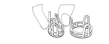
Figure lA shows a hydrated poly(hema) heart valve
stent (left) and an acrylic stent (right), in
uncompressed state.
Figure lB shows a hydrated poly(hema) stent (left)
in compressed state and an acrylic stent (right) in
uncompressed state.
Figure 2 is a diagram of a mould design for solution
casting of a hydrogel stent.
Figure 3 is a sectioned view of a mold of the type
shown in Figure 2.
Figure 4 is a schematic representation of the
components of the mold fixture.
Figure 5 shows stress-strain curves for PVA after
various numbers of freeze-thaw cycles and for aortic root
(white curve).
SUBSTITUTE SHEET(RULE26)
~ . .. . . .
CA 022~649l l998-ll-26
W 097/46177 PCT/CA97/00374 --
Detailed Description of the Invention
The present invention provides an expansible
bioprosthetic valve stent fabricated from a hydrogel and
a method of making such a stent. The stent of the
invention is resilient and expansible and, at the same
time, tough.
Hydrogels are hydrophilic macromolecular or polymer
networks that are capable of imbibing large amounts of
water without dissolving. The networks contain
crosslinks, crystalline regions and entanglements between
the polymer molecules.
Hydrogels have the interesting properties of being
hard and stiff when in the dry state but soft and pliable
when hydrated. These properties make hydrogels ideal
materials for fabrication of bioprosthetic valve stents,
in that they can be milled or machined into any desired
shape when dry and hard, but become pliable and
expansible when hydrated.
Hydrogels are soluble in various non-aqueous
solvents and may therefore also be fashioned into valve
stents by casting in suitable moulds.
In accordance with a preferred embodiment, the
invention comprises a heart valve stent fabricated from a
hydrogel. The heart valve stent maybe for a mitral
valve, an aortic valve or tricuspid valve. It is also
contemplated that the bioprosthetic valve of the present
invention may be used to replace valves of the human body
other than heart valves. For example, in an alternative
embodiment, the valve of the invention maybe modified for
use in the urinary tract to replace a defective sphincter
muscle in order to treat incontinence~ Also, the valve
of the invention may be used in the eye or in the brain
to reduce fluid pressure.
In accordance with a preferred embodiment, the
invention comprises an aortic valve stent fabricated from
a hydrogel.
SUBSTITUTE SHEET (RULE 26)
CA 022~6491 1998-11-26
W O 97/46177 PCT/CA97/00374 --
Any hydrogel that is biocompatible and has
mechanical characteristics that mimic those of the aortic
root (eg. compliance and toughness), would be suitable
for fabrication of the expansible heart valve stent of
the present invention.
A valve stent in accordance with the invention may,
for example, be fabricated from a neutral hydrogel such
as poly(vinyl alcohol), polyacrylamide, poly(N-
vinylpyrolidone), poly(hydroxyethyl methacrylate),
poly(ethylene oxide), poly(ethylene glycol),
poly(ethylene glycol) monomethyl ether, cellulose ( or
other polysaccharides)or from an ionic hydrogel such as
poly(acrylate), polymethacrylate, poly(methylacrylate),
poly(methyl methacrylate) and poly(lactic acid).
It is desirable that a hydrogel for a bioprosthetic
valve stent for an artificial aortic valve should have
the following characteristics when fully hydrated:
1. a strain at ultimate tensile strength (UTS)
value above the maximum strain of the aortic root under
physiological conditions, to ensure that the stent will
expand to the dimensions reached by the aortic root
during systole.
The elastic region of the stent material, ie. the
stress region within which deformation is reversible,
should not be exceeded by the stretch induced by
expansion of the aorta within the physiological blood
pressure range and plastic deformation of the hydrogel
material of the stent should not occur. "Physiological
blood pressure range" means the blood pressure range
encountered in the human body, either in normal or
diseased states.
2. a compliance or elastic modulus similar to that
of the aortic root;
3. a relaxation rate similar to that of the aortic
root.
S~J~;~ 111 ~JTE SHEET (RULE 26)
CA 022~6491 1998-11-26
W 097/46177 PCT/CA97100374
A hydrogel for use in a heart valve stent should
have a strain at UTS of at least about 20~. A strain at
UTS in the range of about 30~ to about 90~ is preferred.
The hydrogel should have a modulus in the range of
about 0.01 to about 10 Mpa; a range of about 0.5 to about
2 is preferred and a range of about 0.1 to 1 is
especially preferred.
The hydrogen should have a relaxation rate of about
3 to about 9 kPa/sec; a range of about 4 to about 8 is
preferred and a range of about 5 to about 7 is especially
preferred.
In accordance with a preferred embodiment, an aortic
valve stent in accordance with the invention is
fabricated from a hydrogel having a strain at UTS in the
range of about 30~ to about 90 ~, , an elastic modulus in
the range of about 0.1 to about 1 Mpa and a relaxation
time in the range of about 5 to about7 kPa/sec.
Maximum systolic pressures encountered ln vivo in
humans (120mm Hg for a human at rest to 400 mm Hg during
intense physical exercise) yield a computed theoretical
stress in the range of 0.32 to 1.00 Mpa, using the
~aplace or "Hoop Stress" equation: stress = pressure x
radius . thickness of aortic root.
A hydrogel heart valve stent is able to mimic
natural radial heart valve expansion at the pressures
encountered during the cardiac cycle in humans. It is
therefore able to reproduce more closely the natural
function of the valve which it replaces.
Any valve stent design may be selected for
fabrication in hydrogel. In accordance with one
embodiment, the stent may be machined to the required
dimensions starting from a block or cylinder of
dehydrated hydrogel. The machining process may be
controlled by computer aided machining. For Example,
this has been done using a three-axis digital milling
machine fitted with a manually rotated vertical indexing
SUBSTITUTE SHEET(RULE26)
CA 022~6491 1998-11-26
W O 97/46177 PCT/CA97/00374
head. In the machining process, a poly(hema) blank of
1.75" x 1.25" was used. It was kept cool during
machining using compressed air. The finished stent was
transferred into water for rehydration (4-6 hours). Upon
rehydration of the machined heart valve stent, it becomes
expansible.
In accordance with a further embodiment, solution
casting provides a convenient method of fabricating valve
stents from a hydrogel, as described in ~xample 4. A
suitable mould cavity is formed around a stent of the
desired design and the mould cavity is used to cast
stents from a selected hydrogel which is dissolved in a
suitable solvent and poured into the mould.
The stent used to form the mould cavity may be of
lS any material; for example, it may be a hydrogel stent
prepared by machining of a block of hydrogel or may be of
plastic or other rigid material.
A solution of a selected hydrogel is poured into the
mould and allowed to solidify, the mould is opened and
the formed hydrogel stent is placed in water to hydrate.
The hydrogel stent is up to 90~ hydrated within 3 to 6
hours but hydration for 24 hours can be carried out to
ensure full hydration. The completeness of hydration can
be monitored by weighing the dry stent, followed by
weighing at intervals during hydration until a constant
weight is achieved.
In accordance with a preferred embodiment, a stent
is made by solution casting from PVA. The PVA stent is
subjected to one freeze-thaw cycle while still in the
mould, to give it sufficient rigidity for removal from
the mould. After removal from the mould, the PVA stent
is subjected to further freeze-thaw cycles until the
desired aorta-mimicing mechanical characteristics are
achieved. Alternatively, the stent can be left in the
mould after the first freeze-thaw cycle and subjected to
further freeze-thaw cycles in the mould until the desired
mechanical characteristics are achieved. The stent is
SUBSTITUTE SHEET (RULE 26)
.
CA 022~649l l998-ll-26
W 097/46177 PCT/CA97/00374 --
then removed from the mould. In a preferred embodiment,
a total of four freeze-thaw cycles are used.
A hydrogel stent in accordance with the invention is
inserted into a conventional Dacron cover, as is done
with a rigid stent. The cover is then attached to valve
leaflets in a conventional manner, as described for
example in U.S.P. 2,123,824.
For example, leaflets may comprise glutaraldehyde-
pretreated bovine pericardial tissue or glutaraldehyde-
pretreated porcine aortic valve cusps.
Examples
The examples are described for the purposes of
illustration and are not intended to limit the scope ofthe invention.
ExamPle 1
Poly(hema) (# 19,206.6) was obtained from Aldrich
Chemical. A 13~ poly(hema) solution in methanol was
prepared, cast as a film onto a Teflon surface and
freeze-dried overnight. The sample was then rehydratea
in distilled water for 24 hours and cut into 5mm x lOmm
strips. Standard uniaxial mechanical testing was
performed using a MTS tensile tester (MTS Corp.,
Minneapolis, MN) and comparing the sample with a
polypropylene sample.
The results are shown in Table 1 and demonstrate
that poly(hema) has high extensibility, high compliance
and an ability to withstand pressures higher than those
encountered physiologically.
ExamPle 2
A heart valve stent was fabricated using poly(hema)
and a well developed CAD/CNC milling apparatus. Total
milling time was about 2 hours. Figure 1 shows one such
stent in both uncompressed and compressed states. The
S~v~ 1 1 1 UTE SHEET (RULE 26)
CA 022~6491 1998-ll-26
W O 97/46177 PCT/CA97/00374 --
compressed state illustrates the expansibility of a
poly(hema) heart valve stent.
Example 3
The mechanical properties of PVA were evaluated
using porcine and human aortic roots as control. A 13~6
solution (w/v) of PVA tDupont Canada Inc: Elvanol HV 99.0
- 99.8~ hydrolysis. (MW 1,000,000) was cast on a Petri
dish and thermally cycled, one cycle comprising -70~C for
11 to 13 hours and ambient temperature (25~C) for 11 to
13 hours. The PVA was cut into rectangular strips (20mm
x 10mm x Smm) for tensile testing using Material Test
System (MTS). The MTS testing was performed in distilled
water at 37~C. The cross-head speeds used were 0.3mm/s,
3mm/s, and 30mm/s (physiological strain rate).
The stress-strain curves (Figure 5) illustrate a
progressive increase in elastic moduli with increasing
number of thermal cycles. The mechanical properties of
the human aorta were closely matched by the PVA exposed
to 4 thermal cycles.
Tables 2 and 3 show further results and indicate the
closeness of the PVA properties to those of human aorta.
The thermal cycling causes an entropic re-ordering
of the molecular chains during thawing. This re-ordering
causes physical cross-links to occur between the carbon
chains. Consequently, this process increases the elastic
moduli of the polymer. Repeated thermal cycling (up to 4
cycles) continually enhances the mechanical properties.
Example 4
A polyethylene stent of a design similar to the
Medtronic stent was designed by computer aided design and
was machined from polyethylene, using computer controlled
machining techniques described in "Machining of
poly(hema) stent" (1992), Evans, D., 4th year Engineering
Thesis, University of Western Ontario. This stent was
then used as a model to form a solution casting mould.
SUBSTITUTE SHEET (RULE 26)
CA 022~649l l998-ll-26
W 097/46177 PCT/CA97/00374 --
The mold design used consisted of a central rod,
used to create the inside diameter of the stent, while
the mold surrounded this central rod, as seen in Figure
2.
This also shows how the mold is expected to be
opened after casting is completed. There would be a
single vertical cut made into the mold material, used to
remove a stent that has been cast. In order to cast PVA
or PolyHEMA in solution, there must be an appropriate
place for the solution to enter the mold cavity. The
mold should also have a means to allow air in the mold
cavity to escape to the atmosphere as it is displaced by
incoming polymer solution. Mold filling was designed to
have a drilled cavity in the bottom section of the mold
center. Three 1/32" holes 120~ apart further up the mold
center would be used to allow a solution to travel from
the cavity in the mold center to the mold cavity itself.
The mold is positioned so that these holes are located at
the lower stent ring, directly under the peaks of the
three valve posts seen in Figure 2.
Dow offers a range of mold making packages
which have different mechanical properties, thereby
creating molds for specific applications. All of the
mold packages consist of a li~uid silicone rubber base
and a catalyst used to cure the silicone base. The
catalyst uses addition polymerization to carry out this
curing process. Table 1 illustrates some of the
important properties of the Silastic mold making packages
and should be used to help one decide on an appropriate
mold material.
SUBSTITUTE SHEET (RULE 26)
CA 022~649l l998-ll-26
W 097/46177 PCT/CA97/00374 --
E J L M S T-2
- Specific Gravity 1.12 1.28 1.271.29 1.12 1.12
Viscosity (poise) 500 900 1000 900 128 550
Durometer Hardness37 56 34 59 26 42
(points)
Tensile Strength 800 900 550 650 1000 800
(psi )
Elongation (~) 350 250 350 250 900 300
Tear Strength 110 90 50 90 140 120
(ppi)
Shrinkage after NIL NIL NIL NI~ NIL NIL
24h (~25~C
Shrinkage after 7 0.1 0.1 0.1 0.1 0.1 0.1
days ~?2 5~C
Table 1: Properties of Dow Corning Silastic mold making
materials.
It was decided that the Silastic E-type mold
material should be used for the solution casting
operation. A relatively low viscosity during mold making
is important as the base-catalyst mixture will be poured
to create the mold. A low viscosity will allow the
mixture to confirm to the stent geometry more easily than
a mixture with a high relative viscosity. Other very
important properties are the elongation as well as
tensile and tear strengths. These mechanical properties
must be relatively high, because the mold will have only
one vertical cut through its volume, and as will be seen
shortly, it must be flexible enough to allow the cast
stent to be extracted from the mold, yet strong enough to
withstand the high degree of elongation anticipated
during stent extraction. Perhaps one of the most
important properties of all the Silastic molding
materials is that none of them undergo any appreciable
dimensional changes during the mold making procedure.
SUBSTITUTE SHEET (RULE 26)
CA 022~6491 1998-11-26
W O 97/46177 PCT/CA97/00374 --
This is important because one would like to alleviate as
many parameters, such as accounting for mold shrinkage.
Two parts of the casting fixture that will be
discussed later, were used to create the mold, this being
the mold center (aluminum) and short piece of aluminum
tubing. Figure 3 illustrates how the mold was made. The
first step was to apply the paraffin wax to the stent in
order to increase its size and thereby increase the size
of the heart valve stent to be cast. The stent used to
create the mold had to be vertically positioned so that
the mold filling holes, as previously described, are
located in the middle of the lower stent ring, and
directly under the peaks of the valve posts. Paraffin
wax was melted at 75~C, and the mold center, with
attached stent, was then dipped into the wax and removed.
After allowing the wax to solidify, a scalpel was used to
trim wax from the stent perimeter, and between stent
features such as the area surrounded by the valve post
and upper ring. This procedure was repeated three times
in order to create a wax layer approximately 0.5mm thick.
The mold center and stent was then placed through a slide
fit hole in a 5/8" thick plate of acrylic that
respectively sat on another place of acrylic without a
hole. This was done because the mold center prior to
casting is press fit 5/8" into an acrylic base. Thus to
offset this, the acrylic plate with a slide fit hole
(with respect to the outside diameter of the mold center)
was used. A 2-l/2" long section of 1-3/4" I.D. tubing
was then centered about the mold center axis, on the
upper acrylic surface. This was used to contain the mold
material as it cures. In order to create the necessary
cavity venting holes, 3/64" monofilament was attached to
the peak of each valve post at one end, and to the top
rim of the aluminum tube at the other. The mold material
was mixed in a lO:l ratio of silicone rubber compound to
catalyst. After thorough mixing, the mold material was
poured into the aluminum tube to a height of
SlJDa ~ JTE SHEET (RULE 26)
CA 022~6491 1998-ll-26
W O 97/46177 PCT/CA97/00374 --
approximately 2 1/8". The entire mold making unit was
put under a vacuum of 80psi for 50 minutes. This was
done to help remove any air bubbles trapped in the mold
material. The mold making unit was then allowed to cure
for 48 hours. The aluminum tube was cut twice lengthwise
(180~C apart) to extract the mold. A scalpel was used to
carefully make one cut lengthwise through the mold
material, in order to extract the stent used to create
the mold, and to provide a means for mold opening and
closing as previously described.
Figure 4 gives a schematic representation of the
components and dimensions of the mold fixture.
The base is composed of two 1~l' thick acrylic plates
used as legs for the rest of the fixture. These legs
have then been bonded to a 1" thick acrylic place used to
mount all the components involved with casting. Acrylic
was chosen for these parts because it is relatively
inexpensive and for the ease with which acrylic can be
machined. A counter bore was put into the center of the
1" thick plate, in order to press fit the mold center
into a permanent position. The four holes surrounding
the central counter bore are used to fit fine threaded
rod, which will be used to tighten the top down onto the
sides of the aluminum tubing during casting. The top is
also made of acrylic, and has a sliding fit hole at its
center to accommodate the mold center during clamping.
The mold center is made of aluminum. Aluminum was chosen
as we require a surface that will not react with any
solvent that it may come into contact with. The bottom
of the mold center was drilled and taped to accept a
standard syringe fixture. The three 1/32" holes that
will allow dissolved hydrogel to enter the mold cavity
were then drilled. After the mold center was used to
make the mold, it was press fit into the counter bore in
the acrylic base.
15g of PVA powder was dissolved in 100 ml distilled
water in a water bath maintained at 70~C.
SU~S ~ JTE SHEET (RULE 26)
CA 022~6491 1998-11-26
W 097/46177 PCTICA97/00374 --
14
A lOmL syringe was used to inject the solution into
the mold. The syringe was attached to the syringe
attachment in the bottom of the mold center. Pressure
was then applied to the syringe plunger to fill the mold
center cavity, the mold cavity, and finally to have the
solution escape through the mold venting holes. Some of
the hydrogel solution was allowed to collect at the top
of the mold after exiting through the venting holes, in
order to be sure that the mold had been completely
filled. Figure 3 illustrates the flow of dissolved
hydrogel through the entire mold.
With the mold filled, the entire casting apparatus
was placed in a freezer at -80~C for 15 hours, at which
time the apparatus was then removed from the freezer to
slowly warm to room temperature (~20~C). This was done
because PVA must undergo a minimum of one freeze/thaw
cycle in order to become rigid enough to be removed from
the mold.
The PVA stent was fully hydrated in water for 24
hours and was then subjected to three further freeze/thaw
cycles, to improve its strength.
SUBSTITUTE SHEET (~ULE 26)
CA 02256491 1998-11-26
W 097/46177 PCT/CA97/00374 --
TABLE 1
Mechanical Properties of Poly(hema) vs. Polypropylene
MaterialElasticModulus Strain atUTS Yield Stress RelaxationRate
(Gpa) (%) (Mpa) (~/O)
Polypropylene 0.6 - 25 ~30 - -
Poly(hema) ~0.1 ~60 20-30 ~6.8
TABLE 2
Modulus Values
Material Modulus (MPa)
Porcine Aorta 5.17
Human Aorta 0.61
PVA - 4 Thermal Cycles 0.61
TABLE 3
Relaxation Rates
Material Relaxation Slopes (kPa/s)
Porcine Aorta 7.69
Human Aorta 5.75
PVA - 4 Thermal Cycles 6.92
SUbS 111 ~JTE SHEET (RULE 26)