Note: Descriptions are shown in the official language in which they were submitted.
CA 02256915 1998-12-22
EGG 2-033
CARDIAC OUTPUT MEASUREMENT WITH METABOLIZABLE
ANALYTE CONTAINING FLUID
BACKGROUND OF THE INVENTION
The determination of cardiac output, or measurement of the blood volumetric
output of the heart is of substantial importance for a variety of medical
situations.
Intensivists utilize such information along with a number of additional
pulmonary
factors to evaluate heart patients within intensive care units. A variety of
approaches
have been developed for measuring this output, all of which exhibit certain
limitations
and/or inaccuracies. In effect, the volumetric aspect of caniiac output
provides
information as to the sufficiency of oxygen delivery to the tissue or the
oxygenation of
such tis$ue. When combined with other measurements, an important evaluation of
the
status of the cardiovascular system of a patient may be achieved.
Currently, the more accepted approach for deriving cardiac output values is an
indicator dilution technique which takes advantage of refinements made earlier
in
pulmonary catheter technology. With the indicator dilution approach, a signal
is
inserted into the blood upstream from the pulmonary artery, and the extent of
signal
dilution can then be correlated with stroke volume or volumetric output of the
heart. Of
these indicator dilution methods, thermodilution is the present technique of
choice, and
in particular, that technique employing a cold liquid injectate as the signal
This
approach is invasive, requiring placement of a Swan-Ganz pulmonary artery
catheter
such that its tip or distal end functions to position a temperature sensor
just beyond the
right ventricle within the pulmonary artery. The indicator employed is a bolus
of cold
isotonic saline which is injected from the indwelling catheter into or near
the right
atrium. Downstream blood temperature then is monitored to obtain a dilution
curve
relating temperature deviation to time, such curves sometimes being referred
to as
"wash out" curves. Combining the area under this thermodilution curve with the
amount of energy subtracted by cooling of the blood provides a measure of the
rate at
which the heart is pumping blood, such rate usually being expressed in liters
per
minute. If cardiac output is high, the area under the thermodilution curve for
a given
applied energy, Q, will be relatively small in accordance with the well-known
Stewart-
Hamilton relationship. Conversely, if cardiac output is low, the area under
the
-1-
CA 02256915 1998-12-22
EGG 2-033
thermodilution curve for a given amount of applied energy, Q, will be
relatively large.
See in this regard:
Ganz, et al., "A New Technique for the Measurement of Cardiac Output
by Thermodilution in Man," American Journal of Cardiology, Vol. 27,
April, 1971, pp 392-396.
In a typical procedure, a cold bolus of saline at ice or room temperature in
an
amount of about 5-10 milliliters is vljected through the catheter as a
measurement
procedure which will require about twa minutes to complete. For purposes of
gaining
accuracy, this procedure is repeated three or four times and readings are
averaged.
Consequently, the procedure requires an elapsed time of 4-5 minutes. In
general, the
first measurement undertaken is discarded inasmuch as the catheter will have
resided in
the bloodstream of the body at a temperature of about 37°C.
Accordingly, the first
measurement procedure typically is employed for the purpose of cooling the
dilution
channel of the catheter, and the remaining measurements then are averaged to
obtain a
single cardiac output value. Thus, up to about 40 ml of fluid is injected into
the
pulmonary system of the patient with each measurement which is undertaken. As
a
consequence, this procedure is carried out typically only one to two times per
hour over
a period of 24 to 72 hours. While practitioners would prefer that the
information be
developed with much greater frequency, the procedure, while considered to be
quite
accurate, will add too much fluid to the cardiovascular system if carried out
too often.
Of course, the accuracy of the procedure is dependent upon an accurate
knowledge of
the temperature, volume, and rate of injection of the liquid bolus. Liquid
volume
measurements during manual infusions are difficult to make with substantial
accuracy.
For example, a syringe may be used for injecting through the catheter with the
result
that the volume may be identified only within several percent of its actual
volume.
Operator error associated with volume measurement and rate of injection also
may be a
problem. Because the pulmonary . catheters employed are somewhat lengthy
(approximately 30 to 40 inches), it is difficult to know precisely the
temperature of the
liquid injectate at the point at which it enters the bloodstream near the
distal end of that
catheter. Heat exchange of the liquid dispensing device such as a syringe with
the
catheter, and the blood and tissue surrounding the catheter upstream of the
point at
which the liquid is actually released into the blood may mean that the
injectate
temperature is known only to within about five percent of its actual
temperature.
Notwithstanding the slowness of measurement and labor intensity of the cold
bolus
technique, it is often referred to as the "gold standard" for cardiac output
measurement
by practitioners. In this regard, other techniques of determining cardiac
output typically
are evaluated by comparison with the cold bolus approach in order to detemune
their
acceptability.
-2-
CA 02256915 1998-12-22
EGG 2-033
Another technique of thermodilution to measure cardiac output employs a pulse
of temperature elevation as the indicator signal. In general, a heating coil
is mounted
upon the indwelling catheter so as to be located near the entrance of the
heart. That coil
is heated for an interval of about three seconds which, in turn, functions to
heat the
blood passing adjacent to it. As is apparent, the amount of heat which can be
generated
from a heater element is limited to avoid a thernlocoagulation of the blood or
damage to
tissue in adjacency with the heater. This limits the extent of the signal
which will be
developed in the presence of what may be considered thermal noise within the
human
body. In this regard, measurement error will be a result of such noise
phenomena
because of the physiological blood temperature variation present in the body.
Such
variations are caused by respirations, coughing, and the effects of certain of
the organs
of the body itself. See in this regard:
Afonzo, S., et al.., "Intravascular and Intracardiac Blood Temperatures
in Man," Journal of Applied Physiology, Vol. 17, pp 706-708, 1962.
See also, U.S. Pat. No. 4,595,01 S.
This thermal noise-based difficulty is not encountered in the cold bolus
technique described above, inasmuch as the caloric content of a cold bolus
measurement is on the order of about 300 calories. By contrast, because of the
limitations on the amount of heat which is generated for the temperature
approach, only
15 or 20 calories are available for the measurement. Investigators have
attempted to
correct for the thermal noise problem through the utilization of filtering
techniques, for
example, utilizing moving averages over 6 to 12 readings. However, where such
corrective filtering approaches are utilized, a sudden downturn in the
hemodynamic
system of a patient will not be observed by the practitioner until it may be
too late. The
effective measurement frequency or interval for this technique is somewhat
extended,
for example about 10 minutes, because of the inaccuracies encountered. In this
regard,
a cardiac output value is achieved only as a consequence of a sequence of
numerous
measurements. In general, the approach does not achieve the accuracy of the
above-
discussed cold bolus technique. Thermodilution techniques involving the use of
electrical resistance heaters are described, for example, in U.S. Pats. Nos.
3,359,974;
4,217,910; 4,240,441; and 5,435,308.
Other approaches to the elimination of an injectant in thermodilution
procedures
have been, for example, to introduce the thermal signal into the flowing blood
by
circulating a liquid within the catheter, such liquid preferably being cooler
than the
blood temperature. See in this regard, U.S. Pat. No. 4,819,655. While,
advantageously, no injectant is utilized with such procedure, the method has
the
disadvanage that only a limited thermal signal is available as compared with
the cold
bolus approach, and, thus, the measurement is susceptible to error due to
physiological
-3-
CA 02256915 1998-12-22
EGG 2-033
temperature variations. As another ex~unple, a technique has been proposed
wherein a
stochastic excitation signal present as a series of thermal pulses of varying
duration is
asserted within the bloodstream, and the resultant output signal downstream,
now
present as blood temperature variation, is measured. The blood flow rate then
is
S extracted by cross-correlating the excitation signal and measured output
signal. See
U.S. Pat. No. 4,507,974.
Dilution and conductivity dilution techniques, also involving injection of an
auxiliary liquid such as a dye or saline solution into the bloodstram are
known. See in
this regard, U.S. Pats. Nos. 3,269,386; 3,304,413; 3,433,935; 3,820,530;
4,572,206; and 5,092,339. A resulting dilution or conductivity dilution curve
will be
seen to be similar to the above-discussed thermodilution curve. Dilution and
conductivity dilution procedures exhibit certain of the deficiencies discussed
in
connection with the injected liquid bolus-based thermodilution approach,
namely
difficulty in precisely controlling the rate of manual injection and measuring
the injectate
volume as well as an unsuitability of the procedure for frequent or repeated
use over
long periods of time. The above-noted dye dilution procedures have been
employed for
a relatively extensive period of time. In general, a dye is injected into the
bloodstream
and then a blood sample is drawn, typically from a major artery, at various
intervals of
time. The technique is quite labor intensive and, because of the extensive
amount of
dye which is required to obtain an accurate measurement. the frequency of
measurement is very low. In particular, if the frequency is attempted to be
enhanced,
then the signal-to-noise ratio encountered becomes unacceptable as the
background
color of the blood continues to change. The saline solution approach involves
the
injection of a hypersonic saline solutian having a much higher salt content
per unit
volume than, for example, typical isotonic saline solution which is about 0.9%
sodium
chloride. Following injection of the hypertonic saline solution, the
electrical resistivity
of the blood is evaluated. The method has been criticized inasmuch as such an
extensive amount of electrolyte is added to the blood for each measurment, the
electrolyte balance in the body becomes adversely affected. Note that the
technique
looks at electrical charges in a direct fashion as they exist in the
bloodstream. Another
indicator-dilution method for determining caliiac output involves the
utilization of a
cation, preferably lithium, which is nat already present in the blood. This
cation is
injected as a bolus into the blood. A canon selective electrode is used to
measure
concentration and subsequently develop a resulting cation dilution curve in a
manner
similar to a thermodilution measurement. Cation-dilution cardiac output
measurement
methods share certain of the same deficiencies as discussed above for liquid-
bolus-
based thermodilution methods. See U.S. Pat. No. 5,395,505.
Ultrasonic echocardiography has been employed for the instant purpose. With
this invasive method, a plurality of microbubbles is introduced into the blood
upstream
-4-
CA 02256915 1998-12-22
EGG 2-033
of the measurement position. As described in U.S. Pat. No. 4,316,391, an
ultrasonic
pulse is generated from a position opposite and spaced from the region of the
flowing
microbubbles, for example, using an ultrasonic transducer/receiver located
outside of
the body. A reflective ultrasonic image, created by reflection of the
ultrasonic pulse
S from the microbubble dispersions is measured and correlated with cardiac
output, i.e.
flow rate, using conventional dilution techniques. This method preferably
employs
microbubbles comprising a gelatin membrane-encased "inert" gas such as
nitrogen or
carbon dioxide to perform each measurement. As a consequence, the method is
not
suitable for performing clinical measurements continuously or even
intermittently for an
extended period of time due to the accumulation of bubble membrane material
that must
be cleared frim the body by the body's own cleansing processes.
A derivation of cardiac output by simultaneously measuring blood velocity and
vessel geometry has been described, for example, in U.S. Pats. Nos. 4,733,669
and
4,869,263. With this approach, a Doppler pulmonary artery catheter system is
provided which develops instantaneous vessel diameter measurements and a
mapping
of instantaneous blood velocity profiles within the main pulmonary artery.
From such
data, an instantaneous cardiac output then is calculated. See in this regard
the following
publication:
"Instantaneous and Continuous Cardiac Output Obtained with a Doppler
Pulmonary Artery Catheter," Journal of the American College of
Cardiology, Vol. 13, No. 6, May, 1989, pp 1382-1392.
A similar approach has been described which involves a technique wherein a
piezoelectric ultrasound transducer is placed in the trachea of a patient in
proximity to
the aorta or pulmonary artery. Ultrasound waves then are transmitted toward
the path
of flow of blood in the artery and are reflected and received. The cross-
sectional size if
the artery is measured, based upon the Doppler frequency difference between
the
transmitted and received waves. Imaging techniques such as X-ray or
radioisotopic
methods also have been used. See generally the following publication:
"Transtracheal Doppler: A New Procedure for Continuous Cardiac
Output Measurement," Anesthesiology, Vol. 70, No. 1, Jan. 1989, pp
134-138.
See additionally, U.S. Pats. Nos. 4,671.295 and 4,722,347.
A pulse contour technique for measuring blood velocity which requires a
secondary calibration is described in the following publication:
"Continuous Cardiac Output Monitoring During Cardiac Surgery,"
Update in Intensive Care and Emergency Medicine, Berlin: Springer
Verlag, 1990, pp 413-417.
-5-
CA 02256915 1998-12-22
EGG 2-033
Another approach employs a so-called "hot wire" anemometer or heated
thermistor as described in U.S. Pat. No. 4,841,981; EP 235811; U.S. Pat. No.
4,685,470; and W088/06426.
Any of the velocity-based measurement techniques for deriving cardiac output
confront a rather basic difficulty not present with indicator dilution
approaches. That
difficulty resides in the necessity for lalowing the geometric cross section
of the vessel
through which blood is flowing. In this regard, the geometry and diametric
extent of
the pulmonary artery is not known and is dynamic, changing with the pulsation
nature
of blood flow. Of course, the velocity measurements themselves must account
for the
surface effect of the interior of the vessel, velocity varying from essenially
a zero value
at the interior surface or lumen of the vessel to a maximum value towards the
interior of
that vessel.
A non-invasive technique evaluating thorasic electrical bioimpedance to derive
cardiac outputs has been studied, far example, using electrocardiographic
signals
(ECG). However, cross-correlation of the results with the well-accepted
thermodilution technique have led to questions of reliability.
For a general discourse looking to alternatives to the current indicator
dilution
method of choice, reference is made to the following publication:
"Alternatives to Swan-Ganz Cardiac Output Monitoring" by Moore, et
al., Surgical Clinics of North America, Vol. 71, No. 4, Aug. 1991, pp
699-721.
What is called for in this hemodynamic field of endeavor is an approach to
cardiac output measurement which permits the generation of a cardiac output
value of
accuracy at least commensurate with the cold bolus technique at a measurement
frequency much higher than currently available, for example, at intervals of 1
to 3
minutes. The technique employed must not be labor intensive in view of the
current
cost constraints encountered by clinicians. Of corresponding importance, the
technique
cannot adversely alter the body stability of the patient, i.e., the blood
component should
not be adversely diluted or changed to the extent that the treatment evokes
iatrogenesis.
BRIEF SUMMARY OF THE INVENTION
The present invention is addressed to apparatus, system, and method for
determining the cardiac output (CO) of the cardiovascular system of the body
of a
patient. Utilizing a catheter-based indicator dilution approach, the system is
capable of
carrying out cardiac output measurements with a highly enhanced measurement
rapidity, without adverse consequences to body hemostasis or stability.
Enhanced CO
measurement rates are achieved by the selection of an analyte containing fluid
as the
dilution injectate which is non-thermal, biocompatible, and importantly,
metabolizable
within the body of the patient. Such analyte selection is combined in the
system with
-6-
CA 02256915 1998-12-22
EGG 2-033
selection of an analyte concentration sensor which is catheter-mounted, having
a rapidly
evaluatable output response exhibiting substantial accuracy. That accuracy is
achieved
without call for a multiple measurement averaging regimen heretofore required,
for
example, in thermal dilution CO measurement techniques.
Implemented with a microprocessor-driven bedside controller, the approach of
the invention avoids labor intensive CO measurement procedures, while making a
variety of cardiovascular parameters available at a display and inconjunction
with
recorded media. The controller also monitors the concentration of analyte-
containing
fluid in the bloodstream to ascertain that concentration level which
corresponds with
hemostasis. That concentration level is one wherein there is an equilibriation
of the
analyte fluid concentration with the carresponding metabolic activity of the
body. As
cardiac output measurements continue under this stable equilibriated
physiological state
of the body, no discernable rise in blood indicator concentration in blood is
evidenced.
In practice, the intensivist inputs a homeostasis threshold value
corresponding with an
analyte-containing fluid concentration in blood for iatrogenesis. The
controller then
monitors the background concentration of analyte-containing fluid in the
bloodstream
which corresponds with hemostasis with respect to the threshold value and
provides a
perceptible output which may include an alarm in the event such threshold is
exceeded.
The analyte concentration sensor mounted with a catheter and the analyte-
containing fluid are selected having a capability for providing a
concentration sensor
output with rapidity effective to derive a cardiac output measurement as often
as about 1
to 3 minutes in conjunction with an infusion interval wherein the analyte-
containing
fluid is injected into the bloodstream which is substantially less than the
measurement
frequency interval. In this regard, the infusion interval is elected as about
2 to 30
seconds. Analyte-containing fluids which may be employed with this approach
are
selected from the group consisting of ammoniacal fluid, heparin, ethanol, a
carbon
dioxide releasing fluid, glucose, and anesthesia agent. Of the above,
ammoniacal fluids
are preferred in combination with an analyte concentration sensor which senses
ammonia gas (NH3) through a fiberoptic assembly performing in conjunction with
a
membrane-covered reactor which is present as an ammonia sensitive dye. The
membrane employed as one impervious to blood but pervious to ammonia (NH3). A
particular advantage in employing an ~unmoniacal fluid is the analyte-
containing fluid
resides in its mixture with both the hemoglobin and plasma components of
blood. As a
consequence, no accommodation is required in the controller analysis for
corrections
with respect to the latter parameter (viz, the blood hematocrit level).
As another aspect of the invention, a method is provided for detemuning the
cardiac output of the cardiovascular system of the body of the patient
comprising the
steps of:
CA 02256915 1998-12-22
EGG 2-033
(a) providing a catheter having a proximal end region extending to a
measurement region, an indicator channel within the catheter having a fluid
input at the
proximal end region and extending to an infusion outlet at the measurement
region from
which analyze-containing fluid may be expressed, an analyte concentration
sensor
mounted with the catheter having a forward assembly contactable with flowing
blood at
the measurement region at a location spaced from the infusion outlet a
dilution
measurement distance, the sensor being responsive to the concentration of an
analyte to
provide an output corresponding with the concentration of analyte in blood,
and having
a capability for providing the output within an infusion interval achieving a
cardiac
output measurement frequency interval of about one to three minutes;
(b) positioning the catheter within the bloodstream of the body
locating the measurement region at the heart regionof the patient in a cardiac
output
orientation wherein the analyte concentration sensor is downstream within the
bloodstream from the infusion outlet;
(c) providing a source of analyte-containing fluid biocompatible
with and metabolizable within the body such analyte being independent of the
thermal
energy content of the fluid and having a predetermined indicator
concentration;
(d) deriving a baseline value corresponding with the concentration
of analyze in the bloodstream from the concentration sensor output;
(e) delivering the analyte-containing fluid from the source into the
indicator channel input at a predetermined mass flow rate for an infusion
interval;
(f) deriving a subsequent value corresponding with the
concentration of analyte in the bloodstream from the concentration sensor
output during
the infusion interval; and
(g) deriving the value for the cardiac output of the heart of the body
by correlating the baseline value, the subsequent value, the predetermined
indicator
concentration, and the predetermined mass flow rate.
Other objects of the invention will, in part, be obvious and will, in part,
appear
hereinafter. The invention, accordingly, comprises the method, system, and
apparatus
possessing the construction, combination of elements, arrangement of parts,
and steps
which are exemplified in the following detailed description.
For a fuller understanding of the nature and objects of the invention,
reference
should be made to the following detailed description taken in connection with
the
accompanying drawings.
BRIEF DESCRIPTION OF THE DRAWINGS
Fig. 1 is a schematic, partially sectional view of a heart showing the
placement
and illustrating the use of a cardiac output measuring catheter according to
the
invention;
_g_
CA 02256915 1998-12-22
EGG 2-033
Fig. 2 is a schematic, partially sectional view of a heart showing the
placement
and illustrating the use of a cardiac output measuring catheter structured for
arterial
insertion;
Fig. 3 illustrates a typical response relating blood indicator concentration
with
time from the start of an analyte-containing fluid infusion for five cardiac
output values;
Fig. 4 is a block diagram illustrating the various sources, metabolism sites,
and
clearance pathways for ammoniacal products in the human body;
Fig. 5 is a curve illustrating the indicator dilution response of blood
temperature
to injection of a cold bolus of liquid in accordance with thermodilution
techniques for
measuring cardiac output;
Fig. 6 illustrates the relationship between measured change in ammoniacal
concentration in blood and cardiac output for known analyte-containing fluid
injection
rates;
Fig. 7 is a graph schematically plotting blood ammoniacal fluid concentration
with respect to a sequence of two cardiac output measurements carried out in
accordance with the invention;
Fig. 8 is a graph schematically relating the concentration of analyte-
containing
fluid in blood with time and showing the development of a hemostatic level of
analyte
concentration in blood;
Fig. 9 is a pictorial view of a catheter employed in connection with a
preferred
embodiment of the invention;
Fig. 10 is a partial sectional and developed view taken along the wedge-shaped
plane 10-10 in Fig. 11;
Fig. 11 is a sectional view taken through the plane 11-11 in Fig. 10;
Fig. 12 is a partial sectional view taken through the plane 12-12 in Fig. 11
and
showing concentration sensor front-end assemblies in schematic fashion as well
as
optical monitoring modules; .
Fig. 13 is a sectional view taken through the plane 13-13 in Fig. 14;
Fig. 14 is a sectional view taken through the plane 14-14 in Fig. 13;
Fig. 15A is a schematic representation of a front-end assembly of a
concentration sensor employed with the invention;
Fig. 15B is a schematic representation of the front-end assembly of a
concentration sensor which may be employed with the invention;
Fig. 16 is a schematic representation of a membrane containing front end
assembly of a concentration sensor which may be employed with the invention;
Fig. 17 is a schematic representation of a membrane-containing front end
assembly of a transmission-type concentration sensor which may be employed
with the
invention;
-9-
CA 02256915 1998-12-22
EGG 2-033
Fig. 18A is a schematic representation of a front end assembly of a .
concentration sensor which may be employed with the invention;
Fig. 18B is a schematic representation of the front end assembly of a
concentration sensor which may be employed with the invention;
Fig. 19 is a schematic representation of a front end assembly for a
concentration
sensor which may be employed with the invention;
Fig. 20A is a schematic representation of the optical components within a
module employed with the front end assembly of Fig. 18;
Fig. 20B is a schematic sectional view taken through the plane 20B-20B shown
in Fig. 20A;
Fig. 21 is a schematic representation of the front end assembly of an optical
pH
sensor which may be employed with the invention;
Fig. 22 is a pictorial view of a catheter incorporating a concentration sensor
with non-optical technology;
Fig. 23 is a partial sectional view taken through the plane 24-24 shown in
Fig.
23;
Fig. 24 is a sectional view taken through the plane 24-24 in Fig. 23;
Fig. 25 is a partial sectional view taken through the plane 25-25 in Fig. 26;
Fig. 26 is a sectional view taken through the plane 26-26 in Fig. 25;
Fig. 27 is a schematic diagram of a Schottky diode-based analyte concentration
sensor;
Fig. 28 is a side view of the sensor of Fig. 27;
Fig. 29 is a sectional view taken through the plane 29-29 in Fig. 27;
Fig. 30 is a schematic representation of an acoustic wave-based analyte
concentration sensor;
Fig. 31 is a pictorial representation of a system according to the invention;
Fig. 32 is a block diagram of ~a control system configured according to the
invention;
Figs. 33A and 33B combine to show a flow chart describing the operation of a
controller shown in Fig. 32;
Fig. 34 is a graph showing cardiac output measurements performed on a pig in
accordance with the invention and in accordance with a thermodilution method;
and
Fig. 35 is a scatter graph compiling data collected in conjunction with the
experiment of Fig. 34.
DETAILED DESCRIPTION OF THE INVENTION
Measurement of cardiac output has been the subject of substantial study and
clinical practice since the 1970's. The approach now presented utilizes the
technologies
evolved from such studies and established sensing technology. In general, a
dilution
-10
CA 02256915 1998-12-22
EGG 2-033
technique is performed utilizing catheters which are located in adjacency to
and within
the heart. The injectate employed with the dilution approach is an analyze-
containing
fluid which, of -importance, is both biocompatible with and metabolizable
within the
body of the patient. T'he term "analyze" as employed herein is considered to
be such a
metabolizable substance which is undergoing analysis. T'he analyte-containing
fluid
may be essentially all analyze or a combination of a species of analyte or
specific analyte
with other components which are metabolized. The total concentration of the
analyte
within the source of analyte-containing fluid utilized, i..e. before
injection, is referred
to as the "indicator concentration". As the analyte-containing fluid is
injected for an
infusion interval of from about 2 to 30 seconds, sensing of the concentration
in blood
of the analyte or a component of the analyte commences to be undertaken. With
frequent measurement intervals, for example between 1 and 3 minutes, the
indicator
concentration builds within the body. However, somewhat simultaneously, the
body
metabolizes the injectate and after an extended sequence of measurements, will
reach a
state of metabolic homeostasis or equilibrium wherein the indicator
concentration
remains constant and below a hemostasis threshold value corresponding with
analyte-
containing fluid indicator concentration for iatrogenesis. The latter is a
level which
would adversely affect the patient. An important complement to the success of
the
approach resides in the selection of an analyte concentration sensor which
will respond
to generate a concentration sensor output within the short infusion interval.
In addition
to being biocompatible with and metabolizable within the body of the patient,
the
analyte-containing fluids of the invention are non-thermal. In this regard,
the analyte-
containing fluid is not used with respect to its thermal characteristics. T'he
analyte is
independent of the thermal energy content of the analyte-containing fluid.
T'he fluids
are selected from the group consisting of ammoniacal fluid, heparin, ethanol,
a carbon
dioxide releasing fluid, glucose, and anesthesia agents. A variety of analyte
concentration sensors employable within the system are described. Because of
the
complementing analyte and analyte concentration sensor approach utilized in
conjunction with the metabolic process of the body, the system may be
automated to
perform under controller-based technology.
A preferred embodiment of the invention employs the well-established
techniques associated with the placement of a pulmonary artery catheter, which
is the
delivery vehicle of choice with current thermodilution techniques. This
preferred
embodiment also employs the noted ammoniacal fluid as the analyte-containing
fluid,
for example, ammonium chloride. T'he indicator or analyte concentration of the
analyte
containing fluid for this selection will be the combined content of ammonia
gas and
ammonium ion. In this regard, ammonia gas (NH3) and ammonium ion (NH4') are in
the equilibrium (NHS + H + -> NH4'). The pKa of this reaction is 9.3, thus at
physiological pH, the ammonium ion, NH4' is mostly present. However, the
preferred
-11
CA 02256915 1998-12-22
EGG 2-033
analyte component for concentration sensing in blood is ammonia gas (NH3). A
particular advantage accruing with the use of an ammoniacal fluid as the
analyte-
containing fluid- or injectate of the procedure is a discovery made during
animal
experimentation that, when utilizing this analyte-containing fluid, the
cardiac output
method does not depend upon an evaluation of hematocrit (HCT). In this regard,
the
components of the analyte-containing fluid enter the red blood cells as well
as plasma in
a uniform way such that by sensing the amount of ammonia (NH3), the system
will
obtain a dilution-based measurement output which is independent of the
hematocrit
component of the blood.
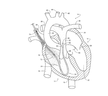
Looking to Fig. 1, a schematic representation of a human heart is identified
generally at 10. In general, the heart 10 performs in two stages or sides,
having a right
side which receives venous-based blood returning from various tissues and
vessels of
the body. This right side of the heart is seen generally at 12 and functions
to pump the
oxygen depleted blood arriving from the venous system to the lungs to be
oxygenated.
Upon being oxygenated and cleared of excess carbon dioxide, the blood is
retttrned
from the lungs and pumped arterially against the vascular resistance of the
entire body
by the left side of the heart which is represented at 14. The pumping chambers
of the
heart are represented in Fig. 1 as a right atrium 16 and a right ventricle 18.
Correspondingly, the left atrium is shown at 20 and the left ventricle at 22.
The right
atrioventricular valve is schematically pornayed at 24, correspondingly, the
left
atrioventricular (mitral) valve is represented at 26. Looking to input to the
right side 12
of the heart 10, the superior vena cava is represented at 28, while the
inferior vena cava
is represented at 30. The output of the right ventricle is shown extending to
the
pulmonary artery 32 which, in turn, extends to a bifurcation represented
generally at 34
to define a left pulmonary artery 36 and a right pulmonary artery 38. Left
ventricle 22
is seen extending to the aorta 40 having an aortic arch from which the left
subclavian
artery extends as shown at 42, the left common carotid artery extends as shown
at 44,
and the brachiocophalic trunk extends as shown at 46. The pulmonary valve is
seen at
48, while the aortic valve is represented of 50. The inferior vena cava 30 as
well as the
superior vena cava 28 both lead into the right atrium 16. Generally, venous
blood
introduced from the inferior vena cava 30 originates from the lower part of
the body,
i.e. the lower limbs, chest, and abdominal cavity. Correspondingly, venous
blood
entering from the superior vena cava 28 is conveyed from the upper anatomy,
i.e.
arms, head, and brain. Another drainage of venous flow not introduced from
these two
major veins evolves from blood draining from the sinuses onto the venous
structure
within the heart. These also mix at the righ atrium 16 and the superior vena
cava 28 as
well as the inferior vena cava 30. In effect, then al of the blood passes
through the
right ventricle to the pulmonary artery 32. By measuring within this region,
accuracy
of caridac output measurement is achieved.
-12-
CA 02256915 1998-12-22
EGG 2-033
A pulmonary artery (PA) catheter adapted to carry out the system and method of
.
the invention is represented generally at 60 at the indwelling location
normally
encountered for heart monitoring including cardiac output (CO) measurement
purposes.
In particular, the catheter 60 is located at the heart 10 in a fashion similar
to that of the
conventional Swan-Ganz flow directed thermodilution catheter. See in this
regard,
Daily, E, "Techniques in Bedside Hemodynamic Monitoring," C.B. Mosby Co.,
1985.
Note that the distal end or tip and measurement region represented generally
at 62 and
configured with a variety of components including a partially inflated balloon
64 is
positioned in the pulmonary artery 32 upstream from the bifurcation 34. At
this
location, the tip region 62 will be immersed in flowing, mixed venous blood,
and from
this measurement location, as noted above, all of the blood of the body
eventually will
flow as it returns to the lungs for oxygenation. Catheters as at 60
conventionally are
mufti-channeled and formed of a soft or compliant material so as not to unduly
interfere
with the valve activities of the right side 12 of heart 10. Typically, the
devices as at 60
will have a diameter of about 7.5 French (0.09 inch) and a length of about 40
inches
extending from an externally disposed proximal end (not shown) to measurement
region 62. The devices are introduced into the body percutaneously, normally
being
entered from the subclavian vein and the jugular vein at the shoulder/neck
region or
alternatively from a femoral vein in the leg. Devices 60 are termed as "flow
directed",
movement into position being achieved as a consequence of blood flow by virtue
of the
partially inflated balloon 64. Correspondingly, the proper positioning of the
tip and
measurement region 62 is confirmed, for example, by the pulmonary blood
pressure
waveforms developed by utilization of an open-ended fluid filled channel or
lumen
extending through catheter 60. This channel is open at the outer tip 66 of the
catheter
60. In this regard, insertion of the catheter 60 is stopped when a pressure
monitor
employed with the blood pressure channel of the catheter exhibits an
appropriate
pressure profile. When appropriately positioned, the distal end will be
located within
the pulmonary artery 38 as illustrated. That same tip and measurement region
62 may
also contain, for example, a temperature sensor and, in a preferred
embodiment, the
forward assemblies of optical fiber components of ammonia and pH optical
sensors,
the outputs of which, respectively, provide signals representing the
concentration of the
ammonia component of the analyte, and the pH of the blood at that measurement
location. Located upstream in the sense of blood flow, an analyte-containing
fluid
injectate or infusion port of catheter 60, shown generally at 70, serves to
infuse or
express a known amount of solution into adjacent blood flow at a controlled
mass flow
rate, which infusion into the bloodstream occurs in the region shown, i.e., at
the
entrance to and within the right atrium. The region of the catheter 60
extending from
the vicinity of the port 70 and the tip thereof at 66 represents a measurement
region
represented generally at 72. Within this measurement region, the forward
assembly of
-13-
CA 02256915 1998-12-22
EGG 2-033
analyte concentration sensor located at the tip region 62 will be at a
location spaced
from the infusion port or outlet 70, a dilution measurement distance
downstream within
the bloodstream. In the figure, a density of "dots" is used to represent the
relative mix
concentration of the analyte in the flowing bloodstream as that analyte is
drawn while
S mixing from the superior vena cava 28 into the right atrium 16, thence
through the right
atrial ventricular valve 24, and into the right ventricle 18. Then, it is seen
as being
progressively diluted in correspondence with flow rate of blood through the
pulmonary
valve 48 to pass the analyte concentration sensor at the tip region 62 as it
extends into
pulmonary artery 32. As is apparent, at the commencement of the injection of
analyte-
containing fluid from port 70, a sensor at tip region 62 will not "see" the
analyte
resulting from the expression of analyte-containing fluid from port 70. The
time delay
before sensing of the injected analyte commences depends upon the cardiac
output of
the heart 10 as well as a need with the system at hand for good mixing of such
analyte
with the flowing blood. The resultant time delay typically will be on the
order of about
2 to 4 seconds.
While the preferred modality for utilizing the present system is with the
right
side of the heart, it can also be employed utilizing a catheter performing in
conjunction
with the left-side 14 of the heart 10. Referring to Fig. 2, the heart 10 is
reproduced
along with the associated components thereof employing the same identifying
numeration as seen in Fig. 1. In the figure, a catheter represented generally
at 80 is
introduced through a major artery into the left half 14 of heart 10. The
catheter 80
extends from an externally-disposed proximal end region (not shown) to an
oppositely
disposed measurement region represented generally at 82. The measurement
region 82
extends from a distal or tip region represented generally at 84 inwardly from
the tip 86.
The tip 86, for this embodiment, is configured to provide an injectate or
diffusion port
or outlet from which analyte-containing fluid is expressed. The outlet or port
at tip 86
is spaced a dilution measurement distance upstream from an analyte
concentration
sensor having a forward assembly contactable with flowing blood at 88. It may
be
noted that no partially inflated balloon as shown at 64 in Fig. 1 is employed
in this
modality. As before, however, a density of "dots" is used to represent the
introduction
and progressive mixing or dilution of the analyte in the flowing blood of the
bloodstream, the diluted concentration of which is measured within the
ascending aorta
with the sensor forward assembly 88.
Returning to Fig. l, the general procedure for determining cardiac output (CO)
35 involves, as a preliminary step, a baseline determination of the
concentration of analyte-
containing fluid in the blood. At the commencement of a procedure, the first
baseline
measurement will be of the analyte which is endogenous to the patient. Where
the
analyte component which is sensed is ammonia gas, the analyte concentration is
represented by the total ammoniacal concentration in the blood, i.e. the
combination of
-14-
CA 02256915 1998-12-22
EGG 2-033
the concentration of ammonia gas (NH3) and ammonium ion (NH4'). That baseline
data or information having been obtained, then analyte-containing fluid
biocompatible
with and metabolizable within the body of the patient is injected into the
bloodstream at
a predetermined mass flow rate through ports as located at 70 in Fig. 1 or at
the tip
region 84 shown in Fig. 2 for an infusion interval. That interval will be
determined by
the rapidity of analyte sensing and the noted dilution of the distance from
the injectate
portal or outlet to the forward assembly of the sensor. During the infusion
interval, a
subsequent sensing of analyte or analyte component concentration level takes
place
which, as before, is converted to total analyte concentration level. By
correlating the
indicator concentration of the analyte-containing fluid source, the mass flow
rate
involved during the infusion interval, the baseline and subsequent analyte
concentrations in blood and other factors which may be called for, cardiac
output then
is derived. For the preferred utilization of ammoniacal fluid as the analyte-
containing
fluid, the value of pH is utilized in the correlation. A particular advantage
associated
with the utilization of an ammoniacal fluid with the procedure is that no
adjustment in
analyte concentration values for hematocrit (HCT) content is required, the
analyte being
taken up both in the plasma and blood cell structure of the blood. Of
additional
importance to the procedure, only one measurement is required to achieve a
value of
cardiac output (CO), averaging of repetitive measurements not being required.
The
terms "mass flow rate" as used herein are meant to include any type of
measured liquid,
e.g. volumetric flow rate where temperature is known.
Looking to Fig. 3, curves 94 through 98 are plotted to reveal values for
cardiac
output (CO) with respect to elapsed time in seconds commencing with the
injection of
analyte containing fluid as further related to the observed change in the
mixed venous
blood indicator, i..e. analyte concentration in blood. Curves 94-98
correspond,
respectively, with cardiac outputs of 10, 8, 6, 4, and 2 liters per minute. It
may be
observed from the figure that for a given injection rate, the lower the
cardiac output
rate, the larger the incremental increase in blood indicator concentration.
This
dependence of the incremental increase in analyte concentration in blood is
due to the
indicator-dilution effect in which the lower the blood flow rate (i.e.,
cardiac output), the
less a given level of analyte-containing fluid injectate will be dispersed and
diluted. In
general, the measurement response time of an ammonia gas sensor employed with
the
preferred embodiment of the system allows reaching an equilibrium value as
shown by
the flattened portion of curves 94-98, or some fraction of the end-point
equilibrium
value, within about 2 to 30 seconds. In effect, the use of a biocompatible and
metabolizable indicator and attendant data retrieval permits a measurement of
CO to be
carried out repetitiously over an extended period of time. The repetition or
updating
rate advantageously may be quite high, thus supplying the intensivist with
substantially
more current CO data.
-15-
CA 02256915 1998-12-22
EGG 2-033
In general, the indicator which is utilized under the precepts of the
invention
may be an anabolite or product of a constructive metabolic process, or a
catabolite, or
product which, by a destructive metabolic process, is converted into an
excreted
compound. In the latter metabolic category, the transformation which occurs
represents
a utility making energy available for organs in use. Desirably, enhanced
measurement
frequencies are made available with the procedure since there is no
substantial
hemodilution nor evoked body system instability. While relatively minor base
line
blood indicator concentration value shifting is encountered, the metabolic
reaction to the
introduced biocompatible analyte-containing fluid fuctions to maintain the
patient in a
stable condition.
Ammoniacal fluid based indicators may be the subject of uptake by certain
organs of the body for further catabolism and excretion, or they may remain in
the body
by anabolism or incorporation into other nitrogenous products. The amount of
such
indicator infused for each cardiac output measurement is based on the
measurement
precision of the sensor, the frequency of cardiac output measurements required
per day,
and the rate of metabolism. For the case of an ammoniacal fluid, the rate of
metabolism
or clearance of ammonia from the blood has been reported to increase with
concentration. See in this regard: Lockwood, A.H., et al., "The Dynamics of
Ammonia Metabolism in Man--Effects of Liver Disease and Hyperammonemia," J.
Clin. Invest., Vol. 63, pp 449-460, 1979). Under resting conditions, most
blood
ammonia/ammonium is of dietary origin. Normal digestive processes generate
ammonia/ammonium from ingested protein, while bacteria in the gastrointestinal
tract
generate ammonia/ammonium by metabolizing protein products of dietary protein
digestion and urea. An illustration of the major organs of amrnonia/ammonium
formation, utilization and circulation is presented in Fig. 4 including the
various forms
of nitrogenous compounds, e.g. ammonia gas (NH;), ammonium ion (NH4') or
related
nitrogenous by-products. Ammonia/ammonium metabolically formed in a given
organ
of the body is generally widely distributed. In Fig. 4, the blood pool or
bloox system
is represented at block 100. Blood pool 100 is depicted supplying glutamine
(GLN) to
the gut or gastrointestinal tract as represented at arrow 102 and block 104.
Ammonia
generated in the gut as at 74 from protein digestion and deamination of
glutamine
(GLN) enters the portal venous circulation as represented at arrows 106 and
108 and is
involved in the liver function as represented at block 110. The metabolic
relationship of
the blood pool or blood system 100 with the liver is represented by arrows 112-
114.
Metabolic interaction with the kidney as at block 116 is represented at arrows
118 and
119, while catabolic ammonium is excreted as represented at arrow 120 and
block 122.
Transport to and from the brain with respect to the blood pool is represented
at block
124 and arrows 126-128. A similar metabolic interrelationship with respect to
skeletal
muscle is represented at block 130 and arrows 132 and 133. Exercise induced
-16
CA 02256915 1998-12-22
EGG 2-033
hyperammonemia will witness a transfer of ammonium ion into the blood supply
as
represented at arrow 134. It may be observed that such relativey short
excursions thus
are readily tolerated by the body. Short duration excursions occur with the
present CO
measurement system. See generally: "Exercise-Induced Hyperammonemia:
Peripheral
and Central Effects," Bannister, et al., Int. J. of Sports Medicine, Vol. 11,
pp 5129-
5142 ( 1990). Under conditions typical of patients in an intensive care unit,
resting
muscles take up ammonia/ammonium from the circulating blood wherein the
substance
enters into protein synthesis via ketoglutaric and glutamic acid. When the
muscle
begins working again, ammonia/ammonium is once again released from the muscle
into
the bloodstream. If additional ammonia/ammonium (in the form of an ammonium
salt
solution) is injected into a peripheral vein, the added ammonia is brought
directly to the
tissue via the blood where it may be retained and eventually used for amino
acid and
protein synthesis. See: Furst, P., et al., "Nitrogen Balance After Intravenous
and Oral
Administration of Ammonia Salts in Man," Journal of Applied Physiology, Vol.
26,
No. 1, pp 13-22 (1969).
The characteristic shapes of curves 94-98 as shown in Fig. 3 may be compared
with a corresponding temperature/time response encountered in a conventional
indicator-dilution approach for developing values of cardiac output, for
example,
procedures involving a brief injection of a cold saline indicator. In Fig. 5 a
mixed
venous blood baseline temperature is represented at dashed line 140 having a
value, for
example, of 37°C. A cold bolus then is injected in the manner discussed
over a time
interval represented within brackets 142 extending to the time line
represented at dashed
line 144. During the interval represented at 142, a 10 ml bolus of isotonic
saline, for
example, at a temperature of 5°C may be injected at the entrance to the
right atrium.
Then, as represented by the temperature characteristic curve 146, a thermistor
or
thermocouple will respond at the region of the pulmonary artery to measure the
relatively rapidly changing indicator value for temperature. By contrast,
systems of
measurement with the present approach may exhibit a relatively slower response
time
inasmuch as the indicator may be injected over a lengthier period and at a
lower rate.
Cardiac output, i.e., volumetric flow rate, is derived empirically as a
function of
the measured or controlled analyte-containing fluid injection rate, m1, and
the measured
increase in blood indicator or analyte concentration. These relationships may
be
plotted. For example considering the preferred amrnoniacal fluid as an analyte-
containing fluid, looking to Fig. 6, the difference in the mixed total
ammoniacal
concentration in blood concentration, C, for a specific rate of analyte-
containing fluid
delivery is plotted with respect to cardiac output in liters per minute. Curve
150 plots
the different values of cardiac output for a range of measured differences in
blood
indicator or analyte concentration with respect to an analyte-containing fluid
injection
mass flow rate of a predetermined value typically derived in milliliters per
second.
-17-
CA 02256915 1998-12-22
EGG 2-033
Lower curve 152 is at a lesser injection mass flow rate. As is apparent, a
family of
such curves will be evolved by a given system. This family of curves may be
represented by the following expression where the analyte-containing fluid is
ammoniacal fluid:
CO(t;) - K * ~ * [IC. - C. (t' ;)] ( 1 )
fC~ (t' ~) - C~ (~)1
where
CO = cardiac output measured at time, t; (liters/minute);
K = constant;
m~ = mass flow rate of injection of ammoniacal fluid (liters/minute);
IC, = total ammoniacal concentration of the analyte-containing fluid
(predetermined indicator concentration) (micromol/liter);
C, (t';) = total ammoniacal concentration of the analyte-containing fluid
in blood measured during period of indicator infusion (blood
indicator concentration) (micromol/liter);
C, (t;) = total ammoniacal concentration of analyte in blood
measured prior to indicator infusion (baseline) (micromol/liter)
The measured volumetric output of the heart often is normalized to the size of
the patient by dividing the measured cardiac output by the patient's "body
surface area,"
BSA (estimated in square meters), the latter parameter generally being derived
based on
the height and weight of the patient. This normalized cardiac output value is
referred to
as the cardiac index, CI, and is given by the expression:
CI(t;) = CO(ti) (2)
BSA
The procedure carried out with the system of the invention is one taking
advantage of the complementary selection of analyte-containing fluid and
analyte
concentration sensor. That matching of system components and the selection of
the
analyte-containing fluid as being metabolizable within the body permits the
carrying out
of rapid measurement of cardiac output with substantial accuracy and without
the need
for averaging procedures. Using an ammoniacal fluid as the analyte-containing
fluid
for demonstration purposes, and referring to Fig. 7, the higher frequency
measurement
approach may be graphically illustrated. In the figure, two of a sequence of
analyte-
containing fluid infusion intervals are represented in conjunction with a time-
related
abscissa, a left-side ordinate representing blood ammoniacal fluid
concentration and a
right ordinate representing ammoniacal fluid or analyte-containing fluid
infusion rate.
With the procedure, following the positioning of a catheter within the
bloodstream of
-18-
CA 02256915 1998-12-22
EGG 2-033
the patient as discussed in connection with Figs. 1 and 2, a baseline analyte
concentration value in the bloodstream is measured with the analyte sensor.
This value
is converted to blood amrr~oniacal fluid concentration and represents a
baseline value
thereof shown as Co at dashed curve portion 160. The initial infusion of
analyte-
S containing fluid or ammoniacal fluid for the instant demonstration then is
carried out for
an infusion interval represented at rectangle 162. The commencement of this
infusion
interval is represented additionally at t,. As the ammoniacal fluid progresses
in the
Bloodstream toward the analyte concentration sensor, there will be no
elevation of the
concentration sensor output. However, as the mixed analyte-containing fluid
reaches
the sensor as illustrated at time t,' in the figure, a very steep increase in
blood indicator
concentration is witnessed as is represented by the curve 164 rising from the
baseline
concentration at 160 to a peak concentration measured during the period of
indicator
infusion and identified by the dashed line level 166. A subsequent analyte
concentration value is developed by the analyte concentration sensor from
which the
ammoniacal fluid concentration in blood is determined. From that value, then
cardiac
output (CO) may be derived as described in conjunction with expression (1)
above.
Note that the curve 164 then relatively rapidly falls as represented at curve
region 168
following the cessation of infusion of analyte-containing fluid, and, further,
as the
infused ammoniacal fluid is metabolized by the body. However, a new
equilibrium
level will be established at a baseline at the curve region 170, that new,
slightly
increased level being represented additionally by dashed line 172.
Two minutes later, the second infusion for cardiac output measurement is
undertaken as represented at rectangle 174. Infusion interval 174 is shown to
commence at tine t3. Following a short interval for perniitting the mixed
analyte-
containing fluid to migrate to the sensor forward assembly, the concentration
sensor
will see a very steep increase in analyte concentration and, consequently,
blood
ammoniacal fluid concentration as represented by curve region 176 at time t3'.
As
before, the curve region 176 will peak as shown at region 178. Note, however,
that
this peak will be slightly higher than the dashed peak line 166. This is
occasioned by
the slight increase in baseline as described at 172. The second infusion
interval is seen
to terminate at time t4 and a rapid fall-off in ammoniacal fluid concentration
in blood
again is achieved as shown at curve region 180. This procedure reiterates over
an
extensive sequence of measurements. At the end of each infusion interval, the
body
again reaches a metabolic equilibrium level with respect to the analyte
concentration at
the newly-established baseline concentration level. This occurs over a
sequence of
measurements until a long-term equilibrium concentration level is reached with
essentially no elevation as a final equilibrium of the metabolic activity and
blood
indicator concentration level is reached. Where the procedure employs
ammoniacal
fluid as the analyte-containing fluid, the peaks in concentration observed
during the
-19
CA 02256915 1998-12-22
EGG 2-033
infusion intervals, as represented at dashed line 166 and curve region 178
will not have
a detrimental affect on the body of the patient. In this regard, it may be
recalled that the
human body will experience ammonia/ammonium ion excursions in the course of
exercise as discussed in connection with arrow 134 in Fig. 4. The system and
method
is capable of carrying out a cardiac output (CO) measurement as often as about
1 to 3
minutes in conjunction with an infusion internal of substantially less than
that
measurement frequency interval. The former infusion interval will be selected
within
about 2 to 30 seconds depending upon the determination of the clinician, the
particular
analyte employed, the concentration sensor utilized, and the mass flow rate of
infusion
of the analyze-containing fluid.
Turning to Fig. 8, a graphical representation of the equilibriation of the
analyte
in blood or blood indicator concentration with metabolic homeostasis of the
body of the
patient is provided. In the figure, the blood indicator concentration or
concentration of
analyte in the blood is represented along the ordinate, while time is
represented along
the abscissa, such time being associated with a sequence of cardiac output
measurements. The figure shows a sequence of blood indicator concentration
spikes,
C~'-CT' and C~'-C~+4' which extend upwardly from respective baseline
concentration
levels Cl-G, and C~,~. The width of each of the spikes corresponds
schematically with
the infusion interval of analyte-containing fluid into the bloodstream. Note
that the
baseline blood indicator concentrations increase with each cardiac output
measurement
as represented at baseline values C1 to about C7. During that period of the
procedure,
a metabolic equilibrium with the concentrations occurs and the concentration
values
elevate above the initial or initial baseline level Co. However, as
represented by the
generally horizontal dashed concentration level line 182, a homeostatic level
of blood
indicator concentration will be reached following a sequence of CO
measurements. At
this point in the procedure, the average rate of infusion will be equal to the
metabolic
rate of the patient. This analyte concentration level corresponding with
metabolic
homeostasis of the body of the patient. As part of the system, the clinician
may provide
as an input to the controls of the system a homeostasis threshold value
corresponding
with a blood indicator concentration level or concentration level of the
analyte
representing a level below iatrogenesis (i.e., a safe concentration level).
Where that
threshold is exceeded, then the procedure is terminated, or a perceptible
output, for
example an alarm, is generated to alert the clinician.
For the preferred embodiment employing an ammoniacal fluid as the analyte
containing fluid, and, for example, employing a CO measurement frequency of 30
measurements per hour representing a measurement of cardiac output after two
minutes, a preferred ammoniacal salt salution infusion rate is 0.5 to 5.0 ml
per cardiac
output measurement, while a more preferred infusion rate is 1.0 to 2.0 ml per
ca~iac
output measurement. The indicator concentration or ammoniacal concentration of
the
-20
CA 02256915 1998-12-22
EGG 2-033
analyte-containing fluid preferably is 10 mmol/liter to 250 mmol/liter, and
more
preferably is 30 mmol/liter to 120 mmolniter. The rate of injection or
infusion of the
analyte-containing fluid for a given cardiac output measurement can be based
on the
previously measured cardiac output value. For example, at higher cardiac
output
S levels, where the amount of dilution of the analyte-containing fluid is
greater, the rate of
the infusion can be greater in order to assure a more accurate cardiac output
measurement. Conversely, at lower cardiac output levels, where the amount of
dilution
of the analyte-containing fluid is smaller, the rate of infusion can be
smaller while still
assuring an accurate cardiac output measurement.
By way of example, the following indicator injection rate may be programmed
into the cardiac outut monitoring system based on an infusion inten~al of 10
seconds:
TABLET
Previous Cardiac Analyte-Containing
Output Fluid Measurement Internal
Measured Value Injection Rate (minute)
(liter/minute) (milliliter/second)
CO < 3.0 0.10 2.0
3.0 CO < 5.0 0.15 2.0
5.0 CO < 7.0 0.20 2.0
7.0 CO < 9.0 0.25 2.5
CO 9.0 0.30 3.0
Using this cardiac output level dependent infusion rate, the amount of analyte-
containing fluid infused per measurement can be selected to assure relatively
uniform
measurement accuracy over the entire range of physiologic cardiac output
values, while
minimizing the total amount of analyte-containing fluid infused into the body.
The
measurement interval can be adjusted according to the infusion rate such that
during
periods of high cardiac output, measurements are performed less frequently to
assure
that the total amount of analyte-containing fluid being infused over a period
of time
does not exceed predetermined limits. For instance, while the measured cardiac
output
level is above 9.0 liters/minute, the measurement interval is 3.0 minutes. At
cardiac
output levels of 7.0 and lower, the measurement internal is 2.0 minutes. This
adjustment in the measurement interval assures that the infusion rate does not
exceed
the ability of the patient's body to metabolize the infused analyte-containing
fluid. As is
apparent, the continuing and frequent measurement of the analyte-containing
fluid level
in the blood and the selection of the noted threshold homeostasis will assure
that such
elected safe limits are not exceeded.
During the monitoring of a given patient, the number of cardiac output
measurements carried out by the system range from less than 50 to greater than
2,000.
-21-
CA 02256915 1998-12-22
EGG 2-033
After some number of measurements, the noted homeostatic level 182 is reached
when
the time-averaged rate of analyte component-containing fluid infusion matches
the rate
of metabolism and and clearance of the- injectate from the bloodstream. The
body's
natural homeostatic process within various organs and tissues serve to
increase the rate
S of metabolism or clearance of the elevated analyte concentration which
results from the
infusions.
The selection of analyte-containing fluid for this indicator/dilution cardiac
output
measurement approach includes balancing the following optimal parameters:
(a) analyte measurement precision--increasing this parameter allows
a smaller amount of analyte-contairu'ng fluid to be infused to achieve a
target
measurement accuracy for each measurement.
(b) background or baseline level of analyte-containing fluid--
selecting an analyte-containing fluid whose baseline or background is low
allows a
greater fractional change in the analyte level for a given rate of analyte
infusion.
(c) metabolism/clearance rate-selecting an analyte-containing fluid in
which the body's rate of metabolism clearance is higher allows more frequent
measurements of cardiac output without significant increase to the baseline
concentration and, importantly, without exceeding safe concentration levels
within the
body.
(d) temporal stability of baseline level of analyte--the greater the
short term stability of the baseline concentration of analyte in blood (i.e.
during the
period between measuring baseline analyte concentration and subsequent analyte
concentration during the infusion interval which typically may range from
several to
tens of seconds), the greater the measurement accuracy for a given rate of
analyte-
containing solution injection (i.e. greater the ratio of signal to noise).
This short-term
stability of the baseline analyte concentration in the blood refers to the
absence of
significant baseline concentration changes due to such transients as: routine
infusion of
intravenous solutions and medicants; movements of the patient in bed;
irregular
breathing; and coughing.
(e) response time of sensor--the faster the response time of the
sensor, the shorter the duration of infusion of the analyte-containing fluid.
The shorter
the duration of the infusion, the smaller the amount of analyte-containing
fluid infused
for each cardiac output measurement (for a target level of measurement
accuracy) and
the smaller amount of analyte-cont<~ining fluid infused for each cardiac
output
measurement (for a target level of measurement accuracy) and the smaller
amount of
analyze-containing solution must be metabolized or cleared by the body.
Now consider the instrumentation employed. As noted above, conventional
catheter designs are utilized. However, the type of analyte concentration
sensors
employed will be seen to fall generally into two categories, optically-based,
utilizing
-22
CA 02256915 1998-12-22
EGG 2-033
fiber optics, and ion selective electrode approaches. In the discourse to
follow, the
analyte concentration sensors are discussed in conjunction with a Swan-Ganz
variety of
pulmonary artery catheter as discussed in connection with Fig. 1.
Looking to Fig. 9, the catheter 60 described earlier in connection with Fig. 1
is
S illustrated at an enhanced level of detail. Catheter 60 incorporates an
optically-based
sensor forward assembly. Specific implementation of such assemblies are
discussed in
figures to follow. Accordingly, the catheter is again represented generally at
60 as
including a tip region 62 incorporating a partially infkated balloon 64 and
outer tip 66.
A measurement region 72 extends from the tip 66 a dilution measurement
distance to an
infusion outlet or port 70 through which analyte-containing fluid is
expressed.
Typically, the infusion port 70 will be positioned about 30 cm behind the tip
66 and is
positioned as discussed in connection with Fig. 1 such that the analyte-
containing fluid
is diffused or expressed into the bloodstream at a location near to and/or
within the right
atrium of the heart. Adjacent the infusion port 70, there is located an
auxiliary port 190
which may be used in conventional fashion to introduce medicants into the
bloodstream. The port 190 also may be employed to carry out a periodic cardiac
output
(CO) measurement utilizing the thermodilution technique with a cold bolus
injection.
Alternatively, a separate port may be provided for the cold bolus injections.
Also
located at the tip region 62 is a temperature sensor 192 which may be provided
as a
thermistor or the like and an open channel or lumen carrying a liquid which is
utilized to
monitor blood pressure at the pulmonary artery. For embodiments wherein an
ammoniacal fluid is used as the analyte-containing fluid, the tip 66 will
incorporate the
forward assemblies of an analyte sensor, for example an ammonia sensor, and a
pH
sensor.
Catheter 60 terminates at a proximal end or end assembly represented generally
at 194 wherein communication is made between its various channels, an analyte-
containing fluid source, and associated control and monitoring features. As
discussed
above, an analyze-containing fluid is supplied at a controlled mass flow rate,
mI, from a
conduit 196 terminating in a fluid transfer connector 198. Fiber optic
assemblies, for
example carrying fiber optics for analyze and for pH sensing extend from their
forward
assemblies at tip region 62 through an assembly 194 and cable 200 to an
optical coupler
202. Optical coupler 202 connects to optical cable (not shown) which connects
to
photodetectors and light emitting diode type light sources as discussed later
herein.
Communication with the auxiliary port 190 is through tubing 204 which
terminates in a
fluid connector 206. Balloon 64 is inflated, for example, with carbon dioxide
via a gas
input at tubing 208 which terminates in a connector and valve assembly 210.
The
columm of liquid channel opening at tip 66 and functioning to measure blood
pressure
extends from the end assembly 194 as tubing 212 which, in turn, terminates in
a
connector 214. Electrical leads which are couplcri with the temperature sensor
192
-23
CA 02256915 1998-12-22
EGG 2-033
extend from end assembly 194 via cable 216 which, in turn, is coupled with an
electrical connector 218. Distance markers are provided on the catheter as
represented,
for example, at 219.
Referring to Figs. 10 and 1 l, the structure of catheter 60 at the tip region
62 is
revealed in sectional fashion. Fig. 10 is a developed view taken along the
wedge
shaped section 10-10 shown in Fig. 1 l, while the latter figure is a sectional
view taken
along the plane 11-11 in Fig. 10. In Fig. 10, the tip 66 is shown to include a
polymeric
collar 220 which functions to block certain of the channels of the catheter 60
and to
form the end component support for the optics and blood pressure related
channels. In
this regard, channel or lumen 222 extends through the catheter 60 and carries
a saline
solution for purposes of transmitting blood pressure witnessed at the tip 66.
Balloon
64 is inflated from an internally disposed port 224 which, in turn, is in gas
flow
communication with a lumen or channel 226. Channel 226 is blocked at the
collar 220
and receives an inflating gas such as carbon dioxide as earier-described. The
two
electrial leads 228 and 230 functioning in conjunction with therniistor or
temperature
sensor 192 extend through a channel such as that at 232 which also is blocked
at tip 66
by the collar 220.
Fig. 11 reveals the presence of two channels at 234 and 236 respectively
carrying fiberoptic assemblies represented generally at 238 and 240 which
function in
the measurement of analyte concentration and pH levels in the blood,
respectively. For
the preferred ammonia analyte measurement component, the pH level value is
called for
to compute the total ammoniacal content, i.e. total ammonium ion and ammonia
gas.
These optical assemblies 238 and 240 extend from forward assemblies of the
concentration and pH sensors. Fig. 11 additionally reveals an analyte-
containing fluid
delivery channel 242 and an auxiliary N channel 244. The latter two channels
are
blocked rearwardly adjacent their outlet pons.
Referring to Fig. 12, the forward assemblies of the optical components at the
tip
region 62 are generally portrayed in block fashion in conjunction with a
schematic
representation of the components supporting them. A variety of adaptations for
the
optical determination of analyte or analyte component concentration and pH
sensing are
addressed in the discourse to follow. Fig. 12 shows that the fiber optic
assemblies 236
and 238 extend to forward assemblies represented in block form, respectively,
at 246
and 248, respectively having a pH sensor and analyte concentration sensor
functions.
Fiberoptic assembly 238 and its associated forward assembly 248 are
operatively
associated with a light source and transducer module represented generally at
block
250. Module 250 includes two channels for providing a light source and
transducing
function as represented at interior blocks 252 and 254. The operative
association
between the analyte sensing fiberoptic assembly 238 and the module 250 is
represented
by dual arrow 256, while the corresponding association between the pH sensing
-24-
CA 02256915 2001-10-31
assembly including fiberoptic assembly 236 and the module 250 is represented
by dual arrow 258 seen
communicating with light source and transducing block 254. Control over the
light source and
transducing functions represented at blocks 252 and 254 is shown asserted from
a monitoring function
represented at block 260 and respective lines 262 and 264. Optically-based
monitoring systems as are
associated with the module 250 and monitor 260 are marketed, for example,
under the trade designation
"ChemCard 2000" by Research International of Woodinville, Washington.
Referring to Figs. 13 and 14, the infusion port or injectate outlet 70 is
revealed as it is
positioned in the analyte-containing fluid delivery channel 242. Note that the
port 70 is located
upstream from a channel plug or block 266, and is formed by locally removing a
portion of the catheter
wall adjacent the lumen or channel 242. The maximum widthwise dimension,
represented in Fig. 13
at L,, of the port 70 will range from 0.1 to 0.8 cm and preferably is within a
range of 0.2 cm to 0.3 cm.
The maximum widthwise extent of the port 70 is represented in Fig. 14 at W,
and will range from 0.05
cm to 0.2 cm, and preferably will fall within a range from about 0.05 cm to
0.1 cm.
While not shown in the drawings, the catheter 60 may be configured to contain
an additional
channel which carries a third fiberoptic assembly which is coextensive in
length with fiberoptic
assemblies 238 and 240. This third channel may be employed to measure oxygen
saturation level of
the blood. Such measurements may be performed using reflectance oximetry
methods as are described
in the following publication: .
Schweiss, J.F., "Continuous Measurements of Blood Oxygen Saturation in the
High
Risk Patient", Vol. 1, Beach International, Inc., San Diego, California, pp 1-
12
(1983).
Additional description for such measurement is described in U.S. Patent
5,788,647 entitled
"Method, System and Apparatus for Evaluating Hemodynamic Parameters" by
Eggers. Alternatively,
one of the fiberoptic assemblies, for example assembly 238, may be employed
for a dual purpose,
including a determination of the oxygen saturation level of blood.
The type of sensor technology employed with the cardiac output monitoring
catheters is selected
in complement with the analyte-containing fluid utilized. Where optically-
based techniques are
employed, a variety of categories for the sensors are available. In all cases,
however, the forward
assemblies of the sensor systems must be within flowing blood as opposed to
being located in cavities
or the like where the blood may be captured and held quiescent. In general,
the optical sensors include:
direct spectrometric sensors; indirect spectrometric sensors; transmission
spectrometric sensors;
transmission/reflectance spectrometric sensors; colorimetric sensors; and
-25-
CA 02256915 1998-12-22
EGG 2-033
fluorometric sensors. These sensors are described in conjunction with
schematic
representations of them in the figures to follow.
Considering initially the direct spectrometric sensors, reference is made to
Figs.
15A and 15B. In the figure, the forward assembly 248 of the analyte
concentration
sensor is revealed. This sensor may, for example, directly measure ammonia gas
as the
analyte. With this arrangement, an optical fiber is shown as represented at
272. Fiber
272 is mounted within the sensor channel, for example, as represented at 234
in Fig.
11. The fiber 272 is surrounded along its lengthwise extent by a sheath 274.
The tip
of the fiber as represented at 276 is coated with a very thin, optically
transparent coating
278. Coating 278 is an anti-coagulant such as heparin which functions to
reduce the
possibility of deposits such as fibrin or blood coatings over the tip 276. The
embodiment of Fig. 15A is one wherein there is a simultaneous transmission of
light at
one or more predetermined wavelengths and reflectance reception of that light.
In this
regard, the bloodstream is schematically represented in general at 280. For
the
preferred embodiment, wherein ammonia gas (NH3) is the analyte, analysis is
made by
light transmission to and reflectance from analyte component (ammonia gas)
particles as
represented at 280. Light transmission is schematically represented in the
figure at 284
and its reflection is represented by the wave arrows 286. The reflected
illumination as
represented by the arrows 286 will exhibit a spectrum which is characteristic
of the
analyte component 282 and the intensity of the spectral portions thereof as
related to the
concentration of the analyte component 282 within the bloodstream 280. In
general,
the diameter of the fiberoptic component 272 is in a range from about 50 to
1,000
microns, and preferably falls in a range of about 100 to 500 microns. A
typical
diameter will be about 250 microns.
The transmission and reception of investigatory light at one or more
predetermined wavelengths also may be carried out using two or more fiber
components. In one approach, two fiber components are positioned in immediate
adjacency. Alternately, one fiberoptic component may provide a transmission
aspect
while a group of such fiber components surmounting a central transmission
fiber
component carries out the opposite or reception function. In such an
arrangement, the
transmitted light and reflected or emitted light are advantageously separated
during their
transmission to and from the bloodstream. In Fig. 15B, the forward sensor
assembly
is again represented in general at 248. The fiberoptic assemblies employed
with the
optical sensor may be singular fibers which are typically formed of plastic or
when
35 formed of glass, typically are provided as bundles or multiple strands of
glass. In the
figure, two optical fibers are schematically represented at 290 and 292. The
lengthwise
extent of each of these fibers is enclosed within a sheath as represented,
respectively, at
294 and 296. Tip surfaces 298 and 300 of respective fibers 290 and 292 are
configured such that the tip surface 298 is slightly canted axially inwardly
as is the
-26
CA 02256915 1998-12-22
EGG 2-033
opposite surface 300. Tip surfaces 298 and 300 additionally may be coated as ;
respectively represented at 302 and 304, with an optically transparent anti-
coagulant
such as heparin. The overall diameter of the transmission/reflection separated
assembly
will be selected as the same as the overall diameter of the single fiber
arrangement of
S Fig. 15A. In the figure, the bloodstream is represented in general at 306,
and the
measured analyte component, for example ammonia gas (NH3) is represented at
308.
With the arrangement shown, light of one or more wavelengths is transmitted
through
fiber assembly 290 as represented by the transmission arrows 310. Resultant
reflection, as represented by transmission arrows 312 is collected and
transmitted by
fiberoptic assembly 292 for analysis. With this sensing forward structure, the
transmitted light and reflected light are advantageously separated during
their
transmission to and from the bloodstream 306, and the analyte component 308
mixed
therewith to enable the more accurate quantitative measures of spectral
intensity and, in
turn, more accurate measurement of the concentration of the analyte component
308. A
concentration of more than one analyte or analyte component in the blood may
be
quantified by the use of appropriate light wavelengths for illumination and
knowledge
of the spectral characteristic of any other analytes of interest. By way of
example, the
direct measurement arrangement may be used to measure both ammonia (NH3)
concentration as well as oxygen saturation level of the blood. As before, the
tip
surfaces or forward assemblies and associated coatings 302 and 304 are
immersed in
flowing blood.
Now considering indirect spectrometric sensor technology, reference is made to
Figs. 16, 17, 18A and 18B. In Fig. 16, the forward assembly of the sensor as
represented generally at 248 includes a fiber~optic transmission/reception
assembly 318
which extends to a tip surface 320. Positioned over the tip surface 320 is a
cap-shaped
membrane 322 having a forward inner surface portion 324 which is spaced from
tip
surface 320 to define a gap 326. A peripheral inner surface 328 of membrane
324 is
sealed to the outer surface 330 of flberoptic assembly 318 to assure the
integrity of the
gap 326. The outer surface 332 of the membrane 322 is in contact with flowing
blood
of the bloodstream represented generally at 334. As before, mixed with the
blood of
the bloodstream 334 is an analyte component, for example ammonia gas,
particles of
which are represented at 336. Membrane 322 is structured to contain
microscopic
pores and functions to minimize or block the ingress of water and other liquid
components within the bloodstream 334 while permitting the analyte component
of
interest, for example ammonia gas, to rapidly diffuse across it due to a
developed
concentration gradient. In effect, a fluid space is developed at the gap 326
containing
measured analyte component as represented at 336'. With the arrangement, an
equilibrium develops between the analyze component 336' and analyte component
336.
One or more wavelengths of light as represented by the transmission arrow 338
are
-27-
CA 02256915 1998-12-22
EGG 2-033
transmitted into gap 326 and reflections from the analyte component 336' as
are
represented by transmission arrows 340 then may be analyzed. The intensity of
the
reflected light is represented by arrows 340, and the concentration of the
analyte
component is correlatable with the intensity of the light at one or more
wavelengths.
Light transmitted as represented at arrows 338 may be of specific wavelength
or a
spectrum of wavelengths may be measured. The advantage of this sensor
structuring
resides in the simplification of spectral analysis, inasmuch as the species of
interest has
been separated from other blood-carrying species. The membrane 324 as well as
the
membrane employed with other embodiments of the invention may be provided as a
Teflon~ barrier, for example, manufactured by W.L. Gore & Associates, Inc. of
Elkton, Maryland. These membranes contain microscopic pores whose size, for
the
ammonia analyte component, preferably is in the range from 0.02 to 3 microns.
The
overall thickness of the membrane 322 will be in the range of from 1 to 100
microns
and, preferably, in the range of 10 to 50 microns. The hydrophobic nature of
the
Teflon4 material serves to minimize ingress of water and other liquid
components
within surrounding blood.
A transmission spectrometric sensor, is illustrated in Fig. 17 where the
optical
sensor forward assembly 248 is schematically revealed for this adaptation. In
the
figure, the fiberoptic assembly is seen to have a general U-shaped
configuration with a
light transmission leg 344 and a return leg 346. Within the assemblage, there
is, as in
the case of the device of Fig. 16, a gap 348 defined between the end face 350
of leg
344 and end face 352 of return leg 346. A surmounting membrane 354 which may
be
of cylindrical shape is positioned across the gap 348 and sealed against the
outer
surfaces 356 and 358 of respective legs 344 and 346. As before, the membrane
354 is
configured having microscopic pores which permit the ingress of analyte
components
from the bloodstream. In this regard, the bloodstream is represented, in
general, at
360, and the analyte components, for example ammonia gas (NH3) are represented
at
362. With the arrangement, when the forward assembly 248 is immersed within
the
flowing bloodstream, the concentration gradient builds between the bloodstream
360
and the gap 348 to provide for the migration of analyte into the latter, such
analyte
being represented at 362'. Light having one or more wavelengths is transmitted
toward
the gap as represented by transmission arrow 364 to be selectively attenuated
by the
analyte 362'. The thus attenuated light then is returned for analysis as
represented by
transmission arrows 366 for analysis quantifying the concentration of analyze
in the gap
348 and, hence, in the bloodstream 360. As in the case of Fig. 15, this
arrangement
has the advantage of isolating the analyte species of interest to simplify
analysis.
Schematic representations of transmission/reflectance spectrometric sensors
are
provided in Figs. 18A and 18B. Looking to Fig. 18A, the forward assembly 248
is
seen to comprise an optical fiber assembly 370 having a side surface 372 and
extending
-28-
CA 02256915 1998-12-22
EGG 2-033
to a tip surface 374. Spaced from the tip surface 374 is a polymeric end piece
376 .
having an inwardly-disposed surface 378 which supports a light reflector
provided as a
coating or the like as seen at 380. The edge surface 382 of end piece 376 is
dimensioned in correspondence with side surface 372 of the assembly 370.
Light reflecting surface 380 is spaced from tip surface 374 a distance
defining a
gap 384 and a cylindrical membrane 386is seen to surround and further define
the gap
384. In this regard, the membrane 386 is sealed to side surfaces 372 and 382.
Forward assembly 248 is immersed in the flowing bloodstream represented, in
general,
at 388. Mixed with the bloodstream 388 is an analyte component as represented
at 390.
With the arrangement, a concentration gradient is developed between the
bloodstream
388 and the gap 384, and the microstructure of the membrane 386 perniits a
migration
of the analyte component into the gap as represented at 390'. Light is
transmitted along
the assembly as represented by transmission arrows 392, whereupon it is
reflected
from the light reflecting surface 380 and returns as represented by
transmission arrow
394. The interaction of this light in crossing the gap 384 then is analyzed to
develop
values for the concentration of analyte component.
Referring to Fig. 18B, alternative structuring of the transmission/reflectance
spectrometric sensor is revealed. The forward assembly 248 is seen to be
structured
incorporating a fiberoptic assembly 400 having a side surface 402 and
extending to a tip
surface 404. Positioned over the forward end of the fiberoptic assembly 400 is
a cap
configured membrane represented generally at 406 having an inwardly disposed
surface
408 and a peripheral, cylindrically-shaped inward surface 410. Supported by
the
inwardly-disposed surface 408 is a light-reflecting component present as a
coating and
shown at 412. The peripheral inward surfaces 410 of the membrane 406 are
sealed to
the side surfaces 402 of fiberoptic assembly 400 to define a gap 414.
Outwardly
disposed surface 416 of membrane 406 is immersed in flowing blood of the
bloodstream as represented in general at 418. Analyte component, as
represented at
420 is mixed with the blood of the bloodstream 418. As before, the membrane
406 is
configured having microscopic pores permitting the migration of the analyte
component
420 into the gap 414 by virtue of the evolution of a concentration gradient
between the
gap and the bloodstream. Other components of the blood essentially are blocked
from
movement into the gap 414. Analyte component which has migrated into the gap
414
are represented at 420'. Analysis of the concentration of analyte component
420',
which is equilibrated with the corresponding concentration of analyte
component 420,
is made by directing light at one or more wavelengths across the gap 414 as
represented
by transmission arrows 422. This light interacts with the analyte component
420' and
is reflected from the reflector component 412 to return for analysis as
represented by
transmission arrows 424.
-29-
CA 02256915 1998-12-22
EGG 2-033
Referring to Fig. 19, a forward assembly is illustrated schematically which
has
a structure common to both colorimetric and fluorometric sensors. The sensor
arrangement includes a fiberoptic assembly which extends to a tip surface 432
and is
surrounded by a sheath 434. Mounted over the sheath and fiberoptic assembly is
a cap-
s shaped membrane 436 having an inwardly-disposed surface 438 and an inwardly-
peripherally disposed surface 440. Surface 440 is sealed to the outer surface
of sheath
434 in a manner spacing the inward surface 438 from the tip surface 432 a
distance
defining a gap 442. Located within this gap is a reactor 444 which, for the
structure
shown, may be an analyte component responsive dye for the preferred
colorimetric
version of the sensor, or a reactor which fluoresces under light stimulation.
The
outward surface 446 of membrane 436 is immersed in flowing blood of the
bloodstream as represented in general at 448 and containing analyte component
as
represented at 450. For the preferred embodiment of the invention, wherein
ammonia
(NH3) is the analyte component and an analyte component-sensitive dye is
employed
for the reactor 444, the membrane 436 is configured having microscopic pores
through
which the analyte 450 may migrate and chemically react with the dye-defined
reactor
444. This will result in a change in coloration of the dye which may be
analyzed by
colorimetric procedures. Accordingly, the reactor 444 is seen stimulated by
light at one
or more wavelengths as represented by light transmission arrow 452. T'he
resultant
light reflected from the reactor dye is represented at transmission arrow 454.
A system
utilizing ammonia as the analyte and an ammonia sensitive dye as the reactor
444 is a
preferred embodiment of the invention. Of the ammonia dyes available for use
as the
reactor 444, bromocreosol green, excited at wavelengths in first band of 380
to 480
nm, in second band of 520 to 680 nm, and third band of 700 to 900 nm;
chlorophenol
red excited at wavelengths in first band of 380 to 420 nm, in a~ second band
of 520 to
620 nm, and in a third band od 650 to 900 nm; bromophenol blue excited at
wavelengths in first band of 380 to 440 nm, in second band of 520 to 640 nm,
and
third band of 700 to 900 nm; m-creosol purple; thymol blue; and Congo red may
also be
considered. T'he light wavelengths for stimulation conventionally are
generated by light
emitting diodes (LEDs) and the wavelengths utilized are based upon the
wavelengths
corresponding to the peak absorption intensity and wavelengths which are
insensitive to
changes in the ammonia concentration. If a plastic fiberoptic assembly is
used, the
preferred third wavelength is about 700 nm. If a glass fiberoptic light
transmitting
assembly is used, the preferred third wavelength of those cited above is
within the
range specified. Dyes serving as reactor 444 quite rapidly reach an
equilibrium with the
analyte component 450. The intensity normalized reflectance of the responding
wavelength of light 454 is utilized to quantitate the concentration of analyze
component
(e.g. ammonia).
-30-
CA 02256915 1998-12-22
EGG 2-033
Where the reactor 444 is provided as an analyte-sensitive fluorescent
material,
then upon excitation by light wavelengths as represented at arrow 452, the
level or
intensity of fluorescence or the rate of quenching when a stimulation source
is
extinguished is correlated with the concentration of analyte component 450.
Referring to Figs. 20A and 208, the light source and transducing function 254
described in conjunction with Fig. 12, representing a component of the optical
coupler
202 described in conjunction with Fig. 9 is revealed in more detail. This
particular
assembly is utilized with the colorimetric embodiment of Fig. 19 wherein the
reactor is
an analyte component-sensitive dye, preferably sensitive to ammonia (NH3). In
Fig.
20A, the fiber assembly 430 is seen extending to a step-down chamber 460. A
singular
fiber optic assembly 430 is positioned in light exchange relationship with an
assemblage of seven fiberoptic components or channels represented generally at
462.
These discrete fiberoptic components include a component 464 which transmits
light at
a wavelength of 450 nm from an LED source 466; a transmitting component
optical
fiber component 468 which transmits light at a wavelength of 615 mm from an
LED
source 470; and a flberoptic component 472 which carries light at a wavelength
of 700
mm from an LED source 474. Reference fiberoptic components 476, 478, and 480
transmit light from respective sources 466, 470, and 474 to a photodiode
reference
function represented at block 482. Light returning from impingement upon the
analyte
component sensitive dye (arrow 454) is collected or gathered and transmitted
by core
gathering fiberoptic components 484-487. Optical components 484-487 are
directed to
a combining input at a photodiode sensor signal represented at block 488.
Looking to Fig. 20B, a cross-section of the assemblage 462 is provided. The
gathering component 484 is seen centrally disposed within the assemlage 462
while
remaining gathering components 485-487 are disposed symmetrically about it.
Transmitting fiberoptic components 464, 468, and 472 have the same diameters
and are
seen to be symmetrically disposed about the centrally disposed collecting
component
484. With this arrangement, about 11 % of the source light from sources 466,
470, and
478 is transmitted to the reactor 442 and about 44% if the light reflected
from reactor
442 is transitted to the photodiode detector 488.
Where the analyte component is, for example ammonia or carbon dioxide, in
order to derive the value of total indicator concentration, i.e. the
concentration of the
analyte fluid in blood, the value of the pH of the blood may be utilized in a
straightforward computation to find total concentration. pH may be measured
with a
variety of techniques using reactors which are chemical or ion selective
electrode-based.
A pH sensitive dye is employed in conjunction with the embodiment described in
conjunction with Figs. 11 and 12. Looking to Fig. 21, the front end assembly
246
represented generally in Fig. 12 is revealed in schematic fashion but at an
enhanced
level of detail. In the figure, the fiberoptic assembly 240 as it is present
at the forward
-31-
CA 02256915 1998-12-22
EGG 2-033
assembly 246 again is represented: The outer cylindrical surface 490 is
covered with a
sheath 492 and the tip surface 494 of the fiberoptic assembly 240 is coated
with a pH
sensitive dye which is applied as a porous coating and represented at 496.
Sealingly
positioned over the tip surface 494 and the dye 496 is a hydrogen ion
permeable
membrane 498 which is cap-shaped having a cylindrical side component 500
sealed to
the assembly 240 and sheath 492. The inner forward surface 502 of the embrane
498
is spaced from the dye layer 496 to accommodate a medium 504 whose pH is in
equilibrium with the pH of the blood within which this forward assembly 246 is
immersed. The pH sensitive dye is interrogated by light at one or more
wavelengths to
detern~ine the value of pH of the blood in the flowing bloodstream. For the
present
embodiment, the forward assembly of the pH sensor is at the tip of the
catheter 60. It
may perform at other locations, for example adjacent the injectate port 70 or
behind the
balloon 64.
Analyte concentration sensing systems can be configured using technologies
other than those which are optically based. Where such alternate approaches
are
utilized, some modification of catheter design is undertaken.
Referring to Fig. 22, a catheter is shown at 510 being structured with a
concentration sensor which is non-optical in design. As before, the
concentration
sensor design may be incorporated in those catheters suitable for carrying out
dilution
method cardiac output measurement, including the embodiments of Figs. 1 and 2.
Device 510, as before, generally is of a Swan-Ganz type having a partially
inflated
balloon S 12 at its tip region 514. The catheter may employ a variety of
analyte
concentration sensor technologies, for example, sensors based upon amperometry
and
voltometry as well as Schottky diode-based technologies and acoustic-wave
based
technologies. Also located at the tip region 514 is a temperature sensor 516
which may
be provided as a thermistor or the like and an open channel or lumen carrying
a saline
solution which is utilized to monitor blood pressure at the pulmonary artery.
This
blood pressure monitoring channel opens at the outward tip surface 518. Spaced
rearwardly from the tip surface 518 a distance selected for dilution
measurement is an
infusion outlet or injectate port 520. Typically, the port 520 will be
positioned about 20
to 30 cm behind the tip region 514 and will have a size selected for achieving
a desired
mass flow rate for the expression of the analyte-containing fluid injectate.
In general,
the port 520 is positioned such that analyte-containing fluid is injected into
the
bloodstream at a location near to and/or within the right atrium of the heart
as discussed
in connection with Fig. 1. Somewhat adjacent to port 520 there is located an
auxiliary
port 522 which may be used in conventional fashion to introduce medicants into
the
bloodstream. Port 522 also may be employed to carry a periodic cardiac output
(CO)
measurement utilizing the long recognized thermodilution technique with a cold
bolus
injection. The forward assembly of the concentration sensor is shown at 524
adjacent
-32-
CA 02256915 1998-12-22
EGG 2-033
the balloon 512. For most implementations of this form of forward assembly, a
membrane of the nature discussed above is employed. This sensor, the
components
within the tip region 514, and the ports 520 and 522 fall within a measurement
region
represented generally at 526 of catheter 510. Catheter 510 is dimensioned in
correspondence with catheter 60 as described in connection with Fig. 1 and, in
similar
fashion, includes distance indicators, certain of which are represented at
528. The
catheter extends to a proximal end region represented generally at 530 and
incorporating
an end assembly 532. From end assembly 532, connection is made with a variety
of
monitoring and control components as well as connection with the control
source of
analyte-containing fluid injectate. In this regard, the analyte-containing
fluid injectate is
supplied at a controlled mass flow rate, m ~, from a conduit 534 terminating
in a
connector 536. Electrical leads extending from thermistor 516 as well as from
concentration sensor forward assembly 524 extend via cable 538 to a connector
540.
Communication with the auxiliary port 522 is through tubing 542 which
terminates in a
fluid connector 544. Balloon 512 is inflated, for example, with carbon
dioxide, via a
gas input at tubing 546 which terminates in a connector and valve assembly
548. The
column of liquid channel extending to tip surface 518 for purposes of blood
pressure
monitoring extends from end assembly 532 via tubing 550 which, in turn,
terminates in
a connector 552.
Referring to Figs. 23 and 24, the structure of the catheter 510 at the forward
assembly 524 of the concentration sensor is revealed. The outer end of the tip
region
514 incorporating temperature sensor 516 is structured identically as the
corresponding
sensor 192 described in connection with Fig. 10. Seen in Fig. 23, in the
region of
balloon 512 is an inflation/deflation channel 554 which is blocked at a plug
556 to
establish an ingress/egress port 558 for carrying out the selective inflation
of the
balloon 512. Extending along the channel at the centerline of the catheter 510
is a blood
pressure channel 560, while directly opposite the inflation channel 554 is an
electrical
lead channel 562. Channel 562 appears in Fig. 24. At this location, the
channel retains
two electrical leads 564 and 566 extending from the temperature sensor 516
(Fig. 22).
Concentration sensor 524 is structured as an ion-specific electrode-based
device, and is
formed having an outwardly-disposed, cylindrically-shaped membrane 568.
Membrane 568 is provided as a microporous, hydrophobic polymer such as the
earlier-
described Teflon~ or polytetrafluoroethylene. In effect, the membrane 568 is
semi-
permeable to the ion of interest. For the case of an ammomiacal fluid
performing as the
analyte-containing fluid, in general, the ammonium ion (NH4') becomes the
analyte
component. Fig. 24 reveals the presence of an inwardly disposed cylindrical
polymeric
wall 570 spaced inwardly from the cylidrical membrane 568 to form a fluid
retaining
annular gap 572 which extends between a cylindrical end plug 574 and a
corresponding
fluid block provided as an outer wal 576. Within gap 572 is an electrolyte or
-33-
CA 02256915 1998-12-22
EGG 2-033
electrically conducting liquid 578. Where the analyte-containing fluid is an
ammoniacal
fluid, the liquid 578 may be a solution containing 0.1 motor ammonium
chloride. That
liquid 578 reaches equilibrium with a blood carried ammonium ion flow across
the
membrane 568 to change or alter the pH of the solution of liquid 578. In
effect, the
device becomes a pH sensor. As is apparent, the liquid 578 must be related to
the
analyte which is being measured. For the ammonium ion analyte component
considered, the higher the concentration of ammonium ion in the bloodstream,
passing
over the membrane 568, a corresponding effect will be observed in the ammonium
ion
concentration in liquid 578. Ion selective electrodes are employed to measure
this ion
concentration within liquid 578. In this regard, the outwardly-disposed
surface of
cylindrical wall 570 is coated at a forward region of the forward assembly 524
with a
pH electrode which may be implemented as a glass electrode selective to the
hydrogen
ion. Such an electrode is shown at 580. Electrode 580 may be a glass
comprising
silicon dioxide, lithium oxide, and calcium oxide in the ratio 68:25:7. Note
in Fig. 23
that electrode 580 extends from cylindrical end plug 574 to an edge or
tem~ination at
582, and is connected to an electrical lead 584 which extends into the
electrical lead
channel 562. A cylindrically-shaped reference electrode 586 completes the
forward
assembly 524 (Fig. 23). This second electrode 586 may be provided as a
metallic
coating, for example, silver/silver chloride. Electrode 586 is spaced from the
glass
electrode 580 but remains operationally associated therewith within the
electrolyte
containing cavity or gap 572. The electrode 586 is connected to a lead 588
which also
is extended into the electrical lead cavity 562 as seen in conjunction with
Fig. 26.
In operation, the electrical leads 584 and 588 are connected across a
potentiometric based sensing system, where the analyte component is ammonia
(NH3).
As blood within the bloodstream within which is mixed the analyte-containing
fluid,
moves across the hydrophobic membrane 568, ammonia gas vapor diffuses through
the
membrane and into the electrolyte 578. 'Changes in the measured potential
between the
glass electrode 580 and the reference electrode 586 are in correspondence with
the
change of pH within the electrolyte 578 and, are proportional to and
correlatable with
the blood ammonia concentration of the bloodstream adjacent the concentration
sensor
524. The value of cardiac output (CO) may be computed in correspondence with
expression (1) above. Sensor 524 may perform in either the above-noted
potentiometric mode wherein voltage across the reference and glass electrode
is
determined, or may operate in amperometric mode wherein the current flow
between
these two electrodes is evaluated during the application of a small D.C.
voltage
difference.
Additionally seen in Fig. 24 is the auxiliary channel 586 communicating with
port 522 (Fig. 22) and with connector 544 through tube 542. The figure also
reveals
the indicator or analyte-containing fluid delivery channel 592 which will have
been
-34
CA 02256915 1998-12-22
EGG 2-033
blocked off just beyond the location of injectate port 520. Blood pressure
channel 560
appears centrally in Fig. 24.
Referring to Figs. 25 and 26, the commencement of the measurement region
526 at the infusion port or inlet 520 is revealed at a higher level of detail.
Catheter 510
now is formed with the enlarged outer cylindrical wall 576 described in
connection with
Fig. 23. That cylindrical wall reappears in the instant figures. The
structuring of the
port 520 is the same as discussed in connection with Fig. 9. Figs. 25 and 26
show that
port 520 extends through the enlarged wall 576 and is in fluid transfer
communication
with the analyte-containing fluid delivery or infusion channel 592. That
channel
receives a controlled mass flow rate of analyte-containing fluid during an
infusion
interval. Note that the delivery channel 592 is plugged or blocked with a plug
594.
Fig. 26 reveals that the electrical lead carrying channel 562 of the catheter
510, as it
extends rearwardly from the electrode%lectrolyte-based analyte concentration
sensor
forward assembly 524 (Fig. 24) incorporates four leads, to wit, 564, 566, 584,
and
588, which terminate at the earlier-desc,~-ibed cable 538 and connector 540.
Now looking to the utilization of Schottky diode-based concentration sensors,
reference is made to Figs. 27-29. In these figures, the concentration sensor
is
represented in schematic fashion. Looking to Fig. 27 the measurement region
600 of a
catheter 602 of a variety described in connection with Figs. 1 and 2 is seen
to
incorporate the front-end assembly 604 which employs a technology based upon
the
interaction of planar Schottky barrier diodes with analytes or analyte
components. In
this embodiment, the sensor 604 is mounted upon, for example, a wall 606
corresponding with a wall as at 576 described in connection with Fig. 26.
Sensor 604
is formed having two metal electrodes configured in spaced relationship and in
interdigitated geometry. These electrodes are provided as a gold electrode 608
configured in conjunction with an aluminum electrode 610. Gold electrode 608
creates
an ohmic contact and aluminum electrode 610 creates a Schottky barrier contact
with
0li(3-octylthiophene)(3POT). The conducting polymer 612 exhibits an electrical
conductivity which is correlatable with the concentration of the analyte or
analyze
component being employed. Conducting polymer 612 may be substituted
polypyrroles, polythiophenes, or polyanilines. Not shown in the drawing is an
analyte
or analyte component permeable membrane as discussed earlier herein which
covers the
active sensor components. These active components provide a noted reactor
function.
The outer surface of the membrane, as before, is in contact with flowing blood
of the
bloodstream. See generally Assadi, A. et al., "Interaction of Planar Polymer
Schottky
Barrier Diodes with Gaseous Substances", Sensors and Actuators, B, Vol. 20, pp
71-
77 ( 1994).
Now considering analyte concentration sensors which are acoustic wave-based,
reference is made to Fig. 30. In the figure, the concentration sensor forward
assembly
-35
CA 02256915 1998-12-22
EGG 2-033
as it would be mounted in the manner of sensor 604 described in connection
with Figs.
27-29 is depicted schematically at 620. The sensing principle of such acoustic
sensors
is based upon the detection of changes of wave velocity and attenuation caused
by
perturbations at the surface of the material in which the wave propagates. If
an acoustic
wave delay line is placed in an oscillator loop as the frequency-determining
element,
velocity shift causes a shift in the delay time of the wave. This results in a
shift of the
oscillation frequency. In the figure, an interdigitated transmission
transducer is shown
at 622 spaced from a reception transducer 624. Sound reflectance from the
analyte or
analyte component being investigated is represented by the arrow 626.
Transducers
_622 and 624 are connected in a delay line oscillator circuit. The latter
circuit includes an
oscillator 628 having an input at line 630 and an output at line 632.
Transducers 622
and 624 are incorporated within a feedback path or delay line, transducer 622
being
coupled via lines 634 and 636 to line 632 and transducer 624 being coupled via
lines
638 and 640 to line 630. Accordingly, the output of the amplifier 628 is fed
back by
the delay line incorporating the transducers where A(w) represents amplifier
gain and
B(w) represents delay line losses. The transducers as well as the oscillator
circuit may
be multi-layer devices constructed using conventional integrated circuit
manufacturing
methods employing a silicon (base), silicon dioxide, aluminum and zinc oxide
(surface). See generally the following publications: Velekoop, et al., M.J.,
et al.,
"Integrated-Circuit-Compatible Design and Technology of Acoustic-Wave-Based
Microsensors", Sensors and Actuators A., vol. 44, pp 249-263 (1994).
Now considering the overall system for developing cardiac output
measurement, reference is made to Fig. 31. In the figure, the system is shown
in
schematic pictorial fashion in general at 650 utilizing a pulmonary artery
catheter as
shown in general at 652. The implementation of this catheter is in
correspondence with
the pulmonary artery catheter described in connection with Fig. 1. At the
input to the
catheter, signal transfer output connections are shown to emanate from a
module 654
and are directed as represented by multi-component cables 656 and 658 to
operational
coupling with a controller represented at 660. Controller 660 is mounted upon
a
conventional IV pole or stand represented generally at 662 and is seen to
include an
array of keys represented at 664 which are utilized for entering or inputting
control
parameters such as cardiac output or cardiac index limits, homeostatic
threshold levels,
and the like. A display 666 is provided adjacent the key array and an analyte-
containing
fluid pump 668 is mounted at the rear of the device along with a strip chart
recorder
670. Analyte-containing fluid is supplied to the pump 668 from a disposable
hanging
bag source 672, and feed tube 674.
Referring to Fig. 32, a block diagram of the system within which the analyte-
containing fluids and complementary sensors perform is represented. This
system, as
represented in general at 680 is operated in conjunction with a microprocessor-
driven
-36-
CA 02256915 1998-12-22
EGG 2-033
controller represented at block 682. The controller includes the conventional
features
for achieving control including read only memory (ROM), random access memory
(RAM), as well as input/output components including programmable interface
adapters
which perform in conjunction with input devices such as a keyboard, and
provide a
display function which may include a printer. In the course of the use of
system 680, a
variety of parameters will be measured and entered into memory. For example,
the
patient's weight and height are entered as represented at block 684 and line
686. This
information is utilized in deriving BSA, the body surface area, in meters2 in
order to
ultimately compute cardiac index (CI) as described in conjunction with
expression (2)
above. The analyte component described in conjunction with Fig. 32 is ammonia
gas
(NHS). For certain of the other analytes, it may be necessary to measure blood
hematocrit and enter it on an intemuttent basis or a continuous basis to
provide a
correction to achieve accurate cardiac output measurement. Two such analytes
requiring this correction are carbon dioxide and ethanol.
The components of the catheter utilized are represented within a boundary 688.
Such components include a blood temperature sensor such as described at 192 in
Fig. 9
and represented at block 690. Data representing blood temperature is
transmitted, as
represented at line 692 to a temperature monitor represented at block 694 and
the
resultant data is inputted to the controller 682 as represented at line 696.
Line 692
corresponds with earlier-described lines 228 and 230 (Fig. 11 ). Similarly,
the blood
pressure channel of the catheter is represented at block 698. The pressure
output of the
blood pressure channel is represented at line 700 as being directed to a blood
pressure
monitor function represented at block 702. This output from blood pressure
monitor
702 is directed, as represented at line 704, to the system controller 682.
The infusion port or injectate outlet of the catheter within boundary 688 is
represented at block 706. Analyte-containing fluid as represented at arrow 708
is
introduced from an analyte-containing fluid flow meter represented at block
710. Input
to the meter 710 is from an analyte-containing fluid flow control represented
at block
712, the output of which is represented at line 714. Flow control 712 performs
in
conjunction with an analyze-containing fluid source represented at block 716
and line
718. Control over the fluid flow control function 712 is provided from the
system
controller 682 as represented at line 720. The control asserted from line 720
is one
corresponding with the mass flow rate of injection of analyte-containing fluid
represented as m~ in expression ( 1 ) above.
The analyze component concentration sensor function within the catheter is
represented at block 722. This sensor is represented as being operatively
associated
with a light source and transducer function represented at block 724 by a line
726. A
blood pH sensor function within the catheter 688 is represented at block 728
as being
associated through line 730 with the light source and transducer function at
block 724.
-37-
CA 02256915 1998-12-22
EGG 2-033
The output of the light source and transducer function 724 is represented by
lines 732
and 734 as being operatively associated with a blood ammonia level and pH
monitoring
system represented at block 736. Outputs to the controller 682 from the
monitoring
system 736 are represented at line 738.
S The measured value of blood pH is used in conjunction with the measured
blood concentration of the analyte component, gaseous ammonia to compute the
total
ammoniacal concentration in the bloodstream at the measuring location. In
particular,
the analyte concentration sensor derives the measured concentration of ammonia
gas in
blood Ca(NH3). This measured value combined with the measured blood pH allows
computation of the total ammoniacal concentration in blood, Ca, by applying
the well
known Henderson-Hasselbalch equation to the equilibriated ammomia gas (NH3)-
ammonium ion (NH4') system. See generally in this regard: Hindfelt, D., "The
Distribution of Ammonia Between Extracellular and Intracellular Compartments
of the
Rat Brain", Clinical Science and Molecular Medicine, vol. 48, pp 33-37, 1975.
The
relative distribution of ammonia gas (NH3) and ammoniim ion (NH4') in solution
is
given by that Henderson-Hasselbalch equation as follows:
pH - pK' = log [C.(NH3)] (3)
Ca NH4
This equation can be restated in terms of the unknown, C,(NH4') as follows:
Ca(NH4') = C,(NH3)/[ 10 exp (pH - pK,)] (4)
where
Ca(NH4') = concentration of ammonium ions (NH4') in blood
(micromole/liter)
C,(NH3) = measured concentration of ammonia gas (NH;) in blood
(micromole/liter)
pH = measured blood pH
pKa = pH level of solution above which all ammoniacal fluid
exists as a gas
(NH5) where pKa = 9.15 (Hindfelt, ibid).
The total ammoniacal content of the blood, C, (total) can be calculated by
controller 682
as follows:
C,(total) = C,(NH3) + C,(NH4') (S)
-38-
CA 02256915 1998-12-22
EGG 2-033
The values of total ammonia concentration of the blood, C, (total) before and
after the
infusion of the analyte-containing fluid are then used in expression (1) to
compute
cardiac output (CO).
Parameters collected and derived by controller 682 are directed to the readout
S function represented by boundary 740. Function 740 includes two readouts
including a
dynamic display or screen as represented at line 742 and block 744. For
permanent
record purposes, as well as developing a cardiac output trendline with time,
such
information also is directed to a printer strip chart recorder represented at
block 746 and
line 748. Data which may be observed at display 744 include cardiac output
(CO)
displayed as computed in conjunction with expression (1) above; "baseline"
ammoniacal concentration (C,) prior to the commencement of an infusion
interval.
Next, cardiac index (CI) is displayed having been computed in conjunction with
expression (2) above. Perceptible indications which may have an alarm status
are
published at the display and an audio signal also may be produced when the
computed
ammoniacal concentration represented by expression (5) exceeds an inputted
homeostasis threshold value corresponding with an ammoniacal concentration in
blood
for iatrogenesis. Where desired, the analyte concentration developed with each
measurement also may be published. Blood temperature data also may be
published as
a valuable parameter, the blood temperature measurement being earned out by,
for
example, temperature sensor 192 as described in connection with Fig. 9.
Referring to Figs. 33A and 33B, a flow chart describing the operation of the
system in conjunction with monitor-controller 682 is revealed. The system is
started as
represented at commencing node 760 and, as represented at line 762 and block
764, the
patient height and weight is entered through the key array. As discussed in
connection
with block 684 in Fig. 32, this information is utilized for the purpose of
computing
cardiac index (CI) as set forth at expression (2). Next, as represented at
line 766 and
block 768, the operator elects an interval for carrying out successful
measurement, that
interval being designated, "MI". The above-described infusion interval is
established in
software by the manufacturer. The operator may select high and low alarm
limits for
cardiac index values, values falling below the lower limit indicating an
inadequate
cardiac output. Correspondingly, certain medical intervention may increase the
cardiac
index above a desired level, thus calling for an alarm condition. The limit,
alternately,
may be established for cardiac output values. However, the cardiac index is a
normalized form evaluation which may be beneficial for this purpose. The
operator
also enters the noted homeostasis threshold value corresponding with
ammoniacal fluid
concentration for iatrogenesis or adverse effects (C~h). As discussed above,
the
metabolic system of the patient will gradually enter into the state of
equilibrium or
homeostasis with respect to the blood indicator or analye concentration in
blood.
-39-
CA 02256915 1998-12-22
EGG 2-033
The program then continues as represented at line 770 and block 772, at which
point the correct date and time are verified by the operator. This assures
that data which
is collected is correlated in time and date with manual records and manual
interventions
which may be carried out.
As represented at line 774 and block 776, the start or continuation of a
sequence
of measurements of hemodynamic parameters ensues. These parameters may have
value in and of themselves or may be employed for the purpose of computing
can3iac
output and cardiac index. As noted earlier herein, a substantial number of
additional
parameters may be measured depending upon the type of concentration sensors
employed and the number of sensors used. Fiberoptic-based sensor may serve
such
functions as additionally sensing blood oxygen levels and the like from which
other
parameters may be computed and displayed. Thus, in addition to the measurement
of
analyte concentration, pH, temperature and blood indicator or analyte
concentration,
other parameters may be developed based upon a supplementing blood oxygen
measurement. Certain of these parameters are listed later herein in connection
with a
carbon dioxide analyte.
Blood pH is measured as represented by line 778 and block 780, and the
baseline concentration of ammonia in blood is measured as represented at line
782 and
block 784. At the commencement of the procedure, this will be carried out
before the
introduction of ammoniacal fluid injectate. Thereafter, the baseline values
will be
developed as described in conjunction with Figs. 7 and 8. Then, as represented
at line
786 and block 788, the controller calculates the baseline ammoniacal
concentration in
blood, Ca(t;). As represented at line 790, the program proceeds to the query
posed at
block 792 wherein a determination is made as to whether the baseline
ammoniacal
concentration in blood is greater than a threshold value, C~,. In the event
that it is, then
as represented at line 794 and node 796, the system is stopped and an alarm
output is
made.
Where the baseline concentration of analyte is not greater than the threshold
value, then as represented at line 798 and block 800, a measured flow of
analyte-
containing fluid, i.e. ammoniacal fluid, is delivered utilizing the infusion
outlet or port
of the catheter.
The program continues as represented at line 802 which reappears in
conjunction with Fig. 33B. Line 802 leads to the procedure represented at
block 804
which provides for the measurement of mixed venous ammonia or ammonia in blood
during the infusion interval at a time, t'~, which is the earlier-noted t~
plus a D interval of
time. At the termination of the infusion interval, as represented at line 806
and block
808, the analyte-containing fluid flow is terminated. Then, as represented at
line 810
and block 812, the controller calculates the ammoniacal concentration in
blood, cardiac
output, and cardiac index as described in conjunction with expressions (1)-
(5).
-40-
CA 02256915 1998-12-22
EGG 2-033
The program then continues as represented at line 814 and block 816. At that
block, the cardiac output and all other measured parameters are computed for
publication. In this regard, as represented at line 818 and block 820, a
screen display is
made of selected parameters in conjunction with time and tabulations of
parameters are
made. Additionally, as repesented at line 822 and symbol 824, a tabular and
strip chart
record can be provided.
The program then continues as represented at line 826 and block 828. At block
828, a determination is made as to whether the procedure should be terminated.
In the
event of an affim~ative response to that inquiry, then as represented at line
830 and
node 832, the system is stopped. In the event of a negative determination with
respect
to the query posed at block 828, then as represented at line 834 and block
836, a
determination is made as to whether the time for a next measurement is at
hand. If that
interval, for example two or three minutes, has not expired, the program loops
as
represented at loop line 838. In the event that it is time for a next
measurement, then as
represented at line 840 and block 842, the index, i, is incremented by l, and
as
represented at line 844, the program reverts to line 774 to commence the
measurement
procedure again.
In the course of carrying out pig experimentation with a pulmonary artery
catheter utiliing ammoniacal fluid as the analyze-containing fluid,
comparisons were
carried out with respect to a standard represented as the cold bolus technique
of them~al
dilution. Pharmacological agents were utilized to raise and lower cardiac
output of the
pig and, in conventional practice, the cold bolus measurements were carried
out four
times and the last.three measurements were averaged. On the other hand,
utilizing the
ammoniacal fluid injected, only one measurement was taken. Resulting cardiac
output
measurements over the interval of the experiment are plotted in Fig. 34. In
the figure,
the cold bolus average measurements are represented by dots and the
measurements
utilizing an ammoniacal fluid injectate are represented by triangles. A
resulting curve is
shown at 850 linking the thermodilution dots. Dashed curves 852 and 854,
respectively, represent the upper bound of a 15°lo envelope and the
lower bounds of
such an envelope, such a range being commonly accepted as an appropriate
accuracy
range for any cold bolus thermodilution triplicate average measurements.
Fig. 35 is a scatter graph configured utilizing the data developed in
conjunction
with Fig. 34. In the scatter graph, the ordinant represents an ammoniacal
fluid-based
measurement and the abscissa represents the corresponding averaged
thermodilution
measurement. Perfect correspondance is represented by the 45° curve
856. A plot of
the averaging of the data is represented by the dashed curve 858.
As noted earlier herein, a carbon dioxide releasing fluid may be employed as
an
analyte-containing fluid. When so employed, the concentration of the carbon
dioxide
analyte is measured by the analyte sensor. While the corresponding
concentration
-41-
CA 02256915 1998-12-22
EGG 2-033
carbonates or the like may be measured, the former analyte component lends
itself more
readily to concentration analysis. Accordingly, where carbon dioxide releasing
fluid is
employed as the injectate, then cardiac output (CO) is computed in accordance
with the
following expression:
K * mCOz (6)
CO = 10 * [CvCOz' (t' .) - CvC)z(ri)]
where CO is cardiac output in liters per minute; K is a constant; mCOz is the
rate of
injection of carbon dioxide in grams per second; CvCOz (t;)' is mixed venous
carbon
dioxide releasing fluid concentration in milliliters of such fluid per
deciliter of blood at
a baseline defining time, t; and CvCOz (t';) is the corresponding mixed venous
carbon
dioxide releasing fluid concentration at subsequent time, t;', during the
infusion
interval.
To derive caniiac output (CO) in accordance with expression (6), the system
requires the measurement of the following five parameters:
(a) Pplaama COz, the partial pressure of carbon dioxide in the plasma
measured utilizing an indwelling COz sensor,
(b) the pH of blood in the bloodstream is measured using an
indwelling pH sensor preferably in the vicinity of the measurement made at
(a);
(c) the temperature of blood, T, within the bloodstream will be
measured using a temperature sensor mounted on the catheter employed.
(d) hematocrit (Hct) which may be used to calculate hemoglobin
(Hgb); and
(e) SvOz, the mixed venous oxygen saturation of the blood in the
bloodstream, as probably measured using an indwelling catheter oximeter or
calculated
from knowledge of above parameters.
In the process of deriving cardiac output (CO) in accordance with expression
(6), the controller carries out solution of supporting expressions. In this
regard, the
concentration of carbon dioxide in plasma is computed in accordance with the
following
expression:
plasma C02 - 2.226 * s * Pplasma COz * [ 1 + IOpH K], (7)
where pH is the pH value of the blood.
The term, pK is coupled in accordance with the following expression:
pK' = 6.086 + [0.042 * (7.4-pH)] + [(38-T) * [0.00472 + [0.00139 * (8)
(7.4-pH)] ] ]
where T is the temperature of the blood.
-42-
CA 02256915 1998-12-22
EGG 2-033
The term s, is computed as follows:L
s = 0.0307 + [0.007 * (37-T)] + [0.00002 * (37-T)Z] (9)
The controller derives a value for mixed venous carbon dioxide content, CvC02
in accordance with the following expression:
CvCOz = [CP,"m, COZ] * [I-0.0899*Hgb[3.352-0.456*SvOz)*(8.142-pH)]
( 10)
where Hgb is hemoglobin level and SvOz is a value corresponding with mixed
venous
oxygen saturation output.
Where carbon
dioxide
is selected
as the
analyte,
the system
may derive
a variety
of parameters
as are
listed
below.
These
parameters
also may
be developed
or utilized
with other
analytes:
CO - measured cardiac output in liters/minute
BSA - body surface area in m2 (calculated based on and
patient's height
weight)
pH - measured blood pH
CI - cardiac index = COBSA (liters/minute 'm2)
T - measured patient temperature in C
SvOz - measured mixed venous oxygen saturation in
%
Sa02 - measured oxygen saturation in arterial plasma
in !o
Pa02 - dissolved oxygen in arterial plasma (mm Hg)
- 10 exp [log Pso + (log (SaO~/(1 - Sa02)))/2.7](11)
- where Pso = 27 mmHg based on normal oxygen
dissociation curve
P v 02 dissolved oxygen in mixed venous plasma (mmHg)
=
- 10 exp [log Pso + (log (SvOi/(1 - SvOz)))/2.7](12)
a - solubility coefficient of oxygen (ml[O~J/dl
[blood] * mmHg)
- 0.0031/[(10 exp (0.024*(38.0-T)))*(10 exp (-0.50(7.40(13)
- pH)))]
Hct - measured blood hematocrit in percent
DysHgb measured blood dyshernoglobin concentration
= in gm/dl
Hgb - 0.3718 (Hct) - 1.30 - DysHgb (gm/dl) (14)
Ca02 - arterial oxygen content (ml[OZ]/dl [blood])
- [Hgb * 1.34 * Sa02] + [Pa02 * a] (15)
where 1.34 has units of ml[OZ]/gm[Hgb]
- oxygen transport (ml/min)
DOz - Ca02 * CO * 10 (
16)
MAP - measured mean arterial pressure in mmHg
MPAP = measured mean pulmonary arterial pressure in
mmHg
CVP - measured central venous pressure in mmHg
-43-
CA 02256915 1998-12-22
EGG 2-033
WP - measured wedge pressure in mmHg
HR - measured heart rate in beats/minute
SVRI - systemic vascular resistance index [(mnHg
* m2)/(1/min)]
- (MAP - CVP)/CI (17)
PVRI - pulmonary vascular resistance index [(mmHg
* m2)/(1/min)]
- (MPAP - WP)/CI ( 1
g)
S I - stroke index (ml/beat/m2)
- CI/(HR * 1000) (19)
LVSWI = left ventricular stroke work index (gm '
m/m2)
- (MAP - WP) * SI * 0.0136 (20)
RVSWI = right ventricular stroke work index (gm ~
m/m2)
(MPAP - CVP) * SI * 0.0136 (21)
Other analyte or analyte component sensors may be provided as follows:
A glucose sensor may be constructed using well-known enzyme-based methods
(e,g., involving glucose oxidase in conjunction with an oxygen senso). In such
a
devices, an immobilized biological/biochemical component interacts with the
analyte to
produce, via an appropriate transducer, a signal proportional to the quantity
or activity
of analyte. The recognition interaction may entail either a binding process
(e.g. for
antibodies) or a biochemical reaction (e.g. enzyme catalysis). Transduction
can be
achieved by any of several detection approaches: optical (e.g. absorbance,
fluorescence, chemiluminescence and bioluminescence, mass measurement (e.g.
piezoelectric and surface acoustic wave), heat and electromechanical-based
measurement. By way of example, the sensor may be constructed based on the
principles first described by Clark and Lyons (Clark, L.C. and Lyons, C.,
"Electrode
System for Continuous Monitoring in Cardiovascular Surgery," Ann. N.Y. Acad.
Science, Vol. 102, p. 29ff [1962]). The concentration of glucose in the blood
is
achieved by means of a dissolved oxygen (p02) sensor used in conjunction with
the
glucose oxidase-catalyzed reaction:
-44-
CA 02256915 1998-12-22
EGG 2-033
Glucose Oxidaze
Glucose + OZ -~ Gluconic acid + H202
The rate of decrease in p02 can be used as a measure of the glucose
concentration.
Yet another approach involves 'the use of an amperometric glucose electrode
which uses a ferrocene derivative as the mediator. In this sensor, dimethyl
ferrocene is
incorporated into a graphite electrode to which glucose oxidase is
immobilized. During
operation, the glucose oxidase that is reduced in the enzymatic reaction is
reoxidized by
electrogenerated ferricinittm ions. 1fie current flowing in this regeneration
process is
proportional to the glucose concentration. (See Cass, A.E.G., et al.,
"Ferrocene-
Mediated Enzyme Electrode for Amperometric Determination of Glucose," Anal.
Chem., Vol. 56, p 667ff (1984]. With the use of an appropriate membrane (e.g.
polyurethane), glucose may be measured at concentrations up to 50 mmol/liter.
One preferred approach for measuring heparin level in blood involves the use
of
an ion-selective electrode in conjunction with a polymer membrane (e.g.,
polyvinyl
chloride) doped with tridodecylmethylammonium chloride as the heparin
complexing
agent. The measured potential between a reference electrode and the heparin-
selective
electrode is correlatable with the concentration of heparin in the blood. In
this regard,
see Yang, V., et al., "A Novel Electrochemical Heparin Sensor," ASAIO Journal,
Vol.
39, No., 3, pp M195-M201 (1993).
An ethanol sensor may be constructed based on principles similar to those
described for glucose sensors. Hydrogen peroxide is a product of the enzymatic
oxidation of glucose or alcohol. Hence, an electrode responsive to hydrogen
peroxide
can be used to quantitate the concentration of the analyte of interest. A
peroxide
electrode that is covered by an enzyme membrane can be used to detect ethanol
concentration in the blood using an oxygen oxido-reductase enzyme appropriate
for
ethanol (e.g. alcohol oxidase). The analyte diffuses to the enzyme layer where
it is
dehydrogenated, thereby producing hydrogen peroxide. The hydrogen peroxide
diffuses to the anode and causes a current proportional to the rate of
hydrogen peroxide
formation. In this regard, see Clark, L.C., "A Family of Polarographic Enzyme
Electrodes and the Measurement of Alcohol," Biotechnology Bioengineering, Vol.
3, p
337ff (1972).
Since certain changes may be made in the above-described system, apparatus,
and method, without departing from the scope of the invention herein involved,
it is
intended that all matter contained in the above description or shown in the
accompanying drawings shall be interpreted as illustrative and not in a
limiting sense.
-45-