Note: Descriptions are shown in the official language in which they were submitted.
CA 02257266 1998-12-02
WO 97/46277
PCT/CA97/00388
ELECTROTHERAPY DEVICE USING LOW FREQUENCY MAGNETIC PULSES
Field of the Invention
This invention relates to magnetic fields and in particular, to the use of
specifically designed low frequency pulsed magnetic fields (Cnps) for
modifying a
= variety of clinical physiological and neurological behaviors and
conditions in
vertebrates and invertebrates.
Background of the Invention
Diverse studies have shown that the behavioral, cellular and physiological
functions of animals can be affected by magnetic stimuli. Weak magnetic fields
exert
a variety of biological effects ranging from alterations in cellular ion flux
to
modifications of animal orientation and learning, and therapeutic actions in
humans.
A number of magnetic field exposures have been shown to reduce exogenous
opiate
(e.g. morphine) and endogenous opioid peptide (e.g. endorphin) mediated
analgesia in
various species, including humans (Kavaliers & Ossenkopp 1991; Prato et al.,
1987;
Betancur et al., 1994; Kavaliers et al., 1994; Del Seppia et al., 1995; and
Papi et al.,
1995). As well, extremely low frequency (ELF) magnetic field exposures are
reported
to modify homing pigeon behavior (Papi et al., 1992) and spatial learning in
rodents
(Kavaliers et al., 1993, 1996) in a manner consistent with alterations in
opioid
function.
There are several theories addressing the mechanism of the effect of low
frequency magnetic field exposure on tissues. For example, low frequency
magnetic
field exposures have been proposed to exert their effect(s) through the
induction of
electric currents (Polk 1992; and Weaver & Astumian 1990). Weak magnetic
fields
have also been proposed to be detected by particles of magnetite in tissue and
by
virtue of this detection have a physiological effect (Kirschvink & Walker
1985);
however, this magnetite based mechanism is not widely believed (Prato et al.,
1996).
Extremely low frequency (ELF) magnetic fields are a physical agent which
have little attenuation in tissue and therefore, can be used to alter
endogenous
processes provided they can be detected and their detection can be coupled to
a
physiological process. It is now shown that magnetic fields may be designed as
time
SUBSTITUTE SHEET (RULE 26)
CA 02257266 2007-11-30
2
varying signals such that they can be used to alter specific targeted
physiological processes
and in this manner can be used to treat/modify various neurological and
physiological
conditions and behaviors. It was therefore an object of the present invention
to provide novel
specific low frequency pulsed magnetic fields having a plurality of the
intermittent
waveforms for use to treat a variety of physiological, neurological and
behavioral disorders in
both vertebrates and in invertebrates.
SUMMARY OF THE INVENTION
The applicants have now designed and characterized complex low frequency
pulsed magnetic
fields (Cnps) and their effects on physiological, neurological and behavioral
conditions. The
low frequency pulsed magnetic fields are specifically designed to target and
alter complex
neuroelectromagnetic applications and to permit the development of therapeutic
strategies in
order to treat and/or alter various physiological, neurological and behavioral
disorders.
Broadly stated, the present invention relates to complex low frequency pulsed
magnetic fields
(Cnps) which are designed and used as a therapeutic treatment for disorders
and behaviours
including: alleviation of pain and anxiety; restoration of balance; improved
learning;
treatment of epilepsy; and depression; and for moderating eating habits.
In some aspects, there is provided the use of a low frequency pulsed magnetic
field to
produce a desired effect in a target tissue of a subject having a
physiological, neurological or
behavioral disorder, each pulse of said low frequency pulsed magnetic field
having a plurality
of intermittent waveforms, wherein said waveforms are designed to initially
mimic an
endogenous electrical activity of target tissue of said subject, and to have a
latency period
between waveforms that varies in a predetermined manner over time, and wherein
the low
frequency pulsed magnetic field initially entrains the electrical activity of
said target tissue
and as a result affects the endogenous electrical activity of said target
tissue.
In some aspects, there is provided a use of a low frequency pulsed magnetic
field to produce
a desired effect in a target tissue of a subject having a physiological,
neurological or
behavioral disorder, each pulse of said low frequency pulsed magnetic field
having a plurality
of intermittent waveforms, wherein said waveforms are designed to initially
generally mimic
CA 02257266 2007-11-30
2a
an endogenous electrical activity of said target tissue, and to have a latency
period between
waveforms that varies in a predetermined manner over time, and wherein the
frequency of
said waveforms decreases over time.
In some aspects, there is provided a use of intermittent specific time varying
low frequency
magnetic fields to produce a desired effect in a target tissue of a subject
having a
physiological, neurological or behavioral disorder, said intermittent magnetic
fields being
separated by refractory periods and having waveforms designed to initially
mimic generally
an endogenous electrical activity of said target tissue and to have a latency
period between
waveforms that varies in a predetermined manner over time, and wherein said
intermittent
magnetic fields initially entrain the electrical activity of said target
tissue and as a result
affect said endogenous electrical activity of said target tissue.
In some aspects, there is provided a use of intermittent specific time varying
low frequency
magnetic fields to produce a desired effect in a target tissue of a subject
having a
physiological, neurological or behavioral disorder, said intermittent magnetic
fields being
separated by refractory periods and having waveforms designed to initially
mimic generally
an endogenous electrical activity of said target tissue, and to have a latency
period between
waveforms that varies in a predetermined manner over time and wherein the
frequency of
said waveforms decreases over time.
In some aspects, there is provided an electrotherapy device comprising a coil
and a controller,
the controller being adapted to energize the coil to produce a low frequency
pulsed magnetic
field, each pulse of said low frequency pulsed magnetic field having a
plurality of
intermittent waveforms, wherein said waveforms are designed to initially mimic
an
endogenous electrical activity of target tissue of said subject, and to have a
latency period
between waveforms that varies in a predetermined manner over time, and wherein
the low
frequency pulsed magnetic field initially entrains the electrical activity of
a target tissue in a
subject.
In accordance with one aspect of the present invention there is provided a
therapeutic method
for treating physiological, neurological and behavioral disorders, the
treatment comprising:
subjecting a mammal to a specific low frequency pulsed magnetic field having a
plurality of
waveforms designed with a length and frequency relative to the target tissue
intermittent with
a build-in variable latency period and a fixed refractory period, for a time
effective to produce
a desired physiological effect.
CA 02257266 2007-11-30
2b
In accordance with another aspect of the present invention, there is provided
the use of a low
frequency pulsed magnetic field (Cnp) having a plurality of intermittent
waveforms that
entrain the electrical activity of a selected target tissue to effect said
tissue's endogenous
activity, for a time effective to produce a desired effect in a target tissue,
for physiological,
neurological and behavioral disorders.
In accordance with yet another aspect of the present invention there is
provided the use of
intermittent specific time varying low frequency pulsed magnetic field (Cnp)
having a
plurality of intermittent waveforms wherein said frequency decreases over
time, for the
treatment of a disorder selected from the group consisting of physiological,
neurological and
behavioral disorders.
The method of the present invention if not completely, at least partially,
averts the
development of tolerance which is typical with repeated administrations of
analgesic drugs
and in particular, opioids. The method also decreases the need to use
pharmacological agents
to treat and alleviate various physiological, neurological and behavioral
conditions. In
addition, the low frequency pulsed magnetic fields can be an electrotherapy
device
comprising at least one coil energized to produce a specific low frequency
pulsed magnetic
field (Cnp) having a plurality of intermittent waveforms that can entrain the
electrical activity
of a selected target tissue to affect said tissue's endogenous electrical
activity.
CA 02257266 1998-12-02
WO 97/46277
PCT/CA97/00388
-3-
designed with specific waveforms to target specific tissues to affect
different
physiological functions without presentation of unwanted side effects.
Other objects, features and advantages of the present invention will become
= apparent from the following detailed description. It should be
understood, however,
that the detailed description and the specific examples while indicating
preferred
embodiments of the invention are given by way of illustration only, since
various
changes and modifications within the spirit and scope of the invention will
become
apparent to those skilled in the art from this detailed description.
Brief Description of the Drawings
The invention will now be described in relation to the drawings in which:
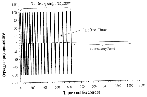
Figure 1 shows a specific low frequency pulsed magnetic field (Cnp) used to
induce analgesia.
Figure 2 shows detail of two of the waveforms of Figure 1. Comparison of the
x-axis between Figure 1 and Figure 2 shows the portion of the time axis which
has
been expanded. Sub-label 1 corresponds to the waveform and sub-label 2 to the
latency period.
Figure 3 shows a Cnp designed to target the vestibular system of rodents. The
top panel corresponds to Cnp in time and the lower panel corresponds to the
magnitude of the Fourier Transform of the Cnp.
Figure 4 shows in detail three waveforms of the vestibular Cnp shown in
Figure 3. As in Figure 2, the x-axis indicates that portion of the time axis
which has
been expanded and relates Figure 3 to Figure 4. Sublabel 1 corresponds to the
waveform and sub-label 2 to the latency period.
Figure 5 shows detail of the Cnp used to target the human vestibular system.
There are differences in the refractory period as compared to the Cnp targeted
for
rodents (See Figure 3). The top panel corresponds to Cnp in time and the lower
panel
to the magnitude of the Fourier Transform of the Cnp.
Figure 6 shows the effect of a Cnp targeted to induce analgesia in a land
snail.
The y-axis corresponds to a measure of analgesia. Basal corresponds to
measurements
SUBSTITUTE SHEET (RULE 26)
CA 02257266 1998-12-02
WO 97/46277
PCT/CA97/00388
-4-
done prior to exposure. The induction of analgesia is only thirty percent when
a
simple magnetic field waveform is applied (30 Hz sinusoidal 15 minute
continuous
exposure with a peak amplitude of 190 [iT and a static field of 76 T parallel
to the
30 Hz field). However when a specific designed magnetic field pulse (Cnp Exp)
is
Figure 7 shows a Cnp pulse designed to target central nervous system targets
which should increase analgesia in land snails. The upper panel corresponds to
Cnp
in time and the lower panel to the magnitude of the Fourier Transform of the
Cnp.
Figure 8 shows that opioid antagonists reduce, but do not block, Cnp induced
analgesia.
Figure 9 shows the effect of a complex neuroelectromagnetic pulse (Cnp),
designed to increase analgesia, consisting of a series of time-varying
extremely low
frequency components (<300Hz) (A) repeated between refractory periods of
several
seconds (B). The Cnps are repeated for the length of the exposure period (15
or 30
static magnetic field set to counter the Earth magnetic field. Sham exposures
consisted of a three-dimensionally (3-D) zeroed Earth magnetic field (3
orthogonal
nested Helmholtz coils tuned to oppose the Earth magnetic field to within
0.1pT,
horizontal component = 14.7pT, vertical component = 43.3pT.)
Figure 10 shows the effects of an acute 15 or 30 min exposure to either a
specific pulsed magnetic field (Cnp) or sham exposure condition on the thermal
(40 C) response latencies of individual hydrated snails (N=120). Response
latencies
were recorded prior to (Pre) and after exposure. Sham 15 and 30 min exposures
were
not significantly different and were combined. Error bars represent the
Standard Error
Figure 11 shows the effects of either (A) 15 min or (B) 30 min daily repeated
exposures to either a specific pulsed magnetic field (Cnp) or sham exposure
condition
on the thermal (40 C) response latencies of individual hydrated snails (N=60).
Response latencies were recorded prior to (Pre) and after (0, 15, 30, 60 min)
exposure.
Response latencies from days 1, 3, 6 and 9 are shown. There were no
significant
SUBSTITUTE SHEET (RULE 26)
CA 02257266 1998-12-02
WO 97/46277
PCT/CA97/00388
-5-
differences within the sham groups, hence the groups were collapsed. Error
bars
= represent the Standard Error of the Mean (SEM), and where not visible are
embedded
within the symbol.
Figure 12 shows the effects of either (A) 15 min or (B) 30 min daily repeated
exposures to either a specific pulsed magnetic field (Cnp) or (C) sham
exposure
condition on the thermal (40 C) response latencies of individual hydrated
snails
(N=60), shown in 3-D perspective. Response latencies were recorded prior to
(Pre)
and after (0, 15, 30, 60 min) exposure. There were no significant differences
within
the sham exposure or pre-exposure latencies.
Figure 13 shows the effects of (A) 15 and (B) 30 min daily repeated acute
exposure to a sham or specific pulsed magnetic field (Cnp) on the thermal (40
C)
response latencies (15 min post-exposure) of individual hydrated snails
(N=60). Day
10 records the effects of condition reversal; in that, the previously sham
exposed
groups were exposed to the Cnp, and vice versa. Error bars represent the
Standard
Error of the Mean (SEM), and where not visible are embedded within the symbol.
Figure 14 shows thermal (40 = C) response latencies of snails (N=60) exposed
to a specific pulsed magnetic field (Cnp) or sham condition for 15 or 30 min
daily for
9 consecutive days. Response latencies were tested on days 1 and 9 prior to
(Pre) and
after (0, 15, 30, 60 min) exposures. There were no significant differences
within the
sham groups, hence the groups were collapsed. Error bars represent the
Standard
Error of the Mean (SEM), and where not visible are embedded within the symbol.
Figure 15 shows thermal (40 C) response latencies of individual snails
(N=30) exposed for 15 min to either a specific pulsed magnetic field (Cnp) or
sham
magnetic field for 9 consecutive days (normal). On day 10 the snails were
exposed to
the Cnp or sham condition while under a novel environment condition (novel).
Response latencies were tested prior to (Pre) and after (0, 15, 30, 60 min)
exposure.
Error bars represent the Standard Error of the Mean (SEM), and where not
visible are
embedded within the symbol. (*P<.01, **P<.001)
Figure 16 shows thermal (40" C) response latencies of individual snails
(N=60), that had been exposed for 15 min daily for 9 consecutive days to
either a
SUBSTITUTE SHEET (RULE 26)
CA 02257266 2003-11-12
-6-
sham or Cnp. Response latencies were tested on day 10 prior to (Pre) and after
being
injected either with the 6 opiate agonist, DPDPE, (0.05 pg/i .0p1 saline) or
saline
vehicle (1 .0p1) at 15, 30, 60 mm intervals. Error bars represent the Standard
Error of
the Mean (SEM), and where not visible are embedded within the symbol.
Figure 17 shows Cnp induced activity in Deer mice. The Cnp shown in Figure
3 was used.
Figure 18 shows Cnp generated interference of human standing balance. The
Cnp shown in Figure 5 was used.
Figure 19 shows the number of rearing behaviors in deer mice in each 5 mm
10 segment of the 10 mm exposure. A rearing behavior is counted when the
animal
rears
up on the hind limbs without touching any of the outside walls of the exposure
container. The Cnp (see Figure 3) exposure produced significantly greater
counts than
either the sham or 60 Hz exposure. The first (0-5) and second (6-10) minute
Cnp
segments are not significantly different. There are no significant differences
within or
between the sham and 60 Hz exposures. Error bars represent the standard error
of the
mean in Cnps.
Figure 20 shows the overall effect of Cnp (see Figure 3) on the rearing
behavior in deer mice.
Figure 21 shows the number of centerline crossings in each 5 mm segment of
20 the 10 mm exposure. A centerline crossing is counted when the entire animal
traverses across the center of the exposure container. The Cnp exposure
produced
significantly greater counts than either the sham or 60 Hz exposure. The first
(0-5)
and second (6-10) minute Cnp segments are significantly different. There are
no
significant differences within or between the sham and 60 Hz exposures. Error
bars
represent the standard error of the mean.
Figure 22 shows the overall effect of the Cnp of Figure 3 on the centerline
crossing activity of deer mice.
Figure 23 shows the number of climbing movements in each 5 mm segment of
the 10 mm exposure. A climbing movement is counted when the animal attempts to
climb or reach up the side of the exposure container with 2 or more limbs
extended
off
CA 02257266 1998-12-02
WO 97/46277
PCT/CA97/00388
-7-
the floor and ends when all four limbs are on the floor. The Cnp exposure of
Figure 3
produced significantly greater counts than either the sham or 60 Hz exposure.
The
first (0-5) and second (6-10) minute Cnp segments are significantly different.
There
are no significant differences within or between the sham and 60 Hz exposures.
Error
Figure 24 shows the overall effect of the Cnp of Figure 3 on the number of
climbing movements in deer mice.
Figure 25 shows the overall effect of the Cnp of Figure 3 on the total
duration
of grooming behaviors in deer mice.
Figure 26 shows the sucrose preference, expressed as the percent of sucrose
drunk out of the total fluid intake, in male and female reproductive deer
mice.
Percents are referred to the day of pairing with Lithium Chloride or saline
solution,
the 3 days following pairing and the two re-test days (10 days after
recovering from
sucrose aversion).
Figure 27 shows the total fluid intake of male and female deer mice before and
after treatment with Lithium Chloride or Saline Solution.
Figure 28 shows the total fluid intake of male and female deer mice after
pairing of the apple juice with a Cnp or a sham magnetic field.
Figure 29 shows the target taste (apple juice or sucrose) preference,
expressed
Detailed Description of the Preferred Embodiments
As hereinbefore mentioned, the present invention provides designed and
characterized low frequency pulsed magnetic fields (Cnps) which have specific
effects
on physiological, neurological and behavioral conditions in vertebrates and
invertebrates. The specific low frequency magnetic fields are designed for
complex
neuroelectromagnetic applications and permit the development of therapeutic
strategies in order to treat and/or alter various physiological, neurological
and
behavioral disorders particularly in mammals and more specifically in humans.
SUBSTITUTE SHEET (RULE 26)
CA 02257266 2003-11-12
8
Magnetic fields have been demonstrated to have various biological effects in
humans, rodents and snails. Such magnetic fields can be detected and this
detection
can be broadly linked to certain physiological processes. It is now
demonstrated that
low frequency pulsed magnetic fields can be designed specifically to alter
specific
targeted physiological processes and in this manner provide a therapeutic
method for
treatment and alleviation of certain conditions without the need for
pharmacological
intervention which is expensive and which poses several problems with respect
to side
effects of certain drugs.
In the present invention it is now demonstrated that the magnetic field
exposure must be designed as a time varying signal such that it can be used to
alter a
specific targeted physiological process. The designed low frequency pulsed
magnetic
field (Cnp) is valid independent of the detection mechanism. However,
different
detection mechanisms may affect how the Cnp is scaled and how it is delivered.
Under certain conditions extremely low frequency (ELF) magnetic fields can be
detected directly according to a resonance model. If the tissue exposed with
the Cnp
pulse detects magnetic fields by the resonance model, then the amplitude of
the Cnp
pulse and possible DC (direct current) offsets are important and must be
specified
with limits below which and above which effects will lessen. On the other
hand, if
magnetic field detection is indirect, ie. the ELF magnetic fields are detected
by tissue-
induced electromotive force (i.e. Faraday's Law of induction) then the effects
will
have a lower threshold below which no effect will be seen and then above this
threshold effects will increase. However, even for indirect detection, a
maximum
threshold will exist above which the induced currents (caused by the induced
EMF)
will be so great that targeted effects will be swamped by large unwanted side
effects.
Therefore, different detection mechanisms might affect amplitude and DC offset
of
the Cnp, but the general design rules will not change. Individual features of
a
generalized Cnp are shown in Figure 1 and Figure 2 and are labeled in letters
running from a. How these features are specified and targeted to a certain
physiological/behavioral effect is described below.
Table 1 represents the 8-bit digital analog values of the specific points used
in
the construction of Figure 1 (Cnp used to induce analgesia). The columns are
CA 02257266 2003-11-12
8a
contiguous, that is, they are essentially one long colomn, that is, they are
essentially
one long column representing all of the serial points of Figure 1. The values
presented in this table can be used by one skilled in the art to replicate the
Cnp used to
induce analgesia using any digital to analog converter.
CA 02257266 1998-12-02
WO 97/46277
PCT/CA97/00388
-9-
Design of Waveform
The low frequency pulsed magnetic fields are comprised of a plurality of
intermittent waveforms. The waveform is designed to look like the
corresponding
= electromagnetic waveform of the target tissue. For example, if the target
tissue were a
part, or parts, of the brain then the waveform would correspond to the
energetic
activity of those parts. If an electroencephalogram (EEG) could record that
activity
then the waveform would mimic the EEG. As seen in Figures 1, 2, 3 and 4 the
waveform is not sinusoidal as this waveform was designed to affect critical
functions
that do not rely on sinusoidal waveforms. Feature la is a rise to a maximum
and
feature lb is designed to stimulate the firing of axons in the tissue type of
interest.
Feature lc is a built in delay to reduce the probability of neuronal
excitation as the
waveform ends.
Latency Period
After each waveform or between successive waveforms there is a delay, a
latency period. This delay is progressively set to increase, or decrease, in
length with
time. This effectively modulates, in time, the frequency of appearance of the
waveform. The specific lengths and progression of the Cnp waveforms are
related to
the target tissue. With respect to the central nervous system (CNS) for
example, there
are a number of characteristic frequencies which relate to: a) frequencies
specific to
the area of the brain; b) frequencies associated with communication/connection
between different brain regions; and c) frequencies and phase offsets
associated with
the co-ordination of different brain regions for a specific function. Now,
although the
waveform has been designed to stimulate neuronal activity for a specific
region,
electrical activity of a region of the CNS will vary between individuals, and
over time,
within an individual. Therefore, to target a function the frequency of
presentation of
the waveform should match the frequency of the target. However, the target is
varying within a frequency bandwidth. These CNS frequencies vary between
approximately 7Hz to 300Hz. (For example: 7Hz corresponds to alpha rhythm;
10Hz
thalamic activity; 15Hz autonomic time; 30Hz intraIaminar thalamus and
temporal
= . .
SUBSTITUTE SHEET (RULE 26)
CA 02257266 1998-12-02
WO 97/46277
PCT/CA97/00388
-10-
regions associated with memory and consciousness; 40Hz connection between
hippocampal and amygdal temporal regions; 45Hz hippocampal endogenous
frequency; 80Hz hippocampal-thalamic communication; 300Hz motor control.)
These frequencies have upper limits due to neuronal electrical properties,
that is: after
a neuron "fires" it is left in a hyperpolarized state and cannot fire again
until it
recovers. Therefore, Feature 2 (see Figure 2) the latency period: a) allows
neurons to
recover so that when the waveform is reapplied the neuron can respond; and b)
its
length is set so that the frequency of presentation of the waveform matches or
approximates the frequencies associated with the target.
Modulation of Latency Period
To change the electrical activity of the target tissue in the CNS, the Cnp
must
"latch on" or more appropriately, entrain, to the appropriate frequency and
either slow
it down or speed it up. The waveform itself does not change substantially,
rather, the
and the rate at which electrical spikes occur in the target tissue. Generally,
for the
CNS, as the frequency of neuronal activity is increased the amount of tissue
involved
per burst of activity decreases. Conversely, as the frequency is decreased a
greater
amount of tissue is synchronized and recruited throughout the CNS. For
example, a)
rate is decreased significantly in humans or animals with epileptic-type
disorders so
much tissue can be recruited that seizures will occur. Therefore, the ramping
up or
ramping down of the rate of presentation of the waveform will: a) ensure that
at least
at some time the applied and endogenous rates will be matched (provided of
course
SUBSTITUTE SHEET (RULE 26)
CA 02257266 1998-12-02
WO 97/46277
PCT/CA97/00388
-11 -
Refractory Period.
As a result of the application of the Cnp the synchrony of the electrical
activity of the target can be disrupted. Before the application of another Cnp
can be
= effectual the tissue must recover its synchrony. It is allowed to do so
by providing a
refractory period between application of successive Cnps where the length of
the
refractory period is determined by the target. For example, if the Cnps are
applied to
a target in humans which is associated with "awareness", then the target will
recover
only after the awareness anticipation time is exceeded (e.g. 1200 ms). Another
example would be the application for the same target, but in rodents without
significant awareness, in which case the refractory period could be reduced to
400 ms.
If the Cnps are to be applied for long periods of time per day, e.g. hours,
then the
refractory periods should be increased to 10 seconds to avoid possible
immunosuppression. Immunosuppression has been show to occur when the CNS is
stimulated chronically and this may be minimized if the refractory periods of
this
stimulation are increased to more than 7 seconds.
Variability in Features
It must be pointed out that the Cnp features are related to the underlying
physiology and that endogenous frequencies vary between individuals and within
an
individual. Therefore, there is tolerance on the feature specifications for
any Cnp
designed for a specific target. For example, in the analgesia pulse shown in
Figures 1
and 2, the features can be varied somewhat and the outcome will remain similar
due
to biological variations in the target. As well, as more and more is learned
about
biological interactions, the Cnp can be modified to take advantage of the new
knowledge to make the Cnp even more specific.
Amplitude and Direction of Application
The amplitude of the Cnp to DC offset, and its direction of application (e.g.
linearly polarized vs. circularly polarized vs. isotropically polarized), is
dependent on
the magnetic field detection mechanism which, may very well differ from one
target
SUBSTITUTE SHEET (RULE 26)
CA 02257266 1998-12-02
WO 97/46277
PCT/CA97/00388
-12-
to another. We have experimentally demonstrated that the amplitude of the Cnp
can
vary significantly and that the Cnp is still effective provided the features
remain
constant for a specific application (Thomas et al, 1997).
Specifically, if magnetic fields are directly detected there will be a window
of
amplitudes and the possible need of a DC offset to the Cnp for it to be
effective.
Further, the relative direction of the DC offset and the time-varying portion
of the Cnp
is important. If the detection mechanism is indirect, that is, induced
currents, then an
induced current feature, such as feature Id in Figure 1 may be added to the
waveform
of the Cnp. This preferably would be a feature with a high value of dB/dt with
frequency components beyond those detectable by the target (i.e. for the CNS,
greater
than approximately 500Hz) but designed to increase the induced EMF in the
target.
For magnetic fields detected indirectly, a DC offset is ineffective but
direction of the
applied Cnp may be important as a time changing magnetic field will induce the
greatest EMF in conductive tissue which projects a maximum area normal to the
direction of the Cnp. We have experimentally verified in a limited
experimental trial
that for some applications the effect is independent of the DC offset.
The present invention is not at the magnetic field detection level, but rather
in
the coupling of a specific low frequency pulsed magnetic field to the target
tissue.
The Cnp design philosophy is not altered if the detection mechanism is
different for
different targets. Rather, the Cnp is used in two "flavours", one for direct
detection
and the other for indirect detection. Theoretically, it may be possible to
produce the
Cnp waveform using other physical entities besides magnetic fields, such as
flashing
light, electrical fields, acoustic waves and peripheral stimulation of nerve
receptors.
However, extremely low frequency (ELF) magnetic fields remain the method of
choice since they penetrate tissue with minimal attenuation and since their
amplitude
can be spatially defined largely independent of the target. Hence, they are
not limited
to specific targets. For example, sound is largely limited to auditory nerves,
light to
optic nerves and electric fields to conductive entry points such as the roof
of the
mouth. Also, the bandwidth of reception may be too low such as that defined by
the
SUBSTITUTE SHEET (RULE 26)
CA 02257266 1998-12-02
WO 97/46277
PCT/CA97/00388
-13-
"flicker fusion rate" of the visual system. Nevertheless, Cnps may be used in
the
future with other stimulation methods to increase target specificity.
= Delivery-Exposure Systems.
Exposure systems which produce variable magnetic field amplitude over the
subject's anatomy would be preferred in situations where the endogenous
frequencies
and waveform of the target overlap with other tissue which could produce
unwanted
"side-effects". Magnetic resonance imaging (MR1) gradient tube and gradient
coil
technology can be easily adapted to produce such spatial variant Cnp exposures
which
can vary in both magnetic field amplitude and direction. Therefore, it is
better to have
two sets of volume coils for each of the three dimensions. One set would
produce the
DC offset eg. Helmholtz configuration (Prato et al, 1996) which would be
needed if
the detection mechanism is a resonance kind. The second would be used to
define
magnetic field gradients eg. Maxwell configuraiton (Carson and Prato, 1996)
This type of exposure system would be ideal for acute and chronic exposures
in which the subject can stay in one position, e.g. treatment of pain while
the subject
is in bed. For mobile subjects, such volume coil configurations would not be
possible
and delivery would preferably be through the use of surface coils either
singly, as say
on the surface of the body, or around the neck or as a Helmholtz pair placed
on either
side of the knee. In this configuration the magnetic field amplitude decreases
rapidly
from the surface coil and matching of target and magnetic field without
exposing
other tissue to an effective Cnp becomes more challenging.
Applications of Cups
Analgesia
The applicants have reported that complicated pulsed magnetic fields (Cnps)
have a pain inhibitory (analgesic) effect. In one embodiment of the present
invention,
the designed Cnps can both increase the analgesic effect of an injection of an
opiate,
eg. Morphine, or actually induce a level of analgesia similar to a moderate
dose of
SUBSTITUTE SHEET (RULE 26)
CA 02257266 1998-12-02
WO 97/46277
PCT/CA97/00388
-14-
morphine. This has a tremendous benefit for the potential of drug-free pain
treatment
which is highly desirable.
Opioid receptors are responsible, among many other functions, for the
mediation of pain. Increase in exogenous/endogenous opioids can induce
analgesia.
The applicants have shown that single sinusoidal ELF magnetic fields can
attenuate
opioid induced analgesia. The applicants have recently demonstrated that the
detection mechanism responsible for this response to ELF magnetic fields is a
resonance model, that is, direct detection (Prato et al, 1996).
Since increases in opioid induced analgesia, rather than decreases, would have
therapeutic value, and since induction of analgesia by ELF magnetic fields
would
have even greater value the applicants have developed a simple pure sinusoidal
waveform specification that would induce mild analgesia. As shown in Figure 6
the
increase was modest (20-30%). However, when the applicants designed a Cnp to
induce analgesia the effect was made much larger (Figure 6).
The analgesia Cnp used and its magnitude Fast Fourier Transform are shown
in Figure 7. This in fact is the Cnp shown as Figure 1 and 2. This "analgesic
pulse"
can be used to: a) increase opioid induced analgesia; and b) significantly
induce
analgesia. In addition, it is now known that analgesia is only partially
opioid
mediated and that another analgesic component is present. This additional
component
corresponds to the modulation of another target tissue or system, as yet
unidentified.
This is probably due to the more general nature of this Cnp, and that the
entire animal
was exposed to identical magnetic fields. The power in the frequency was in
three
bands: 4-16Hz; 22-26Hz; and 28-52Hz. The whole body of the animals (land
snail,
Cepaea nemoralis) were exposed and the purpose was to slow down activity in
the
brain structures which have a high concentration of opioid receptors and are
responsible for the awareness of pain with frequencies in the range of 28-
52Hz. Note,
that when a random pulse was used, in which frequency analysis indicated
constant
power in all frequencies between 0-166Hz, the induction of analgesia was not
seen,
indicating the specificity of even this general Cnp. The slowing up and
disruption of
function in such biological sites in the snail equivalent to the CNS should
have
SUBSTITUTE SHEET (RULE 26)
CA 02257266 1998-12-02
WO 97/46277
PCT/CA97/00388
-15-
profound effects beyond the induction of analgesia. In fact when whole rats
with a
pre-existing condition of status epilepticus were exposed to this waveform,
the result
was increased seizure activity. As previously discussed, when frequencies of
= waveform firings are reduced, more tissue is recruited. In this extreme
case, sufficient
CNS tissue was recruited in the electrically labile rat exposed to this Cnp
resulting in
increased seizure activity.
This Cnp pulse can be made more specific by treating subjects, like humans,
only over specific CNS structures or by incorporating more selectively
designed
waveforms.
The applicant's have previously demonstrated that a short acute exposure to a
specific weak extremely low frequency pulsed magnetic field (Cnp) can induce
significant partly opioid-mediated analgesia in the land snail, Cepaea
nemoralis. In
the first studies individual groups of snails, Cepaea nemoralis, were pre-
injected with
either the general opioid antagonist naloxone or specific antagonists (i_t
naloxazine,
funaltrexamine, 6 naltrindole, ICI-174, 864 or lc nor-binaltorphimine opioid
peptide
specific antagonists), their respective injection vehicles or received no
injection and
then were exposed for 15 minutes to a Cnp or a sham condition. The snails were
then
tested for response latency on a hotplate (40 C). There were no significant
differences in pre-exposure response latencies, or in sham exposure response
latencies, and hence, the individual groups were combined as seen in Figure 8.
All
groups showed a significant degree of induced analgesia as inferred by an
increase in
response latency; however, the general pt and 6 opioid antagonists
significantly
reduced, but did not block, the Cnp induced analgesia.
The time course of Cnp induced analgesia in these snails was also initially
investigated. Individual groups of snails were exposed to Cnps for either 15
or 30
minutes and then tested immediately, at 15, 30 and 60 minutes after the Cnp
exposure
for response latency on a hotplate (40 C). While there was no significant
difference
between the 15 and 30 minute exposures, as compared to the sham exposure,
there
was a significant degree of induced analgesia up to and including 60 minute
post
exposure (Figure 10).
SUBSTITUTE SHEET (RULE 26)
CA 02257266 1998-12-02
WO 97/46277 PCT/CA97/00388
-16-
The effect of Cnp induced analgesia has now been examined for the
development of tolerance to daily repeated acute exposures of 15 or 30 minute
=
duration. Also examined was the effect of acute cross-tolerance to the d
opioid
receptor directed against the DPDPE enkephalin. The results of this study show
that
brief (15 or 30 min) exposure to a specific pulsed magnetic field (Cnp) has
antinociceptive or "analgesic" effects in the land snail Cepaea nemoralis. The
magnitude and duration of this Cnp induced analgesia was reduced, though not
blocked, following repeated daily exposures, in a manner indicative of the
partial
development of tolerance. Both associative (learning related) and non-
associative
(pharmacological related) processes were suggested to be linked with the
expression
and reduction of the analgesic effect of this specific pulsed magnetic field
(Cup)
following repeated exposures. Presentation of novel environmental cues could
ameliorate the expression of this tolerance and nearly re-instate the level of
acute Cnp
exposure induced analgesia. These results are consistent with, and extend,
prior
findings of specific pulsed magnetic fields, including the Cnp, having
behavioral
actions in invertebrate and vertebrate systems. These results also
substantiate and
extend prior reports that the effects of ELF magnetic fields on analgesia and
likely
other behavioral and physiological responses can be modified with repeated
brief
daily exposures.
Exposure for either 15 or 30 min to the Cnp resulted in a significant increase
in the latency of response of Cepaea to an aversive thermal surface,
indicative of the
induction of analgesia. The magnitude of this analgesia was related to the
length of
exposure suggesting a possible duration or dose-related effect of the Cnp. In
previous
studies it was shown that the Cnp induced analgesia is not a generalized or
stress-
related effect of exposure to magnetic fields. Other similar designs of pulsed
magnetic fields were shown to have no significant effects on either basal
nociceptive
sensitivity or opioid-induced analgesia [Thomas et al., 1997j. In addition,
simple
sinusoidal extremely low frequency magnetic fields (<300 Hz) have been shown
either to attenuate or weakly augment opioid-mediated analgesia depending on
the
specific magnetic field exposure characteristics. In the present, as well as
prior
SUBSTITUTE SHEET (RULE 26)
CA 02257266 1998-12-02
WO 97/46277
PCT/CA97/00388
-17-
studies, repeated sham exposures (zeroed or normal Earth magnetic field) and
repeated determinations of response latencies, had no significant effects on
nociceptive sensitivity.
The significantly greater magnitude of analgesia induced by the 30 exposure to
the Cnp as compared to the 15 minute exposure is consistent with the findings
that the
inhibitory effects of an acute magnetic field exposure on opiate analgesia are
affected
by both the duration of exposure and the intensity of the magnetic field.
Results of
more in depth investigations, however, have also shown that the magnitude of
the
inhibitory effects does not scale linearly with either the frequency or
amplitude of the
ELF magnetic field [Prato et al., 1995].
Substantial evidence exists for the presence of multiple endogenous opioid
inhibitory systems. Both naloxone-reversible `opioid' and natoxone-insensitive
'non-
opioid' forms of analgesia have been indicated [Rothman 1996] and are
apparently
phylogenetically conserved and expressed in both rodents and Cepaea [Kavaliers
et
al., 1983]. In prior investigations with Cepaea, it was established that the
Cnp
induced analgesia was of a mixed opioid and non-opioid nature [Thomas et al.,
1997;
Thomas et al., 1997(in press)]. The analgesic effects of the Cnp were reduced,
but not
blocked, by the prototypic opiate antagonist, naloxone, and the d opioid
receptor
directed antagonists, ICI 174,842 or naltrindoIe-5'-isothiocyanate (5'-NTII)
(Table 2
and Thomas et al., 1997 (in press)). However, the analgesic responses were
unaffected by pretreatment with the kappa opioid directed receptor antagonist,
nor-
binaltorphimine. This lack of a complete blockade of the Cnp induced analgesia
by
the opioid antagonists indicates that "non-opioid" as well as opioid mediated
mechanisms are associated with the effects of the Cnp. The neurochemical
mechanisms mediating this non-opioid analgesia remain to be determined.
Typically chronic repeated administrations of opiates result in the
development of tolerance, such that the analgesic effects initially produced
by
substance such as morphine show a progressive decline in intensity until they
are
indistinguishable from the responses of control animals. Similar patterns and
characteristics of morphine tolerance have been established to occur in Cepaea
and
SUBSTITUTE SHEET (RULE 26)
CA 02257266 1998-12-02
WO 97/46277
PCT/CA97/00388
-18-
rodents [Kavaliers et al., 1983; Kavaliers et al., 1985]. Here, it was
determined that
after 6-9 days of daily 15 or 30 mm exposures to the Cnp, tolerance developed
to the
opioid mediated component of the induced analgesia. The pattern of response
and
time course is similar to that for the development of tolerance to
antinociceptive
effects of opioid peptides and opiate agonists in Cepaea and rodents. The
level of
analgesia attained after 6-9 days of daily exposure to the Cnp was similar to
that
recorded in snails treated with either naloxone or specific 6 opioid receptor
directed
antagonists and followed by a single Cnp exposure. In addition, the snails
that had
received the daily exposures to the Cnp displayed a significantly reduced
sensitivity to
the analgesic effects of the specific 6 opioid agonist, DPDPE. This is
suggestive of at
least a partial generalization of tolerance (i.e. cross-tolerance) to the
opioid component
of the Cnp. Determinations of the nociceptive responses of snails that have
become
tolerant to DPDPE and are subsequently exposed to the Cnp are necessary to
explore
more fully the extent of this generalization and the expression of cross-
tolerance
between Cnp and 6 opioids.
In the present experiments there was little evidence of a reduction in the
level
of the "non-opioid" mediated analgesia induced by repeated exposures to the
Cnp.
The analgesia induced by the 15 min and 30 min Cnp exposures was reduced to a
similar level. This raises the intriguing possibility that increased duration
of the Cnp
may selectively augment the opioid mediated analgesia while leaving a
relatively
constant basal non-opioid mediated component. It also suggests that various
components of this specific Cnp may differentially affect the expression and
neurochemical substrates of opioid and non-opioid analgesia.
There have been only limited considerations of the development of tolerance
to naloxone-insensitive non-opioid analgesia. These studies have revealed
either
relatively low or no development of tolerance to non-opioid analgesia. This is
not,
however, completely limited to non-opioid analgesia, as weak tolerance has
also been
reported to the antinociceptive effects of certain opioid activating factors
in rodents.
There is also no apparent cross-tolerance between opioid and non-opioid
analgesia,
with it having been speculated that the presence of opioid analgesia may even
SUBSTITUTE SHEET (RULE 26)
CA 02257266 1998-12-02
WO 97/46277
PCT/CA97/00388
-19-
preclude the development of tolerance to non-opioid analgesia [Rothman 1996].
Similarly, it is possible that the presence of non opioid analgesia may affect
the
expression of opioid systems and limit the expression of complete cross-
tolerance as
suggested here with DPDPE (Figure 16).
It should also be noted that tolerance is considered to be best demonstrated
by
a shift in the dose-response indicative of the need for a higher dose to
produce a
consistent drug effect. In the present study, tolerance is inferred from the
decrease in
analgesia produced by daily repeated 15 or 30 min exposures to the Cnp. The
lack of
supporting evidence for a definitive linear dose-dependent effect of Cnp,
along with
the similar reductions in analgesic effects of the 15 and 30 mm Cnp exposures,
precludes examination of shifts in dose responses.
Opiate tolerance has been proposed to involve both associative and non-
associative components. In prior investigations it was shown that after the
termination of drug treatment, Cepaea that were rendered fully tolerant to
morphine
exhibited dependence and withdrawal symptoms, including hyperalgesia, that are
considered to be consistent with non-associative mechanisms [Tiffany et al.,
1988].
Non-associative tolerance is considered to represent an effect arising solely
from drug exposure. Tolerance is considered to result in part simply from
cellular
adaptations produced by repeated drug stimulation of some physiological system
such
as a particular receptor or second messenger cascade.
Opioids have stimulatory as well as the more conventionally studied inhibitory
effects on neurotransmission that are accepted as the mechanisms underlying
analgesia. There is accumulating evidence that these stimulatory effects may
also be
associated with the development of opioid tolerance. In this regard, daily
acute
exposures of Cepaea to ELF 60 Hz magnetic fields were shown to result in
hypoalgesic or analgesic effects consistent with the antagonism of the
excitatory
hyperalgesic effects of endogenous opioids.
There is also evidence that particular transmitter systems may function to
counteract opioid effects and mediate some aspects of tolerance. In this view,
tolerance may not only result from decreased opiate efficacy, but also
enhanced "anti-
SUBSTITUTE SHEET (RULE 26)
CA 02257266 1998-12-02
WO 97/46277
PCT/CA97/00388
-20-
opiate" influences. The putative anti-opioid peptide, orphanin FQ or
nociceptin,
which exerts its effects through a novel orphan, opioid-like receptor, and has
been
recently implicated in tolerance, has been shown to affect nociceptive
responses in
Cepaea through N1VIDA associated mechanisms. Intriguingly, orphanin FQ has
also
been recently suggested to be involved in opioid mediated electro-acupuncture-
induced analgesia [Tian et al., 1997].
Tolerance has also been shown to involve associative learning. Animals,
including Cepaea, repeatedly receiving morphine in a consistent, distinctive
environment are much more tolerant to the analgesic and thermic effects of
morphine
than when tested in a different, novel, environment. In the present study this
"environmental specificity" was demonstrated for the opioid mediated analgesic
effects of the Cnp. Snails that were exposed to the Cnp while in a novel
environment
displayed an apparent reversal of tolerance, their analgesic responses being
similar to
that of individuals receiving single acute Cnp exposures (Figure 15).
A variety of factors, including ELF magnetic fields, have been shown to
function as salient environmental specific cues and affect the subsequent
expression of
tolerance. This raises the possibility that the Cnp itself may at least
partially serve as
a cue for tolerance development. This may contribute in part to the apparent
lack of a
complete "cross-tolerance" to the analgesic effect of the Cnp to the 5 opioid
agonist
DPDPE.
Associative, environmental or situation specific tolerance has been explained
through classical conditioning, [Tiffany et al., 1981] although habituation
involving
both associative and non-associative components has also been proposed [Baker
et al.,
1985]. According to the conditioning model the distinctive context has become
a
conditioned stimulus that elicits associative tolerance.
In the present study it was found that similar patterns of tolerance developed
whether the snails received nociceptive testing every day or only on the first
and last
days. This suggests that associative factors related to determining the
thermal
response latencies (i.e. hotplate testing) of the snails did not play a major
role in the
development of tolerance. This also minimizes the likelihood that tolerance
arises
SUBSTITUTE SHEET (RULE 26)
CA 02257266 1998-12-02
WO 97/46277
PCT/CA97/00388
-21-
from cues associated with the nociceptive assessment. This is consistent with
the
results of a number of investigations of opiate tolerance in rodents, as well
as
morphine tolerance in Cepaea.
Recent studies have focused on the possible neurochemical mechanisms
involved in associative tolerance. Investigations with laboratory rats have
suggested
that neurotensin and possibly other neuropeptides implicated in memory may
have a
role in the mediation of associative tolerance [Girsel et al., 1996]. This
does not
preclude a role for other neuronal and second messenger systems that have been
implicated in learning in both molluscs and rodents, and been shown to be
sensitive to
various types of magnetic fields.
A number of possible mechanisms have been proposed for the biological
effects of magnetic fields [Kavaliers et al., 1994; Prato et al., 1995]. Among
these,
resonance models have predicated both increases and decreases in opioid
analgesia
along with effects at specific frequencies. These actions have been suggested
to have
effects on calcium and potassium ions and various messenger systems [Kavaliers
et
al., 1996; Kits et al., 1996; Prato et al., 1996; and Kavaliers et al, 1996],
that are
associated with the mediation of opioid actions and learning related
processes. All of
these could contribute to the Cnp induced expression of analgesia and decline
in the
opioid component with repeated exposures.
Vestibular System
The use of Cnp pulses appears to be valuable for affecting various vestibular
components of mammals. With respect to humans, Cnps can be very valuable for
the
alteration of standing balance. Disruptions of the balance system such as
motion
sickness may possibly be treated with the use of Cnps without adverse side-
effects
such as nausea or sleepiness.
The Cnp shown in Figure 3 was used to target the vestibular system in rodents
(activity study; amplitude 100 mT), in deer mice (conditioned taste aversion
study;
amplitude 100 mT), and in rats (conditioned taste aversion study; amplitude 1-
4mT).
The Cnp shown in Figure 5 was also piloted in humans (balance study; amplitude
10-
SUBSTITUTE SHEET (RULE 26)
CA 02257266 1998-12-02
WO 97/46277
PCT/CA97/00388
-22-
60mT). Note that Figure 3 and Figure 5 differ only in the length of the
refractory
period. In humans the refractory period (Feature 4 in Figure 1) was 3 times
longer
than for rodents. The reason is that awareness lasts 3 times as long for
humans who
extrapolate each awareness period (approximately 400 ms) with cognitive
function to
three such periods (approximately 1200 ms).
The rate or waveform presentation was modulated from higher frequencies to
lower frequencies and two different waveforms were used. Figure 5 shows the
magnitude Fourier transform of the Cnp, i.e. it is a magnitude spectrum of the
positive
frequencies and the maximum frequency possible was set to 500Hz by the digital
representation of the Cnp at a separation of! ms. Note, that Figure 5
indicates that
the power in the spectrum is at three major frequency ranges: 100-125; 125-
240; 325-
410. The high frequencies were needed since the vestibular system is a motor
function and, therefore, has endogenous CNS frequencies of the order of 300Hz.
Two different waveforms were used to represent the electromagnetic activity
of the vestibular system. This was necessary to provide a minimum resolution
time
(lms) at the highest frequencies. Initially, a two lobe waveform was used and
then
when the waveform rate was sufficiently reduced and the latency sufficiently
long a
five lobe waveform was used as it was believed to better mimic the underlying
electrical activity of the target tissue.
Modulation of Anxiety
Severe anxiety has been shown to accompany depression. A Cnp has now
been designed which significantly alters anxiety related responses in mice.
A Cnp designed to produce vestibular disturbance in deer mice produced a
marked increase in activity (activity index -= total of escape behaviors such
as
climbing attempts, jumps, centerline crosses) during a 10 minute Cnp exposure
as
compared to a 10 minute sham exposure (Figure 17). The 100 l_tT Cnp exposure
and
sham condition were given while the animals were contained in a Plexiglass
open-
field box. The Cnp exposure also produced a significant decrease in the
duration of
grooming behaviors (Figure 25).
SUBSTITUTE SHEET (RULE 26)
CA 02257266 1998-12-02
WO 97/46277
PCT/CA97/00388
-23-
Modulation of Behavioral Activities
Deer mice were exposed to a specific Cnp designed to interact with the
vestibular system to characterize the effects of Cnp on behavioral activities.
Individual deer mice were exposed to the Cnp or control conditions (sham or 60
Hz)
for a 10 minute period while being videotaped and various behaviors were
monitored.
It was concluded from this study that specific pulsed magnetic fields (Cnps)
may be
designed to affect selectively a variety of behaviors. Acute exposures (5 min)
are
sufficient to produce a significant behavioral effect (Figures 19-25). Cnps
were seen
to affect rearing behaviors and general activity such as climbing and
centerline crosses
compared to the control groups. These result demonstrate that Cnps can be used
to
alter a variety of behavioral activities.
Taste Aversion
Field studies have indicated that deer mice, Peromyscus Maniculatus,
developed long lasting avoidance of poisoned baits, whereas results of an
early
laboratory investigation of conditioned taste aversion (CTA) suggested the
formation
of taste aversions that extinguished rapidly. The applicants have examined in
one set
of experiments the acquisition and extinction of a conditioned taste aversion
(sucrose
paired with LiC1) in male and female deer mice. In another set of experiments,
the
applicants have examined the acquisition and extinction of conditioned taste
aversion
using sucrose alone in Wistar rats and in deer mice. The applicants also
examined the
effects of specific Cnps on taste aversion learning. Together, the results of
these
studies (Figures 26-29) demonstrate that Cnp can be used to modify taste
aversion in
deer mice and in Wistar rats.
A Cnp designed to interfere with vestibular processing was tested for aversive
effects in two independent trials of conditioned taste aversion, or taste
aversion
learning. In one experiment, Wistar rats (N=24) that were exposed to the
specific Cnp
for one hour after being provided with a novel food item (sucrose solution)
consumed
significantly more sucrose solution when tested three days after exposure, as
SUBSTITUTE SHEET (RULE 26)
CA 02257266 1998-12-02
WO 97/46277
PCT/CA97/00388
-24-
compared to sham exposed animals (F1,23=5.99, P=.023, Eta2=.22). In another
experiment, deer mice (Peromyscus maniculatzts) (N=43) were exposed to either
the
specific Cnp or a sham condition for one hour. After exposure, the deer mice
were
given access to water and apple juice simultaneously and the ratio of apple
juice to
total volume consumed (apple juice + water) was recorded. The deer mice
exposed to
the Cnp consumed significantly more apple juice than did the sham exposed mice
(F1.43=3-95, P=.05). Though the exposure systems used in the two experiments
were
vastly different, the same specific Cnp was used. In both cases neither
induced an
aversion to the novel food. Results of prior investigations had shown that the
specific
Cnps were capable of inducing other specific behavioral affects in those
species.
Experiment one utilized a single coil (72 turns of 30WG) wrapped around an
aluminum (1.3m x I. 1m) cage rack (100-7004T Cnp exposure, normal Earth earth
magnetic field sham (Michon et al, 1996)). The exposure system for experiment
two
consisted of three pairs of nested orthogonal Helmholtz coils (Prato et al,
1996)
(100 0.1pT, 3-D 0.1pT zeroed Earth field magnetic sham).
The results of the studies using sucrose and LiCI showed that reproductive
male and female deer mice developed a rapid conditioned taste aversion to a
sucrose
solution that was paired with lithium. There was a complete extinction of the
aversion
after 4-5 days with no evidence of a residual aversion 10 days later which is
a contrast
to the longer lasting aversions generally evident in laboratory rats. There
were also
sex differences in the conditioned taste aversion with male deer mice
displaying a
longer lasting aversion and slower extinction than females.
The Cnp exposure did not elicit a conditioned taste aversion, but rather it
reduced the neophobic responses of males to a novel taste and sex difference
in
baseline taste preferences. Further experiments conducted at Laurentian
University
also revealed that the specific Cnp similarly reduced neophobic responses and
aversions to novel food items in laboratory rats.
Overall, these findings indicate that the effect of the specific Cnp, in at
least a
taste aversion paradigm, is dependent on the "characteristics" of the magnetic
field,
not the exposure system, amplitude, geographical location or species tested.
SUBSTITUTE SHEET (RULE 26)
CA 02257266 1998-12-02
WO 97/46277
PCT/CA97/00388
-25-
Learning
All behaviors, including learning, originate as a pattern of electrical
activity in
= the brain. Using specific Cnps, specific behaviors can be altered
inferring that
specific areas of the brain can be selectively affected. Previous studies
using Cnps
have shown alterations in behaviors such as language, memory, suggestibility,
mood
and understanding. It is anticipated that combinations of specific Cnps will
result in
predictable alterations of memory and learning.
Epilepsy
The use of Cnps has great potential to treat epilepsy safely, a serious
problem
associated with brain trauma.
Depression
The potential to treat depression with Cnps is enormous, in both clinical and
model terms (Baker-Price and Persinger, 1996). Also, related disorders such as
'seasonal affective disorder' may prove to be susceptible to Cnp treatment. It
has
been envisioned that the equipment required for this Cnp treatment would be
portable, about the size of a 'Walkman', and have earphone sized head coils.
The designed pulsed magnetic fields (Cnps) of the present invention can be
used effectively to treat a variety of physiological and psychological
conditions in a
safe and effective manner. Any living organism including humans and animals
can be
subjected to the Cnps of the present invention. By safe and effective as used
herein is
meant providing sufficient potency in order to decrease, prevent, ameliorate
or treat a
a physiological or neurological disorder affecting a subject while avoiding
serious
side effects. A safe and effective amount will vary depending on the age of
the
subject, the physical condition of the subject being treated, the severity of
the
disorder, the duration of treatment and the nature of any concurrent therapy.
The subjection of a subject to effective Cnps exposures of the present
invention is defined as an amount effective, at dosages and for periods of
time
SUBSTITUTE SHEET (RULE 26)
CA 02257266 2003-11-12
-26-
necessary to achieve the desired result. This may also vary according to
factors such
as the disease state, age, sex, and weight of the subject, and the ability of
the Cnps to
elicit a desired response in the subject. Dosage or treatment regima may be
adjusted to
provide the optimum therapeutic response. For example, several divided doses
or
treatments may be administered daily or the dose may be proportionally reduced
as
indicated by the exigencies of the therapeutic situation.
The Cnps of the present invention may be subjected to a mammal alone or in
combination with pharmaceutical agents or other treatment regimes.
EXAMPLES
Example I .Materials and Methods
Animals
Snails were collected from old field sites in London, Ontario which did not
have any overhead or underground electric transmission lines (<0.01 pT ambient
15
magnet fluctuation). Snails were then individually numbered by applying a
small
identifying mark on the apex of the shell using non-toxic colored fingernail
polish.
The individually numbered animals were held in a terrarium (ambient
fluctuating
magnetic fields <0.4pT) under indirect natural, and fluorescent, lighting at
an
approximate 12h light! 12hr dark cycle (LD12:12, L=250¨dW/cm2), at 20 2 C,
with
20 lettuce available ad lib.
Assessment of Nociception
As the activity of gastropods is affected by their state of hydration {Smith
1987], all snails were allowed to fully hydrate under a saturated atmosphere
at
20 2 C before being tested. Individual fully-hydrated snails were placed on a
warmed surface ("hotplate" 40 0.2 C) and the latency of their "avoidance" of
the
thermal stimulus, was determined. The avoidance behavior was a characteristic
elevation of the anterior portion of the filly extended foot, the behavioral
endpoint
being the time the foot reached maximum elevation {Dyakonova et al., 1995].
After
displaying this aversive, or more appropriately, "nociceptive" response
[Kavaliers et
CA 02257266 1998-12-02
WO 97/46277
PCT/CA97/00388
-27-
al., 1983], individual snails were removed from the thermal surface. An
increase in
response latency may be interpreted as an antinociceptive or "analgesic"
response
[Thomas et al., 1997]. The hotplate, which does not produce any magnetic
fields,
consisted of an aluminum water jacket with a stainless steel top (33 x 33cm)
with water
pumped through it from a circulating water bath.
Experimental Apparatus
Groups of 15 snails were placed in translucent polypropylene containers (12
cm square, 5 cm high) in the center of three mutually orthogonal Helmholtz
coils (1.2
m for the coil that generated a vertical field and 1.1 m and 1.0 m for the
coils that
generated horizontal fields). Details of the coils and amplifiers are provided
in Prato
et al.[Prato et al., 1996]. A computer driven 8-bit resolution digital to
analog converter
(S. Koren, Neuroscience Research Group, Laurentian University, Sudbury,
Ontario)
was used to produce the pulsed waveforms. Magnetic fields were measured with a
fluxgate magnetometer (model FGM-3D1) and a field monitor (model ELF-66D),
both
Walker Scientific, Worcester, MA.
Magnetic Field Exposure Conditions
The 15 and 30 min magnetic field exposures consisted of a specific low
frequency pulsed magnetic field (Cnp) (Figure 9) set to 1001.1T peak amplitude
in the
vertical direction.
Sham exposures consisted of a three-dimensionally (3-D) zeroed Earth
magnetic field (Helmholtz coils tuned to oppose the Earth's magnetic field to
within
0.11.1T horizontal component = 14.71.1T, vertical component = 43.311T.)
Results of
prior investigations [Thomas et al., 1997; Thomas et al., 1997 (in press)] had
established that there were no significant differences in the response
latencies of 3-D
zeroed Earth magnetic field sham exposed snails and those that were exposed to
an
ambient Earth magnetic field sham condition.
SUBSTITUTE SHEET (RULE 26)
CA 02257266 1998-12-02
WO 97/46277
PCT/CA97/00388
-28-
Example 2 - Materials and Methods for Taste Aversion Studies
Animals
Male and female sexually mature deer mice (20-25 g and approximately 5
months of age) were housed in mixed-sex sibling groups (3-5 animals per group)
in
polyethylene cages provided with cotton nesting material and Beta chip
bedding.
Food (Purina Rat Chow) and water were available ad libitum. The reproductive
mice
(males scrotal, females cyclic) were held under a reproductively stimulatory
(Desjardins et al. 1986; Nelson et al. 1992), long day, 16 h light: 8 h dark
cycle (light
0600 - 2200 hr) at 20 +7- 2 C. The laboratory bred deer mice (15-20
generations)
were from a population of wild caught animals originally present in the
interior of
British Columbia (Canada) near Kamloops (50* 45' N, 120 ' 30' W).
Additional characteristics of this wild and laboratory population are provided
in Innes and Kavaliers (1987).
Experimental Procedures
There were five phases in this first experiment: a habituation phase; a
conditioning phase: a postconditioning recovery phase, an extinction phase and
a re-
test phase.
Habituation phase
Males (n= 21) and females (n= 22) were individually housed for 10 days with
food and tap water, in standard drinking bottles, available ad libitum. For 4
days the
mice were water deprived overnight (dark period). Each morning they were given
two drinking tubes containing tap water and the total amount of water consumed
over
90 min. was recorded. For the remainder of the day tap water in standard
drinking
bottles was available ad libitum. The drinking tubes consisted of 15 ml
graduated
polypropylene conical tubes (Falcon 2092, Becton Dickinson, Lincoln Park, New
Jersey, USA), with screwable caps that were fitted with stainless steel water
spouts
with ball bearing. With these drinking tubes fluid intake could be accurately
determined to 0.25 ml. Individual body weights were recorded at night
immediately
SUBSTITUTE SHEET (RULE 26)
CA 02257266 1998-12-02
WO 97/46277
PCT/CA97/00388
-29-
after the removal of the water bottles and in the day directly after the
drinking tubes
were removed.
Conditioning phase.
On Day 5 overnight water deprived mice were given the two drinking tubes
each of which contained a 0.3 M sucrose solution. After 90 min. the total
sucrose
intake was recorded and mice were immediately injected intraperitoneally
(i.p.) with
20 ml/Kg of either a 0.15 M LiCI solution or 0.9% isotonic saline solution.
After
injection the water bottles were returned and the mice were then left
undisturbed until
the evening when their bottles were removed and the mice were weighed. Male
and
female mice were randomly assigned to the UCI and saline groups.
Post conditioning recovery phase.
For two days after conditioning (days 6 and 7) mice were kept on their nightly
water deprivation schedule. In the mornings they were presented the two
drinking
tubes with tap water for 90 min. after which water from the standard drinking
bottles
was provided ad libitum. Body weights were recorded twice daily as previously
described.
Extinction phase.
On days 8-11 individual mice were presented-two drinking tubes, one
containing tap water and the other one holding the 0.3 M sucrose solution in
the
morning drinking period. Water and sucrose solution intakes were recorded for
90
min. after which tap water from standard water bottles was available ad
libitum. The
position of the two drinking tubes was varied: half of the mice in each
experimental
group (LiC1 and NACI) had water on the right and the other half had sucrose on
the
right. To correct for possible individual preferences, the position of the
water and
sucrose tubes were reversed on subsequent days. Deer mice were weighed twice
daily.
SUBSTITUTE SHEET (RULE 26)
CA 02257266 1998-12-02
WO 97/46277
PCT/CA97/00388
-30-
Recovering Phase
After extinction of the conditioned taste aversion the male and female deer
mice were left undisturbed for 10 days with ad libitum access to food and tap
water.
Retesting Phase
Individual male and female mice were replaced on the overnight water
deprivation schedule. For two days the water deprived mice were given in the
morning two drinking tubes, one containing water and the other 0.3 M sucrose
and
their total fluid intakes were determined over 90 min. After this 90 min.
period they
were provided with ad libitum access to the standard water bottles.
Total fluid intakes were analyzed by a two way individual analysis
(MANOVA) with sex (two levels; mails and females) and treatment (two levels;
LiC1
and NaC1) as between-subject factors and intake as a repeated-measure within-
subjects factor (eleven levels; Habituation (four days), conditioning (pairing
and 2
post injection days), extinction (four days)). In order to evaluate the
effects of sex and
treatment on the fluid intakes on each day mean comparison were planned a
priori in
the MANOVA model. Since fluid intake displayed a Poissonian distribution the
data
were square-root transformed before analysis. As there were some zero intakes,
0.50
was added to all values before transformation.
Preference data from extinction and re-test phasees were expressed as the
percent of sucrose (arcsin transformed) consumed in the total fluid. These
preference
data were analyzed by a two way MANOVA, with sex (two levels; males and
females) and treatment (two levels; LiC1 and NaC1) as between subject factors
and
percent of sucrose as a repeated-measure within subjects factor (six levels: 4
extinction days + 2 re-test days). In order to evaluate the effects of sex and
treatment
on sucrose preference for each experimental day, mean comparisons were planned
a
priori in the MANOVA model.
Body weights were analyzed by a two way MANOVA with sex (2 levels;
males and females) and treatment (2 levels; LiC1 and NaC1) as between-subject
factors
and intake as a repeated-measure within-subjects factor (21 levels). In order
to
SUBSTITUTE SHEET (RULE 26)
CA 02257266 1998-12-02
WO 97/46277
PCT/CA97/00388
=
-31-
evaluate the effects of sex and treatment on the mice body weights for each
expe6mental day, mean comparisons were planned a priori in the MANOVA model.
All hypothesis tests used ct=0.05 as the criterion for significance.
Magnetic Field generation
In the magnetic and sham field exposure conditions mice were placed in a
Plexiglas box in the centre of three mutually orthogonal HelmoItz coils (1 .2
m
diameter for the coil that generated a vertical and 1.1 m for the coils that
generated
horizontal fields; details of the coils and amplifiers are provided in Prato
et al. (1994).
A computer driver with a 8 bit resolution digital to analog converter produced
the
pulsed waveforms. Magnetic fields were measured with a fluxgate magnetometer
(model FGM - 3D1) and a field monitor (model ELF - 66D ; both Walker
Scientific,
Worchester, MA. USA).
Magnetic field exposure conditions
The magnetic field exposures consisted of a specific low frequency pulsed
magnetic field set to 100+/-0. 1 pT; peak amplitude in the vertical direction.
Sham exposures consisted of a three dimensionally (3-D) zeroed earth field
(Helmoltz coils tuned to oppose the earth's field to within +/-0.1pT;
horizontal
component = 14.7pT, vertical component = 43 .3
Example 3 - Experimental Procedures., Opioid Experiments
Experiment I
Each day for 9 consecutive days, at midphotophase, separate groups (n= 15 per
group, N=120) of hydrated snails were exposed to either the specific pulsed
magnetic
field or sham magnetic field for either 15 or 30 min. On day 10, the exposure
conditions were reversed for each group, with sham animals receiving the Cnp
and
Cnp exposed animals receiving sham exposure. Response latencies of the snails
were
determined prior to (Pre), immediately after (0) and 15, 30 and 60 mm after
exposure.
One individual carried out the Cnp and sham exposures while a second
experimenter,
SUBSTITUTE SHEET (RULE 26)
CA 02257266 1998-12-02
WO 97/46277
PCT/CA97/00388
-32-
in a separate room, determined the response latencies. Results of previous
investigations had established that tolerance to the analgesic effects of
morphine in
Cepaea was evident after 7 days of daily repeated acute treatments [Kavaliers
et al.,
1983].
Experiment 2
Each day for 9 consecutive days, at midphotophase, other separate groups (n=
per group, N=60) of hydrated snails were exposed to either the specific Cnp or
sham magnetic field for 15 or 30 min and then (except for days 1 and 9)
immediately
10 returned to their home container. On days 1 and 9 response latencies of
the snails
were individually determined prior to (Pre), immediately after (0) and 15, 30
and 60
min after exposure, after which they were returned to the home container.
Experiment 3
15 After 9 days of daily repeated acute Cnp or sham exposure (15 min)
and
assessment of nocieeption (pre, 0, 15, 30, 60 min), other groups of snails
(N=30) were
exposed (day 10) to their respective exposure conditions while held in a novel
environment. The novel environment consisted of a modified version of the
previous
polypropylene exposure container. Pieces of coarse garnet sandpaper were
fitted and
glued to the top and bottom inside of the container and then rinsed with
carrot juice
(whole blended carrot). Carrot is assumed to be a novel food item, as the
laboratory
housed snails were not exposed to this food item at any time. In addition,
other naive
snails (N=60) were exposed to either a Cnp or sham condition while housed in
either
the C, "normal" or "novel" exposure environment. The novel environmental
condition had no effect on the magnetic field exposure characteristics.
Experiment 4
After 9 days of daily repeated acute Cnp or sham exposure (30 min) and
assessments of nociception (pre, 0, 15, 30, 60 mm), individual snails (N=60)
were
injected (day 10 with either DPDPE (0,051.1g/1.00 saline, Research
Biochemicals,
SUBSTITUTE SHEET (RULE 26)
CA 02257266 1998-12-02
WO 97/46277
PCT/CA97/00388
-33-
Natick, MA) or 0.9% saline vehicle (1.00). Nociceptive sensitivity was
determined
prior to and 15, 30, 60 min after injection. This dose of DPDPE was
established in a
prior study to elicit an analgesic response comparable in magnitude to that
observed
after a single acute Cnp exposure [Thomas et al., 1997 (in press)]. All
solutions were
injected with a 2.0p1 microsyringe (No. 75, Hamilton, NV) in either the
vicinity of, or
directly in, the mantle cavity into the haemocoel. Injections were made on the
basis
of 1.0g body mass. The body mass of snails, without shells, range from 0.7 to
1.3 g.
Experiment 5
The Thermal response latencies of snails receiving Cnp and treatments with
opiate antagonists.
Snails were injected with either the prototypic opiate antagonist, naloxone
(1.0
ug/1.0 ul saline), the specific 6 antagonist, naltrindole-5'-isothiocyanate
(5'-NTII, 0.1
ug/1.0 IA saline) or saline vehicle (1.0 p,1) prior to being exposed for 15
min to the
specific Cnp. Other groups of snails received either acute sham magnetic
field, acute
Cnp exposure (15 min) or daily repeated (9days) acute Cnp exposure. There were
no
significant differences in response latencies between opiate antagonist
treated animals
(naloxne, 5'-NTII) and those receiving Cnp exposures (acute daily exposure for
9
consecutive days). Acute Cnp exposure produced significantly greater response
latency than all other groups (Tukey's HSD, P<0.05).
Experiment 6
The effect of Cnp exposure on behavioral activities in deer mice.
Individual deer mice (Peromyscus maniculatus) (N=46) were exposed for 10
min (analyzed in 5 min segments), while being videotaped, to either; a normal
Earth
magnetic field, 14.71..LT horizontal and 43.3 uT vertical sham condition, a 3-
D zeroed
Earth magnetic field (+/- 0.1 T) sham condition, 60 Hz (100 +/- 0.1 uT
vertical)
sinusoidal magnetic field or a specific Cnp (100+/-0.1 !IT peak) condition.
The
exposure chamber consisted of a 33 cm Plexiglass cube held within three pairs
of
nested orthogonal Helmholtz coils (1.2m x 1.1m x 1.0m) (Prato et al., 1996).
The
SUBSTITUTE SHEET (RULE 26)
CA 02257266 1998-12-02
WO 97/46277
PCT/CA97/00388
-34-
videotapes were then analyzed by an experimenter blind to the exposure
conditions.
Various behavioral activities were recorded (center-line crossings, climbing
attempts,
rearing and duration of grooming episodes) indicating that the pulsed Cnp
exposed
deer mice had a significantly increased level of activity compared to the
normal Earth
magnetic field, 3-D Earth magnetic field sham and 60 Hz MF exposure
conditions.
There were no significant differences in activity between the normal Earth
magnetic
field, 3-D zeroed Earth magnetic field sham or 60 Hx MF exposure conditions.
Statistical Analysis
Data were analyzed with multivariate, repeated measures, one-way and two-
way analyses of variance (ANOVA) using The Statistical Package for Social
Sciences
(SPSS 7.0). Post-hoc analyses were carried out using Tukey's HSD test. All
hypotheses tests used a = .05 as the criterion for significance.
Example 4 - Experimental Results. Opioid Studies
Experiment I
Acute single exposure to the Cnp elicited a significant (F4.115=268.59,
P<.001,
Eta2=0.90) increase in response latency indicative of the induction of
analgesia at 0,
15, 30 and 60 min post exposure. The 30 min exposures induced a significantly
greater amplitude of analgesia than did the 15 min exposure at 0, 15 and 30
min post
exposure (F4,113=4.71, P<.01, Eta2=0. 14) (Figure 10). In both cases, maximum
analgesia was elicited at 0-15 mm post-exposure with significantly lower
response
latencies at 30 and 60 min post exposure. Repeated daily exposures to the Cnp
resulted in a significant reduction in the levels of analgesia. By the third
day, no
significant differences in the increases in response latency were elicited by
the 15 and
min exposures (Figures 11A, 11B and 12A, 12B).
The analgesic effects of daily repeated acute exposure to a Cnp magnetic field
were highly significant (F1,55=2856.4, P<.001, Eta2=0 .95) (Figure 12A),
consistently
producing a significant increase in response latency. Repeated analysis of
variance
30 revealed a significant reduction in daily induced analgesia (F848=86.29õ
P.001, Eta2
SUBSTITUTE SHEET (RULE 26)
CA 02257266 1998-12-02
WO 97/46277
PCT/CA97/00388
-35-
.94) (days 1 to 9); and a significant reduction in the duration of analgesic
effect
(F4,52=230.66, P<.001, Eta2=0.95) (pre-exposure and 0, 15, 30, 60 min after
daily Cnp
exposure) (Figure 12A). Although the amplitude of Cnp induced analgesia was
significantly reduced after the repeated daily exposure, a significant
analgesia was
present after each Cnp exposure (Tukey's HSD, P<.05) (Figures 12A, 12B).
Maximum reductions in analgesia were evident after day 6 of exposure to the
Cnp,
with no significant further reduction of response latency on subsequent days.
There
were no significant changes in either pre-exposure basal response latencies or
the
nociceptive responses of the sham exposed snails (Figures 12A, 12B, 12C).
Reversal
of the exposure conditions on day 10 produced a significant shift in response
latency
(F452=110.8, P<.001, Eta2=0 .90). The Cnp exposure induced significant
analgesia in
the previously sham exposed snails while the snails now exposed to the sham
condition showed no significant increase in response latency (Figure 13A,
13B).
Results of previous studies [Thomas et al., 1997 (in press) and Table 2 1 had
established that pre-treatment with either the prototypic opiate antagonist,
naloxone,
or the specific 5 receptor directed antagonist, significantly reduced,
but did
not block, the analgesic effect of the Cnp. The saline vehicle had no
significant effect
on response latency. These inhibitory effects of the opiate antagonists on the
amplitude and time course of Cnp induced analgesia were comparable to the
reduction
in response latency and levels of analgesia that were obtained by repeated
daily
exposure (6-9 days) to the Cnp (Table 2).
Experiment 2
Snails that were exposed to the Cnp daily for either 15 or 30 min, but tested
for nociceptive responses only on days 1 and 9, showed a significant reduction
in Cnp
induced analgesia (F1.,,õ--3144.4, P<.001, Eta2 =0.93). The extent of this
reduction
was not significantly different from that seen in snails that received daily
acute Cnp
.
exposures and nociceptive assessments. There were no significant differences
in pre-
exposure or sham exposure latencies.
SUBSTITUTE SHEET (RULE 26)
CA 02257266 1998-12-02
WO 97/46277
PCT/CA97/00388
-36-
Experiment 3
The presence of a novel environment on day 10 of the repeated daily
exposures to the Cnp caused a significant increase in the level of Cnp induced
analgesia (F1,27=250.6, P<. 00 1, Eta2=0.90)(Figure 15). The amplitude of the
analgesic response evident in the novel environment was significantly greater
than on
day 9 of the daily repeated exposures to the Cnp (F1,27=6.98, P<.01, Eta2=0
.24). The
elevated response latencies evident following exposure to the Cnp in the novel
environment on day 10 were not significantly different from those of day 1 of
the
repeated exposures under normal environmental conditions. Other groups of
naive
snails receiving an acute exposure to the Cnp or sham condition (15 min),
while in
either the normal or novel environment, showed elevated response latencies
indicative
of Cnp induced analgesia (F1.58=248.76, P<.001, Eta2=0.90). There were no
significant differences in the levels of analgesia induced in the two
environmental
conditions (F1,58=1.31, P>.50, Eta2=0 .04). There were no significant
differences in
pre-exposure or sham exposure latencies.
Experiment 4
Treatment with the specific 6 opiate agonist, DPDPE, produced a significant
analgesic effect in snails that had received daily repeated sham magnetic
field
exposures for 9 days (Fõ=86.97, P<.001, Eta2=0 .87) (Figure 16). This
analgesic
effect was similar to that previously observed in naive unexposed snails
treated with
DPDPE (0.05pg 1.0p1) [Thomas et al., 1997 (in press)]. Snails that received
acute
(15 min) exposures to the Cnp and were injected with DPDPE also displayed a
significant analgesic response, with increased response latencies at 15 and 30
min
post-injection. However, the magnitude of this analgesia was significantly
lower than
that displayed by the sham exposed DPDPE treated snails (Figure 16). The level
of
analgesia induced by DPDPE in the snails that had received 9 days of daily
repeated
exposures to the Cnp was similar to the analgesic effect elicited by the Cnp
exposure
on day 9 of the repeated exposures. The saline vehicle injection (1.0p1) had
no
SUBSTITUTE SHEET (RULE 26)
CA 02257266 1998-12-02
WO 97/46277
PCT/CA97/00388
-37-
significant effect on response latencies. There were no significant
differences in pre-
exposure or sham exposure latencies.
= Example 5 - Experimental Results Taste Aversion Studies
Total Fluid Intakes
The total fluid intakes across the various experimental phases are shown in
Figure 27. Overall there was a significant interaction of treatment by sex by
time
(F(10090)=2,09; P=0.02). By the 4th day of the habituation phase all of the
overnight
water deprived males and females consumed similar amounts of tap water during
the
90 min. presentation of the drinking tubes. Likewise, an the pairing day males
and
females assigned to both treatment groups drank the same amount of the novel
sucrose solution. The amount of sucrose consumed was not different from that
of the
tap water on the last day of the habituation phase. Similarly, the two sexes
and
treatment groups of deer mice did not differ in the total water intakes in the
post
conditioning (recovery) days as well as on the first two days of the
extinction phase.
On third day of the extinction phase females overall drank significantly mare
than
males (F(1,39)=8.42; P=0.006). This sex difference was highly significant for
the NaC1
treated mice (F(1.,8)=5.50; p=0.02). though not for the LiC1 treated mice
(F(1.19)=3.32;
p=0.09). On the fourth day of the extinction phase there were no significant
male-
female or group differences in total fluid intakes.
On the re-test days the MANOVA showed that total fluid intakes of females
were significantly greater than those of males on both days (main factor sex
RE-
TEST-1: F(1,39)=6.17; P=0.017 / main factor sex RE-TEST-2: F(139)=5.93;
P=0.019).
Percent Sucrose Intake
The percent of sucrose consumed by the deer mice during the extinction and
the retest phases is shown in figure The overall analysis showed a significant
main
effect of treatment (F(, ,08)=1 0.45: P=0.003). a significant interaction of
treatment x
time (F(3,,õ)=3.481; P=0.02) with the interaction treatment x time x sex
approaching
significance (F(3,108)=2.609; P=0.05). The MANOVA showed that on the first and
SUBSTITUTE SHEET (RULE 26)
CA 02257266 2003-11-12
-38-
second days of the extinction phase LiC1 treated male and female mice ingested
a
significantly lower percent of sucrose (first day: 13%; second day.- 28%) than
vehicle
treated males and females (first day: 58%; second day: 66%, with no
significant sex
difference) mice (First extinction day: F (1.36)= 1 9,00; P=0.001 / Second
extinction day:
F(I.36)-= 17,35; P=0.0002). On the third extinction day only LiC1 treated
males still
showed a significant reduction in the percent of sucrose solution ingested
(males:
F(1.18)=4,12- P0.049 / females: F (1.10=1.96; ns). On this day the percent of
sucrose intake
of the LICL treated males was 46%. while that of vehicle treated males was 70%
of
sucrose. Conversely, the sucrose preference of females was equally high in
both the LiCI
(61 %) and vehicle (76%) groups. By day 4 also the males had recovered from
the
conditioned sucrose aversion and the percent of sucrose drunk was not
significantly
different between sexes and treatments (overall mean).
On the re-test phase, ten days after recovering from the taste aversion all of
groups displayed a similar marked preference (80%) for the sucrose solution,
indicating that the greater total fluid intake displayed by females did not
reflect a sex
difference in taste preferences (Figure 28).
Body Weights
There were no significant sex differences or effects of treatment on body 20
weights.
Total Intakes
The total fluid intakes across the various experimental phases are shown in
figure
27. Overall there was a significant main effect of sex (F(1 -95) 4.28, P0.045)
as
well a significant interaction of sex x intake in time (F(5,195)=2.43;
P=0.036). The
MANOVA showed that on the pairing day, when apple juice and the magnetic/sham
field were presented, males and females did not differ in their total intakes
of apple
juice.
On the two days after the magnetic/sham field exposure (sucrose and water 30
presented), there was a significant interaction sex x field condition (POST-i:
CA 02257266 1998-12-02
WO 97/46277
PCT/CA97/00388
-39-
F(1,õ)=4.16; P=0.048 / POST-2: F(1,39)=4.53; P=0.043). Mean comparisons
revealed
that only sham exposed females displayed a greater intake than sham-exposed
males
(Figure 28). This effect was stronger on the first day post-magnetic/sham
field
exposure when sham-exposed females drank more than either magnetic field-
exposed
females and or males of both exposure groups (Sham Females vs Sham males:
F(1,19)=9.55; P=0.004 / Sham Females vs Pulse females: F(,.20)=7.61; P=0.009 /
Sham
females vs Pulse males. F(1.19)=9.19; P=0.004). On the second day following
the
magnetic/sham exposure females of the sham group still consumed a
significantly
greater amount of fluid than the sham-exposed males (F(1,,9)=4.21; P=0.047).
On the
apple juice re-presentation day males and females, in both magnetic and sham
exposed groups, did not differ in their total fluid intake.
Percent of Sucrose
All of groups displayed a similar marked preference (80%) for the sucrose
solution, indicating that the greater total fluid intake displayed by females
did not
reflect a sex difference in taste preferences (Figure 26).
Percent of apple juice
The percent of apple juice consumed by male and female deer mice on the third
day after exposure to the magnetic/sham field are shown in Figure 28. There
was a
significant main effect of treatment (F(I,39)=5.28: P=0.02). Mean comparisons
revealed
that the effect of the pulse on the deer mice reaction to the novel fluid item
was
different in male and female. Magnetic field-exposed female deer mice consumed
the
same percent of apple juice as sham exposed females (F(1,20)=0.32; ns).
Conversely
sham exposed males consumed a significantly lower percent of apple juice than
either
the magnetic field exposed males (F(1,19)=7,765-, P=0.008) or the magnetic
field-
exposed females (F(1.19)=4.87; P=0.03). However, they did not consume less
than
sham-exposed females (F ns).
Figure 28 shows that magnetic field exposed
mice of both sexes consume a high percent of apple juice (males: 79%, females:
75%). In the sham exposed group only females showed a preference for the apple
SUBSTITUTE SHEET (RULE 26)
CA 02257266 1998-12-02
WO 97/46277
PCT/CA97/00388
-40-
juice (68%). while males consumed equal quantities of apple juice and tap
water (51
% of apple juice), suggesting that the magnetic field exposure increased the
initial low
preference of males for the novel taste.
After being re-tested all mice, that at this stage all displayed a marked
preference for the sucrose solution, were then used to examine the effects of
a specific
pulsed magnetic field an taste preferences. There were three phases in this
second
experiment, conditioning phase (pairing of the magnetic field with a novel
taste; apple
juice); post-conditioning phase (sucrose preference) and post-magnetic/sham
pairing
apple juice preference-determination.
Body weights were not determined as the results of previous experiments had
established that the experimental procedures had no significant effect an body
weight.
Conditioning Phase: Novel Fluid (Apple Juice) and Magnetic/Sham Field
Exposure.
On the morning of the first day water deprived mice were given two drinking
tubes both containing pure unsweetened apple juice (McIntosh, Master's Choice,
Canada). Apple juice was a novel fluid which in pilot studies deer mice had
been
shown to readily consume. Apple juice intakes were measured over 90 min. The
mice were then immediately placed in the novel holding cage (four mice per
time, two
males and two females) that was quickly (30 s) moved into the magnetic field
apparatus where they were exposed for 60 min. to either the pulsed magnetic or
sham
field. Each exposure cage was divided in four separate compartments by opaque
Plexiglas partitions that prevented the individual mice from seeing each
other. Thus
four mice per time (2 males and 2 females) were exposed to the same field
condition.
Mice were assigned to the magnetic/sham exposed groups in a quasi-randomized
manner. From each of the LiC1/NaC1 groups of experiment 1 half of the males
and
half of the females were exposed to the magnetic field, while the remaining
animals
underwent the sham exposure. The order of the exposures were quasi-randomized,
with a sham field exposed group following each magnetic field exposure group
of 4
mice. The box was washed with hot water and unscented soap between exposures.
SUBSTITUTE SHEET (RULE 28)
CA 02257266 1998-12-02
WO 97/46277
PCT/CA97/00388
-41-
After being exposed to the magnetic/sham field the mice were returned to their
home
cages with ad libitum food and water. Water was removed overnight.
Post Conditioning- Phase: Percent of Sucrose Intake.
On the two days following the magnetic/sham field exposure mice were placed
an the overnight water deprivation schedules. In the mornings they were
presented
with two drinking tubes, one containing 0.3 M sucrose and the other tap water.
Their
intakes were determined for 90 min. after which mice were provided with ad
libitunz
access to tap water.
Post Pairing Percent of Apple Juice Intake.
On third day after magnetic/sham field exposure overnight water deprived deer
mice were presented with two drinking tubes, one containing water and the
other
apple juice and their fluid intakes over 90 mm. were determined. After this 90
mm.
period of time the mice were provided with ad libitum access to tap water. The
position of the tubes was quasi-randomized: half of the mice in each
experimental
group (Magnetic/sham field exposed; males/females) had water on the right and
the
other half had apple juice on the right.
Total fluid intakes across all the experimental days were analyzed by a two
way MANOVA, with sex (two levels; males and females) and treatment (two
levels;
Magnetic and Sham field) as between-subject factors and intake as a repeated-
measure
within subjects factor (four levels). In order to evaluate the effects of sex
and
treatment on the fluid intake on each experimental day, mean comparisons were
planned a priori in the MANOVA model. Since fluid intake displayed a
Poissonian
distribution the data were square-mot transformed before analysis. As there
were
some zero intakes, 0.50.was added to all values before transformation.
The sucrose preference data were expressed as the percent of sucrose ingested
(arcsin transformed) and were analyzed by a two way MANOVA, with sex (two
levels; males and females) and treatment (two levels; Magnetic and Sham field)
as
between subject factors and percent of sucrose ingested as a repeated-measure
within
SUBSTITUTE SHEET (RULE 26)
CA 02257266 2003-11-12
-42-
subjects factor (two levels, two days after magnetic/sham field exposure). In
order to
evaluate the effects of sex and treatment on the sucrose preference on each
experimental day, mean comparisons were planned a priori in the MANOVA model.
The apple juice preference data were expressed as percent of apple juice
consumed (arcsin transformed) and were analyzed by a two way ANOVA with sex
(two
levels; males and females) and treatment (two levels; Magnetic and Sham Field)
as
between subjects and percent of apple juice as the dependent variable. In
order to
evaluate the effects of sex and treatment on the fluid preference in each
experimental
day, mean comparisons were planned a priori in the ANOVA model.
Although preferred embodiments have been described herein in detail, it is
understood by those skilled in the art that variations and modifications may
be made to
the present invention without departing from the spirit and scope thereof as
defined by
the appended claims.
CA 02257266 1998-12-02
WO 97/46277
PCT/CA97/00388
-43- _
-531 -50 0 96 127 01 -92 -531 0. -128 0
121 -128 0 0 108 72 0 -56 -531 127 -116 0
24 -116 (27 0 120 72 0 0 -128 72 -104 ()
36 -92 2 12 - II 0 -I 6 --0 )
49 -561 72 24 127 -53 0 0 -92 -53 0 if
60 0 -53 36 -128 -128 0 0 -56 -53 127 24
72 0 -33 48 -116 -116 0 0 0 -128 72 36
84 12 -128 60 -104 -92 0 0 0 -116 72 48
96 24 " -116 72 -50 -36 0 0 0 -92 -53 60
108 36 -42 84 0 0 0 0 0 -56 -55 72
120- 48 -56 96 127 0 0 0 01 0 -128 84
127 60 0 108 72 0 0 0 0' 0 -116 96
127 72 0 120 72 0 0 0 0 0 -92 108
-128 84 12 127 -53 0 0 0 0 0 -56 120
-116 96 24 127 -53 0 0 0 0 0 0 127
-104 1081 36 -128 -128 0 0 0 01 0 0 127
-50 1201 48 -116 -1161 0 0 0 01 01
01 0 -128
0 127 60 -104 -921 0 01 0 01 0 -116
12 12 -4 -6 I I 0 el I 0 -
104
72 -1281 84 0 0 0 0 0 0 0 -50
72 -116 96 127 0 0 0 0 01 0 0 0
-53 -10-4 108 72 0 0 0 0 01 0 0 127
-33 -50 120 72 0 0 0 0 0 0 0 72
-12 = 0 - 0 I I 0 0
-116 127 129 51 0 24 0 0 0 0 0 -53
-92 72 -128 128 0 36 0 0 01 0 0 -53
_56 72 -116 -116 0 48 12 0 01 0 0 -128
0 -53 -1-04 -92 0 60 24 0 01 0 0 -116
0 -53 -50 -56 0 72 36 0 01 0 0 -92
12 -128 0 0 0 84 48 0 01 0 0 -56
' 24 -116 127 0 0- 46 60 12 0 0
0 0
36 -921 72 0 0 108 72 24 0 0 0 0
48 -561 72 0 0 120 84 36 01 01 0 0
60 L -53 0 12 127 96 48 Or 01 0 0
721 01 -5 0 24 127 1081 60 121 0 0 0
84 121 -128 0 36 -(28 120 72 24) 01 0 0
96 241 -116 0 481 -116 127 84 361 0 0 0
IOR 361 -92 0 601 -104 127 96 481 0 0 0
I I 48 - 6 0 -50 -12= 1 : Ii I )
127 60 0 0 84 0 -116 120 721 24 0 0
0
127 72 0 0 96 127 -104 127 841 36 0
-128 84 0 0 108 7 -50 127 961 48 0 0
-11 9 0 = 120 72 0 -128 108 60
0 0
-104 MR 0 12 127 -53 127 -116 120 72 0 0
-50 120 0 24 127 -53 72 -10, 127 84 0 0
0 12 0 33 -128 -128 72 -50- 127 96 0 0
127 F27 0 48 -116 -116 -53 0 -128 - tog 0 0
72 -1 = 0- 60 -104 -92 -53 127 -116 120 0 0
77 -11 0 72 -50 -36 -128 72 -104 127 0 0
-53 -10. 0 84 0 0 -116 72 -501 127 0 0
'
-
Table 1
=
SUBSTITUTE SHEET (RULE 26)
CA 02257266 1998-12-02
WO 97/46277 PCT/CA97/00388
-44-
'
(., 01 -128 0 01 0 =
oi 0! -116 0 0 0
,
01 0 -104 0 0 0'
01 0 -0 0 01 0
01 o 0 0 of o
of of 127 12 Or 0
01 0 72 24 01 0
Of 0 72 36 OF 0
of
0 -53 48- 0 0
0! 0 -Si 60 0 0
01 0 -128 72 0 0
01 0 -116 84 0 o
01 0 -92 96 0! 0
of 01 -56 109 0! 0
:0 Cl 0 120 01 0
01 0 ( 0 127 01 0
0, 01 0 127 01 0
0, 01 0 -128 0! 0
01 01 0 -116 01 0
_ o; 01 0 -104 01 0
if 0 0 -50 01 0
121 0 0 0 01 0
241 0 0- 127 12 o
.16 0 0 --',2 24 0
481 0 Cr 72 36 0
601 01 0 -53 48 0
72J 0 0 -53 601 0
841 0 0 -128 72 0
961 0 0 -116 84 0
1081 01 0 -92 96 0
120 01 0 -56 1091 0
127 0 0 0 120 0
127 0 I) 0 127 0
-128 0 0 0- 127 0
-116 0 0 0 -128 0
-104-1 0 0 0 -116 0
-501 0 0 0 -104 0
u 0 0 0 -50 0
127 12 0 0 0 0
72 24 0 0 127 0
721 36 0 0 72 0
-531 48 Cr 0 72 0
- -53 60 0 0 -53 0
-129 . 72 0 0 -33 0
-116 84 0 0 -128 0
¨ -92 96 0 0 -116 0
-56 108 0 0 -92 0
0 ' 120- 0 0 -36 . 0
O 127 0 0 U 0
O 127 0 o o 0
Table 1 (continued)
. SUBSTITUTE SHEET (RULE 26)
CA 02257266 1998-12-02
=
WO 97/46277
PCT/CA97/00388
-45- _ =
Experimental Response Latency
Condition sec sern (n)
Sham 4.9 0.2 (45)
Acute Cnp 10.5 0.4 (15)
Repeated Cnp 7.2 0.3 (15)
Cup + vehicle 9.4 0.3 (48)
Cup + Naloxone 7.7 0.3 (21)
Cnp + 5'-NTII 7.3 0.3 (22)
sem (Standard Error of the Mean)
Table 2
SUBSTITUTE SHEET (RULE 26)
CA 02257266 1998-12-02
WO 97/46277
PCT/CA97/00388
-46- .
REFERENCES
I. Kavaliers, M.; Ossenkopp, K.-P. (1991) Opioid systems and
magnetic field
effects in the land snail, Cepaea nernoralis. Biol. Bull. 180:301-309.
.
2. Prato, F.S., Ossenkopp, K-P., Kaveliers, M., Sestini, E.A. & Teskey, G.
C.
(1987) Attenuation of morphine-induced analgesia in mice by exposure to
magnetic
resonance imaging: Separate effects of the static, radio frequency and time-
varying
magnetic fields. Mag. Res. Imag. 5, 9-14.
3. Betancur, C., Dell'Omo, G. and Alleva, E., (1994) Magnetic field effects
on
stress-induced analgesia in mice: modulation by light, Neurosci. Lett., 182
147-150.
4. Kavaliers, M.; Ossenkopp, K-P.; Prato, F.S.; Carson, J. (1994) Opioid
systems
and the bilogical effects of magnetic fields. In Frey AH (ed): On the nature
of
electromagnetic field interactions with biological systems. Austin, RG Landis
Co.
pp181-190.
5. Del Seppia, C.; Ghione, S.; Luchi, P.; Papi, F. (1995) Exposure to
oscillating
magnetic fields influences sensitivity to electrical stimuli. I: Experiments
on pigeons.
Bioelectromagnetics 16:290-294.
6. Papi, F.; Ghione, S.; Rosa, C.; Del Seppia, C.; Luschi, P. (1995)
Exposure to
oscillating magnetic fields influences sensitivity to electrical stimuli. II:
Experiments
on humans. Bioelectromegnetics. 16:295-300.
7. Papi, F.; Luschi, P. & Limonta, P. (1991) Orientation-disturbing
magnetic
treatment affects the pigeon opioid system. J. exp. Biol. 160, 169-179.
.
.
SUBSTITUTE SHEET (RULE 26)
CA 02257266 1998-12-02
WO 97/46277
PCT/CA97/00388
-47-
8. Kavaliers, M., Eckel, L.A. & Ossenkopp, K-P. (1993) Brief
exposure to 60 Hz
magnetic fields improves sexually dimorphic spatial learning performance in
themeadow vole, Microtus pennsylvanicus. J. comp. Physiol. A 173, 241-248.
9. Kavaliers, M., Ossenkopp, K-P., Prato, F.S. et al. (1996) Spatial
learning in
deer mice: sex differences and the effects of endogenous opiods and 60 Hz
magnetic
fields. J. comp. Physiol A (In the press).
10. Polk, C. (1992) Dosimetry of extremely low frequency magnetic fields.
Bioelectromagnetics Supp. 1, 209-235.
11. Weaver, J. S. & Astumian, R.D. (1990). The response of living cells to
very
weak electric fields; the thermal noise limit. Science, Wash. 247, 459-462.
12. Kirschvink, J.L. & Walker, M.M. (1985). Particle size considerations
for
magnetite-based magnetoreceptors. In Magnetite biomineralisatiotz and
magnetoreception in organisms: a new biomagnetism (ed. J.L. Kirschvink, D.S.
Johnes & B.J. MacFadden), pp. 243-256. New York:Plenum Press.
13. Prato, F.S., Kavaliers, M. & Carson, J.J.L.(1996a) Behavioural evidence
that
magnetic field effects in the land snail, Cepaea nemoralis, might not depend
on
magnetite or induced electric currents. Bioelectromagnetics 17, 123-130.
14. Rothman, R.B.(1996) A review of the role of anti-opioid peptides in
morphine
tolerance and dependence. Synapse. 12:129-136.
15. Kavaliers, M.; Hirst, M. (1983) Tolerance to the morphine-influenced
thermal
response in the terrestrial snail, Cepea nemoralis. Neuropharrnacology.
22(11):1321-
.
1326.
SUBSTITUTE SHEET (RULE 26)
CA 02257266 1998-12-02
WO 97/46277
PCT/CA97/00388
-48-
16. Thomas, A.W.; Kavaliers, M.; Prato, F.S.; Ossenkopp, K-P. (1997)
Antinociceptive effects of a pulsed magnetic field in the land snail, Cepaea
nemoralis.
Neurosci Lett. 222:107-110.
17. Thomas, A.W.; Kavaliers, M.; Prato, F.S.; Ossenkopp, K-P. (in press,
1997)
Pulsed magnetic field induced "analgesia" in the land snail, Cepaea nemoralis,
and
the effects of ji, 5, and K opioid receptor agonists/antagonists. Peptides.
18. Thomas, A.W.; Persinger, M.A. (1997) Daily post-training exposure to
pulsed
magnetic fields that evoke morphine-like analgesia affects consequent
motivation but
not proficiency in maze learning in rats. Electro-and Magnetobiology. 16(1):33-
41.
19. Kavaliers, M.; Hirst, M.; Teskey, G.C. (1983) A functional role for an
opioid
system in snail thermal behavior. Science. 220:99-101.
20. Kavaliers, M.; Ossenkopp, K.-P. (1985) Tolerance to morphine-induced
analgesia in mice: magnetic fields function as environmental specific cues and
reduce
tolerance development. Life Sci. 37:1125-1135.
21. Tiffany, S.T.; Maude-Griffin, P.M. (1988) Tolerance to morphine in the
rat:
associative and non-associative effects. Behav. Neurosci. 102:434-443.
22. Tian, J-H.; Xu, W.; Zhang, W.; Fang, Y.; Grisel, J.E.; Mogil, J.S.;
Grandy,
D.K.; Han, J-S. (1997) Involvement of endogenous Orphanin FQ in
electroacupuncture-induced analgesia. Neuroreport. 8:497-500.
23. Tiffany, S.T.; Baker, T.B.(1981) Morphine tolerance in rats: congreunce
with
a pavlovian paradigm. J Comp. Physiol. Psych. 95:747-762.
SUBSTITUTE SHEET (RULE 26)
CA 02257266 1998-12-02
WO 97/46277
PCT/CA97/00388
-49-
24. Baker, T.B.; Tiffany, S.T. (1985) Morphine tolerance as habituation.
Psychological Reviews. 92-78-108.
25. Girsel, J.E.G.; Watkins, L.R.; Maier, S.F. (1996) Associative and non-
associative mechanisms of morphine analgesia tolerance are neurochemically
distinct
in the rat spinal cord. Psychopharmacology. 128:245-255.
26. Prato, F.S.; Carson, J.L.L.; Ossenkopp, K.-P; Kavaliers, M. (1995)
Possible
mechanisms by which extremely low frequency magnetic fields affect opioid
function. FASEB. J. 9:807-814.
27. Kits, S.K.; Mansvelder, H.D.(1996) Voltage gated calcium channels in
molluscs: classification, CA' dependent inactivation, modulation and
functional
roles. Invertebrate Neuroscience. 2:9-34.
28. Prato, F.S.; Kavaliers, M.; Carson, J.L.L. (1996) Behavioral evidence
that
magnetic field effects in the land snail, Cepaea nemoralis, might not depend
on
magnetite or induced electric currents. Bioelectromagnetics. 17:123-130.
29. Smith, K.H., Jr. (1987) Quantified aspects of pallial fluid and its
affect on the
duration of locomotor activity in the terrestrial gastropod Triolopsis
albolabaris.
Physiol. Zool. 54:407-414.
30. Dyakonova, V.; Elofsson, R.; Carlberg, M.; Sakharov,
D.(1995) Complex
avoidance behavior and its neurochemical regulation in the land snail, Cepaea
nemoralis. Gen. Pharmacol. 26:773-777.
= 31. Michon A, Koren SA, Persinger MA (1996): Perceptual and Motor
Skills
82:619-626.
SUBSTITUTE SHEET (RULE 26)
CA 02257266 1998-12-02
WO 97/46277
PCT/CA97/00388
-50-
32. Baker-Price L.A. and M. A. Persinger, (1996) Weak, but complex pulsed
magnetic fields may reduce depression following traumatic brain injury.
Perceptual
and Motor Skills, 83, 491-498.
33. M. Kaveliers, K.-P. Ossenkopp, F.S. Prato, D.G. L. limes, L.A. M.
Galea,
D.M. Kinsella, T.S. Perrot-Sinal, (1996) Spatial learning in deer mice: sex
differences
and the effects of endogenous opioids and 60 Hz Magnetic fields. J. Corn.
Physio A
179.
SUBSTITUTE SHEET (RULE 26)