Note: Descriptions are shown in the official language in which they were submitted.
CA 02257989 1998-12-09
WO 97/47261 PCT/US97/09503 -
CATHETER APPARATUS AND METHOD FOR CREATING A VASCULAR
BYPASS IN-VIVO
~IELD OF THE INVENTION
The present invention is concer"ed generally with minimally invasive
a, le, ial bypass surgery; and is di. e- ted to a catl ,ete~ i~ation ",etl ,odology for
cr~dting a v~sa ll~r bypass betwecn an unoL sl, ucted artery or vein and an
obstructed artery or vein in-vivo.
BACKGROUND OF THE INVENTION
Coronary artery disease is the single leading cause of human mortality
and is annually (espo"sible for over 900 000 deaths in the United States
alone. Additionally over 3 million Alllerica"s suffer chest pain (angina
pectoris) becPuse of it. Typically the corona, y artery becornes narrowed
over time by the build up of fat cholesle~ol and blood clots. This narrowing of
the artery is called ~, ll ,erosclcrosis; and this condition slows the blood flow to
the heart muscle (myocardium) and leads to angina pectoris due to a lack of
nutrients and ~le~ te oxygen supply. So",elimes it can also coll,~ tely
stop the blood flow to the heart causing per",a(,ent damage to the
myocar~ium the so-called "heart attack."
The conventional lredl",ent procedures for corunary artery disease
vary with the severity of the cûnJition. If the coro"a~y artery ~lise~se is rrild
it is first l(ealed with diet and e~arc-se. If this first line of l(edt",enl is not
effective then the condilion is lrea~ed with "~edications. However even with
",ecl;cAlions if chest pain pe, aisls (which is usually seconda(y to
dcvelopment of serious coronary artery ~;s0~sc) the condilion is often lrealed
with invasive proceclures to improve blood flow to the heart. Currently there
are several types of invasive ~crocedures: (1) Call,eteri~ation techniques by
which cardiologists use balloon cathe~,a dll,erecto",y devices or stents to
reoper, up the blockage of co,unary arteries; or (2) Surgical bypass
techniques by which su, yeol ,s surgically place a graft obtained from a sectionof artery or vein removed from other parts of the body to bypass the
blockage.
Conventionally before the invasive proc~dures are begun corol1a, y
artery angiog,aphy is usually pe,rO""e(J to evaluate the extent and severity of
SUBSTITUTE SltEcT (RULE 26)
CA 022S7989 1998-12-09
WO 97/47261 PCT/US97/09S03 -
the coronary artery blockages. Cardiologists or radiologists thread a thin
catheter through an artery in the leg or arm to engage the coronary arteries.
X-ray dye (co"l(a~l medium) is then injected into the coro,la,y artery through
a portal in the catheter, which makes the coronary arteries visible under X-
ray, so that the position and size of the blockages in the coronary arteries canbe identified. Each year in U.S.A., more than one million individuals with
angina pectoris or heart attack undergo coronary angiographies for
evaluation of such coron~ y artery blockages. Once the blocked arteries are
identified, the physician and surgeons then decide upon the best method to
10 treat them.
One of the medically accepted ways to deal with coronary arterial
biockage is percutaneous transluminal corona~ y angioplasty (PTCA). In this
procedure, cardiologists thread a balloon calheter into the blocked coronary
artery and stretch it by ir,rlatir,g the balloon against the arterial plaques
~5 causing vascular blockage. The PTCA procedure immediately improves
blood flow in the coronary arteries, relieves angina pectoris, and prevents
heart attacks. Approximately 400,000 patients undergo PTCA each year in
the U.S. However, when the arterial blockages are severe or widespread, the
angioplasty procedure may fail or cannot be pel~""ed. In these instances,
20 coronary artery bypass graft (CABG) surgery is then typically pe,ror",ed. In
such bypass surgery, surgeons harvest healthy blood vessels from another
part of the body and use them as vascular grafts to bypass the blocked
corol~ary arteries. Each v~scl~ graft is attached with one of its ends joined
to the aorta and the other end joined to the coronary artery. Approxi",dlely
25 500,000 CABG operations are currently ~e~ rc~ led in the U.S. each year to
relieve s~ ,p(o,ns and improve survival from heart attack.
It is useful here to un~ersta~ Id in depth what a coronary arterial bypass
entails and dema"ds both for the patient and for the cardiac surgeon. In a
standard coronary bypass operation, the surgeon must first make a foot-long
30 incision in the chest and split the breast bone of the patient. The operationrequires the use of a heart-lung machine that keeps the blood circulating
while the heart is being stopped and the surgeon places and attaches the
bypass grafts. To stop the heart, the coronary arteries also have to be
perFused with a cold potassium solution (cardioplegia). In addition, the body
3~ ten~peralure of the patient is lowered by cooling the blood as it circulates
SUBSTITUTE S~EET (RllLE 26)
CA 022~7989 1998-12-09
W O 97/47261 PCTrUS97/09503-
through the heart-lung ")achi"e in order to preserve the heart and other vital
organs.
As the heart is slopped and a heart-lung machine pumps oxygenaled
blood through the patient's body the surgeon makes a tiny opening into the
~ 5 front wall of the target c~ro,)a, y artery with a very fine knife (a, lerioto",y);
takes a previously ~)t.ci~ed sapl~enous vein (a vein from a leg) or an internal
I l Idll ,ma, y artery (an artery from the chest); and sews the previously e~oised
blood vessel to the coro"a~y artery. The most c~l"",Gn blood vessel
harvested for use as a graft is the yl eater (long) sa,. henous vein which is a
t 0 long slrai,yl ,l vein running from just inside the ankle bone to the groin. The
greater sapl)enous vein provides a bypass conduit of the most desired size
shape and length for use with corona, y arteries. The other blood vessel
frequently used as a bypass graft is the left or right internal rna~llllldl y artery
which comes off the subclavian artery and runs alongside the undersurface of
the brea:,lbone (sternum). Typically the internal mammary artery ren,di"s
dtla~ ,ed to the subcl-vian artery proxi"~ally (its upper part) but is freed up
distally (its lower part); and it is then a"as~on,osed to the corona,y artery.
However the saphenous vein graft should be sewn not only to cor~,l ,a, y
artery but also to the aorta since the excised vein is detached at both ends.
Then to create the anasto,nosis at the aorta the ascending lhoracic aorta is
first partially clamped using a curved v~scul~r clamp to occl- ~de the proper
seg" ,ent of the ascending aorta; and a hole is then created through the front
wall of the aorta to a n~ I ,or the vein graft with sutures. The graft byp~-sses the
blockage in the co, ona, y artery and restores adequate blood flow to the
heart. After completion of the y, ~ling the patient is taken off of the heart-
lung ~l~achine and the patient s heart starts bedlirly again. Most of the
patients can leave the l~ospilal in about 6 days after the CABG procedure.
It will be noted that corona~y artery bypass surgery is considered a
more dt:ril litive method for lrealing ~ror,a, y arterial ~ise~se bec~use all
kinds of obstructions cannot be l, ~ated by angioplasty; and be~ ~se a
recurrence of blockages in the ~rona~ y al l~ries even after angioplasty is not
unusual. Also coronary artery bypass surgery usually provides for a longer
~calel)cy of the grafts and the bypassed co,onary arteries in col"p~,ison with
the results of PTCA procedure. I loN~vcr coronaly artery bypass surgery is a
far more complicated plocedure having need of a heart-lung ",acl,ine and a
stoppage of the heart. Also it is clearly the more invasive ~c rocedure and is
SUBSTITUTE SHEET (RULE 26)
CA 022~7989 1998-12-09
WO 97147261 PCT/US97/09503 -
more expensive to ~ue~ rO" " than PTCA. Tl ,ererore cardiac surgeons have
recently developed an alle, I ,alive to the standard bypass surgery namely
minimally invasive bypass oper~lion (MIBO) in order to reduce the risks and
the cost associated with CABG surgery. Also the MIBO is performed without
5 use of a heart-lung macl~ine or the stopping of the heart.
There are several ways that n,.n.lnally invasive coronary bypass
surgeries are being done today. Some versions are modeled after the video-
assisled fiber-optic te~;l " ~iques dc~eloped previously for gallbladder and
other general surgeries. Other teol")i~ues have modified dec~les-old
10 methods to sew arterial grafts onto beating hearts without using heart-lung
",achi"es. In the new and most popular version of the minimally invasive
coronary bypass operation surgeons use a thoracoscope a fiber-optic
device that is similar to a laparoscope. Initially a three-inch incision is madeto the left of the breast bone through which the surgeo"s operate. Three
15 addilional one-inch incisions then are made to insert a video camera knife
surgical stapler and other instruments. In the first stage of the operation
surgeons prepare the internal ma"""~,y artery which courses vertically
behind the rib cage while watching on a video monitor. The internal
mammary artery is freed up distally and is then sewn to the left anterior
20 desce"ding coronary artery. The inler"al mammary artery thus supplies
blood to the coro"ary artery in place of blocked circulation of the heart. The
wall of the chest ror",erly served by the ~a",ma,y artery picks up blood from
elsewhere via collateral blood circulations.
As a bypass graft the left internal "~a"~,na~y artery (LIMA) offers a
25 number of advantages to the coro, lary artery surgery including higher
patency rate; and anat~",ically hislologically and geometrically provides a
more cor ,pa~dble graft than the saphenous vein graft. LIMA is particularly
useful as a graft to the colc,l,a.y arteries such as the left anterior desce".ling
diagonal branches and ramus intermedius arteries (which are located on the
3~ surface of the heart relatively close to the left internal r"al "" ,a~ artery).
However there are several disadva,llages associaled with a CABG operali
with a left internal l,,d,.,,,,aly artery graft which are as follows: (1) technically
this artery is more tedious to take down; (2) sometimes the left internal
man""ary artery is inadequate in size and length; (3) the operation is suitable
35 only for the five percent of cal1did 3les for corona, y artery bypass becauseonly a single left internal l"a"""ary artery is available as a graft; (4)
SUBSTITUTE SHEET (RU~.~ 26)
CA 02257989 1998-12-09
WO 97/47261 PCT/US97/09503 -
-- 5 --
andto,nically the operation is limited mainly to the left ante,ior descending
coronary arter because of its lo~tiol1 ad length; and (5) the maiority of
palie, lls need more than single vessel bypass surgery.
In co""~a, ison coronary a, leries as small as 1 mm in diameter can be
5 rev~sc~ ed by vein gr~li"g and the saphenous vein is longer larger and
more ~ccessi~-le than the left inlen~al n~amma~y artery. Equally in,p~,lanl
although the y,ealer or lesser saphenous veins of the leg are ~refe"e~ the
ce~Jhalic or basilic veins in th ann are available as allel "ali~es when the legveins in the patient are unavailable or are unsuitable. For these reasol ,s the
10 vein graft has today become the standard conduit for myocardial
revascul~ri~ation.
There re",dins however a long-sla"ding and continuing need for a
bypass te~;l ,n;q~le which would allow sulgeons to pe, ror", multiple bypass
procedures using vein grafts as vascular shunts in a mil,in,ally invasive way;
15 and in particular the need remains for a simplemlletllod to place more than
one vein graft proxin,ally to the aorta and distally to the corona,y artery
without using a heart-lung ",acl~ine and without slopl,i"~ the heart. If such a
technique were to be crealed it would be recogni~ed as a ma30r advance in
bypass surgery and be of s~ tial benefit and advanLage for the patient
20 suffering from corona, y artery di .ea .c.
SUMMARY OF THE INVENTION
The present invention has multiple aspect~. A first aspect is a ca~l ,eter
a~.pa,at.ls for creating bypass on-de",and between an u, lobsl, ucted artery
25 and an obstructed artery in-vivo using a previously excised v~sa~l~r segment
as a conduit said vascular bypass catheter apparalus comp, ising:
a catl ,eter sl ~itahle for introduction into and e~lension through the body
in-vivo said call,eler being cG",p,ised of
(a) a hollow tube of fixed axial length having a discrete
30 p(w(illlal end a discrete distal end and at least one internal lumen of
.redete,lllineddia"~eter and
(b) a distal end tip ~d~rted for guidance of said cath~ler in-
vivo to a cl ,osen site wherein an uno~ ucted artery is in anator"ic proximity
to an obstruction Iying within another artery;
SUBSTITUTE SHEET (RULE 26)
CA 022~7989 1998-12-09
W O 97147261 PCTrUS97/09503-
-6 -
an obturator for on-demand introduction and p~ss~ge through said
catheter to a chosen site on the unobstructed artery in-vivo said obturator
comprising
(1 ) an expandable and conLra~;tible puncturing headpiece for
5 puncture of and entry into the lumen of an unol~sl, ucted artery said
puncturing headpiece being ex~,dndable on-demand to a size g~a~er than
the .lia",eter of said in~er"al lumen of said catl,eter and being conlr~ctible on-
demand to a size less than the diameter of said internal lumen of said
catheter
(2) a pe, roraling end tip on said puncturing l-eadpiece to
facilitate the pe, ruralion of an arterial wall at the chosen site in-vivo
(3) an elG"gate-i shaft of fixed axial length integrated with
said puncturing headpiece said elongated shaft being configured for the
carrying and l, dnspo, ~ of the previously excised vascular segment within said
15 i"ter"al lumen of said ~tl ,eter to the chosen vascular site on the
unobstructed artery in-vivo
(4) means for expanding and contracting said puncturing
headpiece of said obturator on-de",a".3; and
a deron~able cuff for positioning over said elongated shaft ~dj~cent to
20 said puncturing headpiece of said obturator together with a previously
excised vascular segment
(i) wherein prior to the pe, roralion of the unobstructed
artery in-vivo by said puncturing t ,e&d,~,icce of said obturator at least a
portion of said de~r,nable cuff has been engayed and joined to one end of
25 the excised vascular segment then carried by said elongated shaft of said
obturator
(ii) and wherein after the pe, rO, aliol1 of the unobstructed
artery in-vivo by said puncturing headpiece of said obturator at least part of
said engaged cuff is extended into the lumen of the unobstructed artery is
30 partially deformed in-situ by an expansion of said puncturing head,~,ioce of
said obturator and said e"gage-i cuff becomes attached via said partial
deformation to the interior of the unobstructed artery
(iii) and whereby said cuff engaged end of the previously
excised v~sclll~r segment become secured to and placed in blood flow
35 communication with the unobstructed artery and serves as vascular conduit
SUBSTITUTE S~IEET (RULE 26)
CA 02257989 1998-12-09
WO 97/47261 PCT/US97/09503 -
means for bypassing an obstruction and r~slo, i"g arterial blood flow from the
unobstructed artery to the obstructed artery.
~ A second aspect of the invention .lefi, 'es a ~tl ,eri~alion ,nethod for
creating a v~sculnr bypass on-der~,and between an unobstructed artery and
5 an obstructed artery in-vivo using a previously excised v~sclJ~r segment as a
conduit said vasu~l~r bypass c~theteri~ation ",eU,od co"".risi"~ the steps of:
providing a cdll ,eter suitable for introduction into and eklension
through the body in-vivo said catl~eter being comprised of
(a) a hollow tube of fixed axial length having a .liscrete
10 proxi",al end a discrele distal end and at least one internal lumen of
pr~deter,ni.,eddiameter and
(b) a distal end tip adapted for guidance of said catl ,eter in-
vivo to a ci ,os~n V~SGIJl~ site wherein an uno~lructed artery is in analor"ic
pro~in,ily to an obstruction Iying within anoll-er artery;
providing an obturator for on--Je,nand introduction and p~ss~e
through said ~tl ,eter to a ~ I ,osen site on the u, lo~sl, ucted artery in-vivo said
obturator COIllyl ising
(1 ) an ex~.andable and contractible puncturing headpiece for
puncture of and entry into the lumen of an ~"obsl, ucted artery said
puncturing headpiece being ek~.andable on-.Jen~and to a size y,ealer than
the dia,~,eter of said internal lumen of said catl,eter and also being
co, Itl a-;tible on-de",ar,d to a size less than the di~,ne~er of said inle" ,al lumen
of said catl ,eter
(2) a pe, roratin~ end tip on said puncturing head~ieoe to
facilitate the pe~ rordtio n of an arterial wall at the cl ,osen v~scl ll~r site in-vivo
(3) an elongaled shaft of fixed axial length inte~,ated with
said puncturing headpiece said elongaled shaft being configured for the
carrying and tra,1spo, l of a previously excised v~scul~r sey"~ within said
internal lumen of said catheter to the chosen site on the unobstructed artery
in-vivo
(4) means for ex~.and."~ and contracting said puncturing
headpiece of said obturator on-de",ar,d;
placing a previously eAcised v~scul~r segment on the elor"3aled shaft
~- 5 Icenl to said puncturing headpiece of said obturator;
positioning a defo"nabl~ cuff over said elongated shaft and one end of
the previously excised vascular segment Iying adjacent to saia puncturing
SUBSTITUTE SHEET (RULE 26)
CA 022~7989 1998-12-09
W O97/47261 PCTrUS97/09503-
headpiece of said obturator such that at least a portion of said defon~,able
cuff engages and is joined to the end of the excised vascular se~",~"l,
pe, ror~ling the ~nol~sll ucted artery at the chose~ ~ site in-vivo using
said puncturing headpiece of said obturator;
e~e"di"g at least part of said engaged cuff into the lumen of the
unobstructed artery;
partially ~lefor"ling said exte~ ,ded cuff in-situ by an expansion of said
puncturing headpiece of said obturator
(i~ whereby said engaged cuff becGmes allacl ,ed via said
partial derol",aLion to the interior of the unobstructed artery,
(ii) and whereby said cuff engaged end of the previously
excised vascular segment become secured to and placed in blood flow
communication with the unobstructed artery; and
joining the other end of the secured vascular segment to the
obstructed artery at a chosen site distal to the obstruction, said joined
segment serving as a v~sc~ r conduit means for bypassing the obstruction
and restoring arterial blood flow from the unobstructed artery to the
obstructed artery.
BRIEF DESCRIPTION OF THE FIGURES
The present invention may be more easily and completely understood
when taken in conjunction with the acco",~anying drawing, in which:
Fig. 1 is an overhead view of a conventionally known first catheter;
Fig. 2 is an overl ,ead view of a conventionally known second catheter;
Figs. 3A and 3B are perspective and cross-sectional views of a single
walt catheter tube of normal thickness,
Figs. 4A and 4B are perspective and cross-sectional views of a single
wall catheter tube of reduced thickness;
Figs. 5A and 5B are perspective and cross-sectional views of a
multiple-wall catheter tube of normal thickness;
Figs. 6A and 6B are perspective and cross-sectional views of a
multiple-wall catheter tube of reduced thickness;
Figs. 7A-7D are cross-sectional views of four different constructions of
dual-lumen catheters;
Fig. 8 is an illustration of the distal end of a conventionally known
guiding catheter;
SUBSTITUTE SHEET (RULE 26)
CA 022~7989 1998-12-09
WO 97/47261 PCTIUS97109503 -
_ 9 _
Fig. 9 is a pel~pe~;ti~re view of a ,..,erer,~d first obturator;
Fig. 10 is a frontal view of the first obturator of Fig. 9;
Fig. 11 is a side view of the puncturing headpiece of the first obturator
shown in Fig. 9;
Fig. 12 is a side view of the puncturing headpiece of Fig. 11 when in a
contracted state;
Fig. 13 is a side view of the puncturing head~,iece of Fig. 11 when in
an expanded state;
Fig. 14 is an ex~.osed view of a l"echar,ical assen~bly used for
ex~d"ding and co"L, acting a puncturing headpiece on-demand in an
obturator;
Fig. 15 is an exposed view of a hydraulic asse,nbly for e,~,~)anding and
conlra.:ti"g a puncturing headpiece on-de",a"d in an obturator;
Fig. 16 is a per~,e~ e view of a second obturator;
1~ Fig. 17 is a frontal view of the second obturator of Fig. 16;
Fig. 1~ is a perspective view of a third obturator;
Fig. 19 is a frontal view of the third obturator of Fig. 18;
Fig. 20 is a side view of an alternative fourth puncturing headpiece of
an obturator;
Fig. 21 is a side view of an alternative fifth puncturing I ,ead,~,iece of an
obturator;
Fig. 22 is a side view of an allernali~/e sixth puncturing he~r~riace of
an obturator;
Fig. 23 is an overhead view of a prefe"ed first defo""able cuff in the
original Ul ~defo""ed state;
Fig. 24 is an overhead view of the pr~rer,ed first deror",able cuff of
Fig. 23 after defor",dlion;
Fig. 25 is an overhead view of a preferr~d second defoll,lable cuff in
the original ~",defor"~ed state;
Fig. 26 is an overhead view of an allerndli~e third deformable cuff in
the original undef~""ed state;
Fig. 27 is an overhead view of an alternative fourth d~for",d~le cuff in
the original u"deror,ned state;
Fig. 28 is a perspective view of a previously excised v~sc~ segment
3~ position on the elongated shaft of the prerened first obturator of Fig. 9;
SUBSTITUTE SHEET (RULE 26)
CA 022~7989 l998-l2-09
W O 97/47261 PCT~US97/09503-
-10 -
Fig. 29 is a perspective view of the prerer, ed first deformable cuff of
Fig. 23 in com~ination with the previously excised vascular segment shown in
Fig. 28;
Fig. 30 is a partially exposed view of the introducer system as a whole;
Figs. 31A-31 F are illusl(dlio"s of the modified Seldinger technique
conventionally used for percutaneous catherteri~alion;
Fig. 32 is a partially exposed view of the introducer system in the
c~re~ position at the exterior wall of an unobstructed b~ood vessel in-vivo;
Fig. 33 is a partially eh~osed view of the prepared obturator
penetrating the vascular wall of the unobstructed blood vessel in-vivo;
Fig. 34 is a partially exposed view of the prepared obturator entering
the internal lumen of the unobstructed blood vessel in-vivo;
Fig. 35 is a partially exposed view of the puncturing headpiece in a
contracted state while disposed within the internal lumen of the u, lubsll uctedblood vessel in-vivo;
Fig. 36 is a partially exposed view of the defûrmable cufl and engaged
over the puncturing headpiece within the internal lumen of the unobstructed
blood vessel in-vivo;
Fig. 37 is a partially exposed view of the subsequently expanded
puncturing headpiece and the deformed cuff disposed within the internal
lumen of the unobstructed blood vessel in-vivo;
Fig. 38 is a partially exposed view of the puncturing headpiece in the
seconda, y co"Iract~d state after deformation of the cuff within the i"ler"al
lumen of the unobstructed blood vessel in-vivo;
Fig. 39 is a partially exposed view of the .leforr"ed cuff and vascular
segment secured fluid-tight to and in blood flow communication with the
internal lumen of the unobstructed blood vessel in-vivo;
Fig. 40 is a partially exposed view of the vascular bypass ~rafted and
secured to the unobstructed blood vessel in-vivo; and
Fig. 41 is a partially e,~,~)osed view of the other open end of the
vascular segment anasIol "osed in the conventionally known manner to an
obstructed blood vessel.
SUBSTITUTE SllEET (I~ULE 26)
CA 022~7989 1998-12-09
W O 97/47261 PCT~US97/09503-
DETAILED DESCRIPTION OF THE INVENTION
The p,ese"l invention is a cath~ter ~uparatLJs and ~LI ,eri,alion
technique for creating a single v~sa ~l~ bypass or multiple v~sc~ ~nr byp~sses
5 on~lema~cl between an unobslnJcted blood vessel such as the aorta and an
obstructed blood vessel such as an obstructed coro"ary artery in-vivo. The
present invention utilizes a previously excis~d v~ca ~ segment as a conduit;
and employs an introducer system using the catl,~ter ap~,aral-Js and excised
vascularl sey",e, h in combination to create single or multiple shunts which
t0 overco,l,e the obstruction and deliver arterial blood from a primary blood
vessel around the obstruction and into a secor)da"/ artery or vein in order to
increase and/or maintain proper blood circulation in the living body. A
number of s~ slAntial advantages and major benefits are tl ,erefo,~ provided
by the presenl invention some of which include the following:
1. The presenl invention provides the means for surgeons to
,.,e,ro(", multiple bypass grafts in a ~r~inimally invasive manner. The
methodology permits the surgeon to utilize previously excised veins as
bypass grafts and allows the surgeon to place each of the vein grafts from a
primary unobstructed artery (such as the aorta) to a secondary obstructed
20 artery (such as the obstructed co,c,nd"~ artery) without using a heart-lung
",achine and without need for slopp;. ,y the heart during the surgery.
2. The present invention simplifies the complexity of conventional
bypass surgery and makes the surgery less invasive. Moreover the
technique provides the ability to create multiple bypass conduits using a
25 catl ,elerization proc~dure which not only sl ,o, le"s the conventional operation
time for surgery but also makes the bypass surgery safer and more cost
effective.
3. The present invention is suitsble for cre~ing a single bypass
graft or multiple bypass grafts in any medical situation condition or
30 pathology in which there is a need for inc,eased blood flow to a speci~ic blood
vessel or v~sc~ r area or body region. The cause or source of the Illedical
problem may be an obstruction in a blood vessel; or a narrowing or thickening
of a blood vessel wall; or a diminution or narrowing of a v~su ll~r section in aparticular blood vessel. Each of these ,nedical conditions has its particular
35 cause origin or source; and each of these pathologies though clirrarel ,t in
origin causes a similar effect overall -- a loss of blood flow and blood
SUBSTITUTE SHEET (RULE 26)
CA 022C77989 l998-l2-09
WO 97/47261 PCT/US97/09503 -
pressure within the blood vessel. Accordingly, the present invention is
deemed useful and desirable to overco~e any of these particular medical
conditions and instances where there is a demGn~l~ ated need for increased
blood pressure and blood volume flow within a particular blood vessel or
blood flow region in the body.
4. The present appara(. s and methodology can be employed to
create a v~ss~ bypass between any two blood vessels. In many irl:~lances,
the v~scl ll~r bypass graft is made between a primary u~lobsl~ ucted artery and
a secondary obstructed artery, a typical exal"ple being a v~sc~ r bypass
10 between the ascending aorta and an obstructed coronary artery. However, a
v~sull~r bypass shunt may atso be c,ealed between any two veins (such as
between the portal vein and the inferior vena cava); or between an artery and
a vein (such as between the superior vena cava and a pul~llona,y artery).
Equally important, although the primary focus of the present invention is the
15 thoracic cavity and the recognized need for vascular bypass conduits among
the blood vessels found therein, the present apparalus and methodology may
be employed anywhere in the human body where there is a need for
increased v~scularization or revascularization of the local region. The sole
limitation, thererore, is a means of ~ccess for the catheter apparatus, the
20 introducer system, and the methodology to be performed by the skilled
surgeon and invasive radiologist.
In order to provide a complete and comprehensive ur,der~la, nling of
the present invention, the detailed description is given as a series of
individual sections presented seriatim. These will include the following: the
25 cor"ponent parts of the ca(l~eter apparalus; the excised blood vessel segmentto be used as a v~-sc~ r conduit; the introducer system utilizing the catheter
a7u7uaral7. s and excised vascular segment in combination; general techniques
of catheter routing and surgical introduction; the methodology and individual
manipulations for creating a vascular graft; and an illustrative summary of the
30 pre~e"ed surgical procedures using the catheter apparalus7 introducer
system, and methodology. Each of these will be desoribe(l and cl ,aracteri~ed
individually.
SUBSTITUTE- SH7tET (RULE 26)
CA 022~7989 1998-12-09
W O 97/47261 PCT~US97/09503-
1. The Component Parts
of the Catheter Apparatus
Three ess~nlial co",pon~rlt parts comprise the catl,eter apparatus
needed to create a vAscl ll~r bypass in-vivo. These are: a flexible guiding
catheter an obturator having a puncturing he~lriece which is expandable
and conl~a~;tible on-de"~a,)d; and a defo,l"able cuff for engaging and
securing a previously excised vasull~rl segment as a graft to an
Ul lo~sl"~cted major blood vessel (such as the aorta). Each of these
component parts will be descl ibed in detail individually.
A. The Flexible Guiding Call ,eter
The in-vivo bypass catl,eteri~alion ",etl,od comprising the p,esent
invention requires that a controlling or guiding flexible catheter be employed
as an essential part of the appar~lus and manipulations. This controlling or
guiding flexible catl ,eter has at least one tubular wall of fixed axial length; has
at least one proxirnal end for entry; has at least one distal end for egress; and
has at least one internal lumen of a volume sufficient to allow for on-de",dl Idcontrolled p~ss~ge ll ,erell)rough of a prepared obturator carrying a cuff and apreviously excise.l v~scl~ sey"l~nl.
Catheters particularly surgical catheters are conventlonally known
and used; and a wide range and variety of guiding cdtl,ete~ are available
which are exll ~n,ely diverse in shape design and specific features. All of
the esser,lial requirements of a guiding flexible call,eter exist as conventional
knowledge and inro",~alion in the relevant te.;hnical field; and all of the
in~u""ation regardir,g caU~eter design and provided in summary fonn
I ,ereir,~rler is publicly known widely disseminated and published in a variety
of auU ,orildli~e texts. The reader is lhereror~ presumed to be both familiar
with and have an in-depth knoY. Iedge and u "~erslancl;"g of the conve, ItiOI ,al
diagnostic and ll ,erapeutic uses of catheters and calhe~ alion techniques.
Merely re~resenlali~/e of the diversity of publications publicly available are
the following each of which is e~,ressly incorporated by r~rerence herein:
Dia~"oslic and TheraPeutic Cardiac Cdtl,e,li~alion second edition (Pepine
Hill and Laml~el l editors) Williams & Wilkins 1994 and the re~ere"ces cited
therein; A Practical Guide To Cardiac Pacing fourth edition (Moses et. al.
editors) Little Brown and Col~"~any 1995 and the rerer~"ces cited therein;
SUBSTITUTE SHEEt (RULE 26)
CA 022~7989 1998-12-09
W O 97/47261 PCT~US97/09503-
-14 -
Abrams An~ioqraPhy, third edition (H. L. Abrams, editor), Little, Brown, and
Company, 1983.
A number of specific types of co, Itl olling or guiding catheters are
known today; but for purposes of practicing the present invention, a number
of newly designed or specifically designed catheters of varying lengths and
sizes suitable for bypass use are exp~led and intended to be developed and
manufactured subsequently. Equally i"~po, lant, minor modifications of the
presently existing ge"erdl categories of catheters are equally appropriate and
are ex~Je~;ied to be found suitable for use when practicing the present
invention. Accordingly, a summary review of the conventionally known
catheter types as well as a summary description of general cdlheter design
and the principles of catheter construction are presented herein.
Catheter Construction and Desiqn:
Presently known specific types of catheters include the following:
central venous call,eter~ which are relatively short (usually 20-60
cel ,li"~eters) in length and are designed for insertion into the internal jugular
or subclavian vein; right heart call ,eters such as the Coumard catheter
designed specifically for right heart cathete, i,alion; transseptal catheters
developed specifically for crossing from right to left atrium through the
interarterial septum at the fossa ovalis; angiographic catheters which are
varied in shape and are frequently used today in the femorial and brachial
approach for cardiac ~ll ,eteri,alion and angiography in any of the major
vessels; co~o,1ary angiographic catheters which include the dirrerenl series of
grouping including Sones, Judkins, Amplatz, multipurpose, and bypass graft
c~tl ,eters; as well as many others developed for specific purposes and
medical conditions.
Merely represe,1lali~re of guiding and controlling flexible catheters,
generally presented herein without regard to their specific past usages or
inlended applications, are those illusll ated by Figs. 1 and 2 respectively. As
exemplified by the catheter of Fig 1, a catheter 2 is seen having a tubular wallof fixed axial length; having two ,c~roxilllal portals 4 and 6 which togetl,er
generate the proximal end 8 for entry into the interiot of the catheter; a single
distal portal 10 and the distal end 12 of the catheter; and an internal lumen
14 (which is not visible in the illusl~liGn).
Another variation commonly known is illustrated by Fig. 2 which shows
a controlling flexible catheter 20 having a tubular wall of fixed axial length;
SUBSTITUTE SHE~T (RULE 26)
CA 02257989 1998-12-09
W O 97/47261 PCTrUS97/09503-
-15 -
three proxi",al portals 21 22 and 23 respectively which collectively form the
proximal end 24 for entry into the inter"al volume of the catl ,e~er and a single
distal portal 25 which desiy"dles the distal snd 26 or tip of the ~U~eter. It
will be appreci~ted and ~" Id~l~lood that Figs. 1 and 2 are ~l~resenled merely
to show the overall y~neral construction and relationsh;p of parts presei~t in
each flexible CGi'ltl ulling catheter suitable for use with the u, esenl
" ,ethodcloyy.
In acco,dance with established principles of convenlional catheter
construction the axial length of the caU ,eter may be composed of one or
several layers in combination. In most multilayered constructions one hollow
tube is sll etched over an~tl ,er to form a bond; and the cor,~pG"ents of the
individual layers detel ,nine the overall ~;l ,aracleri~lics for the c~tl ,eler as a
unitary construction. Most multilayered call1eler~ comprise an inner tube of
teflon over which is anotl ,er layer of nylon woven Dacron or slai(lless steel
braiding. A tube of polyethylene or polyu~ ~U ~ane is then heated and extruded
over the two inner layers to form a bond as the third e~ter"al layer. Other
calheter constructions may consisl of a polyurethane inner core covered by a
layer of stainless steel braiding and a third external jacket layer formed of
polyu, etl ,ane.
Several exa""~les of basic catl,eter construction and design are
illus(,dled by Figs. 3-6 res~ecl,~ely. Figs. 3A and 3B are perspective and
cross-se~tional views of a single tubular wall considered the sla,1dard
minimum construction for a catl ,eter. ~igs. 4A and 4B are per~pedi~re and
cross-sectional views of a thin-walled design for a single layer extruded
cdtheter. In co""~arison Figs. 5A and 5B are perspecti~e and cross-
sectional views of a standafd multilayered catl ,eter construction having a
braided st~-inl~ss steel midlayer in its construction. Finally Figs. 6A and 6
are per~pecti~/e and cross-se-;tio"al views of a thin-walled design for a
multilayered catheter with a braided stainless steel middle layer.
Catl ,eter~ are generall~ sized by e~tter"al and inte" ,al diam~ter and
length. The inte, nal specifiad either by dia" ,~ler (in thousan~llhs of an inch or
milli",eters or French). Many newer thin-walled catheter designs provide a
much larger il,te",al lumen volume to exler"al dia",eter ratio than has been
previously achieved this has resulted in catheters which can accol"",odate
much more volume and allow the pAssA~e of much larger sized articles
through the internal lumen. External diameter is typically expressed in
SUBSTITUTE SHEET (RULE 26)
CA 022~7989 l998-l2-09
W O 97/47261 PCTMS97/09503-
-16 -
French sizes which are obtai"ed by multiplying the actual diameter of the
catheter in millimeters by a factor of 3Ø Conversely by traditional habit the
size of any catheter in millir~et~ra may be calculated by dividing its ~rench
size by a factor of 3Ø French sizes from 5-8 are currently used for
diay, IOalic angiogra~lhy. For purposes of practicing the present invention it
is also desirable that French sizes ranging from 4-16 respectively be
employed unless other specific size require~enls are indicated by the
particular application or circu~alances. In addilion bec~use of the variation
between standard thin-walled and super high-flow cdlheler construction
designs a range and variety of e)~ler"al and internal lumen diameter sizes
exist. To de"~onsl, ale the conventional practice the data of Table 1 is
provided.
Dual-lumen catheters.
A number of dirrere,)l dual-lumen calhelers are known today which
differ in the size and spatial relationsl ,i~ between their individual lumens.
This is iltuallated by Figs. 7A-7D respectively which show dirrerent dual-
iumen constructions for four calheters having similar or identical overall
diameter (French) size.
As shown therein Fig. 7A shows a dual-lumen catheter 30 wherein a
first external tubular wall 32 provides an outer lumen volume 34 into which a
second i"te, llal tubular wall 36 has been co-axially positioned to provide an
inner lumen volume 38. Clearly the construction of catheter 30 is a co-axial
design of multiple tubular walls sp~ced apart and co-axially spaced but
separate internal lumens of differing individual volumes.
In comparison Fig. 7B shows a second kind of construction and
design by dual-lumen calheter 40 having a single external tubular wall 42;
and an centrally disposed inner septum 44 which divides the interior tubular
space into two approximately equally lumen volumes 46 and 48 respectively.
Thus in this construction the dia",eter length and volume of inler"al lumen
46is effectively identical to the dia",eter length and volume of internal lumen
40, and both of these exist and are conlai,)ed within a single co~ llollly-
shared tubularwall.
A third kind of construction is illustrated by ~ig. 7C and shows an
alternative kind of construction and design to Fig. 1 5B. As seen in Fig. 7C
dual-lumen catheter 50 has a single external tubular wall 52. and contains an
SllBSTITUTE SHEET (RUL~ 26)
CA 02257989 1998-12-09
WO 97/47261 PCT/US97/09503 -
asy""net, ically positioned i"~r"al divider 54 which divides the interior tubular
space into two unequal and dir~er~nl lumen volumes 56 and 58 (espec1ively.
Thus in this alt~r"alive construction the discrele volume of internal lumen 56
is markedly smaller than the volume of the adj~~ently positioned internal
5 lumen ~8; and yet both of these inte",al lumens 56 and 58 exist in are
~d; ~cently posilior,ed and are both cont~.n~-l within a CO"""OI ,Iy-shared
single tubular wall.
A fourth construction and design for a dual-lumen catheter is
se, lle~ by Fig. 7D which shows a ~tl ,eter 60 having a single e~t~rnal
10 tubular wall 62 of relatively large size and ll ,ichness. Within the "~at~rial
sl l~ .sl~lce 68 of the tubular wall 60 are two discrele bore holes 64 and 66 ofdi~re(i"g dia",eters which serve as two inten,al lumens of unequal volume.
Internal lumen 64 is clearly the smaller while internal lumen 66 is far greater
in spatial volume. Yet each inte" ,al lumen volume 64 and 66 is ~ cel ,l to
15 the other lies in parallel and follows the other over the axial length of the ~tl ,eter.
In general the tubular body of the ca~l ,eter is gel ,erally straight over
most of its length and may have ~i~rerent bends or curves towards the distal
end or tip. A ~presenlali~e illu~ll atio" of the distal end of a catheter is
20 illusl,ated by Fig. 8. The individual bends in the ~tl,eter are l,aclilionally
called "curves"; and the terms "prir"ary secondary etc. " are a~plied to each
addilional curve further away from the distal tip as is illusl, ated by Fig. 8.
Accor~lingly the primary curve 100 is followed by the seco"dary curve 102
which in turn extends into the Cdti ,eter body 104 g~"erally. The CdU ,~ter tip
25 106 is its most distal sey",e,lt. In ~ddition the catl,eter distal tip 106 may
have any combinéaliol, of a single end hole 110 or an open distal end 108 and
any number of side holes 1 10 1 12 which function as portals for fluids and
gases exiting the distal end of the caU~eter.
ConvenliGnal practice perl"its a number of dirrerer,t distal ends or tips
30 which vary in design and appearance. Merely represenlali~e of these
pe""itled and conve"lional v~, iances in distal end design for call ,eter~
generally are the distal ends of ventricular calheler~ which include: a "pigtail"
design and construction which has a curled-tip format and multiple side
holes; the Lehman ventricular catl ,eter end which provides a number of side
3~ holes in dir~erenl places along the distal end; and the Gensini design which
provides multiple side holes at varying angles. Accordingly for purposes of
SUBSTITUTE SHEET (RULE 26)
CA 022~7989 1998-12-09
W O97/47261 PCT~US97/09503-
-18-
p~CtiCi(Ig the present repair methodology any construction of the catheter
distal end whether having one or more curves or none; and whether or not
there is more than one central portal for exiting the lumen or multiple side
holes are all considered conventional variations in construction design. Any
5 and ali of these distal tip designs and constructions are lhererore deemed to
be enco,n,c assed completely and to lie within the general ~tl ,eter scope of
construction suitable for use with the present invention.
B. The Obturator
The second requisite co"~pone"t part of the call ,eter apparalus is the
obturator. Each embodiment of an obturator preferal,ly comprises four parts:
a puncturing headpiece; a pei foraling end tip on the headpiece; an elongated
shaft integral with the puncturing headpiece; and means for expanding and
cout~acting the size of the puncturing headpiece on-demand. Various
15 embodiments representative of each of these structural components are
individually illusll aled within Figs. 9-15 respectively.
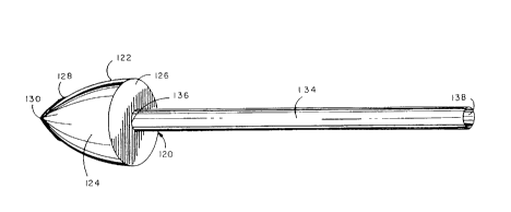
One prefer~ed embodiment of an obturator is illusl,aLed by Figs. 9-13
respectively. As seen therein the obturator 120 con,~rises a puncturing
headpiece 122 which is s~ sl~nlially cone-shaped in configuration and
20 comprises an outer shell 124 and a base plate 126. The outer shell 124 has
deterr"inable dimensions and a girth which can be altered in size. At the
distal end 128 of the puncturing headpiece 122 is a pel roraling end tip 130
which appears as a cross- or star-sl1aped cutting edge for the headpiece 122.
As shown by Fig. 10 the pe"orali"g end tip 130 does not extend over the
25 entire surface area of the outer shell 124; instead the perrorating end tip 130
is limited in size and orientation to the distal end 128. The pe, rorali"g end tip
130 serves as the sharp cutting edge for the obturator 120 as a whole.
Integral with the puncturing headpiece 122 is an elongated shaft 134
whose overall axial length may be varied to acc~,nrnodate the surgeon and
30 the particular medical circumstances of usage. The distal end 136 of the
shaft is irltegrdled with the puncturing headpiece 122 and provides access to
the interior volume of the headpiece bounded by the outer shell 124 and the
base plate 126. The proxi~al end 138 of the elGIlgdled shaft 134 is intended
to be held by the surgeon or invasive radiologist performing the vascular
35 bypass surgery. Accordingly the axial length of the elongated shaft 134 will
vary and acco" ,rr,odate the surgeon; and thus vary from a few inches to a few
SUBSTITUTE SHEET (RULE 26~
CA 02257989 1998-12-09
W 097/47261 PCTrUS97/09S03 -
-19-
, . .
feet in length. The function of the 010l Iyated shaft 134 is for the carrying and
t, anspo, l of a previously ex~-ise :l v~sclJI-r segment to the cl)ose" site on the
unobsl, ucted or primary blood vessel in-vivo. The elongaled shaft 134 acts
to support n,ainlain and convey the excise~ v~scular segment within the
5 lumen of the catheler in a n,an,)er such that the V~SGI ~I-r seg",ent can be
used as a bypass graft.
A critical requirement and feature of the puncturing I ,ead,~ ce 122 and
the obturator 120 as a whole is that means exist for expanding and
contracting the puncturing headpiece on~len~and. This requir~me, It and
10 ~I,dracte,islic is illusl,dted by Figs. 11-13 respectively. As seen within Fig.
11 the puncturing head~iece 122 appea,5 in its initial size identical to that
shown by Fig. 9. The outer shell 124 is su~slanlially cone-sl,aped in
configuration has an initial inte",al volume and has a girth di",el,sion d
equal to the initial .Jia",eter of the base ptate 126. The internal volume of the
15 puncturing headpiece as detel",i"ed by the dimensions of the outer shell
124 and the base plate 126 provides an initial il llel l ,al volume of
dete"".nable quantity.
When the n)ecl ,a"i3m for contracti"g the puncturing headpiece is
activated the consequence is ill~lst,a~d by Fig. 12 in which the di",ensions
20 of the outer shell 124 are diminished and the girth of the headpiece has beenreduced as shown by the red~ced diameter d of the base plate 126. Note
also that as the puncturing l~ead,~iece 122 beco"~es cont,acted in overall
volume and di",ensions the configuration of the puncturing headpiece 122
has conse~- ~enlially become allered and now appears to be spear-like in
25 configuration. Similarly the overall angular di~posili~ " of the pe, ror;dling end
tip 130 serving as the cutting edge will also be slightly altered in overall
appearance as a co~,sequence of COI Itla~Aing the puncturing head,~iece 122.
Alle",ati~/ely when the puncturing he~dpiece 122 is ex~anded the
overall result is shown by Fig. 13. As seen therein the outer shell 124 has
30 been e~,uancleJ in overall dimensio"s and volume; and the girth of the
head,t~iece has been ex~a"ded and can be determined by the dia",eter d" of
the e~-~,d, Ided base plate 126. Note that the overall appearance of the
puncturing headpiece has been altered as a c~nsequence of its expansion
and now appears to be elliptical in shape overall. Similarly the pe, rorali"~
3~ end tip 130 has similarly been altered in appeara"ce and has angularly
SUBSTITUTE SHEET (RULE 26)
CA 022~7989 1998-12-09
W O97/47261 PCTrUS97/09503-
-20 -
expanded somewhat to conro"" with the expanded di",ensions and angularity
of the outer sheli 124.
It will be recognized and appreciated also that throughout the changes
in appearance internal volume and overall size for the conl, acted or
5 expanded puncturing headpiece 122 as shown via Figs.11 12 and 13
res,cecli~/ely the dimensions and overall configuration of the 010ngaled shaft
134 have not been allered meaningfully or sigr,ificanlly. Although this is not
an absolute requirement in each and every embocli."ent of an obturator it is
pr~fer~ed that the eIOI ,yaled shaft 134 particularly at the inleyl dted distal end
10 136 remain consla"t in size and volume as much as possible and be
unaffected subsequent to the on-demand ex~.ansion or contraction of the
puncturing headpiece 122. This prerer~nce and feature will "~di"lain the
ir~leylily and continued viability of the excised vascular seg",enl inten~Jed tobe carried and ll a"s~o, lecJ by the elongated shaft during the bypass grafting
15 proce-lure. Thus to avoid or minimize any physical damage to the vascular
wall of the graft r"alerial it is most desir~ble that the elongated shaft be
maintained in appearance configuration and dime,lsions without change
whenever possible.
An essential feature and component of each obturator is the existence
20 and availability of specific means for expanding and contracting the
puncturing headpiece on-del"~"d. A number of clirrerenl mechanis",s and
means for expanding and contracting the puncturing headpiece of the
obturator are convel lliol ,ally known and easily employed. Merely to
de",ons~,ale some cJirrer~nl and convel,lionally known mecl,anis",s attention
25 is directed to the means illuslldled by Figs.14 and 15 respectively.
The means for expanding and co"l,a~iling the puncturing headpiece
on-demand illu~l,dted by Fig. 14 co"slilute a mechanical ap~roach and
design mechanism which is carried within the i"le" ,al volume of the
puncturing headpiece 122 and the integrated elongated shaft 134. As seen
30 therein a central rod 150 extends through the hollow inlel ior of the elongated
shaft 134 and extencls into the internal volume derined by the outer shell 124
and the base plate 126 of the puncturing headpiece 122. Within the internal
volume of the outer shell 124 a plurality of rotabl2 ribs 152 are joined to the
central rod 150 at the distal end to form a central pivot point 154. Each
35 rotable rib 152 is mobile and pivotable around the central point 154 and forms
an umbrella-like scaffolding structure which can be expanded outwardly or
SUBSTITUTE Sl~EET (RULE 26)
CA 02257989 1998-12-09
WO 97/47261 PCT/US97/09503 -
E 0~ ~ u,
E N ~ ~
tD
t ~-- _ o o o
~ E
. _
C o~ o
c a)~ ~ E O C'
C -C I D ~ o ~ ~
~ ~ ~ ~
o o O O
C O
E ~ ~ '''
m 3 ~ ~D
~ ~ O
~ ~
-- ._ ~ ~ b b ~~
~ U~
E
a) E ~ ~ ~ ~
E ~ c~i N N ~)
E r~
~ . _
x _ c
~ r) 0 N 'J 0::~ c
Y -- (D 1-- a~ o ~ ._
O O ~ ~ ~ ~D
- b b b b b
._
~D
~D
-
- ~D z
D ~
SUBSTITUTE S~lEET (RULE 26)
CA 022~7989 1998-12-09
W O 97/47261 PCTrUS97/09503-
~a -
collapsed inwardly at will. Mounted on the central rod 150 is an expansion
wheel 156. This expansion wheel 156 is centrally mounted on the rod 150; is
moveable over the axial length of the central rod 150; and is controlled in the
direction of axial movement (distally and proximally). The expansion wheel
5 156 c~ rises a center hub 158 and a plurality of hub supports 160, both of
which ",ainlain the expansion wheel in proper posilion as it engages the
plurality of rotable ribs 152. Joined to the central hub 158 of the expansion
wheel 156 are linear movement members 162 which are positioned within the
interior volume of the elongated shaft 134 and have a length sufficient to
10 reach to the proximal end 138 of the elongated shaft 134 for control by the
surgeon or invasive radiologist. The linear movement members 162 engage
the center hub 158 of the expansion wheel 156; and extend or withdraw the
expansion wheel closer to or away from the perforating end tip 130 of the
puncturing headpiece 122. When the expansion wheel is engaged and
15 pushed forward, expansion wheel engages the rotable ribs 152 and expands
the rotable ribs outwardly thereby increasing the overall girth of the
puncturing headpiece as a unit. Alternatively, when the linear movement
members 162 are withdrawn, the expansion wheel recedes towards the
pro~in,al end and the engaged rotable ribs 152 collapse inwardly within the
20 volume of the outer shell 124. The consequence of this movement is a
contraction of the puncturing headpiece 122 as a unit. It will be recognized
and ap~,reciated that this mechanical a,c proach for expanding and col1lracting
the puncturing headpiece is completely conventional in design and operation;
and accordi, lgly, any conventional refinement of these basic coi "ponent parts~25 is co,~sideled to be a variation within the scope of this mechanical system.
As a represe,~la~ed alternative, hydraulic means for expanding and
cGr,l,acting the puncturing headpiece of the obturator on demand is also
provided. In this system, as shown by Fig.15, the internal volume of the
puncturing headpiece 122 and the integrated elongated shaft 134 includes an
30 elastic sack 180 comprised of a fluid containing elastic bubble 182 and a fluid
delivering elastic conduit 184. The outer shell 124 and base plate 126 of the
puncturing headpiece 122 are as previously shown; and the headpiece 122 is
integrated with the elongated shaft 134 as previously described herein.
Within the internal volume of the puncturing headpiece 122, is a fluid
35 containing elastic bubble 182 which is in fluid communication with the elastic
conduit 184 carried within the internal volume of the elongated shaft 134.
SUB~TITIJTE SltEET (RULE 26)
CA 022~7989 1998-12-09
W O 97/47261 PCTrUS97/09503 -
- ~ 3 -
The elastic sack 180 is ro",led of elaslor"eric material (such as rubber
elastic plastic and the like) and is fluid-tight along its seams. The elastic
sack 180 contains any liquid which is co",~>ati~le with the ",~l~rial of the
elastic sack; and it is the inll insic nature of the material rormi. ,g the elastic
5 sack 180 that the mdt~rial exerts a cor"pressio(, force or pressure upon the
fluid conlained within the elastic sack itself. In this way a hydraulic system for
expanding and conl,d.;ling the puncturing headpiece of the obturator is
created.
As fluid is intro~luced through the elastic conduit 180 by the surgeon or
10 invasive radiologist that fluid is conveyed and delivered into the elastic
bubble 182 posilioned within the puncturing headpiece 122. The elasticity of
the bubble 182 exerts a mild co",,~ression force and pressure againsl the
quantity of fluid conlain~J within the bubble interior volume; acc~rdin~Jly the
yl ealer the quantity of fluid within the elastic bubble 182 the larger in overall
~5 volume the elastic bubble beco,),es. Thus as more fluid is delivered through
the conduit 184 into the elastic bubble 182 the larger in overall volume the
elastic bubble l,ecomes; and as the volume of the elastic bubble e,.~.~n.ls the
overall configuration and internal volume of the piercing headpiece 122 also
enlarges. In this ",ar",er by carefully controlling the amount of fluid
20 conveyed through the conduit 184 into the elastic bubble the overall size andconfiguration of the piercing headpiece 122 can be controllably ex~anded.
Subsequently to reduce the overall size and configuration of the puncturing
headpiece 122 a quantity of fluid is pen"itled to be released from the elastic
conduit 184 at the pro,~i",al end by the surgeon or radiologist. Bec~ ~se the
25 i"ate,ial is elastic and exerts a co~pression force against the quantity of fluid
~res~"t within the bubble at any given ",ome"l in time the release of fluid
through the elastic conduit will cause a reduction in overall size for the elastic
bubble 182; and as the overall volume of the elastic bubble is red~ ~ced in
size the puncturing headpiece will consequently be contracted and reduced
30 in configuration and overall volume as well. It will be noted and appreciated also that this hydraulic mechanism for e).~andiny and cont, acting the
puncturing he~driece on d~ma"d is a convel ,liu"ally known fluid system and
techniq.1e; and many conventionally known va, iations and c~anyes in
hydraulic design and fluid control systems are presenlly known and
35 comr"or,ly available for use. Accordingly all hydraulic systems are
SUBSTITUTE S~IEEl (RULE 26)
CA 022~7989 l998-l2-09
W O 97/47261 PCTAJS97/09~03-
_~y_
envisioned as suitable for use as one means for exlJanding and col1l(acti,1g
the puncturing headpiece of the obturator on~e,l,and.
A number of dirrere, It physical e,llbodi",ents for the obturator are also
envisioned and intended for use. Some examples, which are merely
5 illustrative of the range and variety of physical fom,a~s and which serve to
merely illustrate the range and ~legree of dirrerence available for the various
puncturing headpieces of an obturator, are illustrated by Figs. 16-22
respectively. It will be recognized and understood, however, that these
allel "ali"e er,lbodi",ents are merely re,uresenlalive and illustrative of
10 obturators and puncturing headpieces generally and do not signify any
limitation or restriction on their structural construction or design.
The embodiment illusllated by Figs. 16 and 17 respectively shows a
puncturing headpiece 200 which is su~slanlially cone-shaped in overall
appearance and comprises an outer shell 202 and a base plate 206. The
15 distal end 208 of the puncturing headpiece 200 has a pel roraling end tip 210which is also substantially cone-shaped in configuration and appeara"ce and
covers only a small surface area of the outer shell 202. Integral with the
puncturing headpiece is the elongated shaft 134 as described previously
herein; and means for expanding and cont~acli ng the puncturing headpiece
20 200 on-demand are included within the obturator as a integrated unit.
Another embodiment for the puncturing headpiece is illustrated by
Figs.18 and 19 respectively. As shown therein, the puncturing headpiece
220 cG"".rises the outer shell 222 and the base plate 224 integral with the
elongated shaft 134. A particular feature of this embodiment, however, is the
25 distal end 226 seen most clearly within Fig.19 as providing a pel roraling end
tip 230 which is substantially star-shaped and extends over the surface area
of the outer shell 222. The result is to provide a series of grooves 228
alternating with sharp cutting edges 232 over the surface of the outer shell
222. This embodiment for the puncturing headpiece 220 provides a much
30 ~redler area for cutting and pel roraliol1 as a specific feature of the obturator
design.
To demonstrate further the variety and degree of differences
envisioned and in~ended when constructing a puncturing headpiece, the
structural constructions exemplified by Figs. 20-22 respectively are provided.
35 As illustrated by Fig. 20, the puncturing headpiece 250 includes a buttressing
region 254 as a part of the outer shell 252. The buttressing region 254 is a
SUBSTITUTE S~IELT (RULE 26)
CA 022~7989 1998-12-09
W O 97/47261 PCTrUS97/09503-
- ~ 5-
r~;. ,ro,.;ed region for engaging and bel ,ding ",dlerials placed in conta~ withthe outer shell when the puncturing head~iece is ex~Jar,ded. The puncturing
headpiece 250 inc~- ~des a base plate 256 and is integrated with the elongaled
shaft 134 (described previously herein).
In cc:""~a,ison the puncturing hea~,iece 260 exe",pliri~.J by Fig. 21 is
a sharply tapered and contoured embocl""enl in which the outer shell 262
includes a spiral girth zone 264 suitable for derorl,~ing elastic ",at~rials. The
base plate 266 conror",s to and is inleyldled with the spiral zone 264.
Another aller"dti./e embodi",ent of the puncturing headpiece is
illustrated by Fig. 22. In this embodiment the puncturing headpiece 280
co",,~" ises an outer shell 282 and a concave-sl~a~,ed or scalloped zone 284
which is joined to and integrated with the base plate 286. The concave-
shaped configuration of the zone 284 is inle"ded to aid the puncturing
he~drioce as it is ex~.at~decl and cor,l,ac~ed on-de,~and.
C. The Defor",able Cuff
A requisite col"pol ,ent part of the c~tl ,eter ap,~a~tus for credli"g a
vascular bypass graft is the presence and use of a deformable cuff or flange
such as is ill~J~trated by Figs. 23 and 24 respecli~/ely. As illustrated and
embodied therein the defor" ,able cuff 300 is a substantially cylind~ ical-
shaped collar which is open at each of its ends 302 304. The cuff 300 is
hollow; is s!lh5lZ~l ,l;ally round or oval (in cross-sectional view); and has
dete""inaLI~ dimensions initiallywhich can be de~u,,,,ed atwill when
sufficient force is applied to the sidewall 306. As an aide in controlling the
intended .lerol",dtion of the sidewall 306 on-de",d"d via the i"lenliooal
application of exle,r,ally applied forces1 it is most desi~dble that the material
constituting the sidewall 306 of the cuff 300 be pre-stressed along the axis
M' as shown within Fi~. 23; and that the malerial cGnsliluting the sidewall
306 be an open-weave pdllern of resilient matter rather than take form as a
so~id mass of r"alerial. For this reason the sidewall 306 illl,sl,aled within Fig.
23 appears as an open meshwork of wires 308 which are intertwined to form
a s~ sln~ Itially l ,exAgQnal pdtler". This open meshwork of wires 308
provides the desired resiliency fiexibility and defor",dlion capability
(particularly along the axis M ) such that the upper portion of the sidewall
306 can be defor"~ed and flaired outwardly on-de" ~a"d to yield the flaired-lip
deformity 310 shown by Fig. 24.
SUBSTITUTE S~EET (RULE 26)
CA 022~7989 1998-12-09
W O 97/47261 PCT~US97/09503-
~a~-
lt will be recognized and appreciated that the deform cuff shown by
Fig. 24 is merely the result and consequence of exerting force alon~ the
u,~ per,~,osl portion 309 of the open meshwork of wires 308 above the axis M
such that the upper sidewall 309 adjac~,)l to the open end 302 has become
5 expanded outwardly flaired and bent into a curved lip configuration in the
derol ",ed state. Note that the open meshwork of wires constituting the lower
portion 307 of the sidewall 306 at the other open end 304 remains relatively
stable and s!~hst~ntially unaltered in its original shape and state. The
dero~ llldLion has thus been CGI 1ll olled and the forces applied only to the upper
10 portion from the AA axis to cause the outwardly extending flaired lip result.Moreover the resulting flaired lip zone 310 retains structural sller,yll, and
resiliency as an open meshwork of wires despite having been created by
d~ro""alion. The ability of the cuff to be dero" "ed in the manner illustrated
by Figs. 23 and 24 ~especti~rely is a requisite and necessa"/ attribute and
15 characte~islic of each e",bodi",elll and construction for the def~"nable cuff.
The construction and design for the deforlnable cuff in the present
invention is an example of the e, ~ginee, iny principle that structural form
follows intended function. As a requisite component part of the catheter
apparatus and methodology for cr~dli, ~g a vascular bypass in-vivo the
20 intended functions of the deformable cuff are twofold in nature: (1 ) the
de~or"lable cuff is intended to engage and become ~oined to a previously
excised vascular segment which will serve as the bypass graft in-vivo; and (2)
the ~efGr",ablc cuff is intended to be positioned within the i"ler"al lumen of
an unol,sl,ucted major blood vessel (such as the aorta) and beco",e
25 ~leror",ed in-situ such that a portion of the cuff becomes positioned and
secured to the internal lumen (the blood flow channel) of the unobstructed
blood vessel permanently and in a fluid-tight manner. Thus as illustrated by
the embodiment of Figs. 23 and 24 the upper",ost region 309 of the cuff 300
is deror",ed on-demand into a flaired outwardly bent form which is intended to
30 be secured within the lumen of the unobstructed artery or vein while the
undisturbed sidewall portion 307 of the cuff is intended for engage",ent and
juncture to the previously excised vascular segment which will serve as the
bypass graft. However because there is no specific pre-positioning or pre-
alignment of cuff sections or portions as to ultimate or inlended usage it is
35 ir"material and irre~evant structurally as to which end of the deformable cuff
serves which intended function and purpose.
SUBSTITUTE ~HEET (RULE 26)
CA 022~7989 1998-12-09
W O 97/47261 PCTrUS97/09503-
- ' --.?q--
Several attributes and cl ,aracterislics are con""ollly to be shared
among all embodi",enls and constructions of the deformable cuff. These
include the following:
(a) It is only required that the material con~liluting the cuff be
5 ~leror"~able on~",a"~. For convenience and g,edler facility in achieving
such de~""ily in the degree and at the time required it is most desirable that
the material rorl"ing the cuff be an open weave or meshwork rather than a
solid mass which is considered to be more difficult to deform in a controlled
manner. There is however no resl~ i~tion or li~ilalio" at any time or under any
10 intended use circulllsta"ces which necessilates an avoidance of a solid mass
of material either as a single sheet or as a la".inated plank of malerial.
Accordingly the choice of whether to use an open meshwork or a solid mass
of ",aterial is left solely to the ~iscretiol) of the manufacturer and the surgeon.
(b) The defo",)able cuff need only be co",~, ised of resilient
15 flexible but defo,mablc matter. A number of difrer~nl cG",positions and
formulations of material may be usefully employed when making a
de~r"~abl~ cuff suitable for use with the present invention. Among the
desirable materials are those listed within Table 2 below.
(c) After the derorl"able cuff has been manuf~ctl~red using resilient
20 ",ale,ials the completed cuff structure (prior to defor",dlior,) may be .
subsequently covered to advs"lage with one or more biGcori,palible coatings.
These bioco",pdlible coalings are inlended to water-tighten the article and to
faciîilale the seY:in9 of the e~c,sed v~scl~!~ sey",erlt to the cuff as well as to
reduce the inleractiGns of the immune system with the vascular bypass graft
25 after it has been secured to the blood vessels in their appropriate localionsin-vivo. Such biocompdtil~lE coalings are conventionally known; will reduce
the severity and duration of immune reactions which frequently disrupt or
int~r~le with v~scul~r bypass grafts; and are co"siclered desirable in a
majority of use instances in order to ~"ini",i~e the body reaction to v~scl ll_r30 bypass surgery. A represe"lali~e listing of biocompatible coatings deemed
suitable for use with the defor"~aLI~ cuff is provided by Table 3 below.
SUBSTITUTE SHEET (RULE 26)
CA 02257989 1998-12-09
W O 97/47261 PCTrUS97/09503-
-~8-
Table 2: Deformable Materials
Metals and Alloys
stainless steel;
nickel/titaniuim alloys;
aluminum/nickel alloys; and
graphite carbon and metallic blends of carbon
Svnthetic Polvmers
polyamides such as nylon;
polyacrylates such as polyacrylic acid;
polycallJoi~dles such as poly[2,2-bis (4-hydroxyphenyl)] propone; and
polysiloxones.
Table 3 Prosthetic Coatings
High temperature pyrongen-free carbon;
polytetrafluoroethylene (PTFE) and other polyhalogenated carbons;
Fibronection;
collagen;
hydroxyethyl methacrylates (HEMA); and
serum albumins.
SUBSTITUTE SI~EET (RULE 26)
CA 022~7989 1998-12-09
W O 97/47261 PCTAUS97/09503-
--0~ 9--
(d) Although the e"lbo.li",ent of the cuff or collar prior to
dero""alion exemplified by Fig. 23 appears as a geometlically regular and
col)erent structure there is no requ"e",ei ,t or de"~and that either the
structure or configuration of any deformable cuff c~r,ro"" to these
5 par~",et~rs. Accordingly it will be recoy"iced and ~" ,derslood that the
defo",~able cuff structure need not be a co,~,plately el~cir~ling band or collarof dero""able ",~terial. To the cont,~ry a U-sl,ap~d band orflange of
r"alerial where the sidewall does not overlap or join and where a gapped
disla"ce separales the arms of the band or flange is both pel ",illed and
10 envisioned. Moreover although the cylindrical-shaped format of the
defor"~able cuff illuslraled by Fig. 23 is highly desirable there is no
requ,~en,ent that the ~Jian,eter of the cuff prior to clefor",alion be conslan~ or
co"sistent over the entire axial length of the cuff when manufactured. Thus
anisotropic cuff structures as well as isot.upic constructions are inte, Ided and
15 desirable. In this ",anner the cuff in its initial state prior to deformation may
have a variable internal diar,)eter over the axial length of the article in which
one open end may be either y~ealer or lesser in size than the other open end;
and there may be multiple increases and decreases in diameter size
sl~ccessively over the entire axial length of the cuff itself. All of these
20 variations in construction and structure are within the scope of the present
invention.
To illustrate some of the modesl varidliu,~s and dirrere,lces available
and envisioned for a dero,lllabla cuff i,lte"ded for use with the preser,t
invention the alLer"dlive cuff ernbodi",etlts ilh.sl~aled by Figs. 25 26 and 27
25 are provided. As shown within Fig. 25 the defor",able cuff or collar 330
appea,:~ as a cylin.l~ical-sl,aped article having two open ends 332 334 and a
rounded sidewall 336. The body of the sidewall 336 is an open meshwork of
closed loops 338 each closed loop being joined at multiple points along its
peri",eter to at least one other closed loop -- thereby fG, I)~ 9 an open grid
30 meshwork. A notable feature of the cuff construction within Fig. 25 are the
outer edges of the open ends 332 334. Each edge 340 342 is fo",)ed by a
closed loop which is far more easily bent and derolllled than the closed-loop
meshwork in the middle of the sidewall 336. In many ir,slances the
availability of closed-loop edges 340 342 provide an e"or",ûus benefit and
35 adva"iaye in de~-",i"g the cuff in-situ within the internal lumen of an
unobsl, ucted artery or vein. In addition the defor",able cuff 330 has been
SUBSTITUTE S~EET (RULE 26)
CA 022~7989 l99X-12-09
PCTAUS97/09S03-
W O 97/47261
- 30-
pre-stressed substantially at the midiine along the axis BB such that the
upper most portion 339 of the cuff near the open end 332 and the edge 340
are more easily cJero""ed and flaired outwardly as a consequence.
A third embodiment of a defor",able cuff or flange is iliuslraled by Fig.
5 26. As shown therein the defo",~ble cuff 360 is fo",~d primarily as a series
of coiled wires whose overlapping and inle~sec~ing portions have been fused
together to make a unitary article. The defw"1able cuff 360 thus has the two
open ends 362 364 and an open coiled sidewall 336 fo,-"e~ from the
cor,lnlonly fused coits of wire. The open lattice work of coiled wires 368
10 provides the flexible and resilient meshwork suitable for achieving the primary
functions of the defor"ldble cuff. Again the sidewall 366 has been pre-
stressed along the axis CC such that the upper most portion 369 can be bent
and ~lefor"led outwardly on demand using an expansion force.
Finally a fourth alternative e",bodi",ent is provided by Fig. 27 in which
15 the de~rn~able cuff or band 380 is shown having tv,/o open ends 382 and
384. In this instance however the sidewall 386 is comprised of a solid sheet
of material. Two features are included in this embodiment of the deformable
cuff due to its construction using a solid sheet of resilient material as the
sidewall 386 for the cuff. The sidewall 386 has been pre-scored to form
20 cross-hatched squares over the axial length of the sidewall; and the pre-
scored sidewall thus will deform far more easily and bend outwardly as shown
when a e)~a"sion force is applied to the interior of the cuff. Similarly the
sidewall material has been pre-~l(essed along the axis DD such that the
upper most region 389 nearest the opening 382 will bend far more easily and
25 in a controlled fashion when and as required by the user.
Il. The Excised Vascular Segment
To Be Used As A Bypass Graft
The prefer~ed sources of blood vessels suitable for use as a vascular
30 bypass graft are the saphenous veins. These veins constitute the superficial
veins of the lower extremities and comprise both the greater (or long)
saphenous and the lesserl (or short) saphenous veins. Anatori)ically the
long saphenous vein begins on the medial side of the foot and ends in the
fermoral vein below the inguinal li~amenls; and the short saphenous vein
35 begins behind the lateral rnalleous and runs up the back of the leg to end inthe popliteal vein. However if the saphenous veins of the particular patient
SUBSTITUTE SHEET (RULE 26)
CA 022C,7989 1998-12-09
WO 97/47261 PCT/US97/09503 -
--3 1--
are unsuitable or unavailable for any reason~ either the cephalic or the basilicveins are very accept~hle substitutes for use as a v~-scul~r bypass conduit.
However, if these leg or arm veins are not available, synthetic or other
biologic ",vler;31s may also be used as substitutes.
The medical procedure to isolate and excise the saphenous vein of
choice is conve"lionally known and co"si~lered a routine surgical tect ,r,ique.
The sa,vhenous vein is harvested under ge"eral ane~lhesia. An incisiGn is
first made in the medial malleolus, where the saphenous vein is often dilated.
The saphenous vein is identified and then dissected with a single incision
made along its course with scissors. Branches are doubly clal"ped with
he",oslatic clips and divided. The sa,4henous vein is then freed up and
removed from the leg. The leg wound is closed with suhcl ~t~neous sutures
and Ste, i~ll i,v adhesive over the incision. The vascular segment is prepared
on a separale sterile table with adequate light and loupes, and branches are
selectively ligated with 4-0 silk. An oval-tip needle on a syringe is inse~led
into the graft to gently dilate it by administering a balanced electrolyte
solution (pH 7.4, chilled to 7 to 10 C) and 10,000 units/liter of heparin. A
valvulotome is inse, led into the vein graft segment and the valves clipped
with a 3-mm right-angle stainless steel instrument with a highly polished ball
tip on the right angle. The knife edge is ,urotecled and sharply splits the
cusp, causing valvular i"co",pete"ce. Measurements for the approxin,ale
lengths of the grafts may be made with umbilical tapes, and the appropriate
ler,!Jtl Is may be chosen before it is sewn to the cuff and coroual y arteries.
Ill. TheIntroducer~.y;.t~
The introducer system comprises the catheter alJpaldlus including the
defor")able cuff and a previously excised vascular sey",enl in c~",b.. ,dtion;
and it is this introducer system which is utilized by the surgeon to perform therequisite acts and manipulalions by which the excised v~scul-r sey"~el ,l is
30 delivered to and bec~mes secured within the lumen of the unobsl, ucted major
blood vessel (and subsequently ana~omosed to the obstructed blood vessel
at a site distal to the obstruction). For des~ live purposes and for i"creased
clarity of co",,l~rel-ension, this desc,i~,vtiol, will inlenlionally limit itself to the
use of the obturator illustrated by Figs. 9 and 10 respecti~/ely, to the
35 deformable cuff construction and structure illuslraled previously by Figs. 23and 24 respectively, and to the use of a previously excised vascular segment
SUBSTITUTE SHE,LT (RULE 26)
CA 02257989 1998-12-09
PCTnUS97109503-
W O 97/47261
--3~ -
taken from the long or short saphenous vein in the same patient. ~he
introducer system represenls and provides for the intenlional place",el1t and
carriage of the excised v~scl~a- s~ylllenl on the obturator the ~-yage,ne"~
and juncture of the def~r,)lable cu~f to one end of the excised vascular
se~ment; and the proper orientation of the then engaged .leror",~ble
cuff/excised vascular seymei)l on the obturator with respect to its relalio,1ship
to the puncturing headpiece.
The formation of the introducer system begins with the proper
plac~l"ent of the previously e~cised v~sc~ r segment (taken from the
saphenous vein previously) upon the obturator. This manipulation is
illustrated by Fig. 28 in which the excised vascular segment 400 is placed
upon the elongated shaft 134 a~jacenl to but ~l ererably not in direct contact
with the base plate 126 of the puncturing headpiece 122. As shown therein
it is in~ended and prererl~d that the elongated shaft 134 will be inserted into
the internal lumen 402 of the excised vascular segment 400 at the proximal
end 138 held by the surgeon; and then the body of the vascular segment 400
is conveyed over the axial length of the elong~ted shaft 134 until a chosen
position typically 1-2 centimeters from the distal end adJacent to the
puncturing headpiece 122 is reached. In this manner the weight and body of
the excised vascular segment 400 is carried on the elongated shaft 134; and
it is desireable that the diameter of the elongated shaft 134 be only slightly
smaller than the average dia~eter of the i"ler~,al lumen 402 for the vascul~r
segment 400. As a consequence7 the excised vascular segment is ~degu~tely
supported carried and transported by the elongated shaft during the entirety
of the manipulations prior to entry into the body of the living patient as well as
subsequent to the in-vivo pel r~r~lion of the unobsl~ucted major artery or vein.The manipulation illustrated by Fig. 28 is expected to be performed by the
surgeon immediately after excisi"g the vascular seg"~ent from the patient but
prior to beginning the bypass graft surgery itself.
After the excised vasc(ll~r segment 400 has been properly positioned
on the elongated shaft 134 of the obturator 120 the defor"lable cuff
(illustrated by Fig. 23 and desa iL.ed in detail previously herein) is desirablypassed over the puncturing headpiece 122 and over the open end 404 to
cover a small portion of the exterior surface over the excised vaSCUl~3r
se~me"t 400. It is desirable (but not absolutely necessary) that a gap
distance (about 1-2 centimeters) separating the open end 404 from the
SU~STITUTE SltEET (RULE 26)
CA 022~7989 1998-12-09
W O 97/47261 PCTAUS97/09503-
-33-
puncturing headpiece 122 be r"ainlained during the placement of the
defor",dL,I~ cuff -- as this will allow for easier posilior,i"y of the derorn,able
cuff in a pre-chose" aliy"l"el,t and posture and a more controlled
defoln)alion on-derl,a,)d. Once the defor",alJle cuff 300 has been posilioned
over the v~sclJl~r sey",el " to the sAIisr~ 1iGn of the surgeon the lower regionof the cuff covering the end exte, ior surface of the excised v~sa ~I? seglnent
400 must be enyay~d and l.ecor"e joined to the v~sc~ seyl,)enl in a
reliable and safe ",anner.
One prefel ,ed manner of er,ya~~el"er,l and juncture is for the surgeon
to suture the open meshwork sidewall of the cuff directly to the v~scl ~'-= wallof the excised seyll~e,)t. This suturing is easily pe"v,llled by the surgeon
prior to beginning the grarli"y surgery and the sutures 420 will serve as the
physical means for engaging and ~ue""allenll~ joining a portion of the open
meshwork of wires in the sidewall of the cuff to the excised v~-scl ll-r segmentitself. The type of sutures 420 their place" ,e"t their number and the linkage
to the vascular wall of the excised segment are left to the pe, ~onal disc~etionand choice of the surgeon.
Other means for engaga"~enl and juncture of the defor" ,able cuff to
the v~scu~-- wall of the excised sey",ent also are COIlllllOnly available.
These include surgical staples; L.iocG""~alible aJl,esives; enci~cling ligatures;
and a wide range of fasleners and closures. Any and all of these allel "ali~es
may be employed alone or in co"lbination to achieve a reliable el~gagel"enl
and juncture.
After the defor",able cuff has been engaged and joined to one end of
the excised vascular sey",enl then carried upon the elongated shaft of the
obturator the size of the puncturing headpiece 122 should be adjusted in
shape and girth such that the dia",eter of the base plate 124 of the
puncturing headpiece 122 prererably is equal to or slightly y,ealer than the
diar"eter of the open end 302 for the defo""able cuff 300. This manipulation
is also illustrated by Fig. 29 where the size of the base plate 126 is
coeAlensive in diar"eter with the dia",eter of the open end 302 of the
rur",able cuff. In this preferred manner the entirety of the puncturing
l)ead~.ece 122 serves as a front se~;tion or first stage for the introducer
system as a whole.
The wr"ple~e introducer system is illustrated by Fig. 30 in which the
fully prepared obturator carrying the previously excised vascular segment to
SUBSTITUTE Sl IEET (RULE 26)
CA 022~7989 1998-12-09
W O97/47261 PCT~US97/09503
y_
be used as a bypass graft and the deror,l,able cuff have been positioned in
advance; and the prepared obturator placed within the internal lumen of a
cdll ,eter. As seen lher~7in the calheter is exposed in cross-sectional view
and shows the hollow tube 2 of fixed axial length having a disclete ~,r~xi,l,al
5 end (not shown) a discrete distal end 10 and an inler"al lumen 14 of pre-
deten~ined diameter sufficient to house the entirety of the prepared obturato
(illusl,aled by Fig. 29). The distal end tip of the catheter is adapted for
guidance of the c~tl ,eter in-vivo to a ci ,osen site where an Ul ,obslr~cted
artery or vein is in anatomic proxi",ily to an obstruction Iying within another
10 blood vessel; and the prepared obturator of Fig. 29 (including the previouslyexcised blood vessel sey",enl 400 and the deror"~able cuff 300) has been
placed into the internal lumen of the calheter. The introducer system is now
complete. As shown by Fig. 30 the surgeon may now begin the first steps for
surgically delivering the introducer system into the thoracic cavity or other
15 a~,uropriate body region in order to create the vascular bypass graft.
IV. The Routing And Surgical Introduction Of The Cc.~..lling Catheter
Into The Body Of The Living Human
Catheleri~dlio" involves a great deal of technical skill some
20 instrumentation and mature judy,l~ent in order to choose among the
appropriate procedures and the various techniques which are now
conventionally known and available for use. Clearly because the present
technique conslilutes catheter intervention in critically ill palie"ts the
physician or surgeon must be very familiar with the available a"alor"ical
2~ allel "ali~es in order to select the best routing for introducing the call ,eter the
best technique in order to ~Gcess the thoracic cavity of the body where the
obstructed artery and aorta exist and to carefully select the timing and other
operative conditions in order to achieve best results.
In general catheterization can be performed using any duct tube
30 channel or p~ss~geway occurring naturally or surgically created for the
specific purpose. Thus among the naturally occurring passageways in the
body are the anus; the alimentary canal; the rnouth ear nose or throat; a
bronchus of the lun~; the urethra; the vaginal canal and/or cervix; and any
blood vessel of sufficient size of the central circulation in the body. Any of
35 these routings are envisioned and expected to be used when and if
appropriate. However clearly a co,n",only used and the critical route of
SUBSTITUTE SHECT (RULE 26)
CA 022~7989 l998-l2-09
W O97/47261 PCTAUS97/09503 -
-35-
Access is the introduction of ~tl ,eter~ into the thoracic cavity and the arterial
blood circ~ tion A-l; cent to the heart.
For this reason it is useful to briefly sullllllal i~e the tecl ",i~ue currentlyin use for introduction of catheters into the central blood circu!-~ion as an
5 illustrative exar"p e of pref~r,ed ~tl,eteri~ation teol,ni~lues. There are twog~ner~l m~ll ,Gds currently in use. These are: (a) percutaneous introduction
using needles and guidewires; and (b) direct introduction after surgical
isolation of the blood vessel of choice. While either yener~l ",etl,od may be
utilized at any site of the general circulation pradical and anatomical
10 considerali~ns including the site of the flawed tl~erapeutic a~.pliance wil
s~enerally dictate which approacll is most ap~.ro~riate under the individual
circu" ,slances.
The modified Seldin~er Techni~e:
The percutaneous introduction of a cdtl,eter is illusl,aled by the
modified S~ldinger technique which is shown by Figs. 3~A-31 F respe.ti~ely.
Fig. 31A shows a blood vessel being punctured with a small gauge needle.
Once vigorous blood return occurs a flexible guidewire is placed into the
blood vessel via the needle as shown by Fig. 31 B. The needle is then
20 removed from the blood vessel the guidewire is left in place and the hole in
the skin around the guidewire is enlarged with a scalpel as shown by Fig.
31 C. Subsequently a sheath and a dilator is placed over the guidewire as
shown by Fig. 31 D. Tl ,eredrler the sheath and dilator is advanced over the
guidewire and directly into the blood vessel as shown by Fig. 31 E. Finally
25 the dilator and guidewire is removed while the sheath rel,lains in the blood
vessel as illuslraled by ~ig. 31 F. The catl ,eter is then inserted through the
sheath and fed through to reach the desired location.
The other general n~etl ,od for the introduction of catl ,eters into the
blood circulation is a direct surgical cutdown. The surgical cutdown approacl,
30 is generally less favored and is typically used for the brachial or fei~,orala~.p~acl ,. Cutdown ~rucedure is often a complex surgery and is used only
when no direct ~ccess is ge"erally available. A far more complete and fully
des~;, iptive review of both these general cdtl ,eteri~alion techniques is
provided by the texts of: Dia~noslic And Therapeutic Cardiac
35 Catheteri~dlion secondedition 1994 Chaptereight pages90-110andthe
references cited therein.
SUBSTITUTE SHEET (RULE 26)
CA 022~7989 1998-12-09
W O97/47261 PCTrUS97/09503-
-36-
Accordingly, for purposes of practicing the present methodology, any
and all conventionally known genaral cathete~i dliGn procedures and
tecl " ,iq~Jes which are convenli~nally known and in accor~a"ce with good
medical pracLice are explicitly intended to be utilized as necessary in their
5 original format or in a ",odiried form. All of these general catheteri~atio,l
routing and use techniques are thus envisioned and are deemed to be within
the scope of the prese"~ invention.
General rules for choosin~ an apPl opriate site of body entry:
An axio",dlic or general set of rules by which a surgeon or radiologist
can chose a proper or ap,cropriate site of entry for introducing the guiding
calheter into the body of a patient for purposes of creating a v~scl ll_r bypassin-vivo is as follows: (a) always pick the shortest and straightest pathway
possible or available; (b) identify the chosen entry site on an existing and
accessible unobstructed artery or vein, the larger the diameter of the
unobstructed artery or vein the better; and (c) identify the location and
orientation of the obstruction in the obstructed artery or vein and chose an
entry site distal to the obstruction.
A favored a~ roacl, to introducin~ the ~uidinq catheter into the thoracic aorta:Using the ascending aorta appruacl, as a representalive illu~l,alion
and example:
(1 ) Under ge"eral anesthesia, the chest of the patient is prepared
and draped in a sterile fashion.
(2) A three-inch incision is made to the left or right of the breast
bone through which the surgeon operates.
(3) Three additional one-inch incisions then are made to insert a
video camera, knife, surgical stapler, and other instruments.
(4) The ribs are separated, the thoracic cavity is entered, and the
asce"dir,s~ thoracic aorta is exposed.
(5) The introducer system is then positioned at the chosen site on
the ascending thoracic aorta.
SUBSTITUTE SI~EET (FtULE 26
CA 022~7989 l998-l2-09
W O 97/47261 PCTAUS97/09503-
3~
V. The In-Vivo Pla~ t Of The Vascular Bypass Graft
Into The Lumen Of The Unobstructed Major Blood Vessel
The method of the present invention utilizes the introducer system via
5 the catheteri~dlion tech"i~ue to create the v~c~ bypass graft between a
major u(lobsll ucted blood vessel such as the aorta and an obstructed blood
vessel in-vivo using a previously exc~sed v~scul~r segment as a conduit.
This p~cedure is illusllaled by Figs. 3241 collectively. It will be and
a~ preci~ed, however, that while Figs. 32-41 exemplify and illustrate the
10 manipulations of the surgeon and the events in sequence leading to the
creation of a vascular bypass, this desc,ipLion and the figures lllelllselves
present a greatly simplified presenlaliol) and explanation of the medical
proce.Jure, the techl)ical skills required, and the safety measures taken for
the patient's benefit medically.
After the controlling calheter has been routed and surgically
introduced into the body of the living human in the Illall,ler clesclibed
previously herein, the first critical stage for the process is reached as shown
by Fig. 32. The illuatlatioll of Fig. 32 (as well as Figs. 33-41 respectively) are
shown as partially exposed views in order to show more easily the detailed
place"lent and orientation of the prepared obturator carrying the derorlllable
cuff and previously excised v~scul~r segment in combinaliol ,.
As seen within Fig. 32, the major artery such as the aorta 500 is shown
in partial cross-seclional e)~,osed view to reveal the thickness of the arterialwall 502 and the inle" ,al lumen 504.
The catheter and the prepared obturator are as desol il,ed in detail
previously herein and illustrated by Fig. 30. It will be noted that the
puncturing headpiece 122 of the obturator 120 is positioned within the lumen
of the ~theter 2 such that the pe~ roralirly end tip 130 is in direct conlact with
the arterial wall 502 at the c hosen vAsc~ site. The puncturing headpiece
122 is of sulrlci.~"l size such that the entirety of the de~oll,l~ble cuff 300 and
the joined V~SCIJ~-~ seylnent 400 lie directly behind and are in axial alignmentwith the puncturing headpiece 122 and the el~l lgaled shaft 134. When
positioned as shown by Fig. 32, the prepared obturator is properly placed for
piercing and penel(ating the arterial wall on-demand.
When the surgeon extends the pre,~ared obturator within the internal
lumen of the catlleter 2, the result is illusl~aled by Fig. 33. As seen therein,
SUBSTITUTE SHEET (RULE 26)
CA 022~7989 1998-12-09
W O 97147261 PCT~US97/09503-
-3~-
the pe, ~or~li"~ end tip 130 has pierced and punctured the arterial wall 502
and been advanced into the arterial lumen 504. The initial hole in the arterial
wall 502 made by the pe, ror;aling end tip 130 is widened into a passageway
as a consequence of the puncturing headpiece 122 following the entry path
created by the pe, rorali~ ~y end tip. As the puncturing headpiece 122
penetrates the arterial wall 502 the size of the puncture in the arterial wall
becomes widened and enlarged to col,ror") to and accol"",odate the
configuration and the girth of the puncturing headpiece in its entirety. The
configuration and overall size of the puncturing headpiece 122 thus serves as
the means for widening the initial puncture made by the pe, rorati, lg end tip
130 such that the entire girth and overall diameter of the o~turator (complete
with the defor,nable cuff and excised blood vessel segment in cor, lbi"alion)
can subsequently pass through the e,1larged hole in the arterial wall.
As the prepared obturator is extended further across the thickness of
the arterial wall 502 through the enlarged passage the pen~t,a~ing
headpiece 122 is extended farther into the arterial lumen 504 until at least theupper portion of the dero""able cuff 300 has advanced far enough such that
the cuff 300 has itself entered the internal lumen of the blood vessel. This
sequence of events and result is illustrated by Fig. 34.
Subsequently after the upper portion of the deformable cuff 300 has
advanced into the arterial lumen 504 the surgeon holding and controlling the
proximal end of the catheter (not shown) activates the means for co, ll~ dC~il Ig
the girth of the puncturing headpiece 122 while the headpiece lies extended
in the arterial lumen 504. This manipulation and result is illustrated by Fig.
35. As seen therein the puncturing headpiece 122 has been reduced in
overall size and shows a diminished diameter or girth in co",parison to its
initial size as shown previously via Figs. 31-34 respectively. The reduced
overall size and altered configuration of the puncturing headpieoe 122 Iying
disposed within the arterial lumen in-vivo is a critical manipulation and step of
the methodology.
After the puncturing headpiece has been reduced in overall size and
has a diminished girth the overall diameter of the conll acled puncturing
headpiece 122 is smaller in overall diameter and size than the dial"eter of
the deformable cuff disposed directly behind the headpiece. Due to the
reduced size of the puncturing head~iece 122 the deformable cuff and
engaged vascular segment carried upon the elongated shaft 134 of the
SUBSTITUTE SltEET (RULE 26)
CA 022~7989 l998-l2-09
W O 97/47261 PCTAUS97/09503-
-39-
obturator may then be advanced farther through the puncture in the arterial
wall 502 and be further advanced over the puncturing headl.iece 122 into the
arterial lumen 504. This manipulation and result is illus~l~ted by Fig. 36.
It is i" ,po, lanl to recGyni~e and note that a s~ Ihsl~nlial portion of the
5 de~u,,,,a~12 cuff 300 has been extended over the outer shell 124 of the
puncturing headpiece 122 in the ~anner illl~allaled by Fig. 36. Moreover
cGnco",ilanl with the exlensiû,, of the defu~ able cuff 300 into the arterial
lumen 504 the engaged and joined v~sc~ segment 400 is concurrently
advanced and extended through the enlar~ed puncture or hole in the arterial
10 wall 502 at the chùsen site. The degree to which the end of the engaged
v~scl ~- segment 400 is advanced lies at the cJiscriation of the surgeon
pe, ror")ing this n ,ethûdology. If the surgeon so chooses the end of the
e)r.cise~ V:~SC~)~?'Seyllle(ll joined to the def~ able cuff 300 may be e~.lel ,ded
only through the arterial wall 502 but not markedly into the arterial lumen 504
15 itself. In the alle" ,alive the surgeon may choose to advance the end of the
engaged v~Scl l~r sey,nent 400 further and thus position the open end of the
v~scul- seg",ent 400 well within the intemal lumen of the artery itself. The
deyl ee of entry and advancer"ent for the ~Jefor")able cuff and the enyaged
v~scul~r segment thus is the choice and at the discretion of the surgeon at all
20 times.
After the defon"able cuff 300 and the engaged vAscll~-r seg-"ent 400
have been advanced such that each has penet,ated the arterial wall 502 and
a portion of the clefor",a~le cuff 300 has been extended to cover in part the
contracted puncturing headlJiece 122 to the surgeon s per~onal discrelion
25 and accor"r"oddtiûl, the surgeon will then activate the means for expanding
the puncturing he~lri~ce 122. At the time of activating the ex~ ansion means
for the puncturing headpiece 122 it is critical to recognize and appreciate
that the uppermost region of the deror",dble cuff 300 enco",p~.cses and
overlies the outer shell 124 and base plate 126 of the headpiece 122 -- as
30 shown by Fig. 36. It is not necess~ry that the defo,l"able cuff actually cover
and envelope the pe, roralin~ end tip 130 although if the surgeon chooses to
extend the defor",able cuff so far into the internal lumen of the artery the
surgeon may do so at his discl ~tiûn. In each iu~a~ ~ce however when the
means for e~ anding the puncturing headpiece 122 are inactivated the
35 overall size and girth of the puncturing headpiece will markedly increase. In the course of expanding in overall size and dimensions the puncturing
SUBSTITUTE SHEET (RULE 26)
CA 022~7989 1998-12-09
W O 97/47261 PCTrUS97/09503-
- ~ta -
headpiece will then make co~ cl with the upper position of the ¢3efor~"able
cuff 300 which has been pre-positioned to envelope and er,co,l,pass the
headpiece. Thus, as the overall size and girth of the puncturing headpiece
122 increases with time, the outer shell 124 of the headpiece will engage the
5 upper po,lion of the overlying defo"na~l~ cuff 300; and cons~4~ently apply
expansion force to the sides of the de~r",~l.le cuff in-situ.
As the controlled ex~ sion force applied by the increasing girth of the
puncturing headpiece 122 increases, the sidewall 306 of the cuff will clerol,ll,be bent outwardly, and become flaired and flattened out within the internal
10 lumen 504 of the artery ~00. Then, as the puncturing headpiece 122
expands in overall size and girth in ever greater degree, the defolllled
sidewall of the cuff 300 will become more flallened; and will come to lie
against the interior surface of the arterial wall 502; and thereby become
secured to the arterial wall in a per"lanelll manner. This manipulation and
15 result is illustrated by Fig. 37.
As shown within Fig. 37, the puncturing headpiece 122 has been
greatly enlarged and expanded as the headpiece lies within the arterial lumen
504. The controlled ex~ansion of the puncturing headpiece with the
concomitant deformation and flairing of the uppermost portion of the cuff 300
20 occurs within the blood flow channel of the artery and is intended to be
pe, rorlned without s~l~,st~nlially di"~i"ishing the rate of blood through the
lumen of the artery or causing the heart of the patient to stop at any time.
The intentional and controlled dero, m~lion of the cuff 300 along its upper
region as it lies disposed within the arterial lumen ~04 causes a perln~ne"l
25 flairing of the open meshwork of wires 308 for~,ing the sidewall 306. The
de~(",ed sidewall is bent, maneuvered, and flaired solely by the expansion
force of the puncturing headpiece. No other tool, article, or device is needed
or utilized in order to cause a controlled expansion force and to create the
deformation of the cuff while disposed within the central blood channel of the
30 artery in-vivo.
After the cufF has been controlably dercJIr~led within the arterial lumen
504 and become secured to the interior surface of the arterial wall 502 to the
personal satisfa~Aion of the surgeon, the means for COI Itl a~til 19 the puncturing
headpiece 122 are then once again activated. This col,lraclion of the
35 puncturing headpiece 122 serves to di,ninisl, the overall size and
configuration of the headpiece and markedly reduces the girth of the outer
SUBSTITUTE SH~I~T (RULE 26~
CA 02257989 1998-12-09
PCT~US97/09503-
W O97/47261
_y/_
shell 124 such that the overall ~ian~eter or girth of the puncturing headpiece
beco"~es approxin)alely equal to or less than the diameter of the ~lo"yal~d
shaft 134. The degree of collt,action for the puncturing headl-iece 122 is
desirably cl~osen and correlated to the dia~n~ter of the enya~ed v~scl ~t~
5 seylllellt 400 then car,ie~ on the elo"yate~J shaft 134. By c~n~o"";"~ the
overall size and girth of the puncturing l~e~d~iece to a size ap~,rox,",ately
equal to the dia"~eter of the elG"galed shaft 134 the surgeon is confident
that the overall di~",~ter of the ,ur~ ,ly corlt,acted puncturing hea~iece is
now smaller than the internal lumen of the ~ngage-l vascular seg",enl and
10 U,ererore the conlra~ puncturing hea~piece will then be able to enter and
pass con,~ tely through the internal lumen of the engaged v~-scul~r segment
without meaningfully injuring or altering the inte" ~al surface of the blood flow
ch~l ",el itself. The act of reducing the overall size and girth of the puncturing
headpiece with the resulting CGI ,sequence that it may pass through and exit
15 the artery 500 is illusl,dted by Fig. 38.
After the puncturing headpiece has been co, nl acled to a minimal
overall size and girth the entirety of the obturator 120 is withdrawn by the
surgeon holding the ~roxin,al end of the obturator. The reduced size of the
puncturing headpiece 122 shown previously by Fig. 38 will pass through the
20 interior of the deformed cuff 300 which is now secured in part within the
internal lumen of the artery; and also pass through the inle" ,al lumen of the
engaged v~scul~r sey",enl 400 which has been previously joined to the cuff
in per",dnent fashion. The act of removing the obturator is quickly
acco""~lished by the skilled surgeon; and the act of removal serves to isolate
2~ the now defor."ed sidewall 306 of the cuff 300 secured to the inte(ior surface
of the arterial wall 502. The de~"ned cuff 300 and the enya~led vascular
seyn,enl 400 and remain permanently secured and alldol)ed to the blood flow
channel of the major artery in a ,nanner which permits a, lerial blood to enter
through the deformed cuff into the internal lumen of the vascular segment
without meaningful major aller~lion of the primary artery and without major
destruction of v~sa~l- tissues at the site of graft bypass juncture. To ensure
that the placement of the de~rllled cuff and engaged vascular segment is
fluid-tight the surgeon then pr~rerably applies a biocolnpalible adhesive 530
to the exterior surface of the arterial wall 502 at the puncture site. The
35 bi3cor"patible adhesive 530 is desirably spread over the sidewall 306 of the
cuff 300 at the exterior surface of the puncture site as is shown by Fig. 39.
SUBSTITUTE SltEET (RULE 26)
CA 02257989 1998-12-09
PCT~US97tO9503-
W 0 97/47261
~ S~o2 ~
The bioco",pdlible adhesive dries quickly; forms a permanent and fluid-tight
seal; and will not degrade or cause irritation to either the artery or the grafted
vascular segment now to be used as a shunt. Note also that the catheter
tube has desirably also been removed prior to the placement of the
5 bioco~lpdlible adl ~esive at the juncture site on the arterial wall. This caU~eter
removal step is prere"~d in order to have better ~ccess to the derc,r",ed cuff
at the v~scl~l~r site and the point of iuncture.
A number of difrerent biocGr"palible adhesives may be employed to
seal pen"anenlly the puncture site in the manner shown by Fig. 39. A
10 representative but non-exhaustive listing of such biocompatible adhesives is
provided by Table 4 below.
SUBSTITUTE SHEET (RULE 26)
CA 02257989 1998-12-09
PCTAUS97tO9503-
W 0 97/47261
Table 4: Bioc~",patible Adhesives
Adhesives
fibrin glue;
histacryl (butyl-2-cyanoacrylate) tissue adl ,esive;
cyal ,oacl ylates;
liquid silicones;
epoxy resins; and
polyurethal le adhesives.
SUBSTITUTE SHEET (RULE 26)
-
CA 022~7989 1998-12-09
W O97/47261 PCT~US97109503-
The overall result of this procedure is illustrated by Fig. 40 in which the
uppermost region of the cuff sidewall 306 has been deformed and flaired
outwardly into the internal lumen ~04 of the artery 500. The open meshwork
of wires 308 has aided and assisted the ease and speed by which the
deformed sidewall has been bent and extended into the internal arterial
lumen and become secured to the interior surface of the arterial wall 502.
Also, the placement of the biocompatible adhesive 503 at the puncture site
and graft juncture places the byPass conduit in a fluid-ti~ht setting
permanently such that the engaged vascular se~ment 400 has become
attached to and is in blood flow communication with the arterial blood in an
unobstructed manner. The placement and securing of the vascular ~raft to
the major unobstructed artery is thus complete in all respects.
The other end of the excised vascular se~ment 400 is then
conventionally attached to the obstructed blood vessel at a site distal to the
obstruction itself. The manner of joining the other open end of the grafted
vascular segment to the obstructed artery or vein may be achieved
conventionally by anastomosis, with or without sutures and with or without
use of tissue adhesives by the surgeon. It will be noted and appreciated
also, that the sur~eon may in fact intentionally create an aperture in the wall
of the grafted vascular se~ment 400, introduce the obturator 120 into the
internal lumen of the vascular segment; place a second deformable cuff 300
in proper position; and then engage the cuff to the second open end of the
vascular segment in the manner described previously. If the surgeon so
chooses, therefore, the entirety of the introducer system and the catertization
methodology may be repeated at the chosen site on the obstructed blood
vessel. Nevertheless, it is generally expected that in most instances, the
surgeon will prefer to perform conventional anastomosis as the means for
ioining the other open end of the blood vessel segment to the obstructed
artery or vein. This is illustrated by Fig. 41.
The entire catheterization methodology for creating a vascular bypass
graft or shunt has been shown and described in detail via Figs. 33-41
inclusive. Each essential manipulation or required act has been illustrated in
detail and described in depth. Nevertheless, to assure a complete and
comprehensive presentation of the methodology as a whole, a summary
recitation of the preferred surgical procedures using the catheter apparatus,
the introducer system, and the methodology is provided hereinafter.
SUBSTITUTE S!~EET (RULE 26)
CA 022~7989 1998-12-09
W O97/47261 PCTrUS97/09503-
-~5-
Vl. SUMMARY OF THE PREFERRED SURGICAL PROCEDURES USING
THE CATHETER APPARATUS AND METHOD.
The catheter apparatus and methodology comprising the present
5 invention provides an approach designed to allow surgeons do multiple
bypass using vein bypass grafts in a minimally invasive way. This procedure
allows a simpler way to place the vein grafts proximally to the aorta and
distally to the coronary artery without using a heart-lung machine and without
need for stopping the heart. Small incisions are first made between the ribs;
10 a video camera and instruments with long handles are inserted; and, under
the direct visualization, the aorta is punctured to create a proximal graft to
anastomosis (aortotomy) using a specially prepared catheter introducer
system which internally carries a deformable cuff and a previously excised
V~SC~ r segment.
The deformable cuff is made of nitinol wire mesh (or other metals such
as stainless steel or polymers); and is preferably coated with prosthetic
material such as PTFE. The cuff will become anchored by deformation inside
the aortic wall and be secured and blood-leak-proven outside the aortic wall
by subsequently applying a tissue adhesive. This deformed cuff will provide
20 a secure sutureless aortic anastomosis for the bypass vein graft. The
proximal part of the vein graft is preferably sewn to the cuff. The bypass graftis then distally anaslol"osed to the coronary artery, which can be done either
by the conventional way with sutures or by applying tissue adhesive between
the ~fljacent outer walls of the byp~ss~hle coronary artery and the bypass
25 vein graft without sutures.
This unique procedure simplifies the complexity of the conventional
coronary artery bypass surgery and makes the surgery less invasive.
Moreover, this technique provides a critical advantage over the convenlional
bypass surgery (using excised vein grafts), or the thoracoscopic minimally
30 invasive surgery (using an internal mammary vein graft). Also, it will shorten
the operation time and make the coronary bypass surgery safer and more
cost-effective.
SUBSTITUTE Sl IEET (RULE 26)
CA 022~7989 1998-12-09
W O97/47261 PCTrUS97/09503-
-~6-
Thoracotomy and Aorotocoronary Bypass:
After cutting through the muscle and other tissue of the anterior chest,
the surgeon separates a rib from the breast bone and cuts a piece of the
cartilage at the detached end to provide working space for the aortotomy and
5 placement of the proximal graft anastomosis.
The bypassing of the v~scul~r blockage increases blood flow to the
heart. The optimal environment for the vascular anastomosis is a motionless,
dry field. In conventional coronary bypass surgery, this environment can be
obtained by total cardiopulmonary bypass and cardioplegia techniques to
10 arrest the heart. However, in minimally invasive coronary bypass surgery, it
is performed without cardiopulmonary bypass and without stopping the heart.
Instead, the heart beat is slowed down with cardiac medications such as
calcium channel blockers, beta-blockers and hypothermia.
15 Creation of the proximal anastomosis
The ascending aorta is first palpated before creation of the aortotomy
to determine the proper location of the aorta for aortotomy and delivery of the
introducer system. The ascending aorta is preoperatively evaluated by
means of CT scan or MRI to exclude the patient with severe atherosclerosis
20 of the aorta, which may interfere with creation of the aorotomy and increase
possible associated complications such as dissection and embolization of the
plaques. When the ascending aorta is shown to be moderately thick by CT
or MRI, the deformable cuff is larger (7 to 8 mm outer diameter~ than usual (5
to 6 mm outer diameter) and may be placed in the aorta to prevent narrowing
25 at the proximal anastomosis.
This technique involves safe and simple placement of the proximal
anastomosis of the vein graft without clamping of the aorta and without using
heart-lung machine. The proximal part of the ascending thoracic aorta is first
exposed and punctured with an obturator that carries a cuff and a previously
30 excised blood vessel segment within it (Figs. 32-34). The cuff is made of a
nitinol wire mesh (or other metals such as stainless steel or polymers) and
has a flared end, which will firmly anchor its deformed and flared proximal
end against the inner wall of the thoracic aorta. The cufl is desirably covered
with a prosthetic material (such as Dacron and PTFE, etc.) to prevent any
35 leaking of blood through the mesh cuff. Continuous 5-0 Prolene is used for
SUBSTITUTE SHEET (RULE 26)
CA 022~7989 1998-12-09
WO 97/47261 PCT/US97/09503 -
--~7--
the anastomosis between the cuff and the grafts when the saphenous vein is
the usual size (5 to 6 mm).
After the aortic puncture, the proximal end of the cuff vein graft is
deformed and released into the arterial lumen as the obturator is being
5 retracted (Figs. 35-39). The vein graft is slowly pulled back until the cuff is
anchored against the internal wall of the aorta via its deformed flared end.
Once the cuff and the proximal end of the vein graft is internally anchored,
then tissue adhesive (glue) is applied around the exit site of the graft
(between the graft and the adjacent outer wall of the aorta) so that any
10 possibility of leakage of blood will be minimized and also to secure further the
proximal anastomosis. The upper end of the vein graft is clamped to stop
blood flow; and drugs are injected into the lower end to prevent it from going
into spasm while the surgeon works on the coronary anastomosis.
15 Exposure of the coronarY arteries and creation of the distal anastomosis
The sac covering the heart is cut, the thin coronary artery is under
direct view. The patient is given calcium channel blockers and a beta blocker
intravenously to slow the heart, which facilitate that the surgeons thread the
stitches through the artery. The coronary artery vessels to be bypassed is
20 identified and exposed after opening either hemithorax.
With a sharp knife, the surgeons cut into the coronary artery
(arteriotomy). The arteriotomy is then increased to 8 to 12 mm with Pott's or
reversed acute angle scissors. The internal diameter of the coronary artery is
calibrated and the size recorded. The distal part of the graft that has been
25 set aside is sewn to the coronary artery with the same fine sutures that are
used in standard bypass operations (Fig. 41). A continuous suture of 6-0 or
7-0 Prolene is begun in the heel of the vein graft with a narrow mattress stitchand continued to the proximal portion of the coronary artery. Approximately
1-mm bites are taken as the suture line is continued around one side to the
30 distal end. At that point the suture line may be interrupted with one or moresutures. With smaller vessels interrupted sutures are easy to insert and less
likely to constrict the anaslolnosis. With larger vessels (2.5 mm or greater)
the suture line may be continued without interruption around the distal end.
The other end of the original stitch is continued on the contralateral side, and35 the anastomosis is terminated at the midpoint of the arteriotomy.
Anastomotic patency is checked in both directions. A flush of clear solution
SUBSTITUTE S~IEET (RULE 26)
. .
CA 022~7989 l998-l2-09
W O97/47261 PCTAUS97/09503 -
-~8-
through the needle may be of aid during the performance of the distal
anastomosis to keep the anastomotic area free of blood. Alternatively, the
coronary artery and bypass vein grafts can be anastomosed by applying
tissue adhesive (glue) between their adjacent outer walls, without using
5 sutures, which facilitates and expedites the coronary anastomosis. when
application of tissue adhesive make two structures bonded in a side-to-side
fashion, a fenestration in a proper length is made between them by putting an
incision extending from the lumen of vein graft to the lumen of the coronary
artery with a knife inserted via the distal open end of the graft. After this, the
10 open distal end of the vein graft is sewn as a blind end.
This procedure is repeated until all the blocked vessels to be
revascularized are bypassed. After checking for bleeding, the surgeon
closes the chest.
The present invention is not to be limited in scope nor restricted in
15 form except by the claims appended hereto.
SUBSTITUTE SHEE~ (RULE 2