Note: Descriptions are shown in the official language in which they were submitted.
CA 02258989 1998-12-23
WO 98/48300 PCT/IT98/00096
Miniaturised gamma camera with very high spatial resolution
The invention relates to a miniaturised gamma camera with high spatial
resolution for the localisation of tumours, for external diagnostic use or
to be utilised during surgical operations.
{t is well knoxvn that in order to remove a tumour surgically, the surgeon
needs to localise it and for that purpose he/she normally uses the results
obtained with the diagnostic systems employed to identify the tumour
itsel f( radiography, CAT-scans, NMR, scintigraphy).
However, at the time of surgery, after "opening" the part, the surgeon
may still need to locaiise better the point to be cut and removed and,
therefore, he/she may be aided by a so-called "surgical probe": after
injecting into the patient a radio pharmaceutical product that has the
peculiarity of attaching itself preferentially in tumour cells, he/she
detects the gamma radiations emitted by the radioisotope, present in the
molecules of the pharmaceutical product, by means of a probe of the
type of a GEIGER-MULLER counter.
The probe is sensitive to gamma radiation in such a way as to give
analogue signals proportional to the radioisotope concentration detected.
The detected signals are converted into digital signals providing a
luminous or acoustic scale proportional to the intensity of the signal. The
limit is constituted by the impossibility of providing an image in real
time but only the display of the count on areas of interest.
Gamma cameras currently in existence often have very large areas and
are not easily handled during surgical interventions in the course of the
operations. For this purpose, therefore, surgical probes are alternatively
used that are able to localise the tumours but unable to display the
CA 02258989 1998-12-23
WO 98/48300 PCT/IT98/00096
2
receiving areas and hence to effect an imaging to describe the situation
under examination.
For example, if a peritumoral lymph node is enlarged and anti CEA
antibodies have been injected before the intervention, the probe is placed
close to the lymph node: if radioactivity is intense, then the lymph node
is clearly invaded by neoplastic cells expressing CEA. Last generation
probes (CNR patent No. RM95A000451 of 13 July 1995 and
corresponding EPO patent application no. 96924120.7) are already
partially able to express well the localisation of small tumours based on
the rate of counts coming from the areas of interest. The lack of imaging
associated to the situation described above, however, does not make it
easier for the surgeon to act with absolute certain in identifying the parts
to be removed. Also, gamma cameras used in radioimmuno-guided
surgery are not so easy to handle as to allow to reach very small zones
located between organs, for real-time display of any neoplastic
formations and the confirmation of their total elimination after the
surgical intervention for their removal.
Object of the present invention is to obtain a veritable miniaturised
imaging system sensitive to gamma radiation, of reduced size, usable
also for external diagmoses of small tumours (for instance skin
melanomas, thyroid exams, etc.), in such a way that the reduced
dimensions can allow the total ease of handling of the device which can
be held in the palm of a hand, has extremel_y reduced weight with the
ability to display hard to reach areas of interest (between organs). The
use of small detectors (areas of roughly 3 x 3 cm2) able to detect
accumulations of radioactivity with the resolution of about 2 mm is
CA 02258989 2008-02-18
2008/02/181 12:08 PM P. 015/022
3
tharefoxe applicable in this case.
In the ratlioisotopic characterisation of xnelanomas, aud in general of
g1dntwuotror, the use
.5 of such high spatxal resolution detectors is particularly usefid: the
saspended lesiom is
easily identifiable with a physical examination, so the detector can be
positioned in the
location of the suspected lesion and provide a-receoon map, with a response
that=can be
roughlypzedicted as YES/NO.
The same line of reasoning applies to groiz or armpit lymph nodes.
The device according to the invention comprises a Posutaom Seasxtive
Pb.otomultipbier
(PSPM'I) of the last generation coupled cvith a scintillation crystal matrix,
eaoh element
having.2lc 2 mm~ area (or smatler), a collimator of the same -shape a-ad area
as the
crystals, ooupled with suitable electronics far processing the signals from
the phato tube
and a processing software for ieaT=time visualisation of the areas of
irxterest. The
scintillation crystals, all watrix, maybe NaT(T1) or CsT(T1) or other
scintillation crystal.
In accordance with the first aspect of tlie preseat invention, there is
provided a
zniniatazized gamxaaray canlera device, baving a spatial resolutioxz= of less
than 2 mm for
thelocaliza#iom oft=axs and coastxucted and ananged for handheld use for
extemal
diagnastics and during surgical interventionsth,e devioe cona.prising:
20= (a) a catue,ra consrrwted and arranged to behand-held and comprisi~ag:
a, gamnia-ray col'tizn;ator;
Ek scintillatin,g crystal,.produeing an:optaoea szgraa.l wheo, stxuclc by a
gaturn,a ray, the
crystal beuig disposed adjaoent one end of the collimator,
a single photoxnxxY=tiplier tube adjacent the .crystal opposite the gamma ray
collimator, the photomultiplier tube ixausducing the optical signal -into an
electrical signal
on at least one of a plurality of individual oollecting wires, w7ierein the
photomt~ltiplier
=tube has a cross sectxon of 30 millimetersx30 i=nillimeters and a height of
at least 20
miillimeters and the plurality of inclividual couecEo=g vvixes comprUe a multi-
anode charge
collection system comprising at least four collecting wires for detem!u.ing an
X_posi.tion
on an X axis at1.d at least four coltecting wires for determining a Y-position
on a Y axis;
CA 02258989 2008-02-18
2008/02/18A 12:09 PM P. 01 fi/022
3a
a plurality of = signal preamplffiers each connected for amplifying an
electrical
sipal on a respective one of the collecting :wVes; and
,5 claddiuag around the collimator, crystal, photomultiplier tube, aud
prearuplifiers;
and
(tx) an.-electronic device coupled to the.camer.a and comlarising
a hard-wired circuit comprising operatdoual amplifiers, =the circ.uit accepft
a
respective electrical sigual fxom eacb signal preanzplifter and outputting
tbree signals to
-10 . fhree couvertears respectively, each converter having an output
connectable=to a computer,
the tbree.signals compnisimg: . (1) a first signal representing a tume-
iuategration of gsmmarray energy deposited on
the scintillating otS+stat eu.etgy; which fust signal is coupled to affist
analog-to-digital
convercer;
1a = (2) =a second signal representing an X co-orditiate, which second
sxgaal.is coupled
to a second analog to-digXtal converter; and -
(3) a third signal represcniing-a Y ca=ortli,nate, which, third signal is
coupled to a
'third aua.iog-to=digital coavertor..
Preferably, the collimator has acresa 'sectio,u betweein 6 and 10 millimeirers
and= a 7ength
-20 between 3 and 30 millimeters.
Preferabl=y, the sczntWati.ug cr'ystnY comprises amatrix of individual
crystals having a cross
sectionbetween 0:5 and 3 millim,ekms, axLdthe individual crystals are
optically separated
from eadh otherby a separation zone having thic'kness in the xazzge from 3
xtiicrons to 0.5
m~ium,et~r. . 25 Preferably, the cladding includes a coating of inerC material
able to be sterilized
Preferably, the. sciutitlating crystal and the pbotomWtiplie,r are opticalty
coupled by optical
fxbers of inorgauic n.at,erial. -
Preferably, iMe tirae integraation includes a summation Z4n:.1 xii +Z4r.=2 Yn;
the - X-
eoordinate includes a quotient E4n-I X. e¾i nXn; and the Y-coordixiate
includes a quotient
30 Z'~,j Y . &n j nYa. =
CA 02258989 2008-02-18
2008/02/18A 12:09 PM P. 017/022
.3b
To attain the puepose, the object has as its subject a m.iniaturisei# gamma
camera yvit}a hig'h
spatial resolution, able to be used both during surgical interventions and as
an extimnal
t1ia.postic device, with the ability to deteet tissue zones ~=vafied by
tumours, of smatt atea.
Additioz4 features and advantages of the invention shatl be mone readily
appatent from
the description that foliows with reference to the accompanyin.g drawings,
providedpuxel,y
by way of non li-miting exa:nple, wharein: .
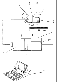
= Figuxs ] is an enlarged scale view of the device wherein the parts
comprising it are
inc~icated;
14 '- Figure i a shows the detail of the detection block;
CA 02258989 2008-02-18
2008/02/181 12:09 PM P. 018/022
4
Figure 2 shows the cletail of the coIIimator;
- Figcre 3 shows the scioti[lating crystal matrix;
- Figare 4'sbows the.shape of the cladding;
FiSme 5 shows the diagram of the photo multiplier and,its dimen.-dons;
- Figure 6 shows the multiplication mechanism of the electrons in the photo
multiplier (metal cha=eJ, dynodes);
Figure 7 shows the electronic'block diagraw required for the operation ofthe
gamma camera;
:. . - Figwtes, 8a and 8b show the operaling cliagam of the operational
amplifiea;
- Figure 8 shows a detail of ffie operating block diagram of the electronies
for-the
conversion of the pulses from the operational amplifiers;
Figure 9 shows details of the signal processi.ag-mechanism:;
15" Fi,gare 10 shows a blor,k etiagram of.the eleettxmics and of the output
signals
towards a personal computer.. -
With refeumoe to the figures, the new gamma eamerais shown,'eornprising:
.a collimator I made. of Z.ead of bigh Z metal (such as W, Au, etc.) able to
let through
onlythe gamma. xadiations acoording to the solic7 angle crossing through
itsholes. In a
practieal embodiment said co]limator presents size equal to -a pazaJielepXped
wi4t side
of 32 mpn and]eight of 30 Tnm or greatw, ..
- a scintiXla#iag cx~stal 2 mac~e ofNal(Tl) (Thallium.-dopec~ sodium iodide)
sensitive to
gmma radiafiions havmg energy raaiging from a few keV to 1 NieV; with total
size
equal to a square with side equaX to 22 ;mm or greater.
25-
CA 02258989 2008-02-18
2008/02/181 12:09 PM P. 019/022
WO 98/48300 ' 1'CTY.IT98/00496
- a cladding 3 constituted by a coating of inert material able to be
sterilised for the part to be introducecl in the patient, constituted by a
patallelepiped with side of35 mm and length ranging from 50 to 80 mm
or more;
5 - a pi~oto multipl ier 4, able to .coilect the optical signal=produced by
.the
scintillation crysUl and amplified into an electrical signal=, Said photo
multiplier is of a compact type camprising eleven thin metal channel
.dynodes encapsulated in a container having .total height of about 3 0 mm,
as shown in p'igure 5, and able to be position-sensitive with a multi-
anode charge collection system. Subsequently, the eight sigrtals exiting
the pboto multiplier ate sent on eight pre-amiplifiers 5. A simplified
eloctronics 4et,6 is nsed to obtaan the sum of the pulses.ejatuag the
pro-mr~]ifiers.
Figures 8aand 8b show a system of eight pre~-amplifiers 5 co=mprising
y 5 'four wire anodes for determining;the posi#ion on the X axis and four
wire=anodes for the Y=position. The electrortic'sy.stern for reading .the
charge colleeted on the_altoiles is accoxnp7ished by noeai3s of eight
independent .pre-amplifiers 5. Subsequently, the pulses are sent to a
block of operational amplifiers 8 wbich perform hardware operations on
the input signals.
Itt regard to the signal processing meclsanism, with reference to figure
9, -from the operational block 8 exit three si grials whieh subsequently
enter three analogue-to-digital coriverters. In detail, the con'vertzr 9.
xepresents the value of the energy of the interacting photon; the
converter 10 represerns the centroid for the X co-Qrdixzate of the position
of the photon and=the converter 1] represents the cemoid of the Y co-
CA 02258989 2008-02-18
2008/02/181 12:10 PM P. 020/022
WO 98148300 PCTlLT98/00096
. , 6
ordinate of the photon.
With reference= to Figure 10, the output, of the signals from the three
converters - is sent on a data acquisition control board 12 and se,ut to a
personal computer. The sigttals related to eaeh co-ordimate of t-he axes
X and Y respeotively are connected to an analogue operationa] device
which.allows simultaneously to perffirrtx suznming operations of the four
- 4-~,
signals for X and of tbe four sigzaals for Y. For.the determination of the X
co-ordinate and..for the Y'co-ordinate; the centroid is computed
respeetively, through only two converters. This hardware computation
. solution for the bhatge disaribution centroid.allows to minimise the data
to be digitised and transmitted to `khe computer. The .aracial point for
data management is the twsfer-rate to -tbe computer which for cost
reduction ressons shall take place using low-cost, standard computers,
-operating systems, and inter.faces. Moreover, dluitig acquisition the
75 computor sball'be able to present the irnage im "near" real =tirne. In
addition. -to having the capability of determining the position of the
incident photon, it shall also be possible to determine its energy -by '
summing the signal exiting the converter 9 which corctains the
. . ~.~
information of the charge-releasect to the {soirrlillation signal. In this way
it will be. possible to. elimioate .a.il those -events caused by- radiation
sGaderitzg which are summed on the final image of the exam lierformed.
With an appropriate er4ergy window, it will be possil~le to correct the
image complete wi,th the "baelCground", reducing the noise caused by
single or multiple interactions in the body tissue. In this way, the energy
'25 window shall di,scrizstiuate only those photons of a given enerU
characteristic of the tracer used. The correction, software skWl enable -
. ,~
CA 02258989 1998-12-23
WO 98/48300 PCT/IT98/00096
7
real-time displaying of the acquisition of information sent by an
appropriate board which is directly connected to the signal converters.
The whole gamma camera is coated, with regard to cladding 3, with inert
and sterilisable material, as described and, for the remaining part which
stays outside the patient, with a parallelepiped with 35 mm side and
length variable between 40 and 80 mm or more.
A suitable presentation software is able to display the information as
images of reception of the tracers injected into the patient, with the same
representation typical of large-area gamma cameras.
By moving the gamma camera in proximity to the regions of interest in
the body of the patient, who has been injected with a radio
pharmaceutical product capable of attaching preferably on tumour cells
and able to emit gamma radiations of an energy ranging from a few keV
to 1 MeV, the surgeon will be able to localise the tumour identifying the
area of maximum signal (emission of gamma radiation) with spatial
resolution of a few millimetres.
This allows the surgeon to intervene with extreme certainty and
precision, only in the specific area involved with the tumour, thereby
reducing surgical damage and risk for the patient.
The high sensitivity of the gamma camera, moreover, allows to use radio
pharmaceuticals at different energies and it enables to mark specific
antibodies for given tumours with different radio isotopes, commonly
used in nuclear medicine.
In possible variations of the invention, the miniaturised gamma camera
can present, as scintillation crystal, a CsI(Na) crystal matrix, where
individual crystals have section of about 1 mm x 1 mm and in any case
CA 02258989 1998-12-23
WO 98/48300 PCT/IT98/00096
8
ranging between 0.5 mm x 0.5 mm and 3 mm x 3 mm and where
individual crystals are optically separated from each other, and the
separation zone between crystal and crystal has thickness of about 0.1
mm and in any case ranging from 3 microns to 0.5 mm. Moreover,
crystals of NaI(T1), Csi(TI), SGO, LSO, YAP:Ce, etc., can also be used
as scintillating crystals.
In an additional variation, the photo multiplier can be replaced with an
analogous one having a greater number of dynodes and a higher number
of charge collection anode wires. As a consequence, the electronics are
also modifiable by the same principle described above, in proportion to
the number of outputs of the photo multiplier.
The dimensions of the photo multiplier used can also be varied, reaching
larger dimensions but always such that they can be considered
miniaturised with respect to a traditional gamma camera and such as to
be usable for the proposed purposes. The principle of the invention is to
obtain a device that makes use of a single photo multiplier for the
computation of the position of the photons emitted, contrary to gamma
cameras of large area which make use of multiple photo multipliers to
reach the same purpose.
Total dimensions may change, always remaining extremely reduced and
such as to allow the surgeon to move the instrument by hand in a simple
and precise manner.
Obviously, moreover, the construction details and the embodiments may
be varied widely with respect to what has been described and shown
purely by way of example, without thereby departing from the scope of
the present invention.
