Note: Descriptions are shown in the official language in which they were submitted.
CA 02259437 2005-07-15
75304-27
1
MULTIPLE MECHANICAL MICROPORATION OF SKIN OR MUCOSA
BACKGROUND OF THE INVENTION
This invention relates to a device and method for
puncturing a selected layer or layers of the skin or mucosa.
More particularly, the invention relates a device and method
for puncturing the stratum corneum or mucosa to diminish the
barrier function thereof and permit a drug to be delivered
to the body or an analyte in the body to be withdrawn for
monitoring. This puncturing of the stratum corneum or
mucosa is minimally invasive, and can be combined with
various other methods, such as use of chemical enhancers,
pressure gradients, sonic gradients, temperature gradients,
and the like for selectively enhancing the inward flux of a
drug to the body or the outward flux of an analyte from the
body.
The stratum corneum is chiefly responsible for the
well-known barrier properties of skin. Thus, it is in this
layer of the skin that presents the greatest barrier to
transdermal flux of drugs or other molecules into the body
and of analytes out of the body. Mucosal tissue also
presents a barrier to flux of molecules into and out of the
body. The stratum corneum, the outer horny layer of the
skin, is a complex structure of compact keratinized cell
remnants separated by lipid domains. Compared to the oral
or gastric mucosa, the stratum corneum is much less
permeable to molecules either external or internal to the
body. The stratum corneum is formed from keratinocytes,
which comprise the
CA 02259437 1998-12-30
WO 98/00193 PCT/US97I11670
-2-
majority of the epidermal cells, that lose their nuclei
and become corneocytes. These dead cells comprise the
stratum corneum, which has a thickness of about 10-30 ~.cm
and, as noted above, is a very resistant waterproof
membrane that protects the body from invasion by
exterior substances and the outward migration of fluids
and dissolved molecules. The stratum corneum is
continuously renewed by shedding of corneum cells during
desquamination and the formation of new corneum cells by
the keratinization process.
Various methods of enhancing the permeability of
the stratum corneum and mucosa have been described. For
example, U.S. Patent No. 5,458,140 and U.S. Patent No.
5,445,611 disclose using ultrasonic energy that is
modulated in intensity, phase, or frequency or a
combination thereof. U.S. Patent No. 4,775,361
discloses a method of administering a drug by ablating
the stratum corneum using pulsed laser light without
significantly damaging the underlying epidermis.
Numerous patents teach the use of chemical enhancers for
improving transdermal flux of a drug through the skin.
E.g, U.S. Patent No. 4,863,970. It would be
advantageous to develop additional methods of permeating
the stratum corneum or mucosa to enhance the transport
of drugs into the body or analytes out of the body,
particularly without the need for expensive or
complicated equipment.
In view of the foregoing, it will be appreciated
that providing a device and method of use thereof for
introducing multiple micropores or perforations in the
stratum corneum or mucosa for enhancing transport of
molecules therethrough would be a significant
advancement in the art.
CA 02259437 2005-07-15
75304-27
3
BRIEF SUMMARY OF THE INVENTION
The present invention provides a simple,
inexpensive device for puncturing the stratum corneum or
mucosa without significantly damaging the underlying tissues
to facilitate transport of molecules therethrough.
The invention also provides a method of enhancing
the passage of molecules through the stratum corneum or
mucosa.
The invention also provides a method for
transdermally or transmucosally delivering a drug.
The invention also provides a method for
transdermally or transmucosally monitoring an analyte.
These and other aspects can be achieved by
providing a device for reducing the barrier properties of
skin or mucosa to the delivery of a substance into the body
or the withdrawal of an analyte from the body comprising:
(a) a base having a lower side and an upper side;
(b) a plurality of puncturing members extending from the
lower side of the base, the puncturing members configured
for puncturing the skin or mucosa to a depth sufficient to
reduce the barrier properties thereof without significantly
damaging underlying tissues;
(c) a plurality of holes extending from the lower side of
the base to the upper side of the base, the holes configured
for permitting a liquid to move therethrough by capillary
action; and
(d) a network of channels configured in the upper side of
the base to interconnect the holes.
CA 02259437 2005-07-15
75304-27
3a
Preferably, the device is fabricated by
microlithography and is composed of a material selected from
the group consisting of silicon, metal, and
CA 02259437 1998-12-30
WO 98/00193 PCT/CTS97/11670
-4-
plastic. It is also preferred that the puncturing
member be in the shape of a pyramid or wedge. The
pyramid or wedge preferably have sharp edges having
corner radii of less than 1 E.cm. The puncturing member
is preferably configured for puncturing the skin or
mucosa to a depth of about 30-50 ,um, and a dimension at
a base thereof is preferably about 10-50 E.cm. The
puncturing members preferably occupy up to about 50% of
the surface area of the lower surface of the base.
The device preferably further comprises a mechanism
for producing vibrations, the vibrations for
facilitating efficient and non-traumatic penetration of
the puncturing members into the skin or mucosa. A
preferred vibration-producing mechanism comprises a
piezo-electric transducer. It is preferred that the
mechanism for producing vibrations produces vibrations
in the range of about 2000 Hz to about 100 MHz.
In another illustrative embodiment of the device,
an external reservoir for holding a liquid drug
composition to be delivered to the body is provided.
Still further, a mechanism for limiting the rate of drug
delivery is preferably included in the device, the
mechanism positioned between the external reservoir and
the puncturing members. Such rate-limiting mechanisms
can include selective permeability membranes and valve
mechanisms. In another preferred embodiment, the device
is disposable.
A method for reducing the barrier function of skin
or mucosa to the delivery of substances into a body or
withdrawal of analytes out of the body, comprises:
(a) providing a device comprising:
a base having a lower side and an upper sides
a plurality of puncturing members extending
from the lower side of the base, the puncturing
members configured for puncturing the skin or
CA 02259437 1998-12-30
WO 98/00193 PCT/LTS97/11670
-5-
mucosa to a depth sufficient to reduce the barrier
properties thereof without significantly damaging
underlying tissues;
a plurality of holes extending from the lower
side of the base to the upper side of the base, the
holes configured for permitting a liquid to move
therethrough by capillary action; and
a network of channels configured in the upper
side of the base to interconnect the holes;
(b) contacting the device with the skin or mucosa
such that the plurality of puncturing members puncture
the skin or mucosa to a depth sufficient to reduce the
barrier properties thereof.
A method of transdermal or transmucosal monitoring
of a selected analyte in a body comprises:
(a) providing a device comprising:
a base having a lower side and an upper side;
a plurality of puncturing members extending
from the lower side of the base, the puncturing
members configured for puncturing said skin or
mucosa to a depth sufficient to reduce the barrier
properties thereof without significantly damaging
underlying tissues;
a plurality of holes extending from the lower
side of the base to the upper side of the base, the
holes configured for permitting a liquid to move
therethrough by capillary action; and
a network of channels configured in the upper
side of the base to interconnect the holes, the
network of channels including a reservoir;
(b) contacting the device with the skin or mucosa
such that the plurality of puncturing members puncture
the skin or mucosa to a depth sufficient to reduce the
barrier properties thereof resulting in seepage of
interstitial fluid to the surface of the skin or mucosa
CA 02259437 2005-07-15
75304-27
6
such that interstitial fluid moves by capillary action
through the holes, through the channels, to the reservoir;
(c) collecting the interstitial fluid from the reservoir;
and
(d) analyzing the interstitial fluid with respect to the
selected analyte.
In a preferred embodiment, the method further
comprises applying suction to increase the rate of
collection of interstitial fluid. Ultrasonic vibrations can
also be applied to the skin or mucosa to increase the rate
of collection of the selected analyte. The ultrasonic
vibrations can be modulated in frequency, intensity, phase,
or a combination thereof, as disclosed in U.S. Patent
No. 5,458,140. The ultrasonic vibrations are preferably in
the range of about 2000 Hz to about 100 MHz. The ultrasonic
vibrations can also enhance the movement of interstitial
fluid by capillary action. In a preferred embodiment of the
invention, the selected analyte is glucose. It is also
preferred to apply an anticoagulant to inhibit obstruction
of the holes or channels.
A method of transdermally or transmucosally
delivering a drug in liquid form to a body comprises:
(a) providing a device comprising:
a base having a lower side and an upper side;
a plurality of puncturing members extending from the lower
side of the base, the puncturing members configured for
puncturing the skin or mucosa to a depth sufficient to
reduce the barrier properties thereof without significantly
damaging underlying tissues;
CA 02259437 2005-07-15
75304-27
7
a plurality of holes extending from the lower side
of the base to the upper side of the base, the holes
configured for permitting a liquid to move therethrough by
capillary action, and
a network of channels configured in the upper side
of the base to interconnect the holes, the network of
channels including a reservoir;
(b) contacting the device with the skin or mucosa such that
the plurality of puncturing members puncture the skin or
mucosa to a depth sufficient to reduce the barrier
properties thereof;
(c) supplying the drug to the reservoir such that said drug
moves from the reservoir, through the channels and holes to
the site of the punctures of the skin or mucosa and thus
into the body.
In a preferred embodiment, pressure is applied to
increase the rate of delivery of the drug to the body.
Applying ultrasonic vibrations to the skin or mucosa also
increases the rate of delivery of the drug to the body. The
ultrasonic vibrations can be modulated in frequency,
intensity, phase, or a combination thereof, as disclosed in
U.S. Patent No. 5,445,611. The ultrasonic vibrations are
preferably in the range of about 2000 Hz to about 100 MHz.
The drug in liquid form can further comprise an anti-
irritant, antiseptic, or analgesic to reduce trauma to the
body due to the application of the device.
BRIEF DESCRIPTION OF THE SEVERAL VIEWS OF THE DRAWINGS
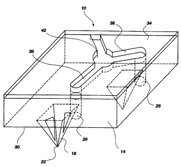
Fig. 1 shows a perspective view of an illustrative
embodiment of the present invention.
CA 02259437 2005-07-15
75304-27
7a
Fig. 2 shows a cross section of a portion of
another illustrative embodiment according to the present
invention.
Fig. 3 shows a perspective view of a portion of
the embodiment of Fig. 2.
CA 02259437 1998-12-30
WO 98/00193 PCT/US97/11670
_g_
FIG.4 shows a top view of a portion of the
embodiment of FIG. 2.
FIG. 5 shows a schematic diagram of a device for
making multiple microporations in skin or mucosa and
collecting interstitial fluid.
FIG. 6 shows a schematic sectional diagram of a
device for making multiple microporations in skin or
mucosa and delivering a drug.
DETAILED DESCRIPTION
Before the present device and method for enhancing
permeability of skin or mucosa for drug delivery or
analyte monitoring are disclosed and described, it is to
be understood that this invention is not limited to the
particular configurations, process steps, and materials
disclosed herein as such configurations, process steps,
and materials may vary somewhat. It is also to be
understood that the terminology employed herein is used
for the purpose of describing particular embodiments
only and is not intended to be limiting since the scope
of the present invention will be limited only by the
appended claims and equivalents thereof.
It must be noted that, as used in this
specification and the appended claims, the singular
forms "a," "an," and "the" include plural referents
unless the context clearly dictates otherwise. Thus,
for example, reference to a device containing "a
puncturing member" includes a device containing two or
more of such members, reference to "a channel" includes
reference to one or more of such channels, and reference
to "an ultrasound transducer" includes reference to two
or more ultrasound transducers.
It has been observed that forming a hole or
micropore, 30 /.~.m across, in the stratum corneum yields
a quick source of about 0.2 microliters of interstitial
CA 02259437 1998-12-30
WO 98/00193 PCT/US97/11670
_g_
fluid seeping through the hole from the underlying
tissue without any additional pumping. Merely
increasing the number of holes introduced through the
stratum corneum would increases the amount of passively
available fluid in a linear fashion. That is, creating
100 holes should produce about 20 microliters of
interstitial fluid. From a practical perspective, using
known approaches to create 100 holes in a controlled
pattern would be challenging and time-consuming.
However, using the mechanical puncturing capabilities of
a mechanical microporation or "bed-of-nails" device
would allow an almost unlimited number of micropores to
be quickly created in any selected pattern. Similarly,
using conventional lancet and needle technologies would
make the needed depth control of the puncture very
tricky and, if the device were to create hundreds of
these holes, the mechanical challenge of building the
device using conventional metal needle technologies
would be formidable. However, by fabricating puncturing
elements en masse such that they protrude from a
substantially planar surface, with sufficient spacing
between each to allow the stratum corneum to come in
contact with this intervening planar surface, the
absolute length of the puncturing elements themselves
would act as an accurate limit for the depth of the
micropore. Also, using a microlithography approach to
fabricate these structures will allow an entire surface
comprised of puncturing elements and the interconnecting
fluid management system to be built very cost
effectively.
One illustrative method would be to utilize the
existing base of manufacturing capabilities developed in
the semiconductor and micro-mechanical industries to
dry-etch an entire 4 inch silicon wafer with a network
of these devices. This master could then be used as the
CA 02259437 2005-07-15
75304-27
basis for an electroplated mold from which thousands of
copies could be produced. For a typical useable surface
area/per device application of 4mm X 4mm, one 4-inch wafer
would yield more than 500 of the devices.
5 A device according to the present invention is
made, for example, by first preparing a master by a dry etch
process on a silicon wafer, as is well known in the art.
Photolithographical processes for etching micrometer-scale
structures into silicon wafers and the like are described in
10 A.T. Wooley & R.A. Mathies, Ultra-high-speed DNA fragment
separations using microfabricated capillary array
electrophoresis chips, 91 Biophysics 11348-52 (1994);
C.S. Effenhauser et al., High-speed separation of antisense
oligonucleotides on a michromachined capillary
electrophoresis device, 66 Anal. Chem. 2949 (1994);
C. Effenhauser et al., 65 Anal. Chem. 2637 (1993); Z.H. Fan
& D.J. Harrison, Michromachining of capillary
electrophoresis injectors and separators on glass chips and
evaluation of flow at capillary intersections, 66 Anal.
Chem. 177-84 (1994); W.H. Ko et al., in Sensors: A
Comprehensive Survey, T. Grandke, W.H. Ko, eds., VCH Press:
Weinheim, Germany, Vol. l, pp. 107-68 (1989); K.E. Petersen,
70 Proc. IEEE 420-57 (1982). The master silicon wafer is
then used to make an electroplated mold, and then the mold
is used to make copies of the device, all by processes well
known in the art.
Also, by coupling the entire device to an
ultrasonic transducer, several known advantages can be
realized simultaneously. For example, ultrasound has been
shown to enhance the smooth cutting ability of scalpels and
other surgical devices and can be expected to facilitate the
CA 02259437 2005-07-15
75304-27
10a
easy, painless penetration of the puncturing elements into
the stratum corneum with very
CA 02259437 1998-12-30
WO 98/00193 PCT/L1S97/11670
-11-
little pressure. The edges of the pyramidally shaped
puncturing elements shown in Figure 1 can easily be
fabricated such that the corner radius is less than 10
nanometers, a sharpness similar to a surgical scalpel.
Second, ultrasound has also been shown to greatly
enhance capillary action, thus the amount of fluid that
could be collected in a device containing a capillary
collection system could be expected to be significantly
greater than that provided by mere passive means.
Third, by using the entire body of the puncturing
elements to provide a conduit for the ultrasonic energy,
a simple method is presented wherein the sonic energy is
placed within the body where it can provide a positive
pressure, and streaming action on the interstitial fluid
from within the body outward towards a collection system
of capillary channels coupling all fluid harvested into
a central reservoir.
FIG. 1 shows a perspective view of an illustrative
device according to the present invention. The device
10 comprises a base 14 with a plurality of puncturing
members 18 extending therefrom. In a preferred
embodiment, the base is substantially planar. Each
puncturing member comprises a sharp point 22 or edge for
puncturing the stratum corneum or mucosa. Since the
stratum corneum can be up to about 30 /.cm thick, it is
preferred that the puncturing element have a height of
about 40-50 /.cm to ensure that the stratum corneum will
be fully breached without significantly damaging the
underlying tissue. A pyramid or wedge shape is a
preferred shape for the puncturing member because of the
ease with which such a shape can be formed by
microfabrication techniques such as microlithography.
In an illustrative puncturing element having a pyramid
shape, the base of the pyramid would preferably have a
square base about 30-40 ~cm on a side.
CA 02259437 1998-12-30
WO 98/00193 PCT/US97/11670
-12-
It is also preferred that the base have a plurality
of holes 26 extending therethrough from the lower side
30, on which the puncturing element are disposed, to the
upper side 34. Preferably, each puncturing element is
adjacent to and paired with at least one hole for
collecting the interstitial fluid that seeps out of the
puncture in the stratum corneum. These holes should be
dimensioned to permit the interstitial fluid to move by
capillary action from the lower side of the device to
the upper side, where the interstitial fluid can be
collected. It is also preferred to interconnect the
holes with capillary channels 38 that are formed in the
upper side of the device. Preferably, such channels
intersect at a reservoir 42. The interstitial fluid
moves by capillarity from the micropore into the hole,
through the channels, and to the reservoir, where the
interstitial fluid is collected, such as with a
micropipet. Additional fluid can be collected by
applying suction to the microporated area of skin or
mucosa.
FIGS. 2-4 show another illustrative embodiment of
the invention. FIG. 2 shows a cross section of a
portion of the device 50 comprising a base 54 with a
puncturing member 58 extending therefrom. The
puncturing member is pyramid-shaped, as in FIG. 1. The
upper side 62 of the base is configured with a V-shaped
channel 66 positioned such that the channel is directly
over the puncturing member and cuts into the volume
circumscribed by the puncturing member. FIG. 3 shows a
perspective view of the device having the V-shaped
channels 66 and interconnecting shallower V-grooves 70.
The channels 66 cut through the lower side 74 of the
base, and thus form openings through which the
interstitial fluid can be taken up by capillary action.
FIG. 4 shows how the V-grooves 70 interconnect the V-
CA 02259437 1998-12-30
WO 98/00193 PCT/US97/11670
-13-
channels for collecting the interstitial fluid. All of
the puncturing members, channels, and grooves shown in
FIGS. 2,3, and 4 are designed to be wedge-shaped,
compatible with being produced in the crystalline
structure of a silicon substrate with a lithographic
'dry-etch' type of process.
FIG. 5 shows an illustrative device 80 for
collecting interstitial fluid according to the present
invention. The device 80 comprises a base 84 having a
plurality of puncturing members 88 extending therefrom.
V-shaped channels and grooves are configured into the
upper side 92 of the base for collecting the
interstitial fluid. A cover plate 96 fits over the base
to cover the network of channels and grooves and to
inhibit evaporation of the interstitial fluid. The
network of channels and grooves leads the interstitial
fluid to a central area, where there is disposed a
capillary tube 100 for receiving the interstitial fluid.
Atop the cover plate is disposed an ultrasonic
transducer 104 and a backing 108 for the tranducer.
The device is pressed against a selected area of
skin or mucosa, and the ultrasonic transducer is
activated to aid in both the puncturing of the tissue
and in enhancing the seepage of the interstitial fluid.
The interstitial fluid is collected by the network of
openings in the base, and is conducted by the network of
channels and grooves to the capillary, which takes up
the fluid by capillary action. The fluid is then
analyzed according to methods known in the art. An
illustrative analyte is glucose, which can be quantified
with various test strips that are available
commercially.
FIG. 6 shows an illustrative drug delivery device
120 comprising a base 124 having a plurality of
puncturing members 128 extending therefrom. A network
CA 02259437 1998-12-30
WO 98/00193 PCT/US97/11670
-14-
of grooves and channels (see FIGS. 2-4) is embedded in
the base for distributing a drug composition 132 from a
reservoir 136. The reservoir is bounded by a housing
138, the base, and a backing plate 144 including an 0-
ring 148. The drug composition flows through the
channels, grooves, and openings in the base to the
surface of the skin or mucosa for entry into the body
through the punctures or perforations. An ultrasound
transducer 140 lies over the drug composition for aiding
in delivery thereof. Above the transducer is the
backing plate 144 including the 0-ring for sealing the
drug in the reservoir. A spring 152 can advantageously
bias the backing plate against the transducer, which
causes the transducer to be kept in fluid contact with
the drug.
The ultrasonic system is utilized not only to
enhance the slicing action of the edges of the
puncturing elements as the penetrate into the stratum
corneum or mucosa, but is then utilized to enhance the
fluid flux of the therapeutic containing solution
through the micro-pores and into the underlying tissues.
In this case, large quantities of large molecular
weight drugs could be delivered transdermally with a
programmable control of the flux rate via variable
activation of the ultrasonic pumping system. In
addition, the sonic energy can be utilized to create
controlled resonant vibrations in specifically shaped
micro-structures such that a micro-pump is created to
facilitate driving the collected fluid from one point to
another within the entire structure. Moreover, chemical
enhancers, air pressure, and other methods known in the
art can be used to enhance the passage of the drug
through the micropores in the skin or mucosa into the
body.