Note: Descriptions are shown in the official language in which they were submitted.
CA 02259742 1999-O1-19
P-3 793 P 1 PATENT
TITLE OF THE INVENTION
METHOD FOR REDUCING INHIBITORS
OF NUCLEIC ACID HYBRIDIZATION
BACKGROUND OF THE INVENTION
The field of the present invention broadly relates to nucleic acid
hybridization. More
specifically, the present invention relates to the reduction of substances in
samples which
inhibit nucleic acid hybridization events. Such events include nucleic acid
probe hybridization
to determine the presence and/or amount of a target nucleic acid, and nucleic
acid primer
hybridization for the initiation of a nucleic acid amplification process.
Nucleic acid amplification processes such as strand displacement amplification
(SDA),
polymerase chain reaction (PCR), ligase chain reaction (LCR), nucleic acid
sequence based
amplification (NASBA), transcription mediated amplification (TMA) and others
are used to
create multiple copies of a particular nucleic acid sequences) of interest
(target sequence)
which is present in lesser copy number in a sample. However, a number of
substances
commonly found in such samples cause inhibition of nucleic acid amplification
processes,
because of inhibition of the hybridization of primers to initiate the
amplification process.
Similarly, such substances inhibit direct nucleic acid probe hybridization
reactions used for the
detection of unamplified target nucleic acids.
An example of such nucleic acid hybridization inhibitory substances are
porphyrin
compounds derived from heme and hematin which are both commonly found in blood
samples
and inhibit PCR. (PCR TechnoloQV, Stockton Press, Henry A. Erlich, Ed. pp 33-
34, 1989).
Protocols using osmotic lysis and pelleting of nucleic and cell debris have
been used to reduce
the amount of these inhibitors.
Salivary samples have also been reported to contain PCR inhibitory substances.
Ochert
et al., PCR Methods and Applications 3, 365-368 (1994). Although the
inhibitory substances
EXPRESS MAIL LABEL NO. ~M397668748
P-3 793 P 1
CA 02259742 1999-O1-19
were not identified, it was found that extended microwaving or boiling of the
salivary sample
totally removed PCR inhibition.
Frickhofen and Young, J. Virol. Methods 35, 65-72 ( 1991 ), report that
heating of
serum samples for 45 seconds at 70°C improves PCR amplification of
viral nucleic acid
sequences. This improvement is theorized to be due to heat inactivation of
serum enzymes
such as aprotinin, leupeptin PMSF and pepstatin which are believed to be
inhibitory to PCR
processes.
Another approach for removing PCR inhibitory substances from serum prior to
amplification of a viral nucleic acid sequence is taught by Zeldis et al., J.
Clin. Invest. 84, 1503
1508 (1989). This approach involves adsorbing the virus to antibody coated
microparticles,
washing the microparticles, and then destroying the remaining proteins which
may be inhibitory
to PCR with proteinase K.
In attempting to detect Treponema pallidum in amniotic fluid, fetal and
neonatal sera
and cerebrospinal fluid by PCR, four different processes were attempted to
remove PCR
inhibitory compounds. Grimprel et al., J. Clin. Microbiol. 29, 1711-1718
(1991). Briefly, the
four processes for removal of PCR inhibitory compounds were: (1) a boiling
method wherein
sample in a tube was placed in a boiling water bath for 10 minutes, cooled on
ice, and then
centrifuged; (2) a low-spin separation method wherein sample was added to
sterile phosphate
buffered saline and subjected to a series of centrifugations, then the pellet
was resuspended and
boiled for 10 minutes, after which it was cooled on ice; (3) an alkaline lysis
extraction method
wherein sample was boiled for 1.5 minutes in 1 M NaCI, 1 N NaOH and 0.1% SDS,
then
neutralized with 0.5 M Tris-HCl (pH 8.0), and then subjected to a series of
extractions with
phenol and chloroform-isoamyl alcohol, and precipitated with isopropyl
alcohol; and (4) a spin
extraction method wherein sample was subjected to low-spin separation as
described in (2)
above, followed by 10 minutes of boiling and one phenol-chloroform extraction
before
precipitation in cold absolute ethanol. The authors reported varying success
of these methods
dependent on the type of samples used.
2
CA 02259742 1999-O1-19
' P-3793P1
With stool samples, polyethylene glycol precipitation was found to remove a
significant
amount of small particles and soluble substances which could be inhibitory to
a reverse
transcriptase-PCR process. Jiang et al., J. Clin. Microbiol. 30, 2529-2534
(1992). Following
the precipitation, an extraction process was performed using the cationic
detergent,
S cetyltrimethylammonium bromide (CTAB) in a high salt concentration in
conjunction with
phenol-chloroform extraction.
A different approach to removal of PCR inhibitory substances from stool
samples is
reported by Wilde et al., J. Clin. Microbiol. 28, 1300-1307 (1990). Before
using PCR to
detect rotavirus nucleic acid from stool samples, the extraction process was
modified with an
added step that utilized chromatographic cellulose fiber powder (CF 11 powder)
to purify the
rotavirus RNA during a series of rapid washing and elution steps.
When performing a study to detect cytomegalovirus (CMV) in urine using PCR, it
was
found that urea is inhibitory to PCR. Khan et al., J. Clin. Pathol. 44, 360-
365 (1991). This
reference reports that the PCR inhibitory effects of urea in urine are
effectively removed by
simple dialysis or ultracentrifizgation.
Another process to remove PCR inhibitory substances from urine before
detection of
CMV nucleic acid is reported by Buffone et al., Clin. Chem. 37, 1945-1949
(1991). This
process occurs subsequent to release of the nucleic acid from the CMV
organisms and uses
fine glass beads to adsorb nucleic acid such that protein and other substances
can be selectively
eluted before recovery of the nucleic acid for amplification.
As evidenced by the references described above, most of the publication
regarding
nucleic acid amplification inhibition has related to PCR. However, these same
substances
which are inhibitory to PCR, as well as a number of other substances commonly
found in
clinical samples such as proteinaceous substances, EDTA, human DNA and iron
have been
found to be inhibitory to SDA, and other nucleic acid amplification processes
as well.
Also, most of these methods to reduce or remove nucleic acid hybridization
inhibiting
substances involve rather time-consuming complicated steps which must be added
to the
3
P-3 793 P 1
CA 02259742 1999-O1-19
sample processing methodology. Another problem with methods which utilize
relatively
severe processing steps or conditions, and/or require separation of target
nucleic acid from
other substances is the loss of some target nucleic acid sequence. Despite the
ability of nucleic
acid amplification processes to make multiple copies of target sequence
(amplicons) from very
few original targets, amplification efficiency and detection ability are
improved if there are
greater numbers of original targets in the sample. The greater detection
ability can be very
important when processing particularly difficult to detect samples such as
acid fast Bacillus
(AFB) smear negative Mycobacterium tuberculosis samples.
SUMMARY OF THE INVENTION
In order to address the problems associated with the presence of substances
inhibitory
to nucleic acid hybridization in samples and thus, achieve the benefits of
more efficient
amplification and improved detection of target nucleic acid sequences, the
present invention
provides a method for reducing the amount of such substances in samples by,
prior to lysis of
cells in the sample which contain nucleic acid to be amplified, contacting the
sample with an
agent which does not effectuate the release of nucleic acid from the cells,
and then separating
the cells from the agent.
Examples of some useful agents for use in the present invention include
chaotropes
such as guanidine thiocyanate, sodium perchlorate and sodium thiocyanate.
Also, the
separation of cells from the agent is generally accomplished by a wash and
centrifugation step
with a solution in which the agent is soluble.
BRIEF DESCRIPTION OF THE DRAWINGS
The various objects, advantages and novel features of the invention will be
more
readily appreciated from the following detailed description when read in
conjunction with the
appended figures, in which:
4
P-3 793 P I
CA 02259742 1999-O1-19
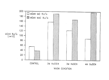
FIG. 1 is a graphical representation of the results of an experiment conducted
to
determine whether the method of the present invention reduces the amount of
nucleic acid
amplification inhibition from a clinical sample as compared to a control
standard sample
processing method.
FIG. 2 is a graphical representation of the results of an experiment conducted
to
determine optimal pH and concentration values for a particular agent used in
the method of the
present invention.
FIG. 3 is a graphical representation of the results of an experiment showing
the
effectiveness of the method of the present invention in reducing the amount of
nucleic acid
hybridization inhibitors from clinical samples.
DETAILED DESCRIPTION OF THE INVENTION
As stated above, the present invention relates to a method for reducing the
amount of
substances which are inhibitory to nucleic acid hybridization processes from
samples containing
cells with nucleic acid which will be subjected to a hybridization process. In
the method, an
agent which solubilizes such substances and does not effectuate the release of
nucleic acid from
cells is contacted with a sample prior to lysis of cells in the sample such
that cells containing
nucleic acid will remain in the sample. Then, such cells are separated from
the agent.
The results of this method were particularly unexpected because of the
complexity of
some of the processes tried by others to remove inhibitory substances as
evidenced by the
descriptions in the Background section above. Also, the agents used in the
method of the
present invention were generally believed by those skilled in the art to be
useful for effectuating
the release of nucleic acid ~rom cells by cell wall lysis or solubilization,
as evidenced by various
references such as U.S. Patent No. 5,482,834 wherein chaotropic salts are
taught to be useful
specifically for the solubilization or lysis of cells. Generally, such agents
were used in
combination with heat to lyse or solubilize cells.
S
CA 02259742 1999-O1-19
P-3 793 P 1
The cell wall lysogenic properties of chaotropes such as guanidinium
thiocyanate
(GuSCN) have also been reported in references such as Hubbard et al.,
Experimental &
Applied Acarolog_y 19, 473-478 (1995), Boom et al., J. Clin. Microbiol. 28 495-
503 (1990),
Reek et al., BioTechniques 19, 282-285 (1995), Chungue et al., J. Med. Virol.
40, 142-145
(1993) and Shah et al., J. Clin. Microbiol. 33, 322-328 (1995). Similarly,
chaotropes and
detergents such as GuSCN have been extensively utilized in the extraction of
nucleic acid from
cells as taught in references such as Delacourt et al., J. Pediatrics 126, 703-
710 (1995), Lee
and Choi, J. Microbiol. & Biotechnol. 5, 181-187 (1995), Cano et al., J. Food
Protection 58,
614-620 (1995), Bartolome et al., J. Hepatol. 17, s90-s93 (1993), Rajagopaian
et al., Lett.
Applied Microbiol. 21, 14-17 (1995) and Shieh et al., J. Virol. Methods 54, 51-
66 (1995).
Thus, the quick and simple method of the present invention wherein cells in a
sample are
contacted (washed) with such an agent prior to cell lysis to remove nucleic
acid hybridization
inhibitory substances was unexpected in view of other processes being used in
the art.
Also, one of the advantages of the method of the present invention is the
ability to
increase the initial yield of target nucleic acid from the cells in a sample.
Although nucleic acid
amplification processes are capable of creating many copies of a target
sequence (amplicons)
from very few initial targets, it is beneficial to start the amplification
process with as many
initial targets as possible. Other processes for removing nucleic acid
hybridization inhibitory
substances subsequent to lysis of the cells are notoriously inefficient,
because they are based on
separation of nucleic acid from other substances in the lysate, and thus, many
initial targets are
not recovered. In the present method, where the inhibitory substances are
removed prior to
cell lysis, such subsequent separation is not necessary, and better yields of
initial target are
achieved.
.. The samples which may be subjected to the method of the present invention
include
virtually all human and veterinary clinical samples such as sputum samples,
blood samples,
urine samples, cerebrospinal fluid ("CSF") samples and others, environmental
samples such as
water, air and soil samples, and food samples. The samples which may be
subjected to the
6
CA 02259742 1999-O1-19
P-3 793 P 1
method of the present invention are suspected of containing cells with a
target nucleic acid
sequence to be subjected to a hybridization process such as direct probe
hybridization or
primer hybridization for initiation of an amplification process.
The types of cells present in the samples subjected to the method of the
present
invention include the cells of virtually all organisms. The method of the
present invention is
particularly useful with samples suspected of containing cells of infectious
organisms. As
shown in the Examples below, the method of the present invention was effective
with the cells
of infectious organisms such as Mycobacterium tuberculosis, Bacillus
stearothermophilus,
Group B streptococcus, Group A streptococcus, E. coli, Candida albicans,
Staphylococcus
epidermidis, Neiserria gonorrhoeae, Chlamydia trachomatis and Enterococcus
faecalis.
Because the primary criterium for determining which types of cells can be
effectively treated
using the method of the present invention is whether the cells are lysed by
contact with the
agent, a routine screening assay can be used by one of ordinary skill in the
art with a
reasonable expectation of success to identify such types of cells.
1 S More specifically, a sample containing the cells of interest is exposed to
the agent of
the method. Subsequently, the cells of the sample are washed to remove any
nucleic acid
which may be present from cells lysed by the agent. Then, the cells are
resuspended and lysed
by application of heat and agitation with particles (beads). Finally, a dye
specific for nucleic
acid is added to the sample, and the amount of dyed nucleic acid from the
treated sample is
compared to a control which was subjected to the same conditions except for
contact with the
agent.
If the amount of nucleic acid from the treated sample is substantially
equivalent to the
amount of nucleic acid from the control, then samples of those cells can be
effectively treated
using the method of the present invention. That is, contact with the agent of
the present
invention did not effectuate lysis of the cells.
Substances which are inhibitory to nucleic acid hybridization processes and
typically
found in such samples include proteinaceous materials and human DNA in human
samples and
7
CA 02259742 2002-09-25
P-3 793 P 1
animal DNA in veterinary samples. As discussed in the Background section
above, these
substances are known to be inhibitory of nucleic acid amplification processes
such as SDA,
PCR, LCR, NASBA, TMA and others.
The first step of the method of the present invention is to contact the sample
with an
agent in which the inhibitory substance is soluble and which will not
effectuate the release of
nucleic acid from cells. This contacting may occur at any time prior to the
lysis of cells to
release target nucleic acid. However, a preferred time for contacting the
cells such an agent is
after some manipulation of the sample to concentrate the location of the cells
or pellet the
cells. Typically, such concentration is a result of centrifugation, but may
also iesult from
IO filtration or selective adsorption.
Such concentration of the cells provides a greater assurance that the agent
will contact
the cells, and permits more efficient use of the agent due to a defined
location of cells. The
contacting of cells with the agent is preferably a relatively brief wash of
the cells. Typically,
the contacting of cells and agent is for up to about five minutes.
I5 Many agents are useful in the method of the present invention. Examples of
such
useful agents include chaotropes and detergents which are well known to those
skilled in the
art such as Triton X-100, Triton X-114, NP-40, Brij 35, Brij*58, Tween 20,
Tweeri 80, octyl
glucoside, octyl thioglucoside, Chaps, sodium iodide, sodium perchlorate,
potassium iodide,
sodium thiocyanate, potassium thiocyanate, guanidine thiocyanate, guanidine
isothiocyanate,
20 sodium trichloroacetate, sodium trifluoroacetate and urea. Other agents
useful in the method
of the present invention can be identified by one of ordinary skill in the art
with a reasonable
expectation of success by performing routine screening assays directed towards
the two
primary characteristics of such agents; degree of solubilization of nucleic
acid hybridization
inhibitory substances and lack of effectuation of release of nucleic acid from
cells.
25 Briefly, an agent is contacted with cells, the cells centrifuged, and
amplification reaction
for a target nucleic acid sequence common to the cells performed on the
supernatant in a
routine screening assay. If the target sequence is amplified, then the agent
does not meet the
* Trademark
8
CA 02259742 1999-O1-19
' P-3793P1
requirements for use in the method of the present invention, because it has
effectuated the
release of nucleic acid from the cells, whereas lack of amplification of the
target sequence and
amplification of an internal control sequence would indicate that the agent
may be useful in the
method of the present invention. Then, the agent which has not effectuated the
release of
nucleic acid from the cells is brought into contact with known nucleic acid
hybridization
inhibitory substances, and the degree of solubilization of the inhibitory
substance quickly
determined in a routine screening assay. Those agents which solubilize the
inhibitory substance
are useful in the method of the present invention.
The concentration and amount of the agent used in the method of the present
invention
is dependent on the type of sample being subjected to the method. Examples of
suitable
concentrations of chaotropes and detergents for use in the method of the
present invention are
presented below in Table 1.
TABLE 1
Chaotrope/Detergent Concentrations
Chaotrope/Detergent Concentrations
Triton X-100 0.024 mM, 0.24 mM, 2.4 mM
Triton X-114 0.021 mM, 0.21 mM, 2.1 mM
NP-40 0.029 mM, 0.29 mM, 2.9 mM
Brij 35 0.009 mM, 0.09 mM, 0.9 mM
Brij 58 0.0077 mM, 0.077 mM, 0.77 mM
Tween 20 0.006 mM, 0.06 mM, 0.6 mM
Tween 80 0.0012 mM, 0.012 mM, 0.12 mM
Octyl glucoside 2.4 mM, 24 mM
Octyl thioglucoside 0.9 mM, 9.0 mM
9
CA 02259742 1999-O1-19
P-3 793 P 1
Chaps 0.9 mM, 9.0 mM
NaI 2M, 3M, 4M, SM, 6M
NaC104 2M, 3M, 4M, SM, 6M
KI 2M, 3M, 4M, SM, 6M
S NaSCN 2M, 3M, 4M, SM, 6M
KSCN 2M, 3M, 4M, SM, 6M
Guanidine Isothiocynate 2M, 3M, 4M, SM, 6M
Sodium trichloroacetate 2M, 3M, 4M, SM, 6M
Sodium trifluoroacetate 2M, 3M, 4M, SM, 6M
Urea 2M, 3M, 4M, SM, 6M
Generally, the volume of the agent used in the method of the present invention
is at
least equal to the volume of sample. More particularly, the volume:volume
ratio of the agent
1 S to the sample is from about 1:1 to about S:1. Also, generally, it is
preferable that the agent be
prepared as a basic solution, with most preferable pHs for particular agents
being determined
by a routine screening assay based, for example, on the experiments presented
in Example 3
hereof, in which a 6.0 M guanidine isothiiocyanate solution at pH 9.0 was
found to be
particularly beneficial for reducing the amount of nucleic acid hybridization
inhibitory
substances. The optimal concentration (molarity) of a particular agent may
also be determined
using the same type of routine screening assay based on the experiments
presented in Example
3 hereof. Also, generally, the agent is brought into contact with the sample
at room
temperature.
When the agent is brought into contact with cells of the sample, nucleic acid
2S hybridization inhibitory substances are solubilized by the agent, thus
washing such substances
from the walls of the cells. Because suitable agents do not effectuate release
of nucleic acid
CA 02259742 1999-O1-19
P-3 793 P 1
from the cells, loss of target nucleic acid when the agent is separated from
the cells is not a
concern.
However, the agents used in the method of the present invention may also
adversely
effect nucleic acid hybridization processes, and thus, such agents are
subsequently separated
from the cells. Such separation may be accomplished by any suitable means such
as filtration
or wash and centrifugation with or without a buffer in which the agent is
soluble. Preferably,
such a buffer is used in order to assure removal of the inhibitory substances
as well as the agent
prior to cell lysis. Thus, when such cells are lysed, the target nucleic acid
is presented in an
environment with minimal amounts of substances which are inhibitory to the
hybridization
process to which the target nucleic acid will be subjected.
A variety of processes are currently used to prepare target nucleic acids in
samples for
hybridization or amplification. For example, sputum samples which are
processed to amplify
mycobacterial nucleic acid sequences are typically subjected to a NALC/NaOH
process. The
method of the present invention may be particularly useful with mycobacterial
samples
subjected to such a NALC/NaOH process due to its selective solubilization of
NALC/NaOH
pellets to reduce clumping of such samples. Similarly, other types of clinical
samples are
subjected to other well known standard processes, for example, centrifugation
for large volume
samples such as blood and urine. The method of the present invention may be
used before, as
part of, or after those standard processes, provided that the method is
practiced prior to lysis
of cells containing the target nucleic acid.
The method of the present invention does not require the quantitative binding
and
releasing of target nucleic acid from a binding surface, and thus permits the
use of more sample
than is conventionally utilized in nucleic acid based hybridization or
amplification assays. This
ability to use more sample confers greater sensitivity to such assays, as
there is more target
nucleic acid present at the initial stages of the assay. The selectivity of
the agents in
solubilizing inhibitory substances, but not concommitantly solubilizing target
nucleic acid or
11
i i
CA 02259742 2002-09-25
P-3793P1
cell walls is one of the characteristics of the agents which contributes to
the results of the
method of the present invention as evidenced by the Examples set forth below.
The following examples illustrate specific embodiments of the invention
described
herein. As would be apparent to skilled artisans, various changes and
modifications are
possible and are contemplated within the scope of the invention described.
EXAMPLE 1
Comparison of Method of the Present Invention
to Control Sample Processing Method
The purpose of this Example was to determine if a method of the present
invention
reduces the amount of nucleic acid amplification inhibition from a clinical
sample and therefore
yields better target detection values compared to a control standard sample
processing method.
MATERIALS
SAMPLE PROCESSING REAGENTS:
~ Sodium Perchlorate (Sigma)
~ Sodium Thiocyanate (Sigma)
~ Guanidine Thiocyanate (Sigma)
~ Reverse Osmosis DIstill'ed ("RODI") H20
~ MycoPrep (BBL)
~ MACfTB Sample Diluent
~ Phosphate buffer solution (BBL)
~ Zirconium Bead Containing Capsules (Becton Dickinson)
~ Clinical sputum samples; Sample ID 785, 657, 634, 8594, 8894,13883,
8396, 13867, 13088,146
~ M. avium complex ("MAC") mycobacterial cells
AMPLIFICATION REAGENTS
~ RODI H20
~ 500 mM KP04
*Trademark
12
. P-3793P 1
CA 02259742 1999-O1-19
50X PBA
SOX dCAG
mg/ml BSA
100 mM DTT
5 50% Trehalose
1 U/ul UDG
192 mM Magnesium
SOX dU
5U/ul UDI
Bst 120U/ul
Bso BI 160U/ul
Internal Amplification Control
("IAC") 103
55% Glycerol
DMSO
Human Placental DNA
Anti-Foam
Target Diluent
DETECTION REAGENTS
~ M. tb. Hybridization mix
~ MAC Probes
~ Hybridization Diluent
~ IAC hybridization mix
~ System Fluid
~ Wash Fluid
~ Assay Device ("AD")
~ LUMIPHOS 530
~ 2.0 ml Labcraft~ tubes
~ MAC Assay Calibrators
~ Assay Calibrators
PROCEDURE:
NaC104 was prepared at 2M in distilled water. NaSCN was prepared at 3M in
distilled
water. GuSCN was prepared at 4M in distilled water. Each of these three
chaotrope solutions
was dispensed into ten 2.0 ml LabCraft~ tubes at 1.0 ml/tube. A 1.0 ml aliquot
of MAC/TB
sample buffer was dispensed into ten 2.0 ml LabCraft~ tubes.
13
P-3793P1
CA 02259742 1999-O1-19
Ten clinical sputum samples were thawed to room temperature. MycoPrep buffer
was
added at an equal volume to the sputum sample volumes, and the samples were
vortexed and
maintained at room temperature for 15 minutes.
Phosphate buffer was then added to each sample to adjust the total volume of
each
sample to 50 ml. The samples were vortexed and centrifuged at 3,000 relative
centrifugal
force (RCF) for 20 minutes. The supernate was decanted and 2.0 ml of Phosphate
buffer was
added to each sample and the samples were vortexed. MAC cells at 75
particles/ml were
spiked into the resulting sample.
A 500 ul aliquot of each sample was then dispensed into all four wash buffer
types
described (sample buffer, 2M NaC104, 3M NaSCN and 4M GuSCN at l.Om1 from
above).
The samples were vortexed and centrifuged at 12,200 RCF for 3.0 minutes. The
supernate
was decanted and the pellet resuspended with 1.0 ml of MAC/TB sample buffer,
and then
centrifuged at 12,200 RCF for 3.0 minutes. Again, the supernate was decanted,
the pellet
resuspended with 1.0 ml of MAC/TB sample buffer, and centrifuged at 12,200 RCF
for 3.0
minutes.
A zirconium beads containing capsule was inserted into each tube, and each
pellet
resuspended with 400 ul of MAC/TB sample buffer. The samples were heated for
30 minutes
at 105°C in a forced hot air oven to lyse mycobacterial cells, and
render any mycobacterial
organisms non-infectious. The samples tubes were then loaded into a Savant
CellPrepT""
instrument which was run on a setting of 5.0 m/s for 45 seconds to separate
nucleic acids from
other cellular components. The samples were then further processed in a MAC/TB
amplification and detection system as follows.
Thermophilic SDA was performed essentially as described in published European
Patent Application No. 0 684 315 in a reaction mixture comprising 25 mM
potassium
phosphate pH 7.6, 100 ~g/mL acetylated bovine serum albumin (BSA), 0.5 mM
dUTP, 0.2
mM each dATP and dGTP, and 1.4 mM 2' deoxycytidine 5'-O-(1-thiotriphosphate)
(a-thio
dCTP), 12% glycerol, 6.5 mM magnesium acetate, 0.5 p,M amplification primers,
0.05 ~M
14
CA 02259742 1999-O1-19
P-3 793 P 1
bumper primers, 50 ng human placental DNA, 12.5 units Bst polymerase, 160
units BsoBI, 1
units uracil-N-glycosylase (UNG) and 2 units uracil-N-glycosylase inhibitor
(Ugi).
Prior to addition of the enzymes and initiation of the amplification reaction,
the samples
were boiled for 2 minutes. The samples were then incubated at 41°C for
2 minutes. and the
UNG was added to degrade any contaminating amplicons. After a 30 minute.
incubation with
UNG the samples were transferred to 52°C for 5 minutes. The enzyme mix
(Bst polymerase,
BsoBI, Ugi and glycerol) was added and amplification was allowed to proceed
for 30 minutes.
at 52°C. The reaction was stopped by boiling for 5 minutes.
The amplification products were detected in a chemiluminescent assay
essentially as
described by C. A. Spargo, et al. (1993. Molec. Cell. Probes 7, 395-404).
Alkaline
phosphatase-labeled detector probes for M. tb., MAC and IAC, biotinylated
capture probes for
M. tb., MAC and IAC, and the samples were added to the well of a microtiter
plate coated
with streptavidin and incubated for 50 minutes. at 37°C. The wells were
then washed three
times with stringency buffer. LUMIPHOS (Lumigen, Inc.) was added and the
reaction was
incubated for 30 minutes. at 37°C. Luminescence was detected in a
luminometer (Dynatech)
and relative light units (RLUs) were recorded.
RESULTS
The results are provided in the table below, as the mean M. tb., MAC and IAC
values
for the ten samples, and in graphical form in FIG. 1.
CHAOTROPE TB (RLU) MAC (RLU) IAC (RLU)
2M NaClO 0.42 189.1 157.4
3M NaSCN 0.42 166.6 122.5
4M GuSCN 0.5, 11.91 196.1 87.7
*
CONTROL 0.6, 2.33* 36.2 54.1
P-3 793 P 1
CA 02259742 1999-O1-19
* Both wash methods had the same sample with positive M. tb. values,
indicating a possible
erroneous diagnosis at the clinical site.
S CONCLUSIONS
The data of this Example indicates that, statistically, there is no difference
in the IAC
values for any of the wash conditions, however it is interesting that the mean
values for all the
agent conditions are higher than the control wash procedure. The data also
show statistically
the 4M GuSCN condition produced higher specific MAC RLU values than any of the
other
three conditions. This indicates that the method of the present invention as
practiced herein
does not damage the mycobacterium and in fact, in the case of 4M GuSCN,
improves the
recovery of the MAC organism and subsequently the target DNA.
EXAMPLE 2
Screening Clinical Samples for
Inhibition of Nucleic Acid Amplification
The purpose of this Example was to screen clinical samples for inhibition of
nucleic
acid amplification.
MATERIALS
SAMPLE PROCESSING REAGENTS
~ MycoPrep Reagent
~ BBL Phosphate buffer, pH 6.8
~ TB/MAC Sample Diluent
~ Smear negative, culture negative sputum from N. Carolina Public Health,
Sample ID
8207, 1514, 13472, 14199, 13847, 3675, 6401, 4691, 13545, 13711, 9939,
12227, 12228, 12161, 8406, 782, 13547, 13448, 13506, 13319, 13420, 243, 103
~ Zirconium Bead Containing Capsules
16
P-3793P1
CA 02259742 1999-O1-19
AMPLIFICATION REAGENTS
Same as for Example 1, and a IN2 Plasmid Control
ASSAY REAGENTS
Same as for Example 1.
PROCEDURE:
Twenty-three clinical sputum samples were thawed to room temperature. Any
sputum
with greater than 12 ml of sputum was split into a separate tube, such that
the range of
volumes for any one processed sputum sample was 7.5-12 ml.
MycoPrep reagent was added to the sputa samples at an equal volume to the
sputum
volume. The samples were vortexed and maintained at room temperature for 1 S
minutes. The
volume of each sputum solution was adjusted to 50 ml with Phosphate buffer.
The solutions were centrifuged at 3,000 RCF for 20 minutes. The supernate was
decanted from each sample pellet and 2.0 ml of Phosphate buffer was added to
each tube. The
samples were aliquoted into 500 ul aliquots in 2.0 ml LabCraft~ tubes. One
sample of each
type was maintained at room temperature and the remaining samples were stored
at -700C.
One ml of MAC/TB ~ sample buffer was added to each sample maintained at room
temperature. The samples were centrifuged at 12,200 RCF for 3.0 minutes. The
supernate
was decanted, 1.0 ml of MAC/TB sample buffer was added to each tube and the
tubes were
centrifuged at 12,200 RCF for 3.0 minutes. The supernate was decanted, a
zirconium bead
containing capsule was added to each tube and 400 ul of MAC/TB sample buffer
was
dispensed into each tube. The tubes were heated in a forced hot air oven at
105°C for 30
minutes to lyse mycobacterial cells, and render any mycobacterial organisms
non-infectious.
The tubes were agitated on a Savant CellPrepTM instrument using setting 5.0
m/s for 45
seconds. Thermophilic Strand Displacement (tSDA) using the liquid MAC/TB
triplex assay
17
CA 02259742 1999-O1-19
P-3793P1
and detection procedures as described in Example 1 were used to generate the
results from this
experiment in Relative Light Units (RLUs).
18
CA 02259742 1999-O1-19
P-3 793 P 1
RESULTS:
The results are presented in the table below.
SAMPLE ID M TB (RLU) MAC (RLU) IAC (RLU)
8207 0.3 5.3 0.3
13506 0.3 3.8 0.7
13472 0.3 3.9 12
14199 0.3 2.1 44
13847 0.3 2.9 116
3675 0.3 2.4 24
6401 0.4 0.9 0.7
4691 0.4 1.3 2
13545 0.4 4.0 16
13711 0.3 2.3 69
9939 0.3 2.0 10
1514 0.3 2.7 50
12228 0.3 0.7 7
12161 0.2 3.5 6
8406 0.4 1.6 8
782 0.3 1.5 0.4
13 547 0.6 2.4 41
13319 0.9 3.0 10
243 0.5 0.7 106
13448 0.4 1.1 0.8
13420 0.4 1.2 54
103 0.8 0.5 123
12227 0.5 1.4 66
19
' P-3793P1
CONCLUSION
CA 02259742 1999-O1-19
Of the twenty-three clinical samples assayed in the MAC/TB system, nine
produced
IAC values of less than 10 RLUs, indicating severe inhibition of the
amplification/detection
S reaction by the clinical sample. These inhibitory specimens produced under
"standard" sample
wash conditions were further processed as shown in the Examples below.
EXAMPLE 3
pH and Molarity Adjustments to
Chaotrope Solution
The purpose of this Example was to determine if the pH and molarity of the
GuSCN
wash can be adjusted to remove more inhibitors from clinical samples.
MATERIALS
SAMPLE PROCESSING REAGENTS
~ Negative NALC pellet sample Nos. 6401, 8406, 782, 13448, 13506 from Example
2
~ Guanidine Isothiocyanate(GuSCN) Gibco BRL
~ MAC/TB sample buffer
~ Zirconium Bead Containing Capsules
~ 500 mM Potassium Phosphate (KP04)
~ SN NaOH Ricca
~ M. tb. mycobacterial cells
AMPLIFICATION REAGENTS
Same as for Example 1.
' P-3793P1
CA 02259742 1999-O1-19
ASSAY REAGENTS
Same as for Example 1.
PROCEDURE
A negative NALC inhibitory pool was prepared by dispensing 1.0 ml of sample
782,
and 500 ul of samples 6401, 8406, 13448 and 13506 into a 2 ml polypropylene
tube. M. tb.
organisms were spiked into the inhibitory pool at 200 particles/ml (7.5
particles/flnal tSDA
reaction). The GuSCN solutions prepared are summarized in the table below. The
pH 7.0 and
9.0 solutions were adjusted to the indicated pH with SN NaOH.
CHAOTROPE SOLUTIONS
6.0 M GuSCN, pH 4.0 M GuSCN, pH 5.6
4.9
6.0 M GuSCN, pH 4.0 M GuSCN, pH 7.0
7.0
6.OMGuSCN,pH9.0 4.OMGuSCN,pH9.0
Each GuSCN solution was dispensed into one 2.0 ml LabCraft~ tube at 1.0
ml/tube.
The spiked negative NALC inhibitory pool was dispensed into each tube
containing GuSCN at
500 ul/tube.
The tubes were vortexed briefly and the tubes were centrifuged at 12,200 RCF
for 3.0
minutes. The supernate was decanted and 1.0 ml of MAC/TB sample buffer was
dispensed
into each tube and the tubes were centrifuged at 12,200 RCF for 3.0 minutes.
The supernate
was decanted and 1.0 ml of MAC/TB sample buiTer was dispensed into each tube.
The tubes
21
CA 02259742 1999-O1-19
P-3 793 P 1
were centrifuged at 12,200 RCF for 3.0 minutes. The supernate was decanted and
a zirconium
bead containing capsule was inserted into each tube.
MAC/TB sample bui~er was decanted into each tube at 400u1/tube. The tubes were
heated in a forced hot air oven at 105°C for 30 minutes. The tubes were
agitated on a Savant
S CellPrepTM instrument using a setting 5.0 m/s for 45 seconds. tSDA using the
liquid MAC/TB
triplex assay and detection procedures described in Example 1 were used to
generate the
results from this experiment in Relative Light Units (RLUs).
RESULTS:
Results are presented in the table below, and in graphical form in FIG. 2.
GuSCN(Ml nH M TB RLUs MAC RLUs IAC RLUs
4 5.7 2.4 0.3 1.9
6 4.9 1.5 0.4 0.7
4 7 2.4 0.4 1.3
6 7 32.8 0.4 6.6
4 9 2.6 0.4 5.3
6 9 1022.7 0.4 320.4
Note: The M. tb., MAC and IAC values are the means of five replicate
amplification/detection samples.
CONCLUSION:
The 6.0 M, pH 9.0 GuSCN condition produced statistically better M. tb. and IAC
RLU
values than the other conditions, without producing non-specific MAC values.
This indicates
22
CA 02259742 1999-O1-19
P-3 793 P 1
that a greater amount of nucleic acid hybridization inhibitors are removed
using the 6.0 M, pH
9.0 GuSCN solution, without releasing target nucleic acid from Mycobacterium.
EXAMPLE 4
GuSCN(at 6 M and pH 9.0) Wash Using M. tb.
Spiked Negative Clinical Samples
The purpose of this Example was to determine if clinical samples spiked with
M. tb.,
and washed with the 6 M GuSCN pH 9.0 solution will remove unwanted nucleic
acid
hybridization inhibitors and allow amplification and detection of specific M.
tb. DNA target.
MATERIALS
SAMPLE PROCESSING REAGENTS
~ Negative NALC pellet samples Nos. 8207, 1514, 13472, 14199, 13847, 3675,
6401,
4691, 13545, 13711, 9939, 12227, 12228, 12161, 8406, 782, 13547, 13448, 13506,
13319, 13420, 243, 103
~ Guanidine Isothiocyanate (GuSCN) Gibco BRL
~ MAC/TB sample buffer
~ Zirconium Bead Containing Capsules
~ 500 mM Potassium Phosphate (KP04)
~ 5 N NaOH Ricca
AMPLIFICATION REAGENTS
Same as for Example 1.
ASSAY REAGENTS
Same as for Example 1.
23
CA 02259742 1999-O1-19
' P-3793P1
PROCEDURE:
GuSCN solution was prepared at 6 M with 200mM KP04 and was adjusted to pH 9.0
with 5 N NaOH as in Example 3. M. tb. organisms were spiked into SOOuI of each
clinical
sample from the Materials section of Example 2 at 200 particles/ml (7.5
particles/amplification
reaction). Some of these samples from Example 2 were inhibitory to nucleic
acid
hybridization. M. tb. was spiked into SOOuI of MAC/TB buffer at 200
particles/ml and was
labeled "sample processing control".
The GuSCN solution was dispensed into one 2.0 ml LabCraft~ tube at l.Om1 and
the
tube was labeled "GuSCN negative control". One ml of the 6 M, pH 9.0 GuSCN
solution was
dispensed into each clinical sample tube.
The tubes were vortexed and centrifuged at 12,200 RCF for 3.0 minutes. The
supernate was decanted and 1.0 ml of MAC/TB buffer was dispensed into each
tube and the
tubes were centrifuged at 12,200 RCF for 3.0 minutes. The supernate was
decanted and 1.0 ml
of MAC/TB sample buffer was dispensed into each tube and the tubes were
centrifuged at
12,200 RCF for 3.0 minutes. The supernate was decanted and a zirconium capsule
was added
to each tube. MAC/TB sample buffer was added to each tube at 400 ul/tube. The
tubes were
heated and agitated as described in the above Examples. tSDA using the liquid
MAC/TB
triplex assay and detection procedures described in Example 1 were used to
generate the
results from this experiment in Relative Light Units (RLUs).
RESULTS
The results are presented in a table below as the mean of three amplification
replicates
from each processed sample and in graphical form in FIG. 3.
24
CA 02259742 1999-O1-19
P-3793P 1
CLINICAL SAMPLE ID M TB (RLUs) IAC (RLUs)
8207 1515.7 298.9
1 S 14 1290. 5 200.8
13472 223.5 79.6
14199 1004.8 263.4
13 847 1342.1 154.3
3 675 294.7 20.2
6401 279.5 71.6
13 545 396. S 43 .6
13711 275.5 32.8
9939 335 134.6
12227 1144.1 18 5.4
12228 297.7 43.6
12161 778.1 66.6
8406 478.6 90.9
4691 344.6 15.3
13547 1271.9 188
13448 3 58. S 16.3
13506 1470.4 101.8
13319 993.7 88
13420 1490.5 305
243 1406.2 117.9
103 1052.2 132.5
782 158.9 171.3
GuSCN NEG. CTRL. 1.8 91.7
M. tb and buffer 568.5 37.9
control
CA 02259742 1999-O1-19
P-3 793P 1
CONCLUSION
Twenty-three IAC values gave acceptable tSDA values of greater than 10 RLUs,
indicating that the amplification reaction was not inhibited. In addition, M
tb. at 7.5
particles/amplification reaction was detected in every spiked sample as
evidenced from specific
to background RLU ratios of less than 88.2:1 for each sample. In Example 2, it
was
demonstrated that five of the clinical samples using no GuSCN wash had
inhibitory IAC values
of less than 10 RLUs. Improvements in nearly all samples (even those which
were shown to be
inhibitory by low signal generation in Example 2) were achieved with the
method of the
present invention as practiced in this Example.
Ti' Y A MpT T'i G
Determination of Types of Cells Effectively
Treated Using the Method of the Present Invention
The purpose of this experiment was determine if treatment of a variety of
different
types of organisms with an agent usefizl in the method of the present
invention would lyse the
organism as evident by a loss of organism DNA prior to centrifugation.
SAMPLE ORGANISMS:
M. tuberculosis
Bacillus stearothermophilus
Group B streptococcus
Group A streptococcus
E. coli
Candida albicans
Staphylococcus epidermidis
N.gonnorhoeae
Chlamydia LGV II
Enterococcus faecalis
26
CA 02259742 1999-O1-19
P-3793P 1
BUFFER REAGENTS
Phosphate buffer
PBSBSA
Capsules containing zirconium beads
AGENTS
GuSCN
NP-40
All organism samples except Chlamydia LGVII were grown and standardized to a
McFarland 10 and each solution was transferred to three centrifuge
tubes/solution at
1.Om1/tube. Chlamydia LGV II at 1.4 x 10⁹ Elementary bodies/ml was transferred
to three
centrifuge tube at 1.0 ml/tube. NP-40 detergent at 0.29 mM was dispensed into
one tube of
each solution type at 0.5 ml/tube. 6.OM GuSCN was dispensed into one tube of
each solution
type at 0.5 ml/tube. The final tube of each set for each organism was a
control tubewhich was
not exposed to an agent. All the tubes were centrifuged at 12,000 g for 3.0
minutes. The
supernate was decanted from each tube and a zirconium bead containing capsule
was added to
each tube. The cell pellets were re-suspended with 1.0 ml of phosphate buffer.
The tubes
were placed in a lysolyzer for 30 minutes at 105°C. The tubes were then
placed in a
FastPrepT"" cell disrupter for 45 seconds at a setting of S.0 m/s.
Each solution from above was assayed for DNA content by preparing DNA
standards
in Tris EDTA buffer and diluting the experimental samples 1:100 in Tris EDTA
buffer.
Oligreen dye was added to standards and samples and the results were measured
in a
fluorometer using an excitation wavelength of 480 and an emission wavelength
of 520.
The results are set forth in the table below.
ORGANISM TREATMENT ng DNA./ML% RECOVERY COMPARED TO CONTROL
B. stearothermophilusGuSCN 5840 31
B. stearothermophilusNP-40 18816 99
B. stearothermophilusControl 18994 -----------
Candida albicansGuSCN 86319 181
Candida albicansNP-40 93564 196
Candida albicansControl 47717 -----------
E. faecalis GuSCN 96043 147
E. faecalis NP-40 97408 149
E. faecalis Control 65400 -----------
27
CA 02259742 1999-O1-19
P-3 793 P 1
Chlamydia LGV GuSCN 29550 43
II
Chlamydia LGV NP-40 56454 83
II
Chlamydia LGV Control 68362 --
II
M. tuberculosisGuSCN 53251 102
M. tuberculosisNP-40 64224 123
M. tuberculosisControl 52211 --
E. coli GuSCN 148905 95
E. coli NP-40 190594 122
E. coli Control 156150 --
N. gonnorheae GuSCN 13801 5.4
N. gonnorheae NP-40 163606 63.7
N. gonnorheae Control 256855 --
Group B streptococcusGuSCN 89847 78
Group B streptococcusNP-40 116079 100
Group B streptococcusControl 115932 --
Group A streptococcusGuSCN 133237 70
Group A streptococcusNP-40 166085 88
Group A streptococcusConVol 115932 --
S. epidermidis GuSCN 22067 64
S. epidermidis NP-40 29361 85
S. epidermidis Control 34443 --
Although samples of B. stearothermophilus, Chlamydia LGV II and N. gonorrhoeae
produced results that were lower when the organism was treated with GuSCN than
with NP-
40, NP-40 treatment of the organisms resulted in no dramatic loss in recovery
of organism
DNA for any sample. Thus, one of ordinary skill in the art would have a
reasonable
expectation of success in determining, without undue experimentation, which
types of
organisms are susceptible to the method of the present invention for reducing
the amount of
inhibitory substances from samples of various organisms. In general, the
method used above is
effective as a quick screening method to determine which organisms will lyse
using treatments
in accordance with the method of the present invention.
While the invention has been described with some specificity, modifications
apparent to
those with ordinary skill in the art may be made without departing from the
scope of the
invention. Various features of the invention are set forth in the following
claims.
28