Note: Descriptions are shown in the official language in which they were submitted.
CA 02260209 1999-O1-08
WO 98/02209 PCT/US97/12443
MINIMALLY INVASIVE IMPL.~NTABLE DEVICE FOR
~VIONITORING PHYSIO1 WGIC EVENTS
This invention relates to an implantable: monitoring device for sensing
physiologic events with minimally invasive intrusion into an animal or patient
body,
and is particularly well suited for long term monitoring of body events like
ElectroCardioGrams (ECG's) and in monitoring other body physiologic events
related
to heart function. By enabling easy monitoring; and recording of physiologic
events in
the patient's body, such events can then be studied at leisure outside the
body,
providing research, diagnostic and therapeutic opportunities not otherwise
available.
Bacl~ground of the Invention
Syncopal events and arrhythmias of the; heart are particularly problematic for
diagnostic physicians to observe in living patients. These events, can be of
short
duration and sudden onset, coming with little or no warning, and may happen
very
infrequently. Holter monitors are well known for monitoring electrocardiograms
periods of time amounting to days or perhaps .a week, but these are bulky and
interfere
with the patient's normal life, making them impractical for long term use.
Further,
patient compliance cannot always be guaranteed, and is a common problem in use
of
the Holter devices. Problems with external monitors and associated recorders
also
include inability of some patients to abide the attendant skin irntation.
Bulky or
expensive special purpose devices rnay need to be available and maintained.
Removal is required for showering, and so on.. Any time a living body needs to
have
a long term monitoring of a physiologic event that is intermittent or
infrequent or
both, all these problems come into focus. Therefore, there exists a need for
minimally
intrusive long-term monitoring of the patient's physiologic events and status.
This is
particularly indicated in, but not limited to patients with cardiac
arrhythmias and
vasovagal syncope to provide sufficient evidence for diagnostic purposes and
for
research into the causes and effects of such events. Patients have come to
accept long
term implants of small items for many things, including birth control, for
example,
like the "Norplant" (TM of Wyeth Laboratories) devices which secrete birth
control
hormones for perhaps a year before they need replacing. Accordingly it is
believed
CA 02260209 1999-O1-08
WO 98/02209 PCT/US97/12443
2
that small device implants for long term implant will be well tolerated by the
patient
population to be served by this invention.
Many attempts to address some of these problems have been made and met
with limited success. The problem has been long existing. The Instromedics
approach is seen in the Mills, et al patents (U.S. Pat Nos. 5,333,616;
5,289,824 and
5,111,396) for a wrist worn monitor for ECG's which include features like
patient
triggering and microprocessor determination of event types (QRS detection).
Wrist
worn devices are also shown in the Righter patents issued to assignee Ralin,
including
US Pat. Nos. 5,226,425 and 5,365,935. Jacobsen, et al in U.S. Pat. No.
5,513,645
describes multiple resolution storage for ECG's (ELA Medical is the assignee),
and
Snell's 5,518,001 vaguely describes a patient triggered recording device with
multiple
sensors and patient triggering(assigned to Pacesetter). InControl's approach
is seen in
the Yomatov patents, U.S. Nos. 5,411,031 and 5,313, 953 which seems to
concentrate
on beat to beat timing records, suggests the use of an arrhythmia detector,
and does
mention the possibility of leadless electrodes for monitoring cardiac signals.
Examples of an external monitor/recorders can be found in Segalowitz' patents,
including U.S. Pat. No. 5,511,553, and Salo's 5,417,717. Another well known
event
recorder is the "King of Hearts" (TM of Instramedix) which records pre-event
and post-
event data.
Monitoring can be done using implantable pulse generators such as
pacemakers and other heart stimulating devices or devices with leads in the
heart for
capturing physiologic parameters, including the ECG. However, the expense and
risk
from implanting a pacemaker or changing out one without these functions is
something both patients and physicians would prefer to avoid. Such devices, in
addition to performing therapeutic operations, may monitor and transmit
cardiac
electrical signals (e.g., intracardiac electrograms) to an external diagnostic
devices
typically with leads fixed in the patient's heart, to observe electrical
activity of a heart.
It is common for implanted cardiac stimulation devices to send intracardiac
electrogram signals to a monitoring device, such as an external programmer, to
allow
a user to analyze the interaction between the heart and the implanted device.
Often the
CA 02260209 1999-O1-08
WO 98/02209 PCT/US97/I2443
3
user can designate that the communication from the implantable device to the
programmer include a transmission of codes which signal the occurrence of a
cardiac
event such as the delivery of a stimulation pulse or a spontaneous cardiac
depolarization.
For example, U.S. Pat. No. 4,223,678, entitled "Arrhythmia Recorder for Use
with an Implantable Defibrillator", issued to Larger et al. on Sept. 23, 1980,
discloses
an arrhythmia record/playback component within an implantable defibrillator.
ECG
data is converted from analog to digital (A/D) form and stored in a first-in,
first-out
memory. When the defibrillator detects an arrhythmia event, it disables the
memory
so that no further ECG data is recorded in the memory until a command is
received
from an external monitoring device. This command requests the implantable
defibrillator to transmit the stored ECG data to the monitoring device via
telemetry.
Langer et al. in U.S. Pat. No. 4,407,288, entitled. "Implantable Heart
Stimulator and
Stimulation Method", issued Oct. 4, 1983, discloses a programmable,
microprocessor
based implantable defibrillator which senses and loads ECG data into a memory
via a
direct memory access operation. A processor analyzes this ECG data in the
memory to
detect the occurrence of an arrhythmia event afflicting a patient's heart.
Upon such an
event, the defibrillator may generate a therapy to terminate the arrhythmia
event and
store the ECG data sequence of the event, for transmission to an external
monitoring
device and later study. In normal circumstances, when no arrhythmia event is
occurring, the defibrillator continuously overwriites the ECG data in the
memory.
U.S. Pat. No. 4,556,063, entitled "Telemetry System for a Medical Device",
granted to D. L. Thompson et al, 1985, teaches a pulse interval telemetry
system
capable of transmitting analog data, such as sensed intracardiac electrogram
signals,
without converting analog data to a digital numc;ric value. The Thompson et
al.
telemetry system is capable of sequentially transmitting both digital and
analog data,
individually and serially, in either an analog or a digital format, to a
remote receiver.
The features and capabilities of these pacemaker/defibrillator devices is now
well
known, but the problems in long term monitoring for events and adequate
recordation
remain.
CA 02260209 1999-O1-08
WO 98/02209 PCT/US97/12443
4
In the December 1992 Vol. 15 edition of PACE (15:588), a feasibility study
was done for implantable arrhythmia monitors and reported in an article by
Leitch et
al. Subcutaneous, Bipolar "Pseudo-ECG" Recordings using an Implantable
Monitoring System and at chaired poster presentation of the North American
Society
of Pacing and Electrophysiology (NASPE) an implantable monitoring system was
described using the pacemaker that had been altered to use a point on the can
as an
electrode and to have an electrode mounted into the connector block thereof.
This
was presented to NASPE in Munich in 1994 by Brian Lee of Medtronic, Inc. A
photograph of the device shown in that poster presentation was published by
the
American Heart Association Inc. in 1995 by Andrew Krahn, M.D. in an article
entitled "The Etiology of Syncope in Patients with Negative Tilt Table and
Electrophysiological Testing", pp. 1820 of CIRCULATION, 1995; 1992. The
initial
thinking for this started in NASPE 1991 in an Abstract published in PACE,
1991,
14:677 authored and titled: Leitch, JW, Klein, GJ, Yee, Le , Kallok, M, Combs,
B, Bennett, T: Feasibility of an implantable arrhythmia Monitor.
Further, a leadless implantable sensor for cardiac emergency warning was
described in U.S. Patent No. 5,404,887 issued to Knowlan et al. which detects
heart
events through impedance measurement sensed using a coil. See also Yomato et
al,
US Patent No. 5,313,953 which describes (in Fig. 26) a large but leadless
implant.
With sufficient hardware and connections to the bode, numerous other
physiologic parameters may be sensed as is pointed out in U.S. Patent No.
5,464,434
issued to Alt and U.S. Patent No. 5,464,431 issued to Adams et al.
Accordingly, there still exists a need for a more acceptable recording and
monitoring device capable to maintain a data record over a long period of time
and
highlighting or least capturing those physiologic events that are of interest
to a
diagnostic, research or therapeutic study. Further, it has heretofore been
unreasonably
expensive and overly invasive to the patient to implant monitors for simple
recording
functions. Many of the features of this invention are designed to ameliorate
both
these problems.
CA 02260209 1999-O1-08
W0 98/02209 PCT/US97/12443
Brief Descri~ion of the Drawings
Figures 1 and 2 are the exterior side view, interior block diagram,
respectively
of a prior art device.
Figure 3 is a block diagram illustrating the main circuit and assembly of a
5 device in accord with a preferred embodiment.
Figures 3A-D are block diagrams of preferred embodiment circuits of the
implanted device used for monitoring and storing ECGs.
Figures 4a, 4b, and 4c are exposed front, side, and back views, respectively
of
a preferred embodiment of the invention.
Figure S is an illustration of a preferred embodiment of the invention,
showing (in dotted line), locations for fin/wing and stubby lead features.
Figures 6a and 6b are front and side views of preferred embodiment cross-
sections taken from Fig. 5.
Figures 7A, and 7B are front, and cross section views of another preferred
embodiment of the invention.
Figure 8 is a front view of another embodiment of the invention.
Figure 9 is a drawing of a patient body segment with specific locations
referenced thereon.
Figure l0A and l OB are front and back views of a testing ECG device for use
with this invention.
Figure 11 is a block diagram of the looping memory and its control circuitry
in
accord with a preferred embodiment of the invention.
Figure 12 is a flow chart of the functioniing of the recordation of triggered
events in a preferred embodiment of the invention.
Figures 13a, 13b, 14a and 14b are front .and side views of alternate
embodiments of the invention.
Figure 15 is a rough sketch of an insertion tool for implanting the device in
accord with this invention.
CA 02260209 2003-09-22
66742-688
6
Summary of the Invention
In accordance with one aspect of the invention,
there is provided a minimally invasive implant for
implantation beneath a skin and into a living body
comprising: a shell housing means having an inside and an
outside, said outside forming a shape and having electrodes
for sensing a physiologic parameter of said body, said
electrodes located on said outside such that a substantially
fixed spacing is maintained between said electrode areas
comprising electrodes on said outside's shape for electrical
connection with the body and wherein said housing shape has
a longitudinal dimension exceeding a transverse dimension,
said transverse dimension being of a size suitable for
insertion into said body with minimal opening requirements
to the skin of said body, said minimal opening size being
substantially no greater than 1.4 cm in maximum diameter,
said inside having a power source and electronic circuitry
powered thereby comprising at least an input means for
sensing an electrical signal due to a physiologic parameter
of said body connected to at least one of said electrodes, a
memory means connected to accept output from said means for
sensing, triggerable to use segments of said memory means by
a trigger means and to store said output as digital data
representative of said physiologic parameter, and telemetry
means connected to transmit a representation of said data
from said memory means upon activation of said telemetry
means by a receiver means outside said body.
In accordance with a second aspect, there is
provided a device for recording physiologic events for
implant into a body having a long narrow shape with a
longitudinal dimension substantially greater than a
transverse dimension and wherein a transverse cross-section
CA 02260209 2004-03-29
66742-688
6a
of the device is substantially elliptical or otherwise
substantially flat wherein a substantially flat surface
resulting from said substantially elliptical or otherwise
flat cross section provides resistance to turning while
implanted and having two electrodes and having a memory for
said recording.
In accordance with a third aspect, there is
provided a system for locating the appropriate insertion
positioning for an implantable device having two electrodes
for monitoring an ECG in a body such that use of said system
indicates the substantially optimum ECG monitoring position
for implant prior to implant in a non-invasive manner, said
system having electrode means for measuring ECG signals on
the surface of a body, means for moving the electrodes to
various other positions on the body and means for comparing
the ECG output signal at said various locations.
In accordance with a fourth aspect, there is
provided a device having a mechanism for insertion of an
implantable device which is for recording physiological data
automatically, characterized by means for holding said
implantable device, means for piercing a patient's body to
bring said means for holding into proximity to the inside of
the patient's body and means actuatable by a user for urging
said device into said patient's body.
Objects of this invention include providing a
minimally intrusive implantable system capable of
communicating with an external device and having electrodes
separated by a fixed distance to measure a subcutaneous
electrogram including a signal input means, here shown as an
amplifier, a looping memory, and a circuit for controlling
the memory, the device having an external configuration and
dimensions maximally adapted to such needs.
CA 02260209 2004-03-29
66742-688
6b
Numerous features are included to facilitate the
implantation, management, and orientation in the body of the
implanted device. A preferred data compression scheme is
also disclosed as is automatic selection of time periods pre
and post triggering.
In its presently most preferred embodiment it
provides for long term ECG monitoring. It has the capacity
to use manual or automatic triggers or both to cause the
memory to store events in reserved areas of a looping
memory, preferably in identifiable memory partitions. It
can accept limited programming or mode control and can read
out sections of or all of the memory when prompted from the
outside by a physician or other user, provided they have the
appropriate external device to initiate and receive such
transmissions from the implanted inventive device.
Detailed Description of the Preferred Embodiment
Prior to this invention the only consistent use of
implantable electrode sensing systems employed leads located
in the heart because of the quality of the signal obtained
that way. Subcutaneous electrodes (below the skin) have not
been demonstrated to be highly effective in producing good
monitoring devices, and have not found commercial medical
success. A well known example of a system having leads
which also contained more than a single electrical contact
in the body of the pacemaker was described in U.S. Patent
No. 5,331,966 issued to Bennett et al. in 1994. In column 8
of that patent, several other implantable recording systems
are described. The data recording system described in this
invention requires only two electrode surfaces.
CA 02260209 1999-O1-08
WO 98/02209 PCT/US97I12443
7
The closest prior art is described with reference to Fig. 1 and appeared at a
NASPE (North American Society of Pacing and Electrophysiology) conference as a
poster presentation in 1994. The device I O was provided with two suture holes
13 and
two spaced apart non-lead or leadless electrodes 12 at one and one-quarter
inches
distance center to center. The device was coated with paralene indicated by
arrow 11
so that the only area of exposure on the body of the pacer can 19 is the
exposed area at
the electrode 12a. The other electrode is a metal plug electrode 12b mounted
in a
connector block 19.
In Fig. 2 the same electrodes 12 supplied signals into the circuitry inside
the
housing or "can" 18 (Fig. 1) by first entering a ~~nalog to digital conversion
and
amplifier circuit 14. Data from this circuit 14 was fed to a microcontroller
15 which
provided functions of data compression, telemel:ry control and event capture
triggered
by patient operation. Telemetry block 16 and RAM memory storage 17 were also
provided in this device. The device described in the Yamato et al patent,
(IJS, No.
IS 5,313,953, Fig. 26)is quite complex and in any case, built for deeper
implant than is
this invention in its preferred uses.
Practical considerations in adopting preferred structure design
A small and easy-to-implant, primarily leadless device or one having a very
short lead-like structure, device will require a minimal incision size, which
is good for
the patient. This can vary if the physician wants to use sutures to hold the
device in
place or for other reasons as needed. Between l /2 and 1 inch incisions are
preferred
to avoid trauma and scarring. If significant concern exists regarding
scarring, both
ends can be tapered.
For ease of insertion the device should lie easy to self position, and
preferably
elongate in shape to maximize signal strength for a given volume by having
electrodes
spaced at far ends of the length or longitudinal ~ucis of the device. The
larger the
device the more electronics and larger the battery volume can be. Both the
functionality provided by extra electronic circuits and battery volume may be
traded
for enhanced useful life and minimal complexity when considering the optimum
device size. Although it is preferred that the electrodes be widely spaced on
opposite
CA 02260209 1999-O1-08
WO 98/02209 PCT/LJS97/12443
8
ends of an elongate device, variations to this theme may be acceptable for
alternative
monitoring missions. The primary mission of the preferred implant is long term
ECG
event monitoring.
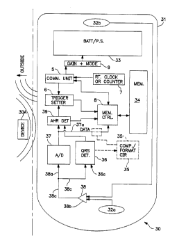
Refer now to Fig. 3 in which a circuit model 30 is illustrated in an outline
of
an implantable device shell 31. Electrodes 32a and 32b bring signal from the
body to
an input mechanism 38, here drawn as a differential amplifier for simplicity
only, the
output of which is fed to a QRS detector 36 and an AID converter 37. Both
these
circuits 36 and 37 supply output to an arrhythmia detector 39, which in this
preferred
embodiment supplies the autotrigger signal to the trigger setting circuit 6.
The data
output from the analog to Digital converter may be converted, compressed ,
formatted
and marked or reformulated if desired in a circuit 35 before the data is ready
for input
into the memory 34. The Memory control circuits 8 receives input from the A/D
converter, with or without conversion and so forth from circuit 35, from the
auto
triggering determination circuit (here seen as the arrhythmia detection
circuit) 39
(which may include input directly from the QRS detector if desired) as well as
signals
from the trigger setter circuit 6. The trigger setter circuit may also be
controlled by a
communications unit S which operates to receive and decode signals from the
outside
of the implant 30 that are telemetered or otherwise communicated in by a user.
This
communications unit S will also be able to communicate with the memory
controller
to request the offloading of memory data for analysis by an outside device. It
should
contain an antenna a or other transceiver device or circuitry to communicate
with an
outside device such as device 30A. A clock or counter circuit 7 reports the
time since
start or real time to the outside interrogator device 30A contemporaneously
with a
data offloading session so that the events recorded in memory 34 may be
temporally
pinpointed.
Alternatives to this overall design may be considered, for example by using a
microprocessor to accomplish some or all of the functions of circuits 6,8, 39,
and 35
but it is believed that such a design will not provide the power and size
savings taught
by use of the preferred design.
CA 02260209 1999-O1-08
WO 98/02209 PCT/US97/12443
9
Figs. 4a-c illustrate one preferred form 4~ of the invention. In this form it
has
an outer titanium shell 40, in a plastic cap means 44, which together form the
exterior
of the device. The cap means 44 may be composed of material similar to those
used
for pacemaker connector blocks as it is in the is case. The two electrodes, 44
and 49,
provide metal surface contacts to the body. Electrode 49 is formed as a whole
in a
paralene coating over the metal body 40, of the device. The metal electrode 42
is
connected via a feedthrough 43 which is itself electrically connected to the
circuit
board 41. Circuit board 41 contains all the elecl:ronics required for the
device function
and is connected to a battery BA for power. An integrated circuit 46 houses
circuitry
and intelligence required for the function and the memory M is packaged on the
other
side of the circuit board. In this preferred form, the invention uses a
communications
circuit 45 having a telemetry antenna both to indicate from outside the body
that a
read out is requested of the device, and for communicating data out from said
device.
Programming of the device or mode setting will also use the communications
circuit
45. In this form also a suture hole 45 is provided through the cap means 44.
Electrode 49 is connected by a conductive connection (not shown in this fig.)
to the
circuit board. In this embodiment the length "1''' is 2-3/8" and "w" is 3/4".
These
measurements can be varied within the constraints described. Electrode spacing
here
is about 1-3/4", center to center.
Presently less preferred three or more electrode embodiments are also
described with reference to Figs. S-8. A third electrode, like electrode 56,
can be used
to optimize signal strength responsive to changea in device position, heart
position, or
body position. A transistor or other switch means can switch the electrode
configuration automatically based on a determination of signal strength or
direction
from an outside device through the communications circuit. In order to retain
the
elongated shape yet provide a well spaced orthogonal position, the third
electrode can
be mounted on a self positioning (flexible, rigid, or semi-rigid) stubby lead.
An
additional variation from the most preferred design could provide for a wing
or fin-
shaped member 57 or more than one wing (57, 56) that extend substantially in
one
plane from the main body of the device. Ideally this would be approximately in
the
' CA 02260209 1999-O1-08
P-302ti.00 PCT
same plane as the other tZVo electrodes(53 and 5'~). Unless they are
constructed so as
to spring from t:na main body outward after insertion into the intended body
area,
wings like 57 or 58 will require a larger incision than the e~,u~re~tly most
preferred
device, a smooth babied device. The illustzation of the device 50 in Fig 5
without the
S dotted line external parts 55, 57, 5g, and 60, would be such a most
preferred form.
Some ot?:er features are also significant and should be noted. .a single
sutur;.
hole 54 (or t<vo or more if desired) can be praviiic;d ih the cap. Additional
suture
append<~.ges, like :irg 50, having 3 S11t1LrC hole 60a, m;~ v additionally be
provided for
more stability. Additionally, a suture may secure the stubby lead to the
patient's
10 tissue if desired. T hese suttee holding means al;'.ow the device to be
fzxedly held in
one orientation in the body of the user, whether intramuscuiar or strictly
subct!taneous. Intramuscsiar pocket implantation is advantrxgeous in that the
device
rray be protected from the outside world by a lager of muscle. which will
prov(~e
cosmetic benefits to the patient as well., The e:cact sites of llnplant may
LS advantageously be varied from patient to patient for various reasons appar~-
rtt to the
physician. Implant just under the skin row appears to provide the signal most
tree cf
skeletal muscle myepotentiai or body movement si"taI interference.
~notl;er important feature ofthe shape is to have one end cf the elongated
device tapered to aid in easily inserting under the: skin during
in?plant.'insertion (as in
a ble~nt dissection proced~trel. 'his self placing tapered tip helps
°nsu:e that chf~. de~~.e°
stays positioned properly in ai.igrunen~ with the principal cardiac -~~e;aors
whetver they
be the principal R-wave, or P-wave vector or best for both, especially whe:e
t~x~c
sutures would be used at the cap end. It is belie:o;d that this taper feahue
v;zll be better
than just a blunt placement with an instrument. Another preferred m~tf:od of
implant
?5 could be injection of the tapered end into the body, using a device similar
to that
described in the US Patent No. 5,520,660, the Implant Plunger. A5 a secondary
feature
the other end from tLe insertion tip may be blunt o; otherwise formed to
assist in
providing a better directing and pushing surface riuring inserticn. .~ rough
sketch of
' an alternate tool is provided in Fig 15. in which a handle unit with a blade
154 makes
an insertion into the opening 151 created in the sS;in, l:oldino the implant
152 hefween
~MEPIpFp STET
'.) ~i v ~!)b'b6EiF:~. fifl f~ b+ ~-f;f-:~.f: 61 ~ r, I~3 . ~f:: ~.i. . . flli-
ff -f~ I : i:i1 ',;'_J1-I:)'V.'-1: IlN-V~1:1:.\() \ ' \J?I
CA 02260209 1999-O1-08
WO 98/02209 PCT/(JS97/12443
11
a recess behind the blade and a pushing member 157 until a handle releases the
pushing member. The handle 155 may advantageously tie the end of a suture into
the
patient beneath the skin with tool 153, which is then retrieved by
manipulation of a
wire 157, thus accomplishing insertion and securing the implant at the far end
159,
rather than at the cap end of the implant. Many variations on this injection
and
insertion theme can be accommodated within the teachings of this document.
These kinds of instrument assisted insertion are herein referred to generally
as
insertion via a "trocar" concept. In general this "trocar" concept involves
any
instrument which encloses the implantable device and contacts the surface of
the body
or point of incision, starts the incision and allows the device to be inserted
thereinto.
The trocar is used to make a starting hole/incisi~on using a sharp point
and/or cutting
edge first. The physician then uses the mechanical advantage provided by the
trocar
to stretch the incision wide enough to allow the implantable device to fit
through the
incision and then pushes the device under the skin (or into the muscle, etc.)
in one
motion. The incision could be enlarged to facilitate suturing if desired.
A preferred form of insertion tool should be fitted with a smooth protective
chamber (preferably plastic lined) just wider than the implantable device (
but of
approximately the same cross-section) to slip the implantable device into,
tapered end
toward the insertion end of the tool. The battom~ of the chamber could be
shaped to fit
the taper of the implantable device and would move out of the way when the
implantable device was pushed by hand or an injecting plunger. The outside of
the
bottom of the chamber would come to a sharp point and possibly have cutting
edges
tapered back on both sides from the sharp point., but may not need to cut to
the full
width, instead it could stretch the initial opening to allow insertion of the
implantable
device with a push.
Suturing to hold the implant device in place could be done automatically or
with surgical staples by some means associated with the tool, the device could
be left
in the pocket, or it could be held in place by a coating of its surface with a
sticky
substance or one that adheres to body tissue like silicone rubber, or it could
be
CA 02260209 1999-O1-08
WO 98/02209 PCT/US97/12443
12
inserted with a properly shaped Parsonnett pocket, although this would likely
interfere
with the gathering of signal through the electrodes.
While considering the features of the embodiments illustrated by Fig. 5, it is
well to note the electrode configuration. Here the electrode 53 is a
conductive or
metal plate compatible with the patient's body that is on one surface of the
cap unit
51, the cap being delineated by dotted line 52. One can construct the device
50 as a
solid container having out of body compatible materials. For examples,
titanium or
other metal or alloy, coated with compatible insulator but exposed for at
electrode
areas or fitted with conductive electrodes, ceramic having conductive areas
thereon,
etc. One should have two surface electrode areas separated by a distance
(functionally
similar, therefore, to electrodes 53 and 59 in Fig. 5) for the device to work.
This
distance should be at least far enough to achieve good signal but not too far
so as to
make the size of the implant too large to accommodate. The first devices had
electrode
separation distances of just over 1-3/4"
center to center and we currently believe the best separation distance to be
approximately that. This distance can range between 1/2 and 2-1/2 inches, or
even
near 4" before becoming impractical.
In the presently preferred embodiment the cross-section is an easy-to-insert
rounded rectangular or oval shape to the potential of the device turning over
after
implant. Fig. 6A shape 61 and Fig 6B, shape 62 illustrate this concept and the
reader
may use any similarly functional cross-sections. Our studies have determined
that
electrodes which are faced outward, toward the skin of the patient, are
preferable to
face in or other electrode orientations. This may be due to less muscle
exposure or
movement or other noise sources.
Additional features are illustrated which can assist in preventing medically
unintended movement of the device. In Fig. 7A the electrodes are placed so as
to be
matched on opposite sides of the rectangular, round, or ovoid shaped device
and
electrically connected in parallel on opposite sides to retain the same signal
in spite of
flipping or other movement. (The internal circuitry would operate like the op-
amp 75
to produce output 76 from electrodes 71-74 as shown to produce this effect.)
In
CA 02260209 1999-O1-08
WO 98/02209 PCT/US97/12443
13
surface pacemaker implants, patient populations have been known to play with
their
implants, often unconsciously and this has been a common enough problem in the
pacemaker art to have obtained the name itwid~dler's syndrome." These features
address this problem. The device of 7A is seen in cross-section in Fig. 7B.
Another feature in a preferred embodilr~ent employs circumferential electrodes
on a cylindrically shaped device. In Fig. 8 this device can be seen to also
have a
body 69 that is tapered on one end 81 and blunt on the other 82. The effect
again is
to provide a constant signal in spite of likely unwanted rotation of the
device, because
the electrodes each extend around the device circumference. Here the electrode
area
positions are illustrated for each end, 65 and 6fi for end 81 and positions
66, 67 for
end 82. This approach trades-off the protection from muscle noise of the
rectangular
outward-facing device.
Additional designs for the device shape: which would be employed if the
circuitry and power needs could be reduced in size are shown in Figs. 13a,
device 130
and 14 a, device 140 with side views in the corresponding Figs. 13 and 14b.
These
devices have three electrodes each, l, 2, and 3, to adjust orientation to the
best signal
if desired, however two electrode forms and foams with windows W for sensors
are
also contemplated.
Procedure for Non-Invasively Deternninin~ Om it
I~,plant Position and Orientation Prior to the Irn_plant.
One of the preferred ways to use the invention is to be careful to assure that
the device is implanted in substantially the optimal position and orientation.
For
obtaining the best ECG signals with a two electrode device this is especially
important. A simple and noninvasive determination of the proper position and
orientation prior to implant can be made by merely employing an external ECG
measurement device using external electrodes (of any of a number of standard
types
well known in the field of cardiology ). By observing the ECG at orthogonal
electrode orientations in roughly the positions preferred by the
physician/patient, the
signal amplitudes both P and R wave can be monitored until a good positioning
is
CA 02260209 1999-O1-08
WO 98/02209 PCT/US97/12443
14
found and the signals are optimal. It is preferred that these measurements be
made in
several typical patient postures to account for posture variability as well.
The electrodes should be approximately spaced with the same spacing (within
a factor of two or so) as the implantable device and with approximately the
same
diameter electrodes as the implantable device (a factor of two or so as well).
(The
diameter of the external electrodes in most ECG systems will be smaller than
the edge
to edge spacing of the electrodes by greater than roughly a factor of two or
so). We
outline two approaches here.
Annroach 1 ~ Standard ECG Electrodes: a standard ECG Monitoring System
can be used with the standard electrodes and electrode preparation of the
skin. The
electrodes are then placed in orthogonal patterns of the proper electrode
spacing over
each candidate implant site (as described in the above paragraph) per Fig. 9.
Orthogonal measurements over each candidate implant site (here illustrated as
1, 2, or 3, for example locations for three electrodes each though two could
be used)
can be used to determine the optimal orientation.
One can either simply look at the signal amplitudes using the orthogonal
electrodes and assume a similar implant orientation will be substantially as
"good."
One may try again until a satisfactory signal at a given location and
orientation is
obtained.
For a more exact orientation to produce the absolutely best and largest R-wave
one can do simple vector arithmetic in the following manner:
If the two orthogonally oriented electrode pairs with a common electrode
produce R-wave Amplitudes A and B, the optimal orientation will be at the
angle = Arc-Tangent(B/A), where this angle is taken from the common
electrode to the electrode producing R-wave amplitude A. The same procedure
can be followed for optimizing the P-wave amplitude. One can also use
similar calculations to determine the best compromise angle for P and R
waves.
CA 02260209 1999-O1-08
WO-98/02209 PCTIUS97/12443
This Standard ECG approach has the advantage; of being possible using commonly
found ECG Monitoring systems, but has the disadvantage of requiring surface
preparation of the skin, as well as additional calculations or repeated tries
if the "best"
orientation is desired.
S ~~uroach 2: Hand-Held Device with Fi:~ed Electrode Probes: In this approach
a special device similar to hand-held emergency heart monitors provided by
several
manufacturers can be used to probe the surface locations and orthogonal
orientations
that are desired in order to fmd the optimum orientation. This device needs to
be
customized to have electrode probes which are roughly the same spacing as the
10 implantable device and looks like Figure 10A. The ECG is either displayed
on an
attached recording device or display or on a built-in display such as an LCD
monitor.
The procedure can also use a customized hand-lheld portable ECG monitor with
only
slight modifications to produce a satisfactory result. For example the
Micromedical
Industries Inc. (Northbrook Illinois) Paceview(t:m) with the modification
shown in
15 Fig. lOB could be used. It has a raised electrode assembly constructed on
points 93
which support posts 94 and electrodes 95, configured so as to maintain the
proper test
position of the electrodes for the device being considered for implantation.
This
added structure is on back side 92. Because these additional structures have a
spacing
similar to that of the implantable device, the readout on side 91 will produce
fine
results for placement and orientation data.
This device 90 has the advantage of not requiring the placement of surface
electrodes over the implant site, is fast enough to allow a simple sequential
test at each
orientation and implant site, and has no wiring or external equipment
required. The
ECG can be seen in real time in monitor window 96.
Functional considerations for the preferred embodiments.
In Fig. 3 the inventive system is described as stated above. The external
device 30A is preferably a device that is commonly called a "programmer" in
the
pacemaker art, because it's usual function is to communicate with and program
implanted devices. Software modifications and modifications to the telemetry
system
of device 30A to accommodate communication with and analysis of data from
device
CA 02260209 1999-O1-08
WO 98/02209 PCT/US9'7/12443
16
30 can be made as required. Such modifications will vary with the programmer
type
and are within the discretion of the manufacturer and thus will not be
illustrated here.
Using a programmer will avoid having to have additional devices cluttering the
operating room or clinic by creating a separate and distinct external
communications
device for this invention. The functionality necessary for mere ECG monitoring
and
event triggering is minimal, so in the preferred embodiments that only monitor
some
form of ECG or other limited sensory input, a microprocessor can be and is
done
away with altogether by using particularized functional circuits instead of
doing the
ft~nctions in software.
In Fig 3A, a block diagram of an analog to digital conversion circuit for use
in
this invention is shown. The clock input may advantageously use an output from
the
clock circuit 7, input 7i . The input 38c is the analog input signal from
input circuit
38, and the converted output is a stream of 8 bit digital data words on line
37a,
sequenced by a timing line 37b.
Fig 3B illustrates the basic parts of circuit 38, additionally indicating the
input
of gain set bits which can modify the value of the output of the low noise
bipolar
amplifier for output at line 38c, the input to the QRS detector. In this
invention QRS
detection is done on the analog signal, advantageously saving more complex
detection
after digital conversion.
In Fig 3C QRS detect circuit 36 has a 2nd order bandpass filter with a center
frequency preferably in the 20-25Hz range. It includes a transconductance amp
A1,
summing amp/comparitor A2 and resistors Rbpl-3, capacitors Cbpl-4 and
selectable
resistor R sense connected as shown. R sense is preferably adjusted during
manufacture. Additional control is provided for QRS sensitivity at line 36c,
since the
gain is delectable for this input.
A simple arrhythmia detection circuit 39 is included with this preferred
embodiment, and illustrated in Fig 3D. The output from circuit 36 is monitored
at a
200 millisecond blanking interval circuit, controlled by a clock input 7i2. In
the
preferred embodiment, a high rate can be selected amongst 4, with two
selection bits
dedicated to do so at input 9d and the low and flatline trigger rates each
have one bit
CA 02260209 1999-O1-08
WO 98/02209 PCT/US97/12443
17
to turn them on or off provided by inputs 9d. These inputs designated 9d
preferably
come from a register that holds the gain the mode and the rate settings,
illustrated as
register 9 in Fig 3. Such features may be progr~unmable through communication
with
the implanted device by an external device. Preferred timing for the high rate
triggers
is 140, 162 and 182 beats per minute, requiring 8 consecutive beats at such a
rate to
initiate the trigger. Additionally the trigger may be programmed off. The low
rate
counter/comparitor may be programmable to detect low rates of 40 or 30 bpm,
requiring 4 consecutive low rate intervals to trigger. Additionally a flat-
line trigger
can be set to occur after 3 or 4 and one half seconds of no QRS detection.
For embodiments that include more sensors and/or electronics, an additional
sensor could be added to benefit the patient. One particularly useful would be
an
activity sensor based on a single or mufti-axis accelerometer, which indicates
the level
of patient activity and his orientation. By checking for output that indicates
the
occurrence of a VVS (VasoVagal Syncope) episode, (for example, the patient
falling
from an episode) such an addition offers an improved trigger for events that
might
otherwise be missed by an arrhythmia detector ..et up like in Fig 3D. Such a
sensor
trigger could replace the circuitry of 3D.
Additional circuits may be provided to ;>upport additional functions if
desired,
however in order to reduce size and power consumption and extend the life of
the
device and reduce the intrusion into the body of the wearer, auxiliary
circuits should
be kept to a minimum. Such additional circuits could support oxygen sensing,
pressure sensing, respiration sensing, and any other kind of sensing that can
be
demonstrated to have been known for implanted. devices. They may each have
their
own auto triggers based on sensor output, or depend on manual triggers.
Additionally, activity sensing or positional sensing devices can provide
additional
input for recordation and or autotriggerring functions. As new sensors become
available they may also be incorporated into these designs.
In considering size, the maximum dimension of the device need be only the
minimum required dimension for good signal to be obtained from the two
electrode
areas. In our studies we have found useable signal for ECG monitoring at a
distance
CA 02260209 1999-O1-08
P-3026.60 PCT
Is
of about 11? inch(1 cm). Tl:e best minimum electrode distance fog ;.utTetit
electronics
at reasonaole prices appears to be from 3h+ ir,.ches to 2 inc~~!es.
FCG_recordin_~ f~tionz '~1'~ r for o,~~tbodil~ents._
The most important functivti o f the si~rnk ie versions of this invention is
the
long tezan ECG monitoring of ehe subcutar;eous ~Ur 111tr~.ITlL7~Gt1i~3C?C.CG.
The d~vic~:
cor_tisnuously:ecords and monitors the subcutaneous ECG in ar endleas loop of
r'le:::OT1'. 1.1' !tS ~.'r:2::~I1~' rllad° L!le d=VlCt.', :5 t: igo?red
t0 5~1V~ ~r°ta?:? .rl LrlemOi T the ~ X52
X minutes or seconds of ECG data b y the patient subsequent to f~;elinn
s}mptorns or
interest (e.g. syncope, palpitations, etc.).
In the ioreferr::d embodiment with 12(3 k: of memory the device can :;tore 42
ur
~~ 1 ?,'~i;~tlT,.eS Of E~'G.. '.~hioh. car, ~~wi~set aver nt~!.oading i7~,'
t;'l~~!~tT'>r tit ?~! ~ltn
., rn aI
de-ice for analysis anc ;iispla~.'. In one form there are four modes settable
for p;tient
nigger onl;.' and in anoth°r form there are auti~triggers. In the
patient c~nlj~(aiso called
"manual"1!zig4er modes, tho patient oa~-t capture ettrar One or twee eVeC~tS
l)etu'een
~ifloadings at either ne compression or at a compression ratio of 1;~ or scone
oth~z~
device si~pported ratio. When setting the zn.otle of the implant, the
physician oz'
attendant can d~eide whether to r~c;at-d data ir.~ a comer essed mode or not
in tl.e
preferred e:r~bodiment. If heater detail alf tine trgg~~rsd ECG is required
than can be
?~) ~oveiop:d frorn cctrpvesse;l 3ata ;itorwge, t'a~ n11~'SlC';a't sho;:Jd
se::.~ci :.~;.'Ii-~:~?TIY'_'~~.5~~
__- recard:ng, thereby limiting the tli-ne a~-ailabl~ to rerord_ I:. some
emr~OOt_~nerltS 5anlr~le
rate may be modified as well, but this is not preferred.
Compression is preferably cane using a kna :vn compression algorithms
implemented in hardware. '!Zany types are laiawn and software compression
could be
?5 used if desired too. An excellent and easy to inzplert~ent example is found
in the
article ~ ~-!~hvLh~m;a Detection Prc~~am For an f~-rbul atory, EC G Monaor by
vfueller,
copyright 1978, ISA, ISHI'J ~75e45. Using this algorithm in one er.~tbod:ment
we
have used a pre-trigger time of record of a mx~cimurn of ~~00 5ecer~ds and a
maximum
post trigger record of ? ZO seconds, .;rtd 3t the higher sa:upled or less
cuzn.pre.ssed rate
30 of 1200160 for a sinele event and 3ti0160
i~I~Er~IDED SHEE~f
,. "~n W r~ ' r--,..r.;,. -r~~,...,?,~.,r ,~.~~,.._ ~CC~ . ;rV
! Jt : ~!)-t~~6~l.iOf:U. (i7~ (.i b+ ~--f;E;i~l: G I ~i i. f ~) ,..t.: : c.i.
. ~ fill -fF -s~ I : U.() \;=)F(;)!Vcl. I(h'-l''d:1 ; 1O 1 \J?l
CA 02260209 1999-O1-08
WO 98/02209 PCT/US97/12443
19
seconds for three events. These time values are obviously only examples and
the
reader can set whatever time he or his physician feels is appropriate within
the ambit
of this invention. After such a record is made the device memory locations are
full
and will be overwritten by the next triggered event since in the preferred
embodiment
S the memory is maintained in a continuous loop.
Additional modes include those with pure autotriggering, which can mirror the
patient triggered only modes if desired. It should be considered that with
autotriggered events, the determination by the dlevice of an event worth
recording and
the subsequent activation of the trigger by the device itself will be faster
than the
patient finding his device for activation or othemvise activating the device,
so the pre
trigger time record can be smaller. In one preferred embodiment the memory is
segmented to allow for 14 autotriggers and 3 manual triggers. Further detail
regarding
modes is described with reference to Figs. 11 and 12.
The patient activated triggering of a preserved form of the recorded ECG
signal can be carried out by using a small handheld external device which may
be of
any number of different forms. A first way is through a handheld battery-
powered
device which uses a coded radio-frequency tele~metered signal through the skin
to the
device, on the press of a button. A simpler deviice a small handheld used to
close a
magnetic switch within the implanted device to trigger it by holding the
magnet close
or patting the area of the body that has the implant a set number of times
with the
magnet. Other methods for triggering ECG data retention in memory ( each of
which
has it's own advantages for implementation) are; to use physical tapping or
slapping of
the finger or hand on the skin over the device in. a particular cadence and/or
number of
taps (advantage is that no triggering device is nE:eded. With such methods the
disadvantage is that the patient needs to memol7ize the triggering sequence.
Matched
voice activation with a known command is possible but the complexity at this
time of
discerning voice commands precludes such activation for the present time, but
could
be in future devices using this invention. Another approach is light
activation through
the skin using a light source and receiver, auditory/sonic activation using a
handheld
auditory/sonic source held over the skin with a microphone receiver in the
device. All
CA 02260209 1999-O1-08
WO 98/02209 PCT/US97/12443
these methods are patient activated and require patient compliance or
cooperation, a
feature this device was designed to avoid. Accordingly in conjunction with one
of
these patient triggers or alone, an automatic activation or trigger for
holding a chunk
of memory should be included. This could be activated by automatic recognition
of
5 an arrhythmia, a heartbeat too fast or too slow, or for any other condition
the device
may be set up to find.
If a patient trigger is used it is advantageous provide feedback to the
patient
regarding whether the attempt to trigger long term storage of the event was
successful.
To accomplish this the implant should telemeter out a signal that indicates it
has
10 recognized a valid trigger. (This of course requires additional circuitry
and usage of
the limited available power supply.) The external triggering device then
notifies the
patient via the triggering device or through some known alarm mechanism
whether
they have or have not properly triggered the implanted device. This
notification can be
one of any combination of a number of feedback methods including: one or two
visual
15 sources such LED's, an auditory source such as a beeping speaker in one or
two tones,
or a tactile source such as a vibration. See also US Patent No. 5,518,001 for
other
potential trigger-indicator ideas for a hand-held patient activated trigger
device.
Features and Construction of the ,pr_eferred embodiment imnlantable devices.
Refernng now to Figure 11 in which a block diagram of a functional model
20 110 of the controller and memory 111 of a preferred embodiment device is
illustrated.
The memory is generally organized as a continuous loop of, preferably, 8 bit
addresses starting at address 0 and looping back around to address 0 through
Iine 124.
By telemetry or hard-wired input during manufacture 120, a mode selector 121
is set
so as to divide the memory 111 into working segments 111 a-d. The address of
the
start of each of these segments is indicated with lines 112.
Since this device is used for recording physiologic data, after the data is
compressed, converted, formatted and is in appropriate digital form, it is
continually
recorded in the memory 111. The address value at the tip of arrow 122 in the
combined memory space 111 d, 1 i lc is monitored by a program counter register
113.
CA 02260209 1999-O1-08
P-3x26.00 PCT
21
T'Ze size of each memory segment set in a gi:.~en mode limits the amount of
data available for each triggered event. In the preferred embodiment, using
only one
progr~n counter set of registers, the flexibility to accommodate two different
ttiaaer
lengths can be limited. Alternate forms of memory allocation are available.
For
example organizing the entire looping memon~ as one unit and marking each
trigger
~.vouid :illorv mere flexinility but increase the overhead. See for example
the memory
structure in Fnigra, U~'at. No. 5,339,824, Fi2;. ?.
To itse a single program ccutiter. the actual trigger address ~~~:rus th;;
time (;<
r?tern.ory location storage events j required to ha.~: a already stored the
amount o f data
I0 rteodod for pre event analysis for that trigger is stored as a value in the
triggor location
:eg'tster I 16 of Fib. I I . if 3 larger time for tire trigger recording is
required b;r a
trtf~.'.~~'°r ~JCC;I.'..T'I1'.'lf~' :'~Z.L::~~ ~~I'! '°Irn~a~;
>;.'i!?'f?.. Ar'd ~':'C,'rii. ~ :,y,: j ~~,..rT;~l ~,-; ~,=Pr fi.~~~' .. ~'~,-
.
occv:rrerce ~f an auto trigged, the value in the trigger register cnn be
decremcnted,
true yielding a larger pr a trigber tir,~e period in the allocated :nea~orv
segnuent for this
I ~ event. A priority system for ;vhe~l~er tc extend th.e pre trigger record
is simple to
implement but again would require additional hard~.~are and Js ilot preferred.
i.,n .fact
the Gimplest construction ignores any row triggers once a trigger is set until
the results
of comparing the program counter with the trie';er register correspoads to a
match in
~'?ltle.
L1J ~ 5 ~rl~.r~4 f ~.A.. i , 1 ~ ~sf'.iTl~ ~-.
t 1 ~ ~ O ~3l '_7br ~' ;,3t3 _! ~T 1 :TlaI:G31 t~ ~~'°C; ,. .. ~ L;;,'~
ia.T: 'll:'.0
trigge:ed one because upon reco:~erilg $om do ::vent ti:e patient ha.; enoz:~h
time to
rzc;over, get their -:vits about them. and find the triggering device. Manual
triggering
may therefore be set to record in double or multiple sized segments. Fig. 11
'.;
segments 11 I c and d are joined by looping amour 122 to dive effect to this
concept.
Because the memory size is preferably auite limited a time record or first-in-
. first-out pool record should be kept on order that the ne:vest triggers
record only over
the oldest vents segments. An additional preferred feature allo-.vs for a mode
that
prevents recording over any triggered went segn:(ent. This is preferably
implemented
~;.~E~~DED SHEEN
' 1i lf:~!)~[rtyil;E:c'. fil3'fl;(.+ nr ~.;a:i.:: ('(~' C'.(f3 . .',E.::r-
..<.~~~.~~~f;~- ..,....,.,. ,~..~~,\. ~l<_.~.-. , .:
t. t -fJt: c'.() ~~iIIJ~;~3;1t1-V~1~3:~\''()r\ ,\:O
CA 02260209 1999-O1-08
WO 98/02209 PCT/US97/12443
22
by a counter which fills for each segment used and has storage for the set
number of
looping segments. When it is full recording of new events stops.
When a trigger is activated and under the control program of the device is
allowed, a signal 115 is permitted by some control gate 117 to allow the
program
counter address to be loaded into a trigger location address register 116.
After
loading, each subsequent clock cycle or set of clock cycles depending on the
configuration of the device will load the trigger location from 116 into a
comparator
118 to compare this location with the program counter address stored in
register 113.
When comparator 118 fords that they match, an appropriate output is generated
to
start the next loop via control circuit 119. This control circuit 119 will
cause the
mode selector to point to the next available loop location effectively placing
that into
the program counter 113.
The diagrammatic algorithm 100 to indicate the flow of this information is
found in the illustration of Fig. 12 in which an electrode signal 101 is input
filtered,
converted from analog input to digital values, compressed and formatted if
desired in
step 102 so as to be in appropriate form to store in a memory location
designated by a
program counter pointer.
This data word's form could be containing a value representing input signal
compressed at various available ratios, and may be mixed with other
information like
data provided by another sensor or clock data. The data stored will of course
carry
information related to the signal taken at the sampling rate. Thus lower
sampling
rates to save power will adversely affect the usefulness or detail of the
data. Whatever
its preferred form, each data point stored as a word is referred to as a
chunk.
Output form step 102 provides the next chunk of data to the next memory
location in step 103.
Device checks to see if there is any trigger pending after storing each chunk
of
data in step 104. If not, the next chunk of data is stored. If there is, the
device
preferably checks to see if there is another trigger already set and if so
either ignores it
or resets the value of the reserved looping memory area (like areas 111 a-d in
Fig. 11 )
to accommodate a larger trigger or it ignores the trigger if it is smaller or
if it indicates
CA 02260209 1999-O1-08
WO 98/02209 PCT/ITS97/12443
23
a smaller value needs to be stored. If on the other hand, no trigger is
already set, then
a new trigger location is recorded in the trigger location memory and then the
next
memory location is written with the next chunk of data. At step 107 if the
trigger
location is equal in value to the program counter, the device knows that it
has gone
through the entire loop reserved by the mode selector for this particular
event record
and then moves on to the next loop location, step 108.
It should be recognized that any of the inventive concepts taught herein may
be applied to implantable devices to supplement their other functions, such as
a
supplemental recording system for a pacemaker, implantable drug pump, et
cetera.
Further, known enhancements to telemetric corrnnunication can be used to
automatically activate offloading of data to a device located in the patient's
home.
Such a device could send its received communications to the attending care
giver/physician's office at some convenient time:, telephonically or otherwise
so as to
enable close compliance with prescribed follow-up of patient conditions. This
invention is not understood to be limited in scope except by the following
claims.