Note: Descriptions are shown in the official language in which they were submitted.
CA 02260703 1999-02-04
-1-
TITLE
DISPOSAL CONTAINER FOR DETECTING,
DISTINGUISHING AND COUNTING OBJECTS
Background of the Invention
1. Field of the Invention
The invention relates to a container which detects, distinguishes and
counts marked objects placed into the container in an operating room using a
detection
device which responds to an alternating electromagnetic field.
2. Description of the Prior Art
Despite precautions, surgeons still occasionally leave surgical objects
such as sponges and, less frequently, small surgical tools in their patients
after an
operation. Areas which are badly injured tend to have a great amount of blood
which
may cover the surgical obj ects, making the obj ects hard to locate. Also, obj
ects may
find their way under an organ. This is most likely to occur in surgical areas
such as the
abdomen which is large and has many organs.
The prior art discloses use of X-ray opaque material positioned on the
surgical devices in order that after the surgery is completed and the wound is
closed, an
X-ray can be taken to insure that no surgical objects were left within the
patient.
Although this detection method is effective, it is cumbersome. Most operating
rooms
do not have X-ray machines. Hence, the patient must be taken to another room.
There
the patient often must be moved from his gurney to an X-ray table for X-rays
to be
taken. If a surgical object is detected after an X-ray has been taken, the
patient must be
returned to the operating room. Then, the cavity or incision must be reopened
to
remove the surgical object and then reclosed. This second surgery can cause a
great
deal of trauma to the patient, preventing optimum healing. Examples of
surgical
sponges which are marked by radiopaque material are disclosed in United States
Patent
CA 02260703 1999-02-04
-2-
No. 2,190,432 to Lewison, United States Patent No. 2,698,270 to Mesek, United
States
Patent No. 4,185,626 to Jones et al., and United States Patent No. 4,205,680
to
Marshall.
Manual counting of the sponges and other surgical objects after the
surgery is completed is also used to prevent those objects from being left in
body
cavities. This is not a foolproof method. Fatigue, poor handwriting, and
misreading of
numbers will occur during operations lasting 4 to 12 hours when dealing with
badly
damaged patients. Consequently, miscounts occur as a result of human error.
Surgical objects are counted not only to insure that no such objects are
left in the human body, but also to prevent such objects from being
inadvertently
discarded. There have been instances where a surgical item costing several
thousand
dollars has been discarded with the surgical trash.
Presently, many operating rooms follow a practice of placing all used
and soiled sponges, drains, packs and other objects in a holding container. At
the end
of the operation the container is emptied and the objects that had been
contained therein
are counted. Blood on the discarded objects may contain infectious disease
viruses.
Therefore, each time a soiled surgical object is handled there is a risk of
accidental self
inoculation. Consequently it is desirable to minimize the handling of such
objects, not
handle each discarded object twice as in this current practice.
Because of the risks of infection and in an effort to minimize human
error, the art has developed several methods of marking objects for automatic
detection.
Many of those methods rely upon markers which respond to a magnetic or
electromagnetic field.
Greenberg in United States Patent No. 3,587,583 attempts to overcome
the problems of leaving surgical objects within the human body. He proposes to
mark
the surgical obj ect with a permanently magnetized material. A surgeon
performs an
CA 02260703 1999-02-04
-3-
operation in the normal manner. Before closing the incision the surgeon probes
for the
presence of a surgical object with a magnetic field detector means which
generates an
electric signal which is modified in the presence of a magnetic field. If the
marked
object is present, the magnetic field of the magnetic marker is sensed by the
magnetic
field detector means, which modifies the electric signal. Yet, an operating
room has
many types of equipment which generate permanent magnetic fields. The presence
of
those fields can activate the magnetic field detector means, giving false
detection.
Because of its unreliability in an operating room, Greenberg's device is not a
practical
solution to the problem.
In United States Patent No. 5,057,095, Fabian proposes to mark surgical
instruments with a marker adapted to produce identifying signal
characteristics when
exposed to an alternating magnetic field. He discloses three types of resonant
markers
that are able to resonate at a certain preselected frequency. The first marker
is a
magnetomechanical device comprised of a permanent magnet overlaying a
magnetostrictive metal strip in a plastic housing. The magnetostrictive strip
vibrates
when the marker is exposed to an alternating electromagnetic field, and its
resonance is
detected when the frequency of the applied field reaches a predetermined
value.
However, such devices are very sensitive to pressure and stress, which will
inhibit
them. Since a body cavity is under some pressure and the marker may be
stressed
during surgery, this type of marker is not reliable for use as a marker for
surgical
objects. The second proposed type is an electromechanical circuit comprised of
an air
coil, with or without a ferrite core, and a resonant structure such as a
piezoelectric
crystal. As the first type, this type of marker can be adversely affected by
pressure and
stress because its principle of detection relies on a electro-mechanical
resonance;
therefore, a piezoelectric crystal type marker is also unsatisfactory. The
third type of
marker proposed by Fabian is an electromagnetic LCR circuit. This type of
marker can
CA 02260703 1999-02-04
-4-
be either built out of discreet components or made of a flexible printed
circuit. In the
former case, this unit is expensive to build and bulky, and it is impractical
for surgical
sponges. In the latter case, due to its high electrical resonance frequency
this type of
marker can be adversely affected by the presence of metal objects and
conductive
media. Because the human body is conductive, this type of marker is also
impractical
for surgical sponges. Consequently, none of the markers proposed by Fabian,
nor the
Greenberg marker, has been available on the market.
In United States Patent No. 5,045,071, McCormick teaches about the use
of magnetic materials for accurately locating the position of a catheter which
has been
inserted into a blood vessel. At column 9, lines 12-16, the patent cross
references U.S.
Patents 4,416,289, 4,431,005 and 4,445,501 for an explanation of the general
method of
detection. At column 5, lines 41-52, the '005 patent explains that a
distortion of the
magnetic field indicates the presence of the catheter. Thus, the McCormick
patent
teaches that merely a change in the magnetic field is a sufficient indicator
of the
position of the marked object. However, McCormick's measurements can be
affected
by the presence of other nearby magnetic and conductive materials. Hence,
McCormick's technique can and likely will provide "false positives" as to the
presence
or the position of the marked object.
In my United States Patent No. 5,456,718 I disclose a marker for
surgical objects made of a selected nonmagnetostrictive, soft magnetic
material which
will emit known specific selected harmonic frequencies when exposed to an
alternating
magnetic field. I further taught that a variety of devices could be used to
detect the
presence of a magnetized market attached to an surgical object left in a
patient.
However, I did not teach nor then realize that a container could be provided
for used
surgical objects which could detect, distinguish and count the objects as they
are placed
in the container. Consequently, it would not be necessary to routinely subject
patients
CA 02260703 1999-02-04
-S-
to a detection device as they left the operating room. Only which a count
indicating
that an object was missing would there be a need to scan the patient.
Thus, there is a container for receiving, detecting distinguishing and
counting surgical objects as they are retired from use an operating theater
where the
objects have been marked with a material that can be readily identified before
the
patient leaves the operating suite. The detector in the container must not
give false
positives or otherwise be ineffective in the presence of magnetic and
electromagnetic
fields of the type commonly produced in the operating room. Such a container
and
method should also be useful outside the operating room in environments where
objects
must be counted or detected and in which magnetic or electromagnetic fields
are
present.
3. Techniques for Detecting Electromagnetic Material
There are different ways of providing and detecting what we can call
generically an "electromagnetic marker." The cited prior art references all
use materials
which respond to an electromagnetic field. In order for a material to respond
to an
electromagnetic field and therefore to create "detectable changes" of the
electromagnetic field, a material has to have at least one of the physical
properties of
electrical conductivity, moderate to high magnetic permeability, and
magnetostriction
(in general associated with moderate magnetic permeability). Moderate magnetic
permeability is defined as a permeability comprised of between 5,000 and
20,000 and
high magnetic permeability as a permeability above 20,000. In each case, the
response
to the electromagnetic field and, therefore, the creation of "detectable
changes" of the
electromagnetic field are heavily dependent upon the geometry and size of the
marker.
In addition, the response to the electromagnetic field depends upon the
intensity and
frequency of the electromagnetic field.
CA 02260703 1999-02-04
-6-
In general, a magnetic material subject to an electromagnetic field of
known and fixed frequency fo responds to the applied electromagnetic field by
creating
"changes" of the intensity of the applied field and by creating harmonics of
the
frequency fo. If the material is electrically conductive, it responds by
creating not only
"changes" of the intensity of the applied field but also "changes" of the
phase of the
field. In addition, if the material is magnetostrictive, the electromagnetic
field creates
strains or stress in the material, and the material responds to it by creating
a frequency-
dependent "change" of the intensity and of the phase of the applied field.
Therefore,
there are three methods of detection. First, one can simply look for a change
of
intensity and/or phase in an applied magnetic field, a method which can only
be used
for detection of the position of an object at a distance comparable to the
size of the
object. McCormick uses this method. Second, one could look for the frequency
of the
applied field to reach a predetermined value that is the electromechnical
resonance
frequency of the marker. Fabian discloses a magnetomechanical device which
uses this
technique. Finally, one could look for particular harmonics generated by a
material in
the presence of an applied magnetic field. This method has never been used in
a
medical environment. Indeed, the teaching of Heltemes in United States Patent
No.
4,857,891 indicates that the art has generally failed to recognize that "open-
strip"
markers made of selected nonmagnetostrictive materials which generate specific
harmonic frequencies upon application of a unidirectional electromagnetic
field can be
used to identify the presence of particular articles.
Heltemes discloses a Magnetic Marker for Electronic Article
Surveillance Systems having multiple filaments randomly dispersed in a sheet-
like
substrate so as to be substantially parallel to the plane thereof. "The
filaments are
selected of low coercive force, high permeability material, and the random
orientation
results in certain filaments intersecting with them being magnetically coupled
to other
CA 02260703 1999-02-04
filaments to thereby collect and concentrate lines of flux associated with an
applied
field of an EAS system into filaments parallel to the field."
To take advantage of a high magnetic permeability material a marker has
to be elongated (fiber, long strip, with an aspect ratio length/square root or
cross-
sectional area of a least 200). Heltemes complies only partially to this
requirement, but
randomly distributes the fibers. In this respect Heltemes defeats this purpose
because
the applied electromagnetic field has to be parallel to the magnetic fibers to
generate a
high enough level of high harmonics to be recognized as marker specific.
Moreover,
Heltemes' marker is not very well suited for generating high harmonics.
Consequently,
Heltemes, like others in the prior art, failed to recognize that markers could
be created
for detection of surgical objects which generate specific, detectable,
selected harmonic
frequencies.
Summary of the Invention
I provide a container for receiving and detecting objects which have an
attached marker. The container has a generator that produces an alternating
electromagnetic field. The marker responds to the presence of that alternating
electromagnetic field. That response is detectable as discrete pulses of radio
frequencies. The marker is comprised of at least one elongated member made of
nonmagnetostrictive, soft magnetic material encapsulated in biocompatible
material.
Preferably, the magnetic material is an amorphous metal. However, crystalline
materials can be used if the encapsulation material has a high flexibility and
plasticity.
Detection is made using a pulse detection technique similar to that used
m magnetic resonance imaging (MRI), a common and safe diagnostic procedure. As
an
object is placed into the container, the object is exposed to an alternating
electromagnetic field of about 3-4 Oe (oersteds) for a specific time. This
field will
cause the elongated member to become magnetized and consequently to generate
CA 02260703 1999-02-04
_$_
harmonics of the frequency of the applied alternating electromagnetic field. A
detector
placed nearby will detect the reflected wave form from the marker as sharp
signal peaks
at the specific frequency of the applied alternating electromagnetic field.
My marker is particularly useful for surgical sponges. However, other
surgical tools (e.g., forceps, scalpel), plastic or rubber/polymer surgical
implement or
equipment used during surgery (e.g., plastic drain, suction tubes), and
implants (e.g.,
artificial veins, artificial arteries, knee replacement) may also be marked.
When the object is a surgical sponge, the marker can be woven into the
sponge. When the surgical object is a surgical tool such as forceps, the
marker is
placed on the tool by an adhesive means.
A microprocessor and memory can be connected to the detector. As
objects are detected they are counted. The total is then recorded and can be
displayed.
A keypad can be provided to allow a user to enter the number of objects that
should be
placed into the container. A program would then sound an alarm if too few
objects
were received.
Other details, objects and advantages of the invention will become
apparent as the following description of a present preferred embodiment
thereof and a
present preferred method of practicing the same proceeds.
Brief Description of the Drawings
In the accompanying drawings I have shown a present preferred
embodiment of the invention in which:
Figure 1 is a perspective view partially cut away of a present preferred
marker;
Figure 2 is a perspective view partially cut away of a second preferred
marker;
CA 02260703 1999-02-04
-9-
Figure 3 is a perspective view of a surgical sponge having one and
optionally two markers woven through the sponge;
Figure 4 is a diagram of a detector of my marker;
Figure S is a graph of responses of a detector in the presence of my
marker;
Figure 6 is a perspective view of a pair of forceps having a marker
positioned on the exterior handle surface;
Figure 7 is a perspective view of a container having mounted thereon a
detector with associated components for detecting, distinguishing and counting
marked
objects as they are placed into the container; and
Detailed Description of the Preferred Embodiments
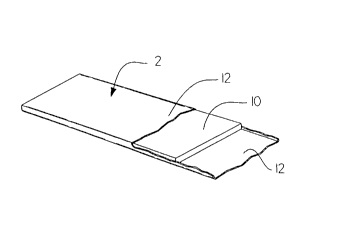
As shown in Figure 1, my marker 2 is comprised of an elongated body
of soft magnetic material which is nonmagnetostrictive. It is characteristic
of many
ferromagnetic materials that even the slightest applied mechanical strain
tends to cold
work the material and degrade its permeability and other magnetic properties.
Nonmagnetostrictive magnetic materials are insensitive to strain; they have
the required
magnetic properties for my marker but they are also sensitive to work
hardening.
Amorphous materials are very difficult to cold work. Preferably, the material
of my
marker is a nonmagnetostrictive amorphous material with very soft magnetic
properties. It should have high magnetic permeability, a low coercive field,
and
induction saturation should be as high as possible. One suitable material is
sold by
Allied under the trademark "Metglas." The composition of the alloy is recited
in
United States Patent No. 4,298,862 to Gregor et al. Thus, for example, Allied
compositions identified as types 2826MB@ or 2705M may be preferably used.
Another suitable amorphous material is Vitrovac 60252 soft magnetic amorphous
alloy
with zero magnetostriction sold by Vacuumschmelze GmbH of Hanau, Germany.
CA 02260703 1999-02-04
- 10-
Although the marker body could be any dimension and shape, I prefer to make
the
elongated body 10 as a ribbon or a strip or a set of parallel ribbons, strips,
fibers or
filaments. The elongated geometry of these members allows me to take advantage
of
the high magnetic permeability of the materials. Preferably, the elongated
members
will have an aspect ratio (length/square root of cross-sectional area) of at
least 200.
Such a sufficiently large value for the aspect ratio will assure generation of
a detectable
signal according to the teachings of U.S. Pat. No. 3,665,449. If the direction
of an
applied electromagnetic field is essentially parallel to the marker, and the
intensity of
the applied electromagnetic field is greater than a minimum field or
threshold, then the
marker will generate high harmonics of the frequency of the applied
electromagnetic
field. That is, the marker will emit a spectrum of harmonics whose intensity
decreases
slowly with the order of the harmonics in the spectrum. In particular, the 9th
or the
11 th harmonics should be detectable. Higher harmonics could also be
detectable. I
prefer the ribbon 10 to have a width less than 2 mm, preferably about 0.5 mm,
a
thickness ranging between 0.01 to 0.03 mm and a length of about 5 to 7 cm.
Body 10
could be longer or shorter, but preferably not shorter than 3 mm.
The width and thickness of the ribbon 10 can be selected to obtain
flexibility so that when the marker is attached to an object such as a
surgical sponge, it
will not adversely affect the flexibility of the object. I prefer to
encapsulate the
elongated body in a polymeric material that is compatible with the human body.
Suitable coating materials are nylon and delrin plastic. The coating 12
prevents the
body from oxidizing or reacting with body fluids and covers any sharp edges or
corners
of the ribbon 10.
In order to maximize the ratio of the length to the square root of the
cross-sectional area, it has been found desirable that the material be as thin
and as
narrow as practical, depending upon off setting production cost
considerations. It is
CA 02260703 1999-02-04
-11-
evident that fibers possess the required geometry. The metallic fiber which I
prefer to
use can be made in accordance with the teachings of U. S. Patents No.
5,003,291,
5,015,992, 5,015,993 and 5,027,886 by Strom-Olsen and Rudkowski. These fibers
are
nonmagnetostrictive, amorphous magnetic materials with very high magnetic
permeability. Typically, the diameter of such fibers ranges between .O1 and
.04 mm.
I further provide marking means in the form of a flexible marker made
of fiber laminated between two layers of polymeric materials. In order for
detection to
occur, one to approximately ten fibers should be placed parallel to each
other, and each
fiber should be approximately 1 to 3 inches in length. Such a multiple fiber
marker
assembly is shown in Figure 2. As shown, the marker of this invention
comprises two
layers 23 in the form of a web or ribbon of polymeric materials. I provide a
plurality of
fibers 11 a, 11 b and 11 c arranged parallel to each other and secured between
the two
layers 23 by adhesion or lamination means. As mentioned earlier and for the
same
reasons, I prefer to make use of a polymeric material that is compatible with
the human
body.
However, a marker made of a ribbon or fiber should be less than the
length or the width of the surgical object or not so long that it would
require significant
bending or multiple folding for attachment to the object to be marked. When
one is
outside this preferred range, a response signal from the marker will be less
characteristic and therefore more difficult to identify.
Although my markers would be X-ray detectable, one could impregnate
the coating or layers with an X-ray opaque material such as a barium compound
to
improve detectability. Another possibility is to use an X-ray opaque coating
material
which is currently being used on medical products. Use of such material not
only
improves the detectability of the marker, but should also reduce the time
needed to
obtain government and hospital approval of use of the marker.
CA 02260703 1999-02-04
-12-
One could also use nonmagnetostrictive crystalline magnetic materials
such as Permalloy alloy for the marker body 10. If a crystalline material is
used, the
cover layer of polymeric material should be designed to minimize the transfer
of
mechanical stress induced upon bending or folding the marker.
The surgical sponge 4 shown in Figure 3 is a gauze sponge having a
marker 2 woven throughout the sponge. It is understood that other means of
attachment
or securing the marker 2 to the surgical sponge 4 can be used. The marker can
be
connected to the sponge by means of pressure, heat, adhesive or the like.
Either the marker of Figure 1 or the marker of Figure 2 could be used. I
prefer to use markers which are shorter than a length or a diagonal of the
sponge to
avoid bending or folding. Optionally, I may use a second marker 32, shown in
chainline, positioned at a right angle to the first marker 2. This
configuration assures
that the sponge will be detected regardless of its orientation with respect to
the detector.
One could use more than two markers on an object. But, as the number of
markers
increases, the response of the marked object to a detector will likely be less
distinctive.
Therefore, I prefer to use one or two markers positioned as in Figure 3. This
arrangement provides a distinctive response such as shown in Figure 5.
It is relatively easy to detect an object marked with my marker. Once
the surgery procedure is completed, the surgeon exposes the surgical cavity to
an
alternating electromagnetic field, using the detection system. Preferably, the
patient
will be on a nonmagnetic gurney or examination table. A very low
electromagnetic
field of approximately 3 to 4 Oe is all that is required. Such a field is
relatively easy to
establish and will not harm the patient or the equipment which is normally
found in an
operating room. If desired, one may use as little as 1.5 Oe and as much as 6
Oe or
higher. However, it is normally not practical to exceed 10 Oe because such
high fields
would interfere with other equipment present in an operating room. This
magnetic field
CA 02260703 1999-02-04
-13-
will cause the marker body attached to any surgical obj ects in the surgical
cavity to
emit specific harmonic frequencies of the frequency of the applied
electromagnetic field
corresponding to the selected marker material. That emission will cause a
change in the
alternating electromagnetic field, which change can be correlated to the
presence of
only the selected nonmagnetostrictive, soft magnetic marker material. The
detection
system can be designed to measure all changes in the applied electromagnetic
field and
then look for the specific change which would be caused by the presence of the
marker.
Alternatively, the detection system could be designed to measure only a change
or
changes of the type which would be caused by the presence of the selected
marker
material or materials.
A number of techniques and variety of devices could be used to detect
the presence of the magnetized marker body attached to a surgical obj ect left
in the
patient. Those techniques and devices should be apparent to those skilled in
the art.
The detection system circuitry with which the marker 2 is associated can be
any system
capable of ( 1 ) generating within an interrogation zone an incident
alternating
electromagnetic field, and (2) detecting magnetic field variations at selected
harmonic
frequencies of the frequency of the applied electromagnetic field produced in
the
vicinity of the interrogation zone by the presence of the marker therewithin.
Such
systems typically include means for transmitting a varying electrical current
from an
oscillator and amplifier through conductive coils that form a frame antenna
capable of
developing an alternating magnetic field.
A fairly simple detector is illustrated in Figure 4. A flat search coil 14
has a first section 13 wound in a clockwise direction from points A to B and a
second
section 15 with the same number of windings running in a counter-clockwise
direction
from points B to C. A frame inductor antenna 16 is placed near the search
coil. If an
AC current is passed through the inductor antenna, an alternating magnetic
field will be
CA 02260703 1999-02-04
- 14-
created. That field will induce a voltage through the search coil. One can
detect the
voltage from points A to B and plot it on coordinates 34 in Figure S as a sine
wave 23.
The voltage from points B to C can be plotted as a sine wave 25, 180 degrees
out of
phase from sine wave 25. If the two waves are plotted simultaneously, they
will cancel
each other and yield a straight line over the time-axis. In the event a
magnetized
marker moves within the interrogation zone of coil 14, it will change the AC
magnetic
field received by the coil 14 and modify waves 23 and 25. For a single marker,
that
modification can be seen as a series of peaks 27 also shown in Figure 5 on
coordinates
35. The points along the time-axis where peaks occur depend upon the size and
composition of the marker. If too many markers are used, the peaks would
flatten and
approach a sine wave. Therefore, I prefer not to use more than two markers,
preferably
oriented as in Figure 3. The marker produces peaks at particular points along
the x-
axis. Thus, the detector looks for a response at those intervals. Only if a
response
occurs at the chosen points is a detection made. It is possible that equipment
in the
operating room, such as CRTs, will generate electromagnetic fields which will
cause a
detector response. However, the chances that such interference will produce
peaks at
the selected intervals is small. Hence, false detections are remote
possibilities, and they
can be eliminated by predetecting and electronically canceling the signals of
such
equipment.
My markers can be used in other surgical equipment such as forceps,
scalpels and hemostats. Figure 6 shows a pair of forceps 6 which have a marker
positioned along the handle. My markers can also be used to mark surgical
implants.
Hence, a physician could learn about an implant in the patient's past medical
history
using my detection method. The area under suspicion of an implant will be
excited at a
specific frequency. If an implant exists in that area, it will emit energy
over a specific
spectrum of frequency which corresponds to the specific implant material. For
this
CA 02260703 1999-02-04
-15-
diagnostic process to work, a standard must be used for all implants. Standard
material
should be used for each specific implant differing the material from one
implant to
another. In that event, the response will identify the specific implant.
Marked objects can be detected and counted at the time of disposal using
a waste container of the type shown in Figure 7. A detector 42 is placed on
the top 43
of the waste container near opening 41. Another detector 44 is placed on the
container
40, opposite the first detector 42. At least one of the detectors includes an
electromagnetic field generator. Whenever a marked object is dropped into the
container 40 the marker on the object will emit a signal that is detected by
the detector.
The detector will then emit a corresponding signal to a control system to
indicate that a
marked object has been discarded. Preferably, the control system is attached
to or
within the container as indicated by chain line box 45. The control system
causes a
display 47 to show the number of marked items that have been discarded.
Although not
shown, the control system could be connected to an external computer which
utilizes or
stores the number of discarded, marked items. This information could be used
for
patient billing or other purposes. The container 40 may have a warning light
46 or
LED display which is illuminated whenever an object having a particular marker
is
detected. If surgical sponges which are to be discarded have a marker that
evokes one
response and other objects are marked with a marker that prompts a different
response,
the control system can be designed to illuminate the light only when marked
objects
which are not to be discarded pass the detector.
The detectors 42 and 44 that are placed on the container 40, could also
be used in other locations, such as at a doorway or disposal chute, to detect
and count
the passage of marked objects.
In Figure 7 I have shown certain preferred orientations and geometries
of the elements that generate the electromagnetic field and detect the
harmonics
CA 02260703 1999-02-04
- 16-
produced by the marker. Those skilled in the art will recognize that there are
many
other possible embodiments. For example, a frame inductor antenna can be
placed
opposite a search coil. Two frame inductor antennas could be placed at right
angles to
two search coil detectors. The electromagnetic field could be created by two
frame
inductor antennas placed near a search coil detector. Other types of coils
could be used,
either for generating the electromagnetic field or for detecting the signal
from a marker.
I have shown and described the present preferred embodiments of the
invention. It is to be distinctly understood that the invention is not limited
thereto, but
may be otherwise variously embodied within the scope of the following claims.