Note: Descriptions are shown in the official language in which they were submitted.
CA 02261170 2005-02-07
74702-78
I
DESCRIPTION
BALLOON DISSECTING INSTRUMENTS
Technical Field
This invention relates to methods and devices for endoscopic vascular
surgery, in particular tv methods and devices for dissecting tissue to create
a
working space adjacent a blood vessel.
Back~rvund of the Invention
Numerous surgical procedures have been developed to replace arteries that
have become blocked by disease. The aortocoronary bypass surgery is perhaps
the
i5 most important of these bypass operations. The coronary arteries supply
blood to
the heart. As a result of aging and disease, coronary arteries may become
blocked
by plaque deposits, stenosis, or cholesterol. Iwsome instances, these
blockages can
be treated with atherectomy, angioplasty or stent placement, and coronary
bypass
surgery is not required. Coronary bypass surgery is required when these other
methods of treatment cannot be used or have failed to clear the blocked
artery. In
CA 02261170 1999-O1-21
WO 98/04314 PCT/US97/06571
2
coronary bypass surgery, a vein is harvested from elsewhere in the body and
grafted
into place between the aorta and the coronary artery below the point of
blockage.
An illustration of this surgery is shown in Fig. l, which shows the heart 1
and the
right anterior coronary artery 2 and the left anterior coronary artery 3 which
supply
blood to the heart. The right anterior coronary artery 2 is blocked in its
proximal
segment at 2a, as shown. This blockage has been bypassed by grafting a segment
of vein 4 between the aorta 5 and the distal segment 2b of the right anterior
coronary artery 2. Similarly, the left anterior coronary artery 3 may be
blocked, and
may require bypass with a length of vein 4a between the aorta and the distal
segment 3b of the left anterior artery. The operation requires access to the
heart,
which means that the chest cavity must be opened completely.
The coronary bypass surgery requires a length of vein or artery for the graft.
It is preferred to use a vein taken from the patient undergoing the bypass
surgery.
The patient is a ready source of suitable veins that will not be rejected by
the body
after transplantation and grafting onto the aorta and coronary artery. The
saphenous
vein in the leg is the best substitute for small arteries such as the coronary
arteries,
and it is the preferred vein for use in coronary bypass surgery. This is
because the
saphenous vein is typically 3 to S mm in diameter, about the same size as the
coronary arteries. Also, the venous system of the legs is sufficiently
redundant so
that after removal of the saphenous vein, other veins that remain in the leg
are
adequate to provide return blood flow. The cephalic vein in the arm is an
alternative that is sometimes used.
A typical operation previously required to harvest the saphenous vein is
illustrated in Fig. 2. The surgeon cuts into the leg to allow access to the
saphenous
CA 02261170 1999-O1-21
WO 9$/04314 PCT/US97106571
3
vein and cuts the vein from the leg. To expose the saphenous vein 6, the
surgeon
makes a series of incisions from the groin 7 to the knee 8 or the ankle 9,
leaving
a one or more skin bridges 10 along the line of the incisions. Some surgeons
make
one continuous incision from the groin to the knee or ankle. Handling of the
vein
S must be kept to a minimum, but the vein must be dissected free from
connective
tissue. After exposing the vein, the surgeon grasps it with his fingers while
stripping off the surrounding tissues with dissecting scissors or other
scraping
instruments. The surgeon uses his fingers and blunt dissection tools to pull
and lift
(or mobilize) the vein from the surrounding tissue. The vein is mobilized or
pulled
as far as possible through each incision. To reach under the skin bridges, the
surgeon lifts the skin with retractors and digs the vein free. While stripping
the
vein, the surgeon will encounter the various tributary veins that feed into
the
saphenous vein. These tributaries must be ligated and divided. To divide and
ligate
tributaries that lie under the skin bridges, the surgeon may need to cut one
end of
the saphenous vein and pull it under the skin bridge to gently pull the vein
out from
under the skin bridge until the tributary is sufficiently exposed so that it
may be
ligated and divided. When the vein has been completely mobilized, the surgeon
cuts
the proximal and distal ends of the vein and removes the vein from the leg.
After
removal, the vein is prepared for implantation into the graft site, and the
long
incisions made in the leg are stitched closed.
The procedure described above can be used to harvest veins for a femoral
popliteal bypass, in which an occluded femoral artery is bypassed from above
the
occlusion to the popliteal artery near the level of the knee. The procedure
can also
be used to harvest veins for the revascularization of the superior mesenteric
artery
CA 02261170 1999-O1-21
WO 98/04314 PCT/US97/06571
4
which supplies blood to the abdominal cavity and intestines. In this case, the
harvested vein is inserted between the aorta to the distal and patent
(unblocked)
section of the mesenteric artery. For bypass grafts of the lower popliteal
branches
in the calf, the procedure can be used to harvest the umbilical vein. The
harvested
vein can also be used for a vein loop in the arm (for dialysis) between the
cephalic
vein and brachial artery.
As can be seen from the description above, the vein harvesting operation is
very traumatic in its own right. In the case of coronary artery bypass, this
operation is carried out immediately before the open chest operation required
to
graft the harvested vein into the coronary arteries. The vein harvesting
operation
is often the most troublesome part of the operation. The long incisions
created in
the leg can be slow to heal and very painful. Complications resulting from the
vein
harvesting operation can also hinder the patient's recovery from the entire
operation.
The method of vein harvesting presented herein is accomplished with
laparoscopic procedures. This allows the veins to be harvested in an operation
that
requires only a few small incisions. Endoscopic surgical techniques for
operations
such as gall bladder removal and hernia repair are now common. The surgeon
performing the operation makes a few small incisions and inserts long tools,
including forceps, scissors, and staplers, into the incision and deep into the
body.
Viewing the tools through a laparoscope or a video display from the
laparoscope,
the surgeon can perform a wide variety or maneuvers, including cutting and
suturing
operations, necessary for a wide variety of surgical procedures and
operations.
Minimally invasive procedures for vein removal have been proposed. U.S.
Patent No. 5,373,840 to Knighton, entitled, "Endoscope and Method for Vein
CA 02261170 2005-02-07
74702-78
Removal," shows a method of cutting the saphenous vein at one end, and
grasping
the vein with graspers or forceps, then sliding a ring over the vein while
holding it.
Knighton uses a dissecting tool with an annular cutting ring, and requires
that the
saphenous vein be overrun or progressively surrounded with the dissecting tool
and
5 the endoscope, so that after the endoscope has been inserted as far as it
will go, the
entire dissected portion of the vein has been pulled into the lumen of the
endoscope.
As shown in figures 1 and 10 of Knighton, the method requires deployment of
forceps inside the annular dissection loop, and it requires deployment of the
loop
and graspers inside the endoscope lumen. The blood vessel must be cut and
gasped
by the forceps before it can be dissected by the dissecting ring.
Disclosure of the Invention
The methods and devices disclosed herein allow surgeons to harvest veins,
or dissect along other elongate structures without making fang incisions
through the
skin to access the structure as previously required. The present devices
permit
minimally invasive procedures which, in the case of a saphenous vein harvest,
require just two small incisions, one at either end of the saphenous vein, to
be
performed. The procedure is accomplished with laparoscopic instruments under
the
guidance of a laparoscope.
CA 02261170 2005-02-07
74702-78
5a
According to a broad aspect of the invention,
there is provided a surgical apparatus comprising: an
elongate shaft having proximal and distal ends, said
elongate shaft having sufficient rigidity to push through
body tissue free of naturally occurring openings in the body
tissue alongside an elongate structure; an elongate balloon
capable of assuming deflated and inflated states on said
elongate shaft, said elongate balloon having an axial length
substantially greater than a transverse diameter of said
elongate balloon when said elongate balloon is in said
inflated state, said balloon defining an inflatable interior
space into which a fluid is introduced to inflate said
balloon; said distal end of said elongate shaft located
inside said inflatable interior space; and a fluid
passageway in communication with said inflatable space in
said elongate balloon for communicating an inflation fluid
to said elongate balloon.
According to another broad aspect of the
invention, there is provided a pushable balloon dissection
apparatus comprising: a tubular member having proximal and
distal ends, said tubular member having sufficient rigidity
to push through body tissue free of naturally occurring
openings alongside an elongate structure; an elongate
balloon capable of assuming deflated and inflated states on
said tubular member, said elongate balloon having an axial
length substantially greater than a transverse diameter of
said elongate balloon when said elongate balloon is in said
inflated state, said balloon defining an inflatable interior
space into which a fluid is introduced to inflate said
balloon; said distal end of said tubular member located in
said inflatable interior space; a balloon cover surrounding
said elongate balloon; and means for inflating said elongate
balloon.
CA 02261170 2005-02-07
74702-78
5b
In a first preferred embodiment, a blunt loaded
balloon dissector has an elongate balloon of any suitable
length which may be formed of an elastic or non-elastic
material. The balloon may be of double walled construction
and may be provided with a central lumen which may receive a
guide rod, scope or other surgical instrument. The device
may have a support tube secured to the inner wall
CA 02261170 1999-O1-21
WO 98/04314 PCT/US97/06571
6
of the balloon to provide columnar support for the apparatus. The support tube
receives the guide rod, scope or other surgical instrument and may have a stop
member to translate pushing force applied to the guide rod or scope to pushing
force
on the apparatus. By using the guide rod or scope as a pushing member the
apparatus may be advanced alongside the vessel it is desired to dissect free
from
attached tissue. A balloon cover which may be elastic or resilient is provided
to
surround the balloon and facilitate compression of the balloon after it is
deflated.
In another embodiment of the invention, another pushable balloon dissection
device is provided which also may utilize an elongate balloon. The balloon in
this
embodiment may have a central lumen to receive a scope or other laparoscopic
instrument. The apparatus has a guide tube which receives a guide rod with a
slender metal rod and enlarged tip. The guide rod is utilized as a pushing
member.
A resilient balloon cover may also be provided in this embodiment to compress
the
balloon upon deflation.
In yet another preferred embodiment of the invention, a pushable balloon
dissection apparatus may have an elongate balloon disposed over an elongate
shaft
or tubular member such that the shaft or tubular member resides within the
interior
space of the balloon. The balloon dissector may be advanced between the tissue
planes it is desired to dissect and then inflated to create a tunnel alongside
a vessel
or other elongate structure. The balloon may then be serially deflated,
further
advanced and reinflated to enlarge the tunnel. When the apparatus is provided
with
a tubular member, a laparoscope may be inserted into the bore of the tubular
member and utilized as a pushing member to advance the apparatus and to
provide
observation of the procedure. A resilient balloon cover may also be utilized
in this
CA 02261170 1999-O1-21
WO 98/04314 PCT/US97/06571
7
embodiment to assist in deflating and compressing the balloon to facilitate
redeployment of the apparatus.
The method of vein harvesting disclosed herein utilizes an elongate tubular
balloon to dissect a tunnel alongside the vein to be harvested. The elongate
balloon
may be wrapped around a guide rod or endoscope and inserted through a small
incision in the leg and pushed along the vein to create a small tunnel over
the vein.
The elongate balloon may be provided with a balloon cover which may be a
separate
removable cover or attached to the balloon. When the balloon is in place
adjacent
the vein to be dissected, the removable balloon cover (if provided) may be
removed
and the balloon inflated to enlarge the tunnel and create a working space for
insertion of endoscopic instruments. The guide rod or endoscope may be removed
to allow other endoscopic instruments to be passed into the tunnel through the
balloon.
In a preferred method of harvesting the saphenous vein, the surgeon makes
one small incision at each end of the saphenous vein. After making the
incisions,
the surgeon inserts a tunneling instrument or blunt dissector which carries a
long
balloon into one incision and advances or pushes the dissector along the
saphenous
vein to make a small tunnel along the saphenous vein. The surgeon then
inflates the
long balloon to enlarge the tunnel. When the tunnel is enlarged to an
appropriate
size, the surgeon removes the balloon and seals the tunnel at both ends. The
surgeon may then injects carbon dioxide into the tunnel at sufficient pressure
(typically 5-15 mm Hg) to inflate the tunnel and create room for laparoscopic
instruments. The surgeon then inserts a laparoscope through the seal to
provide a
view of the procedure, and inserts a laparoscopic vein harvesting device, such
as
CA 02261170 2005-02-07
74702-78
8
one of the hooked vein harvesting devices disclosed in U.S. patent no.
5,601,581 entitled,
"Methods and Devices for Blood Vessel Harvesting", into the leg to dissect~the
connective tissue from the vein, identify side branches, and remove the vein
from the leg.
After the vein is loosened or dissected free from its channel in the leg, the
surgeon
can cut the proximal and distal ends of the vein and easily pull the vein from
the
leg. The small skin incisions are then stitched so that they may heal. : The
small
incisions heal much more readily, with fewer complications and far less pain,
than
the open procedures now in use.
Brief Description of the Drawings
Fig. 1 is a front view of the heart showing a vein grafted from the aorta to
the ,right anterior coronary artery, bypassing the proximal segment of the
right
anterior coronary artery.
Fig. 2 is a view of the leg showing the incisions necessary for harvesting
the.
saphenous vein using a traditional open procedure.
Figs. 3, 3a and 3b are views of the leg showing the incisions necessary for
harvesting the saphenous vein according to the methods presented herein.
Fig. 4 shows a. balloon dissector according to tt~e invention uninflated and
ready for insertion.
Fig. 5 shows a balloon dissector according eo the invention in its inflated
state.
CA 02261170 1999-O1-21
WO 98104314 PCT/US97/06571
9
Fig. 6 is a cross-section of an alternate embodiment of a balloon dissector
according to the invention in its uninflated state.
Fig. 7 is an isometric view of a balloon dissector according to the invention,
illustrating the balloon dissector in its expanded state.
Fig. 8 is a view of the balloon dissector illustrated in Fig. 7 with a
resilient
balloon cover surrounding the elongate balloon.
Fig. 9 is an isometric view of another embodiment of a balloon dissector
according to the invention with visualization capability illustrated in its
expanded state.
Fig. 10 is a view of the balloon dissector illustrated in Fig. 9 with a
resilient
IO balloon cover surrounding the elongate balloon.
Best Mode for Carr ~~in~ out the Invention
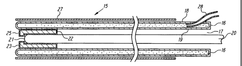
Fig. 4 shows an embodiment of a balloon loaded blunt dissector 15 in its
uninflated state, with a balloon 16 packed inside the device. The balloon 16
is a
nonelastic balloon or bladder and is cylindrical or tubular with a central
lumen 17.
i5 The balloon 16 has two walls 18 and 19 and may be described as a double
walled
balloon tube. The balloon 16 may be made of polyethylene, polyurethane,
polyamide and other nonelastic materials as well as latex and other elastic
materials.
The balloon 16 may be any suitable length, for example 12 to 24 inches long,
to
provide of a tunnel of convenient length when harvesting the saphenous vein.
The
20 balloon 16 may be any convenient diameter or width, for example 2 to 3
inches, to
allow laparoscopic instruments to fit and operate conveniently within the
tunnel
created by the balloon 16. The balloon tube 16 may have any suitable cross-
sectional shape.
CA 02261170 1999-O1-21
WO 98/04314 PCT/US97/06571
A guide rod 20 with a blunt or rounded tip 21 is disposed in the central
lumen 17 of the double walled balloon tube 16. The guide rod 20 is used as a
pushing member to push the balloon 16 through body tissue. A support tube 22
may be provided to give some columnar support to the device and provide a stop
5 member or coupling member to translate pushing force applied to the guide
rod 20
to pushing action on the balloon tube 16. The support tube 22 may be secured
to
the inner wall of the balloon tube 16 in any suitable fashion. The support
tube 22
may have an overhanging lip 23 which obstructs passage of the guide rod 20 or
endoscope 29 (if provided). Alternatively, the guide rod 20 or endoscope 29
can
10 be fitted with a stop collar 30 to engage the support tube 22 (as shown in
Fig. 5).
The support tube 22 may have a square tip 25 as in Fig. 4 or a rounded tip 26
as
shown in Fig. 5. The guide rod 20 and support tube 22 are used to push the
balloon
16 along the saphenous vein or other desired pathway between tissue layers.
Use
of the support tube 22 permits the guide rod 20 or endoscope 29, if utilized
as the
pushing member, to be removably received by the apparatus 15. This allows the
apparatus 15 to use fairly expensive and nondisposable devices such as the
endoscope as the pushing member. If visualization is not needed or desired,
the
balloon 16 may be sealed to a disposable pushing member and may be coupled to
the pushing member with adhesives, heat sealing or integral construction or
any
other coupling means. A balloon cover 27 surrounds the balloon tube 16 and
provides a protective sheath during placement of the balloon loaded dissector
15.
The balloon cover 27 may be a thin sheath of polyethylene or other plastic
film, or
it may be a more rigid tube of PVC, PTFE, PETG, polyethylene or other plastic.
CA 02261170 1999-O1-21
WO 98/04314 PCT/US97/06571
11
The balloon cover 27 may be elastic or resilient so that it serves to compress
the balloon 16, so that the balloon 16 quickly and automatically collapses
upon
deflation. The balloon cover 16 may be made resilient by choosing a resilient
material such as a thin sheet of polyethylene which is sufficiently resilient
and
elastic under the pressure used to inflate the balloon 16. The balloon 16
itself may
also be made of polyethylene, and may be a thick polyethylene which is
nonelastomeric under range of pressure used to inflate the balloon 16. When
the
balloon 16 and balloon cover 27 are made of the same material or a miscible
material, the balloon 16 may be heat sealed to the balloon cover 27 at various
points
to prevent the balloon cover 27 from inadvertently slipping off the balloon
16.
When the balloon 16 and balloon cover 27 are made of different or immiscible
materials, they may be attached with adhesive or through the use of other
suitable
fasteners.
In the preferred embodiment of a method of using the devices disclosed
herein, the surgeon uses a balloon loaded dissector to create a working space
under
the skin and over the saphenous vein suitable for laparoscopic techniques. The
surgeon makes one or more incisions as shown in Fig. 3, to expose the
saphenous
vein. These incisions are referred to as cut-downs. An incision at the knee
12, an
incision at the groin 13, or an incision close to the ankle 14 can be used. In
Fig.
3, the saphenous vein 6 can be seen through the cut-downs 12, 13 and 14. It
will
be apparent from the description that the use of three or four incisions to
harvest the
entire saphenous vein is merely a matter of convenience, and those
particularly
skilled in laparoscopic procedures may require fewer incisions, and smaller
incisions
than illustrated may be required. After insertion, the balloon loaded blunt
CA 02261170 1999-O1-21
WO 98/04314 PCT/US97/06571
12
dissector 15 is pushed along the blood vessel until the balloon tube I6 is
located
over the desired length of the saphenous vein. When the balloon 16 is properly
in
place it occupies a narrow tunnel over the saphenous vein. When in place, the
balloon 16 is inflated through inflation tube 28. As shown in Fig. 5, the
outer walls
expand under inflation and the balloon cover 27 stretches as the balloon 16 is
inflated. The expansion of the balloon 16 enlarges the tunnel. The outer
diameter
of the balloon tube 16 defines the size of the tunnel that is created, and the
outer
diameter may be controlled during manufacture and during inflation. Also as
shown
in Fig. 5, the guide rod 20 may be conveniently replaced with an endoscope 29
which can also serve as the pushing member. The endoscope 29 can be chosen to
have an outer diameter matching the support tube, or it can be provided with a
stop
collar 30, both constructions serving to couple the endoscope 29 to the
balloon tube
16 so that pushing on the endoscope 29 serves to push the balloon 16 into the
body.
When the balloon 16 is deflated through the inflation tube 28, the balloon
cover 27 serves to compress and collapse the balloon 16 and squeeze the
inflation
fluid out of the balloon 16, thus returning the balloon 16 to the collapsed
state
shown in Fig. 4. After the balloon 16 has been collapsed by the elastic force
of the
balloon cover 27, the device 15 may be further advanced or pulled-back from
its
position in the body, and repositioned at another area of interest. When the
balloon
16 is repositioned, it may be reinflated to enlarge the tunnel. The balloon 16
may
be repeatedly inflated and deflated in this manner. Alternatively, the balloon
cover
27 may be removed by pulling it proximally out of the incision to allow the
balloon
16 to expand.
CA 02261170 1999-O1-21
WO 98/04314 PCT/US97/06571
13
Fig. 6 shows an alternate embodiment of a balloon loaded blunt dissector.
The guide rod 31 is provided with a slender metal rod 32 fitted with an
enlarged tip
or olive tip 33. The guide rod 31 may be replaced by a scope if visualization
is
desired. The balloon 34 is a long slender cylindrical balloon, with or without
a
central lumen. A guide tube 35 is attached to the outside of the balloon 34
and the
guide rod 31 fits through the guide tube 35. The balloon 34 is uninflated in
Fig.
6, and the balloon 34 and guide tube 35 are shown inside the balloon cover 27.
The
balloon 34 of Fig. 6 is used in the same way as the balloon 16 of Figs. 4 and
5.
In operation, the apparatus is slipped over an endoscope {if utilized) or
guide
rod 31 and the balloon cover 27 is slipped over the apparatus. It is expected
that
use of an endoscope will be preferred, because it allows for visualization of
the
anatomy at its distal tip as the apparatus pushes through the fat layer
overlying the
saphenous vein. The apparatus is inserted either directly into the incision or
is
introduced through a cannula. After the guide rod 31 and balloon 34 are in
place
over the blood vessel, the balloon cover 27 can be pulled out of the incision,
and
may be provided with a weakened section to facilitate removal. The balloon
cover
27 may be pulled back gradually as the balloon 34 is inserted to uncover that
portion
of the balloon 34 which is inside the body, and the balloon 34 can be inflated
to
dissect a larger tunnel in the early stages of insertion. The balloon cover 27
may
also be left in place and, if made of a resilient material, can be used to
compress
the balloon 34 after deflation to facilitate repositioning of the assembly.
After full insertion, in a preferred method of use, the balloon 34 may be left
in place in the tunnel while the endoscope is utilized to view the interior
surfaces
of the body at the tip of the apparatus, as shown in Fig. 3a. If the balloon
34 is
CA 02261170 1999-O1-21
WO 98/04314 PCT/US97/06571
14
provided with a central lumen, endoscopic instruments may be passed through
said
central lumen to perform surgical procedures on body parts such as the
saphenous
vein and communicating veins of the leg. In situations where it is desirable
to
insufflate the tunnel created by the balloon 34, the balloon 34 may be
deflated and
pulled out of the tunnel through cut-down 13, and a cannula port 36 with
insufflation tube 37 may be inserted into the same cut-down as shown in Fig.
3b.
A secondary endoscopic access port 38 may be inserted into the knee incision
12 to
pass a variety of instruments into the work space.
The step of removing the balloon cover 27 may be avoided if the balloon
cover 27 is perforated along a number of longitudinal lines and sealed to the
balloon
34 along longitudinal lines, so that expansion of the balloon 34 tears the
balloon
cover 27 to allow expansion, but the pieces stay fixed to the balloon 34 so
that they
may be removed easily.
Another embodiment of a balloon dissection apparatus 50 is illustrated in
Fig. 7. In this embodiment, the balloon dissector 50 includes a handle 52, a
tunneling rod 54 which may be provided with a blunt tip 56, and an elongate
balloon 58 having an inflation harness 60 extending from the balloon 58. The
tunneling shaft 54 is formed of a suitable material such as surgical stainless
steel to
provide adequate rigidity for the shaft 54 to serve as a blunt obturator for
tunneling
between tissue layers. When the balloon dissector 50 is configured as an
extraluminal balloon dissector to harvest the saphenous vein in the leg, for
example,
the tunneling shaft 54 may have a length of about 12 inches, and a diameter of
approximately 1/8 inch. Alternatively, the tunneling shaft 54 may be formed
from
a semi-flexible material, such as plastic, for example, to accommodate
situations
CA 02261170 1999-O1-21
WO 98/04314 PCT/US97/06571
where it is desirable to provide a tunneling shaft with the capability to
navigate
somewhat torturous passages within the body. The tunneling shaft 54 may be
mounted in the handle 52 using any suitable fastening system, e.g., gluing or
a
compression fit. The blunt tip 56, which may be integrally formed with the
5 tunneling shaft 54 or a separate member, provides a blunt distal end on the
tunneling
shaft 54. Although an olive-shaped tip 56 is illustrated, other shapes
providing a
blunt surface are possible. The blunt tip 56 may also be omitted.
The elongate balloon 58 has distal and proximal ends 59 and 53 and a neck
portion 57 extending from the proximal end 53 of the balloon 58. For the
purposes
10 of this application, an elongate balloon is defined as a balloon having an
axial length
substantially greater than its transverse diameter when the balloon is
inflated. As
illustrated in Fig. 7, the elongate balloon 58 is mounted over the tunneling
shaft 54
such that the tunneling shaft 54 lies within the interior space 63 of the
balloon 58.
The distal end 59 of the balloon 58 is preferably provided with a nipple or
pocket
15 61 that may mate against the distal tip 56 of the tunneling shaft 54 to
help protect
against stretching or tearing of the distal tip of the balloon during
tunneling. As
will be described below, this construction permits the tunneling shaft 54 to
be used
as a pushing member to advance the deflated balloon 58 alongside a blood
vessel or
other elongate structure it is desired to dissect free of connected tissues.
The neck portion 57 of the balloon 58 may be secured inside the handle 52
in a fluid-tight fashion. Alternatively, the neck portion 57 may be secured to
the
tunneling shaft 54 by any suitable bonding system such as gluing or clamping.
When the neck portion 57 is mounted in the handle 52 as illustrated in Fig. 7,
the
balloon inflation harness 60 may extend from the neck portion of the elongate
CA 02261170 2005-02-07
74702-78
16
balloon 57 to provide a fluid passageway into the interior space 63 of the
balloon
58.
One example of a suitable balloon inflation assembly is illustrated in Fig. ,
7
in the form of balloon inflation harness 60. Balloon inflation harness 60
includes
a tube 68 which extends from the balloon 58 and is connected to a wye fitting
70.
A luer-type fitting with check valve 67, is connected to one port of the wye
fitting
70 and an evacuation fitting 69 is connected to the other port of the wye
fitting 70.
A pinch clamp 66 is provided to close off the fluid passageway from the wye
fitting
70 to the evacuation fitting 69. The balloon inflation harness 60 illustrated
is of the
same type as described in U.S. patent no. 5,?72,680, filed on December 12,
1995. Of
course, numerous other suitable balloon inflation assemblies are possible.
In the Fig. 7 example, the elongate balloon 58 is inflate by closing the
pinch clamp 66 and injecting a suitable inflation fluid, preferably sali~
solution,
although other fluids such as air may be utilized instead, through the fitting
67 into
the balloon inflation lumen 68, which is in communication with the interior
space
63 of the balloon 58.
As one alternative to the balloon inflation harness 60, the handle 52 may be
provided with an inflation port which is in communication with the proximal
end of
the neck portion 57 of the elongate balloon 58. In this alternative
embodiment,
inflation fluid is injected through the inflation port in the handle 52
directly into the
neck portion 57 of the balloon 58.
CA 02261170 1999-O1-21
WO 98/04314 PCT/US97/06571
17
The balloon 58 is preferably constructed of a medical grade nonelastomeric
material of suitable type such as a polyurethane according to known
fabrication
techniques so as to have a predetermined elongate shape. Although all
nonelastomeric materials exhibit some degree of elasticity, for the purposes
of this
S application, a nonelastomeric material is one which remains substantially
inelastic
over the desired inflation pressure ranges utilized for the particular
procedure.
Although an elastomeric balloon may be utilized with the balloon dissector 50,
it is
preferable to use an inelastic balloon so that the expansion envelope of the
balloon
can be more precisely predicted. In contrast, an elastomeric balloon tends to
inflate
following the path of least tissue resistance and localized differences in
tissue
resistance may cause an undesirable aneurysm in the balloon leading to
nonuniform
dissection.
In a preferred method of use, an incision is made in the body proximate the
elongate structure within the body it is desired to dissect free of connective
tissue.
The surgeon identifies the appropriate tissue planes dissection is desired to
occur
along and the balloon dissector 50 is inserted, either directly or with the
aid of a
cannula, through the incision into the body and advanced following the
identified
tissue planes. The surgeon uses the handle 52 and pushes the balloon dissector
50
as a blunt obturator following the tissue planes adjacent the elongate
structure until
a desired location for deployment of the dissector 50 is reached. As the
dissector
50 is advanced within the body, its progress may be monitored through the skin
by
direct observation and/or manual palpation. Depending on the procedure,
varying
amounts of the overall length of the elongate balloon 58 may be disposed
within the
incision.
CA 02261170 2005-02-07
74702-78
18
Once the desired location for dissection has been reached through blunt
tunneling, the elongate balloon S8 may be inflated by closing the pinch clamp
66
and communicating an inflation fluid through the balloon inflation harness 60
into
the interior space 63 of the balloon S8. As it inflates, the balloon ~8
expands into
S the predetermined elongate shape and dissects tissue away from the elongate
structure to create a tunnel alongside tlx elongate structure. . After the
tunnel has
been created, the dissector SO may be deflated by releasing the pinch clamp 66
and
applying vacuum to the male evacuation fitting 69. After deflation, the
dissector SO
may be removed from the body through the incision, or further advanced
alongside
the elongate structure and reinflated to enlarge the tunnel.
After the dissector has been removed from the body, a cannula and skin seal
assembly of the type disclosed in U.S. patent no. 5,77,680, for example,
may be inserted into the incision and the skin seal advanced into the incision
to
create a substantiaDly gas-tight seal with the incision. The space created by
the
1S balloon dissector SO may then be insufflated by injecting an appropriate
insufflation
gas through a port provided on the cannula should an insufflated operating
space be
desired.
Use of the dissector SO specifically contemplates multiple serial balloon
dissections. The dissector SO may be advanced into the body in stages along
the
identified natural tissue planes by repeatedly advancing and deploying the
dissector
SO to dissect a tunnel of the desired length alongside the elongate structure.
The
process of advancing the dissector S0, inflating the balloon S8, and deflating
the
balloon S8 may be repeated in serial fashion until the desired tunnel has been
created. In the embodiment of the balloon dissector SO illustrated in Fig. 7,
a
CA 02261170 1999-O1-21
WO 98104314 PCT/US97/06571
19
vacuum may be drawn on the balloon 58 to deflate and contract the balloon 58
so
that the dissector 50 can be repositioned and redeployed as needed.
The balloon dissector 50 is illustrated in Fig. 8 with an elastomeric balloon
cover 72 that surrounds the elongate balloon 58. The dissector 50 is in all
other
respects identical to the Fig. 7 embodiment. The cover 72 preferably has a
diameter
such that when the balloon 58 is in a deflated condition, the cover 72
compresses
the balloon 58 around the shaft 54. The use of the cover 72 thus aids in the
serial
redeployment of the balloon 58 by automatically causing the balloon to return
to a
compressed state upon deflation. The elastomeric cover 72 may be formed from a
resilient material such as a thin sheet of polyurethane which is sufficiently
resilient
and elastic under the pressure used to inflate the balloon 58, or it may
formed from
an elastomer such as silicon or latex rubber.
The cover 72 may be bonded to the balloon 58 at various points using known
bonding techniques to prevent the cover 72 from inadvertently slipping off the
balloon 58. Alternatively, the cover 72 may have a diameter closely matching
the
diameter of the shaft 54 so that the cover 72 is retained in position by its
elastic
compression on the shaft 54, or by friction. The cover 72 may also completely
cover the elongate balloon 58 and be secured in the handle 52. In this case,
the
cover 72 may be inflated independently of the elongate balloon 58 to provide a
balloon with elastomeric characteristics, i.e., localized tissue expansion
following
the path of least tissue resistance.
As an alternative embodiment, the elongate balloon 58 may be elastic and the
cover 72 may be inelastic such that the inelastic cover 72 acts to limit
expansion of
the elongate balloon 58.
CA 02261170 1999-O1-21
WO 98/04314 PCT/US97/06571
It is also possible to utilize a separate removable cover of the type
disclosed
in co-pending application serial No. 08/570,766, for example, or an integral
balloon
cover which separates upon expansion of the balloon as described with
reference to
previous embodiments.
5 Another embodiment of a balloon dissector 80 is illustrated in Fig. 9. The
balloon dissector 80 differs from the balloon dissector 50 only in that the
tubular
member 82 replaces the tunneling shaft 54 and an opening in communication with
the bore in the tubular member 82 is provided in the handle 52. In this
embodiment, the tubular member 82 serves as a scope cover to provide the
dissector
10 80 with visualization capability when used in conjunction with a
Iaparoscope. The
tubular member 82 has an internal diameter, such as 10 mm for example, sized
to
receive a conventional Iaparoscope. The tubular member 80 may be formed of any
suitable material, such as plastic for example.
The tubular member 82 may have an open distal end 84 as illustrated in Fig.
15 9 to permit observation with the laparoscope through the open distal end
84. As
shown in Fig. 9, the open distal end 84 of the tubular member 82 may be cut
away
at a 45 degree angle, for example, and provided with a lip 86 which serves to
capture the distal end of the Iaparoscope when inserted to prevent the
laparoscope
from extending beyond the open distal end 84. Alternatively, the open distal
end
20 84 of the tubular member 82 may be squared off and the lip 86 provided by
rolling
the open distal end inward. The distal end of the tubular member 82 may also
be
closed and rounded if visualization through an open distal end is not desired.
An instrument seal, which may comprise an area of reduced internal
diameter in the tubular member 82, may be provided to prevent the balloon
inflation
CA 02261170 2005-02-07
74702-78
21'
fluid from leaking out of the handle 52 during balloan inflation.
Alternatively, or
in combination with the area of reduced diameter in the tubular member 82., an
instrument seal of the type illustrated in U.S. patent rro. 5,772,680 may be
provided in the handle 52.
In addition'to the balloon inflation options described with regard to the
Figs.
7 and 8 embodiments, the elongate balloon 58 of the dissector 80 may be
inflate
by injecting the inflation fluid through an inflation port in the handle 52
directly into
a lumen in the tubular member 82. If the tubular member 82 has the presently
preferred open .distal end construction, the inflation fluid may be
communicated
through the bore in the tubular member 82 and out the open distal end 84 into
the
interior space 63 of the balloon 5$. If a closed-ended tubular member 82 is
utilized,
inflation may be through a separate lumen formed in the wall of the tubular
member
82 which opens into the interior space 63 of the balloon 58.
To prepare for tunneling dissection, a laparoscope, which may be a
conventional 10 mm laparoscope, for example, is inserted through the handle 52
and
advanced into the bore of the tubular member 82 until the distal extremity of
the
laparoscope is captured by the lip 86 in the open distal end 84 of the tubular
member 82. The lip 86 in the open distaD end 84 thus prevents the laparoscope
from being advanced beyond the open distal end of the tubular member 82.
Although an angled scope will provide the best visualizatian through the open
distal
end 84 of the tubular member 82, a straight scope may also be utilized.
The balloon dissector 80 is then inserted through the incision and tunneled
bluntly following the desired tissue planes using the laparoscope and tubular
member
82 as a blunt obturator. ~ The procedure to be followed is the same as
previously
CA 02261170 1999-O1-21
WO 98/04314 PCT/US97/06571
22
described with regard to the Fig. 7 and 8 embodiments with the exception that
the
progress of the operation may be observed through the laparoscope during
tunneling
dissection. During the tunneling stage, the distal end of the laparoscope
looks out
through the open distal end 84 of the tubular member 82 obstructed by only a
single
and preferably transparent layer of the elongate balloon 58. Moreover, when
the
balloon 58 is inflated to create a tunnel alongside the desired elongate
structure, the
Iaparoscope may be utilized to observe the progress of the dissection.
Fig. 10 illustrates the addition of a resilient cover 72 to the balloon
dissector
80 having visualization capabilities. The cover 72 may be of the same
construction
as previously described and may function in an equivalent manner to
automatically
compress the elongate balloon 58 about the tubular member 82 upon deflation.
The balloon loaded devices disclosed herein can be used in other procedures
besides dissection for vein harvesting. The description of the devices in that
-
environment is intended to be illustrative of the device only. It is readily
apparent
that the devices and methods may be used for tunneling and enlarging working
spaces over other long structures in the body. Various arteries and veins must
be
exposed and mobilized for other operations, such as popliteal bypass, or a
dialysis
vein loop. In these operations, a vein must be harvested, and the sites at
which the
vein will be attached or anastomosed must also be uncovered. The balloon
loaded
devices may also be used to gain access to any blood vessel for any type of
vascular
surgery. For example, communicating veins or perforators in the calf may be
exposed by dissecting the muscles deep within the calf to expose these blood
vessels
to accomplish a Linton procedure laparoscopically. The devices and methods may
be used to expose those portions of the arteries to which grafts will be
placed.
CA 02261170 1999-O1-21
WO 98/04314 PCT/US97/06571
23
Other vessels may be dissected from surrounding tissue, such as fallopian
tubes, spermatic cords, bile ducts, intestines and others. These vessels may
be
dissected and mobilized laparoscopically using the devices and techniques
described
above. Embodiments of the device may also be used to retrofit a scope with a
balloon dissector to dissect a tunnel under direct vision. Alternatively, the
balloon
can be used to guide or support a scope within an existing space that needs
periodic
dilation to permit advancement of the scope. For example, a colonoscope may be
fitted with one of the balloon devices disclosed herein and the device used to
facilitate insertion of the colonoscope into the colon, especially around the
splenic
flexure, by inflating the balloon when the tip of the colonoscope approaches
the
splenic flexure. A urethral scope may be fitted with the devices disclosed
herein to
facilitate insertion of the scope into the urethra, which often requires
dilation before
insertion of a scope. The disclosed devices may be used in combination with a
scope as an anchor, which while inflated serves to hold the scope in place
within the
body. While the preferred embodiments of the devices and methods have been
described, they are merely illustrative of the principles of the invention.
Other
embodiments and configurations may be devised without departing from the
spirit
of the inventions and the scope of the appended claims.