Note: Descriptions are shown in the official language in which they were submitted.
CA 02262812 2009-06-03
51501-1
HOLLOW FIBER BIOREACTOR COMPRISING A HYDROGEL FLOW
RESTRICTOR
Field of the Invention
The present invention is in the field of bioreactors. More particularly, the
present invention relates to bioreactors loaded with animal cells at a density
which
approaches that of normal animal tissue.
Background
Hollow fiber bioreactor cartridges typically include a housing, often
cylindrical in shape, that contain a plurality of hollow fibers or filaments.
The
filaments are formed of a material which allows molecular transport through
the filament wall. Such materials typically include polysulfones, cellulose
acetates and
the like. The cartridges usually define two spaces: an intrafilament space
(defined by
the lumens of the filaments) and an extrafilament space (defined by exterior
of the
filaments and the interior of the cartridge housing). The intrafilament space
defines a
filament flow path which communicates with at least a filament inlet port and
a
filament outlet port, and the extrafilament space defines an extrafilament
flow path
which communicates with at least a housing inlet port and a housing outlet
port.
Communication between the flow paths is limited to molecular transport through
the
walls of the filaments.
The use of bioreactors containing live cells is known in the art. Typically,
the
cells are loaded into the extrafilament space and often contained or
encapsulated
within a supporting matrix. Numerous types of cells have been used, including
hepatocytes.
In one mode of use, a biological fluid to be treated, such as blood, is
circulated
through the filament flow path. If the bioreactor is to act as an artificial
liver, the cells
are hepatocytes, often harvested from pigs. As the blood flows through the
filaments,
molecular transport occurs across the filament walls, thereby removing
contaminants
= 1
CA 02262812 1999-01-12
WO 98/53046 PCT/US98/10111
from the blood. Such systems are often configured with the blood traveling in
a fluid
circuit from a patient, to the bioreactor and back to the patient. If desired,
although
not typical, a nutrient solution for the cells can circulated through the
extrafilament
flow path simultaneously with blood flow through the filaments. The flows may
be
countercurrent, co-current or crosscurrent flows.
One problem that exists with bioreactors of the type described above is that
it
is very difficult to construct them with adequate cell uniformity and cell
loading
density. Thus, bioreactors of the types commonly in use include cells which
are
seeded into the reactor at low densities and then allowed to grow to
confluence.
Alternatively, cells are sometimes encapsulated or attached to biocarriers and
injected
into the bioreactor interior. In each of these embodiments, it has been found
to be
very difficult to obtain a cell density greater than about 105 cells/ml. Since
the
resulting bioreactors have cell densities several orders of magnitude less
than those of
normal animal tissue, less than ideal results have been obtained.
As noted above, it has been found to be very difficult to load bioreactors to
high cell densities. The basis for this difficulty is as follows. Bioreactor
cartridges
may include thousands of hollow filaments which are assembled into a
cylindrical
bundle and then inserted into the cartridge housing. In order to maximize the
area of
the transport membrane, the filaments are loaded into the housing at very high
packing densities. The resulting extrafilament space comprises thousands of
very
small, narrow passageways between the densely packed filaments. The resulting
geometry makes it very difficult to pump viscous fluids through the
extrafilament
flow path. While lower viscosity fluids can be used, such fluids often do not
offer a
sufficiently high cell density to produce a device that is viable in clinical
settings.
However, fluids capable of providing cell densities approaching those of
normal
animal tissue (i.e., 108 cells per milliliter) suffer from an inability to
sufficiently
infiltrate the extrafilament space. In particular, the use of such high
density fluids is
often subject to flow shunting in which the fluid will stream directly along
the interior
2
CA 02262812 1999-01-12
WO 98/53046 PCT/US98/10111
wall of the housing, resulting in cell loading of only about 10 to 20% of the
available
extrafilament space.
Thus, a need exists for a bioreactor that is capable of maintaining living
animal
cells at a density approaching that of normal animal tissue. A need also
exists for a
bioreactor that is adapted to be filled with a high cell density in a manner
that is
simple and results in uniform cell distribution.
Summary of the Invention
The present invention relates to a bioreactor which includes a flow restrictor
to
aid in the uniform, high density loading of living animal cells. More
particularly, the
present invention relates to a bioreactor having an elongate housing defining
a central
axis. A plurality of elongate hollow filaments are positioned within the
housing
substantially parallel to the central axis. The filaments define an
extrafilamentary
space within the housing and are formed of a material which allows molecular
transport across the filament wall. Cells, such as hepatocytes, inhabit the
extrafilamentary space at a density approaching that of normal tissue. The
bioreactor
is also provided with a filament inlet port and a filament outlet port, each
communicating through the hollow filaments to define a filament flow path, as
well as
a housing inlet port and a housing outlet port, each communicating through the
extrafilamentary space to define an extrafilament flow path. The extrafilament
flow
path is isolated from the filament flow path such that a material in one path
may enter
the other path only by molecular transport through the hollow filament walls.
Additionally, the device is provided with a flow restrictor positioned in the
extrafilament flow path to maintain a substantially uniform flow across the
extrafilament flow path.
The flow restrictor has been found to be useful in reducing or eliminating the
flow shunting problems associated with the bioreactors of the prior art. In
particular,
it has been found that if a large, uniform flow resistance is provided in the
3
CA 02262812 2009-06-03
51501-1
extrafilament flow path, preferably at least adjacent to the
housing outlet port, near plug-flow conditions can be
achieved in the extrafilament flow path. Such flow
conditions are conducive to uniform, high density cell
loading in the extrafilament space.
The flow restrictor is preferably a hydrogel plug
positioned in the extrafilament space in such a manner as to
restrict fluid flow through the housing outlet port and in
the extrafilament space adjacent to the housing outlet port.
The plug is preferably formed in situ by introducing a
gellable material into the extrafilament space, causing the
material to migrate to a position adjacent to the housing
outlet port, and gelling the material. Since the plug is
positioned only in the extrafilament space, it does not
restrict flow through the hollow filaments, and thus allows
a high volume of a fluid to be treated to be passed through
the device during clinical use.
The invention also relates to a bioreactor which
comprises: a) an elongate housing defining a central axis;
b) a plurality of elongate hollow filaments each positioned
within the housing substantially parallel to the central
axis and defining an extrafilamentary space within the
housing, each of the hollow filaments formed of a material
which allows molecular transport therethrough; c) a cell
population positioned within the housing, the cell
population occupying the extrafilamentary space and
comprising living cells; d) a filament inlet port and a
filament outlet port, said ports communicating through the
hollow filaments to define a filament flow path; e) a
housing inlet port and a housing outlet port, said ports
communicating through the cell population to define an
extrafilament flow path, the extrafilament flow path being
isolated from the filament flow path such that a material in
4
CA 02262812 2009-06-03
51501-1
one path may enter the other path only by molecular
transport through the material comprising the hollow
filaments; and f) a hydrogel plug positioned in the
extrafilament flow path to maintain a substantially uniform
flow across the extrafilament flow path.
The invention further relates to a method of
fabricating a bioreactor having a plug, the method
comprising: a) providing a hollow filament bioreactor
cartridge, the cartridge comprising a housing containing a
plurality of elongate hollow filaments each positioned
within the housing substantially parallel to the central
axis and defining an extrafilamentary space within the
housing, each of the hollow filaments formed of a material
which allows molecular transport therethrough, the housing
further comprising a filament inlet port and a filament
outlet port, said ports communicating through the hollow
filaments to define a filament flow path, and a housing
inlet port and a housing outlet port, said ports
communicating through the extrafilamentary space to define
an extrafilament flow path, the extrafilament flow path
being isolated from the filament flow path such that a
material in one path may enter the other path only by
molecular transport through the material comprising the
hollow filaments; and b) introducing a volume of a gellable
hydrogel material into the housing in a manner such that it
becomes positioned at least adjacent to the housing outlet
port, the volume such that, upon gelling, the resulting gel
will form a hydrogel plug positioned in the extrafilament
flow path to maintain a substantially uniform flow across
the extrafilament flow path.
The invention still further relates to a
bioreactor which comprises: a) an elongate housing defining
a central axis; b) a plurality of elongate hollow filaments
4a
CA 02262812 2009-06-03
51501-1
each positioned within the housing substantially parallel to
the central axis and defining an extrafilamentary space
within the housing, each of the hollow filaments formed of a
material which allows molecular transport therethrough; c) a
filament inlet port and a filament outlet port, said ports
communicating through the hollow filaments to define a
filament flow path; d) a housing inlet port and a housing
outlet port, said ports communicating through the
extrafilamentary space to define an extrafilament flow path,
the extrafilament flow path being isolated from the filament
flow path such that a material in one path may enter the
other path only by molecular transport through the material
comprising the hollow filaments; and e) a hydrogel plug
positioned in the extrafilament flow path to maintain a
substantially uniform flow across the extrafilament flow
path.
Brief Description of the Drawings
FIG. 1 is a schematic representation of a
bioreactor of the present invention.
FIG. 2 is a schematic representation of one
embodiment of a method for positioning a flow restrictor in
a bioreactor.
FIGS. 3A and 3B are a schematic representations of
a second embodiment of a method for positioning a flow
restrictor in a bioreactor.
FIG. 4 is a schematic representation of a method
for loading a bioreactor with living cells.
FIG. 5 is a schematic representation of a method
of use of the bioreactor of the present invention.
4b
CA 02262812 2009-06-03
51501-1
FIG. 6 is a schematic representation of a method
for maintaining a bioreactor loaded with living cells during
a culture phase.
FIG. 7 is a Kaplan-Meier plot showing survival
differences in treated and untreated test subjects.
4c
CA 02262812 1999-01-12
WO 98/53046 PCT/US98/10111
FIG. 8 is a plot showing intracranial pressures vs. time in treated and
untreated
test subjects.
FIG. 9 is a plot showing cerebral perfusion pressures vs. time in treated and
untreated test subjects.
FIG. 10 is a plot showing ammonia levels in treated and untreated test
subjects.
FIG. 11 is a plot showing the ratio of branched amino acid levels to aromatic
amino acid levels in treated and untreated test subjects.
FIG. 12 is a plot showing in vivo oxygen consumption rates for the bioreactors
of the present invention.
Detailed Description
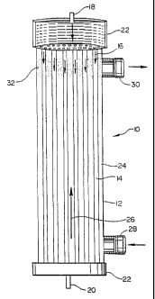
A bioreactor of the present invention in depicted schematically in FIG. 1. In
FIG. 1, the bioreactor 10 includes a housing 12 containing a plurality of
hollow
filaments 14. The housing 12 defines a central axis, and the filaments 14 are
positioned in the housing in a manner such that they run generally parallel to
the
central axis. The housing may be formed of any of a wide variety of
biocompatible
materials, including but not limited to acrylics, polycarbonates,
polysulfones, styrene-
acrylonitriles, and the like. Housing materials which are transparent to allow
visualization of the interior of the device are preferred. Furthermore, if
photoactive
processes, discussed below, are to be used in the fabrication of the device,
the use of a
transparent housing greatly simplifies the fabrication process.
The filaments 14 are hollow tubes having a central lumen. Typically, the
filaments have an external diameter on the order of about 210 to 250 microns,
however, filaments having external diameters as large as about 1 to 2 mm can
be used
as well. The filaments typically have a lumen diameter on the order of about
190 to
220 microns, however diameters of up to about 800 to 1800 microns can also be
used.
The housing typically has a length in the range of approximately 20 to 30 cm
to
5
CA 02262812 1999-01-12
WO 98/53046 PCT/US98/10111
accommodate filaments having a length in the range of approximately 16 to 26
cm.
The housing typically has an internal diameter of approximately 5 to 10 cm,
allowing
it to accommodate approximately 10,000 to 20,000 filaments. Of course, it is
noted
that the above dimensions are provided for reference only, and are not
intended to
limit the scope of the invention.
The filaments are formed of a biocompatible material through which
molecular transfer can occur. For example, the filaments may be formed from
polysulfones, cellulose acetates and the like. Other filament materials
include
polyacrylonitriles, polymethylmethacrylates (PMMA), ethylene polyvinyl alcohol
copolymers (EVAL) as well as other materials routinely used to make
hemodialysis
and similar membranes. The lumens of the filaments define a filament flow
path,
depicted by arrows 16, which is in communication with a filament inlet port 18
and a
filament outlet port 20. The filament inlet and outlet ports are preferably
positioned in
end caps 22 sealingly positioned at opposite ends of the housing 12.
The space between the exterior of the filaments 14 and the interior of the
housing 12 is referred to herein as the extrafilament space 24. The
extrafilament
space 24 defines an extrafilament flow path, depicted by arrow 26 which is in
communication with a housing inlet port 28 and a housing outlet port 30. The
housing inlet and outlet ports are preferably located adjacent to the opposite
ends of
the housing 12. It is noted that the arrangement of the filament inlet and
outlet ports
and the housing inlet and outlet ports depicted in FIG. I is such that
countercurrent
flow between the filament flow path and the extrafilament flow path is
established. It
should be noted that the filament inlet port 18 may serve as an outlet and the
filament
outlet port 20 may serve as an inlet, thereby reversing the direction of the
filament
flow path 16 and establishing cocurrent flow between the filament flow path
and the
extrafilament flow path. As will be discussed in detail below, the
extrafilament space
24 contains living cells present at a density approximating that of normal
animal
tissue. It is noted that for some cells, such as hepatocytes, extracellular
matrices, such
6
CA 02262812 1999-01-12
WO 98/53046 PCT/US98/10111
as collagen, fibronectin, laminin and the like be used to better support the
cells. In
such situations, the extracellular matrix can be mixed with the cells and
infused into
the reactor simultaneously with infusion of the cells themselves.
A flow restrictor 32 is positioned at least adjacent to the housing outlet
port
30. The restrictor typically comprises a biocompatible hydrogel. As will be
discussed
in detail below, the restrictor can be formed in situ using various methods.
The
restrictor serves as a partial barrier in the extrafilament flow path, and as
such,
establishes plug-flow conditions in the extrafilament space. Such conditions
have
been found to greatly assist in providing uniform, high density cell loading
in the
extrafilament space. As used herein, the term "at least adjacent to the
housing outlet
port" is intended to mean that the flow restrictor is positioned in a manner
such that it
restricts the flow of fluid through the housing outlet. Such positioning may
be simply
in the region of the housing at which the port is located, or it may encompass
a larger
area, extending a short distance into the extrafilament space toward the
housing inlet
port 28.
As noted above, the flow restrictor 32 may be formed of any of a wide variety
of biocompatible hydrogels. These include, but are not limited to collagen,
agarose,
calcium alginate. chitosan acetate, polyacrylamides, and combinations thereof.
It is
preferred that the hydrogel comprise at least about 90% water and less than
about 10%
polymer. In general, the flow restrictor preferably comprises a biocompatible
hydrogel which will gel under gentle conditions, at temperatures below about
50 C,
without the use of organic solvents. Gels formed by various methods including
photo-activation, aqueous catalysis, and the like can be used.
In the case of collagen, the gel is preferably formed of Type-I bovine
collagen
such as Vitrogen-100 available from Collagen Corporation of Palo Alto, CA. The
material comes as a stock 3.0 mg/ml solution and has been shown to maintain
gellability even when diluted 1:10 with a tissue culture medium.
7
CA 02262812 1999-01-12
WO 98/53046 PCT/US98/10111
Agarose is a purified form of agar, and is available from a wide variety of
sources, such as Sigma Chemical Company of St. Louis, MO. Agarose is typically
obtained in a powdered form which can be dissolved in a warm solution of water
or
tissue culture medium. Agarose will gel at room temperature in concentrations
of
about 0.1 to 3.0% w/v.
Calcium alginate is a biocompatible hydrogel that can be made in a wide
variety of concentrations. It is known that concentrations of about 1.0 to
4.0% can
successfully encapsulate mammalian cells, including hepatocytes. Calcium
alginate is
made from sodium alginate, a water soluble liquid precursor, that has been
exposed to
calcium chloride.
Chitosan acetate may be used in a wide variety of concentrations mixed with
phosphate-free tissue culture media or saline solutions. When the solution is
mixed
with tripolyphosphate solution, a biocompatible hydrogel is formed.
Polyacrylamides are well-known for their ability to form electrophoresis gels.
This gelling ability also makes them well-suited for flow restrictor
applications such
as those of the present invention.
The flow restrictor may be formed in situ using a variety of methods. In one
method, depicted in FIG. 2, the gellable material forming the plug is
introduced and
positioned by simply injecting it into the housing at the desired location. In
FIG. 2,
the bioreactor 10 is placed in a fluid circuit with a reservoir 40 of a
sterile fluid
medium, such as sterile water or Williams' E medium (available from Gibco,
Grand
Island, NY), and a pump 42. The circuit allows the sterile fluid to be
circulated
through the filament flow path of the bioreactor 10 during formation of the
flow
restrictor. A syringe pump 44 containing a gellable material in its fluid
precursor
form is placed in fluid communication with the housing outlet port 30 of the
bioreactor. The syringe pump contains a volume of gellable material that, upon
gelling, will form a flow restrictor of the desired dimensions at least
adjacent to the
housing outlet port 30. Upon activation of the syringe pump, the gellable
material is
8
CA 02262812 1999-01-12
WO 98/53046 PCTIUS98/10111
caused to enter the housing through the housing outlet port 30, and flow
downward,
by gravity, to form a liquid slug in the extrafilament space adjacent to the
housing
outlet port. The liquid slug is then gelled to form the flow restrictor.
Prior to, during or subsequent to the introduction of the gellable material
into
the housing, the sterile fluid can be circulated through the filament flow
path. Such
circulation can be used to rinse undesirable substances from the fibers, such
as
glycerin or isopropyl myristate prior to formation of the hydrogel plug in the
extrafilament space. Additionally, if the gelling is carried out by altering
the
temperature of the gellable material, the temperature of the sterile fluid in
the filament
flow path circuit can be controlled to activate and regulate the gelling
process.
A detailed description of one embodiment of the process is provided in
Example 1. Additionally, while it is noted that Example I relates to the
formation of
a collagen plug, it should be understood that the process is intended to have
applicability to the formation of other hydrogel plugs. Thus, plugs comprising
alginate, chitosan, polyacrylamides, and other two part hydrogels are
contemplated as
well. For example, in the case of alginate, a sodium alginate solution could
be
substituted for the collagen solution and additional calcium chloride would be
added
to the Williams' E medium. Thus, in one embodiment, the hydrogel is intended
to be
any hydrogel that can be formed by introducing a gellable polymer into the
extrafilament space and circulating a gelling catalyst through the filament
flow path.
Of course, in such a system, the gellable polymer must be incapable of passing
through the filament walls, whereas the catalyst must have that ability.
As an alternative, the gellable material may be positioned in the bioreactor
using centrifugation. In this method, depicted in FIGS. 3A and 3B, a plurality
of
bioreactors 10 are positioned within a centrifuge 50. Prior to placement in
the
centrifuge, the intrafilament space is pressurized to 200 to 300 mmHg and the
intrafilament access ports are sealed to allow the intrafilament space to
remain
pressurized during hydrogel injection. This prevents the hydrogel, in its
liquid state,
9
CA 02262812 1999-01-12
WO 98/53046 PCT/US98/10111
from crossing the filament walls into the filament lumens before the hydrogel
has an
opportunity to gel. The centrifuge 50 is depicted generally as having a drive
motor
52, a rotating shaft 54, and a basket 56. A nest 58, which is rotated by the
shaft 54, is
used to hold the bioreactors. Positioned above the nest 58 is a potting boat
60 which
is in fluid communication with the bioreactors through their housing outlet
ports 30.
An injector 62 is used to inject the gellable material 64 into the potting
boat 60 from
which it then enters the bioreactors. In use, the centrifuge is activated,
causing the
nest, and attached bioreactors, to rotate at approximately 250 to 500
revolutions per
minute. Since the lengths, diameters, and filament packing densities will vary
among
the numerous bioreactor configurations that are envisioned, some
experimentation
may be required to find an optimum rotation speed for each bioreactor type and
each
hydrogel precursor. The gellable material 64 is injected into the rotating
potting boat
from the injector. Due to the rotation, the gellable material is caused to
flow
outwardly from the middle of the potting boat toward its ends. The gellable
material
then exits the potting boat and enters the extrafilament space of the
bioreactors
through the housing outlet port on each. Again, as a result of the rotation,
the gellable
material is caused to move outwardly, thereby filling the ends of the
bioreactors
adjacent to the housing outlet ports. In this method, the hydrogel formation
should
occur as a result of a temperature change, such as by injecting hot agarose
and
allowing it to cool and set before stopping the centrifuge, or by using a
hydrogel that
has been precatalyzed, yet has a pregelling time, such as a polyacrylamide.
The use of
centrifugation is particularly desired in circumstances where the viscosity of
the
gellable material is so high as to prevent gravity from adequately positioning
the
material as in FIG. 2.
Once the plug has been formed, the bioreactor is in condition for the
insertion
of the living cells and, if necessary, their supporting matrix. In one
preferred
embodiment, the living cells comprise living mammalian cells, such as
hepatocytes.
The cells preferably are introduced into the bioreactor at a loading density
of at least
CA 02262812 1999-01-12
WO 98/53046 PCT/US98/10111
about 10' cells per milliliter, more preferably at a loading density of at
least about 108
cells per milliliter. Although not necessary, the cells may be maintained or
encapsulated within a gel matrix such as a collagen matrix or the like. In
such a case,
the cell-supporting matrix may be, but need not be, of the same gel used to
form the
flow restrictor. It should be noted that although the use of hepatocytes is
described in
detail throughout, the present invention is not intended to be limited to use
with that
specific cell type. Rather, any of a wide variety of cells, both primary and
transformed. may be used in the present system including, but not limited to,
hybridomas. chinese hamster ovary (CHO) cells, baby hamster kidney (BHK)
cells,
endothelial cells, epithelial cells, and fibroblasts.
In one preferred embodiment depicted in FIG. 4, living cells are introduced
into the bioreactor using a method similar to that used to position the flow
restrictor.
More particularly, the bioreactor 10 is placed in a fluid circuit with a
reservoir 70 of a
cold, sterile fluid medium, such as William's E medium, and a pump 72. The
circuit
allows the cold, sterile fluid to be circulated through the filament flow path
of the
bioreactor 10 during introduction of cells into the bioreactor. Similar to the
procedure
shown in FIG. 2, the cold, sterile fluid can be circulated from the reservoir
70 by
pump 72 into the filament inlet port 18, along the filament flow path, out the
filament
outlet port 22 and back to the reservoir. The primary purpose of these steps
is to keep
the bioreactor cold (approx. 4 to 10 C) while the cells are being loaded.
Alternatively, the cell loading operation can be conducted in a controlled,
chilled
environment, such as in a refrigerator or a cold room.
While circulation of the cold, sterile fluid through the filament flow path is
proceeding, the extrafilament space is loaded with cells and, if necessary, a
cell-
supporting matrix as follows. A reservoir 80 containing the cells is provided.
The
reservoir is preferably chilled to help maintain cell viability. The solution
is
transferred from the reservoir 80 by a pump 82 driven by a motor 84. The cell-
containing solution enters the bioreactor 10 through the housing inlet port
28. The
11
CA 02262812 1999-01-12
WO 98/53046 PCTIUS98/1011 I
solution then fills the extrafilament space. As more solution is pumped into
the
bioreactor, a portion of the solution, is caused to pass through the flow
restrictor 32
and the housing outlet port 30. The flow restrictor preferably acts as a
filter to trap
cells and prevent them from exiting the extrafilament space, while allowing
the fluid
being displaced by the cells to exit. To assist in the loading process, by
reducing
pressure pulses and differential pressures across the plug, a second pump head
86
connected to the same motor 84 can be used to simultaneously withdraw the same
volume of fluid as the volume of cells being pumped into the extrafilament
space of
the bioreactor. Fluid exiting the bioreactor is channeled into a waste
receptacle 88.
Because the flow restrictor 32 is a hydrogel, it should be apparent that it
will
impede fluid flow therethrough and causes the significant pressure drop across
the
restriction. This restriction also causes an increase of the pressure in the
extrafilament
space which contributes to plug-flow conditions through that space. As a
result of the
plug-flow conditions, the cell-containing solution is caused to uniformly
disperse
throughout the extrafilament space, and uniformly distribute cells
therethrough. As a
result, shunt flow is avoided, and a dense, uniform cell-loading is caused to
occur.
For determining how much of the cell-containing solution to use, in one
preferred
embodiment, the amount is determined to be the volume of the extrafilament
space
less the volume of the flow restrictor. Thus, in a reactor in which the
extrafilament
space has a volume of about 140 ml and the restrictor occupies a volume of
about 40
ml, 100 ml of the cell-containing solution would be used.
If the cells used in the present invention are freshly isolated hepatocytes, a
period of 16 to 24 hours of culture is preferably provided to allow the
hepatocytes to
recover from the trauma of the enzymatic digestion of the liver. Rather than
causing
the cells to multiply and fill the extrafilament space, however, the purpose
of the
culture phase as used in the present invention is to allow the cells to
recover while
establishing natural physiologic-like conditions of pH, temperature and media
composition to allow the cells to remain alive and productive.
12
CA 02262812 1999-01-12
WO 98/53046 PCT/US98/10111
The resulting, cell-loaded bioreactor offers numerous uses. For example, if
the
cells are hepatocytes, the bioreactor may be used to sustain a patient
undergoing full
or partial liver failure until liver function returns or a suitable transplant
can be
provided. The bioreactor acts as an artificial liver and purifies the blood.
Once the cells have been loaded into the bioreactor, the bioreactor can be
placed into any of several types of culture circuits. Examples of such culture
circuits
are described in U.S. Patent No. 3,883,393 (Knazek), U.S. Patent No. 4,220,725
(Knazek), and U.S. Patent No. 4,804,628 (Cracauer). If the cells have been
genetically altered to secrete a biologic product, it is possible, using the
present
invention, to begin harvesting cell secreted product almost immediately. This
may be
contrasted with conventional systems in which it is often necessary to wait
days or
weeks for the cells to undergo multiple divisions and fill the reactor prior
to secreting
useful amounts of biologic products.
A system in which the bioreactor contains hepatocyes and is used to purify
blood is depicted schematically in FIG. 5. In FIG. 5, blood from a patient 100
is
pumped by a blood pump 102 and mixed with heparin 104 which has been pumped by
a heparin pump 106. Pressure monitoring apparatus 108 is a safety device. If a
problem occurs with the patient, the patient's blood access site. or the
tubing leading
from the patient, an increase in negative pressure will be detected and can be
used to
activate an alarm. The heparinized blood is then heated in a blood warmer 110
to a
desired temperature and the introduced into an oxygenator 112. The oxygenator
112
is provided with sources of oxygen 114, carbon dioxide 116 and nitrogen 118
each
regulated by a flow controller (120, 122 and 124, respectively) so that a
desired blood
gas content and blood pH can be achieved and maintained. The oxygenator
includes
pressure and temperature monitoring equipment, and other blood handling
subsystems
commonly used in the art. The oxygenated blood is monitored by a pH probe 126
and
then passed through the filament flow path of the bioreactor 10. As the blood
passes
through the filament flow path, numerous impurities of the type normally
removed by
13
CA 02262812 1999-01-12
WO 98/53046 PCT/US98/10111
the liver are caused to diffuse through the filament walls to the hepatocytes.
The
hepatocytes provide liver function of the type normally occurring in vivo to
the blood.
During this process, the hepatocytes can be maintained by a nutrient medium
which
travels through the extrafilament flow path. The nutrient medium is provided
by a
nutrient reservoir which includes pressure monitoring subsystems to determine
the
amount of nutrient remaining in the nutrient reservoir. Spent nutrient media
exits the
bioreactor and travels to a media waste receptacle 130. It should be noted,
however,
that the use of a circulating nutrient solution, particularly during treatment
of a
patient, is entirely optional, and need not be performed. Treated blood
exiting the
bioreactor is passed through an air/foam detector and then back to the
patient.
It is believed that systems of the type described and depicted in FIG. 5 may
be
used to maintain patients undergoing partial or full liver failure for up to
until a
suitable donor liver can be found or until the patient's liver recovers
acceptable
function. In this latter case, several intermittent treatments over the course
of several
days or weeks may be required. This offers the benefit of providing sufficient
time
for the patient's liver to return to normal function or to locate a donor
organ sufficient
for transplantation. Of course, it should be noted that the bioreactors of the
present
invention are not intended to be limited strictly to liver function, but
rather, that such
reactors may be suitable for a broad range of clinical applications in which
it is
desirable to provide extracorporeal systems for maintaining living cells at
densities
and functionalities approaching those of normal animal tissue.
Examples
Example 1: Formation of a Collagen Hydro el Plug in a Hepatocyte Bioreactor
In this example, reference numerals refer to the reference numeral of the
Figures, particularly FIGS. I and 2.
One liter of filter sterilized Williams' E medium, pH 7.40, at 4 C was
introduced into a reservoir 40. Sterile tubing was used to connect the
reservoir to a
14
CA 02262812 1999-01-12
WO 98/53046 PCT/US98/10111
pump 42 and from the pump to one filament port of the bioreactor. Additional
sterile
tubing was connected from the other filament port of the bioreactor to a
temporary
sterile waste receptacle. Approximately 45 ml of sterile Vitrogen-100 was
drawn into
a sterile 60 ml syringe, and the syringe was connected, via sterile tubing to
the
housing inlet port 28. A sterile hydrophobic air filter and sphygmomanometer
was
connected to the housing outlet port 30. The sphygmomanometer was arranged to
communicate with the extrafilament space through the filter in order to
maintain
sterility of the bioreactor.
The extrafilament space was pressurized to approximately 200 to 300 mmHg
pressure, and the pump 42 was started. Approximately 500 ml of Williams' E
medium was allowed to flow through the bioreactor and into the sterile waste
receptacle before further steps were carried out. This was done in order to
rinse the
filaments and to remove any glycerin or other unwanted contaminants from their
interior. The pump was then stopped and the outlet tube from the reactor was
clamped and transferred from the sterile waste receptacle to the reservoir 40.
The
extrafilament space was then depressurized and maintained at ambient
atmospheric
pressure.
The collagen-filled syringe was then positioned in a syringe pump 44, and
about 40 ml of Vitrogen-100 was pumped into the extrafilament space of the
bioreactor. The collagen was pumped into the bioreactor at a rate of about 40
ml per
hour. Once the collagen had been pumped into the bioreactor, the tube
connecting the
syringe to the extrafilament inlet port was clamped off. The other
extrafilament port
was closed off as well, by closing the needle valve on the sphygmomanometer.
The
intralumenal outlet tubing was unclamped and the intralumenal circulation pump
was
turned on at a flow rate of approximately 30 ml per minute.
As Williams' E medium (pH 7.4) flowed through the filaments, the pH of the
collagen in the extrafilament space was caused to rise from about 2.0 to
neutral. In so
doing, the collagen was caused to gel. During the initial phase of the
gelling, the
CA 02262812 1999-01-12
WO 98/53046 PCT/US98/10111
pressure in the extrafilament flow path was closely controlled, by adjustment
of the
sphygmomanometer needle valve. The close control was required to prevent
ultrafiltration, a condition in which media can cross from the filament flow
path into
the extrafilament space through the filament walls. This condition is
undesirable as it
dilutes and changes the volume of the hydrogel solution in the extrafilament
space.
This was accomplished by carefully observing the level of the collagen
solution in the
extrafilament space and adjusting the pressure in that space as described
above.
Although not wishing to be bound by any particular theory, it is believed that
the
pressure rises primarily as a result of CO2 gas that evolves as hydrochloric
acid in the
collagen solution is neutralized by sodium bicarbonate resident in the
Williams' E
medium.
After the Williams' E medium was circulated at a rate of about 30 ml/min for
approximately 30 to 45 minutes, the rate of the intrafilament circulation pump
was
doubled to about 60 ml/min for about 30 minutes, and the reservoir 40 was
placed in a
37 C water bath. The rate of the intrafilament circulation pump was doubled
every 30
minutes until a flow rate of about 240 ml/min was achieved. At that point,
gellation
of the collagen was substantially complete. The remainder of the extrafilament
space,
not occupied by the collagen plug, was allowed to fill, via ultrafiltration,
with
Williams' E medium by venting the extrafilament space until the medium reached
the
hydrophobic air filter. The medium was then allowed to circulate for several
hours to
ensure that the collagen plug was fully set, and to neutralize any residual
hydrochloric
acid.
Example 2: Hepatocvte Harvest and Isolation
Porcine hepatocytes were harvested from 7 to 12 kg male pigs (Midwest
Research Swine, Gibbon, MN) using a two step collagenase technique. The
animals
were anesthetized with ketamine intramuscularly, intubated, and sterilely
prepared and
draped. A midline incision was used to enter the abdomen. The infrahepatic
inferior
16
CA 02262812 1999-01-12
WO 98/53046 PCT/US98/10111
vena cava (IVC) and suprahepatic IVC above the diaphragm were isolated and
encircled. The protal triad was then dissected, ligated and all structures
were divided
except the hepatic artery and the portal vein, which were isolated and looped.
Heparin, at 300 I.U. per kg , was given. After 3 minutes, the cannula,
connected by
sterile silicone tubing to a peristaltic pump, was passed into the surgical
field. The
hepatic artery was then ligated and divided, and the portal vein was
cannulated. Five
liters of PER-I solution (calcium-free hydroxyethylpiperazine-ethanesulfonic
acid
(HEPES) buffered solution (143 mM NaCl, 6.7 mM KC1, 10 mM HEPES, 100 mg%
ethylene glycol-bis-aminoethyl ether (EGTA), pH 7.4), at a rate of 1 liter per
kg body
weight was perfused through a silicone oxygenator and heat exchanger (Avecor
Cardiovascular, Minneapolis, MN) and then directly into the portal vein to
wash out
the blood. The suprahepatic IVC was then ligated and divided, and the
infrahepatic
IVC was divided to allow outflow of the perfusate. During the initial
perfusion, the
liver was transferred to a sterile tray. Two liters of PER II (HEPES buffered
solution
(67 mM NaCl, 6.7 mM KCI, 4.8 mM CaCl2, 100 mM HEPES, pH 7.6), 1 gm/liter
Collagenase-D (Boehringer Mannheim, Indianapolis, IN)) was then recirculated
through the liver, at the same flow rate as the PER-I, for 10 to 15 minutes
until the
liver was soft and well digested, as determined by palpation and visual
inspection.
The liver was removed to a laminar flow hood and the capsule was incised on
all lobes. Next, the liver was skeletonized bluntly with gentle agitation in 5
C
Williams'-E medium to free the hepatocytes. The cell laden media was filtered
through a Buchner funnel which contained a single layer of sterile gauze to
remove
any tissue fragments, into sterile 250 ml centrifuge tubes which were spun at
500 to
700 RPM for 5 minutes to form a soft cell pellet. The pellet was resuspended
in fresh
medium and centrifuged two more times. Hepatocyte viability and cell number
was
assessed by using a hemocytometer and Trypan Blue dye exclusion methods.
17
CA 02262812 1999-01-12
WO 98/53046 PCT/US98/10111
Example 3: Bioreactor Loading
Approximately 100 ml of hepatocyte cell pellet was transferred to a sterile
250
ml polyethylene bottle. 25 ml of Vitrogen-1 00 was added to the cells and the
bottle
was gently swirled to mix the collagen and cells. 100 ml of this solution was
pipetted,
50 ml into each of two 60 cc syringes (Becton Dickinson, Franklin Lakes, NJ)
and
connected to a circuit similar to the one shown in Fig. 4. The syringes were
placed on
ice to keep the cells cool. The cell loading pump 84 was set to a rate of 100
ml/hr to
inject the cells into the extrafilament space over a one hour period. The
intrafilament
pump 72 was set to a flow rate of 300 ml/min to keep the bioreactor cool
during cell
loading. After all of the hepatocyte solution was pumped into the
extrafilament space,
sterile caps were placed on all of the ports and the bioreactor was
transferred to an
incubator in which CO2 and humidity were controlled.
Example 4: Bioreactor Culture
An example of the fluid circuits employed during the culturing phase of the
bioreactor manufacture is depicted schematically in FIG. 6. The hepatocyte
containing bioreactor was placed in the culture circuit and maintained for
approximately 18 hours in the incubator to allow the cells to rest. During
this time the
cells can recover from the stress of harvest and re-establish cell to cell
contact and
remodel their local environment.
The incubator CO, was maintained at 5.5%, temperature at 37 C and the
humidity at 70-80%. The bioreactor was maintained in a circuit of the type
shown in
FIG. 6. The intrafilament circuit was comprised of a 1 liter reservoir 150
containing
Williams'-E medium formulated as shown in Table 1:
18
CA 02262812 1999-01-12
WO 98/53046 PCT/US98/10111
TABLE 1: Intrafilament Medium
Component Amount per liter Source/Quality
Wm's-E Powder with Glutamine 10.79 g Life Technologies
Sodium Bicarbonate 2.2 grams Sigma Chemical
Penicillin 40,000 Units Life Technologies
Streptomycin 400 mg Life Technologies
Transferrin 6.25 mg Sigma
Epidermal Growth Factor 5.05 ug Sigma
Insulin 4 mg / 111.8 IU Sigma
Glucagon 0.4 mg Sigma
Selenium 2.854 ug Sigma
Dexamethasone 329.5 ug Sigma
A peristaltic pump 152 operating at 700-900 ml/min was used to pump media
from the I liter reservoir 150 through a silicone membrane oxygenator 154,
where the
media is oxygenated and pH adjusted to 7.2. Oxygenation and pH control via the
oxygenator was achieved by using an air pump (MEDO USA, Hanover Park, IL) (not
shown) to circulate the incubator gasses through the gas path of the
oxygenator. From
the oxygenator, the media was delivered into the intraluminal space of the
bioreactor
10, and then returned to the I liter reservoir 150 from the outlet 18 of the
intraluminal
space of the bioreactor. Sample points S, and Sz were used to periodically
withdraw
media, before and after the bioreactor, for blood gas analysis of pH and
oxygen
content. Recirculation was maintained until the bioreactor was removed for
connection to a liver injured dog.
On the extrafilament side of the bioreactor was a second circulation path
which included a 500 ml reservoir 156 of media formulated as shown in Table 2:
19
CA 02262812 1999-01-12
WO 98/53046 PCT/US98/10111
TABLE 2: Extrafilament Medium
Component Amount per liter Source/Quality
Wm's-E Powder with Glutamine 10.79 g Life Technologies
Sodium Bicarbonate 2.2 g Sigma Chemical
Penicillin 40000 Units Life Technologies
Streptomycin 40 mg Life Technologies
Bovine Serum Albumin- 10% 500 mg (HSA) Sigma
linoleic acid
Transferrin 6.25 mg Sigma
Epidermal Growth Factor 5.05 ug Sigma
Insulin 4 mg / 111.8 IU Sigma
Glucagon 0.4 mg Sigma
Selenium 2.854 ug Sigma
Dexamethasone 329.5 ug Sigma
Circulation of the extrafilament media was not started until 12 hours after
the
bioreactor had been installed in the incubator. The circulation rate was 30
ml/hour
and was continued until the bioreactor was removed for use on the liver
injured dog.
Example 5: Canine Liver Iniury and Bioreactor Treatment
Evaluation of the hepatocyte-containing bioreactor for use as a bioartificial
liver was conducted using a lethal canine galactosamine liver failure model
similar to
that described by Sielaff et al. (Hepatology, 21:(3), 1995). Modifications
from the
protocol as published by Sielaff included replacement of halothane with
isofluorane
as the anesthetic and more frequent laboratory testing to manage the animal
proactively. In addition, the source animals were purpose-bred, well-
conditioned
hunter hounds instead of the mongrel animals reported in the Sielaff studies.
As a
result of these changes, the animals lived somewhat longer than reported and
the
studies were consequently extended to 60 hours.
Dogs receiving D-galactosamine (1.5 gram/kilogram body weight) were
allowed to proceed into liver failure for 24 hours at which point
hemoperfusion with
the bioartificial liver was initiated. The vascular access device was a dual
lumen
CA 02262812 1999-01-12
WO 98/53046 PCT/US98/10111
catheter surgically implanted in the right internal jugular vein prior to
administration
of the D-galactosamine.
The treatment objective was to hemoperfuse at the highest possible flow rate
until the animal expired or reached the end of the 60 hour study period and
was
electively euthanized. In practice, initial flow rates typically exceeded 200
ml/min.
This rate dropped as the animal became increasingly edematous and the patency
of the
vascular access deteriorated. In no case was the catheter lost to clotting. In
some
studies, blood flow rate dropped to less than 100 ml/min 50 hours after drug
administration or after 26 hours of continuous hemoperfusion.
Four animals were treated in this manner with bioreactors charged with 60 to
80 grams of primary porcine hepatocytes. The animals were managed according to
the same intervention schedule as the control subjects. Two of the four
treated
animals survived to the end of the study period. In contrast none of the
untreated
animals did. The survival difference is shown in the form of the Kaplan-Meier
plot of
FIG. 7.
The five untreated control animals and four treated animals included in the
data set were evaluated by the Kaplin-Meier survival statistic. The results do
not
reach statistical significance (p=0.06) due to sample size and to the
arbitrary
termination condition for the study.
TABLE 3: Statistical Evaluation
Variable Group Time
mean 47.8
Untreated sem 2.9
Survival n 5
Time mean 55.8
Treated sem 2.5
n 4
t-test 0.080
Statistic Log Rank 0.065
Breslow 0.064
Tarone-Ware 0.063
21
CA 02262812 1999-01-12
WO 98/53046 PCT/US98/10111
Animals were fitted with LADD superdural Intracranial pressure monitors.
These were monitored continuously throughout the study but data were recorded
hourly. The results for each of the four treated animals are shown in FIG. 8
along
with the mean of these data. The animals supported with the bioartificial
liver showed
an oscillatory ICP pattern similar to that exhibited by the control animals.
As before,
the sampling interval chosen obscures the surges in ICP observed by the
attending
staff.
The average reading of the five control and four treated animals are compared
in FIG. 8. Statistical tests were not applied to these data due to a variety
of
confounding factors present in the data set. These factors include the fact
that more
frequent and aggressive medical intervention occurred in the untreated animals
(as
permitted under the management guidelines), than was evidently necessary with
the
treated animals. Bias is also introduced because the longer lived untreated
animals
have on average lower ICP values than those expiring earlier in the study.
With both ICP data and mean arterial pressure (MAP) data available, it is
possible to calculate the cerebral perfusion pressure (CPP) in these study
animals. As
shown in FIG. 9, CPP declines in treated animals as it does in the untreated
ones.
Furthermore, those animals with the lower values appear to have shorter
survival
times.
The arithmetic means of the CPP values for treated and untreated dogs are
plotted together in FIG. 9. Decreasing blood flow to the brain is present in
both
groups of animals. The rise in CPP in the untreated animals between 30 and 40
hours
after administration of the drug may be due to more aggressive intervention to
correct
falling blood pressure in the untreated population.
As shown in FIG. 10, the rise in arterial ammonia levels is moderated in
treated animals. The effect is not statistically significant at 40 and 48
hours, because
of the large dispersion in the data from the untreated animals. The
convergence of data
22
CA 02262812 1999-01-12
WO 98/53046 PCT/US98/10111
at 56 hours is a result of the fact that the sicker untreated animals, those
with higher
values of ammonia, have shorter survival times.
The ratio of branched chain amino acids (BCAA) and aromatic amino acids
(AAA) declines in both treated and untreated animals, as shown in FIG. 11. The
difference reaches statistical significance at 48 hours. Intermediate values
were not
obtained and it is not clear if the magnitude of this change is sufficient to
generate a
meaningful patient benefit. The rate of oxygen consumption in the bioreactor
before
and after connection to a dog is shown in FIG. 12.
Equivalents
Various modifications and alterations to this invention will become apparent
to those skilled in the art without departing from the scope and spirit of
this invention.
It should be understood that this invention is not intended to be unduly
limited by the
illustrative embodiments and examples set forth herein and that such examples
and
embodiments are presented by way of example only with the scope of the
invention
intended to be limited only by the claims set forth herein as follows.
23