Note: Descriptions are shown in the official language in which they were submitted.
CA 02262873 1999-01-13
!
ROTATIONAI ATHF~F.CTOMY DEVICE
TFCHNICA~ FIF.I,D
The invention relates to devices and methods for removing tissue from body
passageways, such as removal of atherosclerotic plaque from arteries, utili7ing a
rotational atherectomy device.
BACKGROUND OF THF INVFNTION
A variety of techniques and instruments have been developed for use in the
removal or repair of tissue in arteries and similar body passageways. A frequentobjective of such techniques and instruments is the removal of atherosclerotic plaques
10 in a patient's arteries. Atherosclerosis is characterized by the buildup of fatty deposits
(atheromas) in the intimal layer (under the endothelium) of a patient's blood vessels.
Very often over time, what initially is deposited as {elatively soft, cholesterol-rich
atheromatous material hardens into a calcified atherosclerotic plaque. Such atheromas
restrict the flow of blood, and therefore often are referred to as stenotic lesions or
15 stenoses, the blocking material being referred to as stenotic material. If left untreated,
such stenoses can cause angina, hypertension, myocardial infarction, strokes and the
like.
Several kinds of atherectomy devices have been developed for attempting to
remove some or all of such stenotic material. In one type of device, such as that
20 shown in U.S. Pat. No. 4,990,134 (Auth), a rotating burr covered with an abrasive
cutting material such as diamond grit (diamond particles or dust) is carried at the
distal end of a flexible drive shaft. The rotating burr is rigid and inflexible, however,
making navigation around tight bends or curves in an artery more difficult, and
making the removal of stenotic lesions in such bends or curves equally difficult.
U.S. Pat. No. 5,314,438 (Shturman) shows another atherectomy device having
a drive shaft with a section of the drive shaft having an enlarged diameter, at least a
segment of this enlarged diameter section being covered with an abrasive material to
define an abrasive segment of the drive shaft. When rotated at high speeds, the
abrasive segment is capable of removing stenotic tissue from an artery. In some of the
30 embodiments depicted in the Shturman patent, wire tums of the enlarged diameter
segment of the drive shaft are supported by a bushing. Even though this bushing may
CA 02262873 1999-01-13
.
_ 2
be made of a flexible material, nevertheless it decreases somewhat the flexibility of
the enlarged diameter abrasive segment of the drive shaft.
Unless a bushing within the enlarged diameter section is utilized, Applicants
have found that adjacent wire strands of this section can fall out of alignment with one
5 another when the enlarged diameter portion of the drive shaft is bent around a curve of
a relatively small radius.
SUMMA~Y OF THF ~NVFNTION
The invention provides an atherectomy device comprised of a flexible,
elongated drive shaft having an enlarged diameter tissue removal section which
10 retains substantial flexibility while reducing the tendency of the wire turns to fall out
of alignment with one another. The tissue removal section of the drive shaft of this
atherectomy device includes proximal and distal portions comprised of helically
wound wire, wire turns of the proximal portion of the tissue removal section having
diameters that progressively increase distally at a generally constant rate thereby
15 forming generally the shape of a cone. Wire turns of the distal portion of the enlarged
di~meter tissue removal section have di~meters that gradually decrease distally
thereby forming a generally convex distal portion.
The conical shape of the proximal portion of the tissue removal section
subst~nti~lly reduces the tendency of the wire tums to fall out of aligrlment with one
20 another, without the need to utilize a bushing, thereby preserving substantial
flexibility in the enlarged diameter section.
At least part of the tissue removal section includes an external coating of an
abrasive material, secured to the wire tums of the drive shaft by a suitable binder, to
define an abrasive segment of the drive shaR. Preferably the binder also secures some
25 of the adjacent wire turns of the tissue removal section to one another, most preferably
throughout a distal portion of the tissue removal section.
In a preferred embodiment, the drive shaft of the atherectomy device includes
a reduced diameter segment, such segment being located near the tissue removal
section of the drive shaft to function as a bearing for rotation of the drive shafl about a
30 guide wire. The reduced clearance between the guide wire and the inner surface of the
.reduced diameter segment is less than in other portions of the drive shaft and is
CA 02262873 1999-01-13
,,
intended to reduce vibrations of the tissue removal section and facilitate smooth
rotation of the drive shaft and its tissue removal section about the guide wire when the
atherectomy device is rotated at high speeds. Two or more of such reduced diameter
segments may be included, preferably at least one being located distally of the tissue
5 removal section of the drive shaft, and at least one being located proximally of the
tissue removal section of the drive shaft. Preferably such reduced diameter segments
are located within about one inch from the enlarged diameter tissue removal section,
and most preferably within about a quarter inch from such tissue removal section.
The maximum outer diameter and length of the abrasive segment of the
10 enlarged diameter tissue removal section may be selected so that, at operational
rotational speeds and under load, at least some of the wire turns of the proximal
portion of the enlarged diameter tissue removal section unwind from their at-rest
diameter to an effective outer diameter which is the same as or larger than the
maximum outer diameter of the abrasive segment.
The drive shaft of the atherectomy device may include a distal end segment
having an outer diameter which de~ ases distally to define a generally convex outer
surface. At least a portion of this distal end segment may be provided with an
external coating of an abrasive material to define a second abrasive segment at the
very distal end of the drive shaft. Such an abrasive coated distal end segment
facilitates passage of the rotating drive shaft of the atherectomy device across even
very tight stenoses.
BRIFF DF.~C~IPTION OF THF DRAWINGS
Figure I is a perspective view of an atherectomy device of the invention;
Figure 2 is a broken-away, longitudinal cross-sectional view of the enlarged
diameter tissue removal section of the atherectomy device shown in Figure l;
Figure 3 is a broken-away, longitudinal cross-sectional view of the enlarged
diameter tissue removal section of a prior art atherectomy device;
Figure 4 is a broken-away, longitudinal cross-sectional view of the
atherectomy device of Figure 2 depicted in a slightly curved configuration;
Figure 5 is a broken-away, longitudinal cross-sectional view of the prior art
atherectomy device of Figure 3 depicted in a slightly curved configuration;
CA 02262873 1999-01-13
t
Figure 6 is a broken-away, longitudinal cross-sectional view of the
atherectomy device of Figure 2 depicted in a more tightly curved configuration;
Figure 7 is a broken-away, longitudinal cross-sectional view of the prior art
atherectomy device of Figure 3 depicted in a more tightly curved configuration;
Figure 8 is a broken-away, longitudinal cross-sectional view of a mandrel used
in manufacturing a rotational atherectomy device of the invention;
Figure 9 is a broken-away, longitudinal cross-sectional view of a modified
embodiment of the invention having a distal end segrnent coated with abrasive
material to define a second abrasive segment at the very distal end of the atherectomy
I O device;
Figure 9A is an enlarged view of the distal section of the atherectomy device
of Figure 9;
Figure 10 is a broken-away, longitudinal cross-sectional view of a modified
embodiment of the invention having a distal end segment partially coated with
abrasive material to define a second abrasive segrnent at the very distal end of the
atherectomy device;
Figure I OA is an enlarged view of the distal section of the atherectomy device
of Figure 10;
Figure 11 is a broken-away, longitudinal cross-sectional view of a modified
embodiment of the invention having a layer of tubing covering the section of the drive
shaft in between the two abrasive segrnents of the atherectomy device;
Figure I IA is an enlarged view of the distal section of the atherectomy device
of Figure I l;
Figure 12 is a broken-away, longitudinal cross-sectional view of a modified
embodiment of the invention having a distal section with a diameter smaller than the
diameter of the proximal section of the atherectomy device;
Figure 12A is an enlarged view of the distal section of the atherectomy device
of Figure 12;
Figure 1 2B is an enlarged view of portions of the proximal section of the
atherectomy device of Figure 12;
CA 02262873 1999-01-13
- S
Figure 13 is a broken-away, longitudinal cross-sectional view of a modified
embodiment of the invention having a distal section with a reduced diameter, and a
single reduced diameter segment located proximally of the enlarged diameter tissue
removal section;
Figure 1 3A is an enlarged view of the distal section of the atherectomy device
of Figure 13;
Figure 1 3B is an enlarged view of portions of the proximal section of the
atherectomy device of Figure 13;
Figure 14 is a perspective view of a clamp used in m~nuf~cturing the
rotational atherectomy device shown in Figure 13, which has a distal section with a
reduced diameter, and a single reduced diameter segment located proximally of the
enlarged diameter tissue removal section;
Figure 14A is a longitudinal cross-sectional view of the clamp of Figure 14;
Figure 14B is an enlarged view showing in longitudinal cross-section details
I S of a portion of Figure 14A;
Figure 15 is an enlarged cross-sectional view, partially broken away, of Figure
14A, taken along lines 15-15 thereof;
Figure 16 is a broken-away, longitudinal cross-sectional view of a modified
embodiment of the invention with a distal section having a segment with a reduced
diameter, and a similar reduced diameter segment located proximally of the enlarged
diameter tissue removal section;
Figure 1 6A is an enlarged view of the distal section of the atherectomy device
of Figure 16;
Figure 1 6B is an enlarged view of portions of the proximal section of the
atherectomy device of Figure 16;
Figure 17 is a broken-away, longitudinal cross-sectional view of a modified
embodiment of the invention with a distal section having two segments with a reduced
diameter, and a similar reduced diameter segment located proximally of the enlarged
diameter tissue removal section;
Figure 1 7A is an enlarged view of the distal section of the atherectomy device
of Figure 17;
CA 02262873 1999-01-13
~_ 6
Figure 1 7B is an enlarged view of portions of the proximal section of the
atherectomy device of Figure l 7;
Figure 18 is a broken-away, longitudinal cross-sectional view of a modified
embodiment of the invention with a distal section having two segments with a reduced
5 diameter, and two similar reduced diameter segments located proximally of the
enlarged diameter tissue removal section;
Figure 19 is a broken-away, longitudinal cross-sectional view of a modified
embodiment of the invention similar to Figure 13, but wound from a single strand of
wire;
Figure 20 is a broken-away, longitudinal cross-sectional view of an
atherectomy device of the invention, showing both the extent of coverage by the
abrasive material of the enlarged diameter tissue removal section of the rotational
atherectomy device, and a moved position of the wire tums of the enlarged diameter
tissue removal section of the device when the wire tums are unwinding under load;
Figure 21 is a broken-away, longitudinal cross-sectional view of another
embodiment of an athele~,to.,.y device similar to Figure 20, the wire tums, in their
moved position, being depicted as unwinding to a slightly larger diameter;
Figure 22 is a broken-away, longitudinal cross-sectional view of an
atherectomy device similar to Figure 20, with the proximal end of the abrasive
20 segment terrnin~ing at the maximum diameter of the enlarged diameter tissue
removal section;
Figure 23 is a broken-away, longitudinal cross-sectional view of an
atherectomy device similar to Figure 21, with the proximal end of the abrasive
segment terrnin~ting at the maximum diameter of the enlarged diameter tissue
25 removal section;
Figure 24 is a broken-away, longitudinal cross-sectional view of an
atherectomy device of the invention, with the proximal end of the abrasive segment
temminating distally of the maximum diameter of the enlarged diameter tissue removal
section;
CA 02262873 1999-01-13
'
Figure 25 is an enlarged longitudinal cross-sectional view of a portion of a
rotational atherectomy device of the invention, illustrating changes in the longitudinal
cross-sectional profile of its enlarged diameter tissue removal section;
Figure 26 is a broken-away, longitudinal cross-sectional view of another
5 atherectomy device of the invention, illustrating a slightly different profile of the
enlarged diameter tissue removal section;
Figure 27 is an enlarged view of a portion of the atherectomy device of Figure
26, illustrating the changes in the longitudinal cross-sectional profile of its enlarged
diameter tissue remo.val section;
Figure 28 is a broken-away, longitudinal cross-sectional view of another
atherectomy device of the invention, illustrating a different profile of the enlarged
diameter tissue removal section;
Figure 29 is a broken-away, longitudinal cross-sectional view of another
atherectomy device of the invention, illustrating yet another profile of the enlarged
diameter tissue removal section;
Figures 30A and 30B are broken-away, longitudinal cross-sectional views of
atherectomy devices of the invention, illustrating proportionality of such devices of
different sizes;
Figures 3 IA and 3 IB are broken-away, longitudinal cross-sectional views of
atherectomy devices of the invention, each having a distal end segment coated with
abrasive material to define a distal end abrasive segment;
Figure 32 is a broken-away, longitudinal cross-sectional view of a distal
section of a modified atherectomy device of the invention with wire turns of the distal
section having a thin extemal coating of a radio-opaque material;
Figure 33 is a broken-away, longitudinal cross-sectional view of a distal
section of a modified embodiment similar to Figure 32, but with the radio-opaquematerial coating the entire circumference of wire tums in the distal section of the
atherectomy device;
Figure 34 is a broken-away, longitudinal cross-sectional view of a distal
section of another modified embodiment similar to Figure 33, but with the radio-
CA 02262873 1999-01-13
opaque material coating all but the inner surface of wire tums in the distal section of
the atherectomy device;
Figure 35 is a broken-away, longitudinal cross-sectional view of a distal
section of a modified atherectomy device of the invention with the distal end
5 encapsulated by a generally cylindrical ring made from a radio-opaque material;
Figure 36 is a broken-away, longitudinal cross-sectional view of the distal
section of a modified atherectomy device of the invention with the distal end rounded
off;
Figure 37 is a broken-away, longitudinal cross-sectional view of the
10 embodiment of Figure 36 with the distal end coated with a bonding material to secure
the wire tums to one another;
Figure 38 is a broken-away, longitudinal cross-sectional view of the distal
section of a modified atherectomy device of the invention with the distal end trimmed
off "square";
Figure 39 is a broken-away, longitudinal cross-sectional view of the
embodiment of Figure 38 with the distal end coated with a bonding material to secure
the wire tums to one another;
Figure 40 is a broken-away, longitudinal cross-sectional view of the distal
section of a modified atherectomy device of the invention with the distal end
20 electroplated before being trimmed to its finished length;
Figure 41 is a broken-away, longitudinal cross-sectional view ofthe
embodiment of Figure 40 with the distal end trimmed to its finished length;
Figure 42 is a broken-away, longitudinal cross-sectional view of a modified
embodiment of the invention having a drive shaft with two distally tapered segments;
25 and
Figure 43 is a broken-away, longitudinal cross-sectional view of a mandrel
used in manufacturing the atherectomy device of Figure 42.
CA 02262873 1999-01-13
)
DFTAILED DFSCRIPTIO~ OF THE INVFNTION
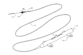
Figure I illustrates a typical rotational atherectomy device of the invention.
The device includes a handle portion 10, an elongated, flexible drive shaft 20 having
an enlarged diameter tissue removal section 28, and an elongated c~heter 13
extending distally from the handle portion 10. The drive shaft 20 is constructed from
helically coiled wire, preferably multifilar. The catheter 13 has a lumen in which
most of the length of the drive shaft 20 is disposed, the enlarged diameter tissue
removal section 28 extending distally beyond the distal end of the catheter 13. The
drive shaft 20 also contains an inner lumen, pemnitting the drive shaft 20 to beadvanced and rotated over a guide wire 15.
The handle 10 desirably contains a turbine (or similar rotational drive
mech~nicm) for rotating the drive shafl 20 at high speeds. The handle 10 typically
may be connected to a power source (such as compressed air), a source of physiologic
solution (used for cooling and lubrication), through suitable tubing, which are not
illustrated for the sake of clarity (details regarding such handles and ~c50cj~ted
instrumentation are well know in the industry, and are described, e.g., in U.S. Pat. No.
5,314,407, issued to Auth). The handle 10 also desirably includes a control knob 11
for advancing and retldclhlg the turbine and drive shaft 20 with respect to the catheter
13 and the body of the handle.
Figure 2 shows more details of the enlarged diameter tissue removal section
28. The section 28 includes proximal and distal portions. Wire turns 31 of the
proximal portion 30 of the tissue removal section 28 have diameters that progressively
increase distally at a generally constant rate, thereby forming generally the shape of a
cone. The conical shape of the proximal portion 30 of the tissue removal section 28
gives desirable perfommance characteristics, which will be discussed in greater detail
below. Wire tums of the distal portion 40 have diameters that gradually decreasedistally (preferably at a varying rate) thereby fomming a generally convex distal
portion 40.
At least part of the tissue removal section 28 (preferably the distal portion 40of the tissue removal section 28) includes an extemal coating of an abrasive material
24 to define an abrasive segment of the drive shaft 20. The abrasive material may be
CA 02262873 1999-01-13
'~' 10
any suitable material, such as diamond powder, fused silica, titanium nitride, tungsten
carbide, aluminum oxide, boron carbide, or other ceramic materials. Preferably the
abrasive material is comprised of diamond chips (or diamond dust particles) attached
directly to the wire tums of the drive shaft 20 by a suitable binder 26--such attachment
5 may be achieved using well known techniques, such as conventional electroplating or
fusion technologies (see, e.g., U.S. Pat. No. 4,018,576).
Preferably a portion of the drive shafl 20 proximal to the enlarged tissue
removal section 28 is encased in a thin, flexible, low friction sheath or coating 22. In
a preferred embodiment, the sheath or coating 22 is sufficiently long so that its
10 proximal end remains disposed inside the catheter 13 even when the drive shafl 20,
with its enlarged diameter tissue removal section 28 is fully advanced distally with
respect to the catheter 13. Applicants have successfully utilized heat shrinkable
polytetrafluoroethylene tubing to make such sheath 22. Such sheath or coating 22 may
be made from other suitable materials.
Figure 3 depicts an enlarged diameter tissue removal section 28' of a prior art
atherectomy device similar to that described in U.S. Pat. No. 5,314,438 (Shturman).
In both Figures 2 and 3, the enlarged diameter tissue removal sections 28 and 28' are
shown in a generally straight (i.e., "at rest") configuration.
Figures 4 and 5 illustrate the differences in certain performance characteristics
of the atherectomy device of the invention in comparison to the prior art device. In
these figures, each of the devices has been bent into a curved configuration with a
radius of curvature which is relatively large. Each of the devices is illustrated as
being constructed from tri-filar helical windings of wire, and all but the abrasive
segments of both devices are generally flexible.
Notice that in Figure 5 (the prior art version), adjacent windings in the
proximal portion of the enlarged diameter section 28' have slipped past one another,
coming out of smooth alignment. This phenomena is not seen in the distal portion of
the enlarged diameter section 28' because the binder used to secure the abrasiveparticles to the turns of the drive shafl also serves to secure adjacent wire turns to one
another, thus keeping such wire tums in relative alignment with one another.
CA 02262873 1999-01-13
t~ r
11
Figure 4 illustrates an advantage of the invention over the prior art. Applicants
have found that by providing the proximal portion 30 of the enlarged diameter section
28 of the drive shaft 20 with a generally conical shape, the wire tums 31 tend to stay
in alignment as this portion of the drive shaft is bent into a curved configuration.
S Alignment of the wire tums 31 in Figure 4 can easily be co.,lpared to the
misalignment of the wire turns in Figure 5 by reference to the hypothetical center lines
32 and 32'. The atherectomy device of Figure 4 illustrates a device having an
enlarged diameter tissue removal section 28 with a maximum diameter of about
2.1 mm, bent into a curved configuration with a radius of curvature of about lOmm.
Figures 6 and 7 illustrate this effect even more dramatically, as both an
atherectomy device of the invention (Figure 6) and a prior art atherectomy device
(Figure 7) are bent into a curved configuration with a smaller radius of curvature. The
misalignment of wire turns in the prior art device (Figure 7) becomes more severe,
while the wire turns of the device of the invention (Figure 6) stay well-aligned. The
I S atherectomy device of Figure 6 illustrates a device having an enlarged diameter tissue
removal section 28 with a m~irn--n~ meter of about 2. lmm, bent into a curved
configuration with a radius of curvature of about Smm.
Helically wound multifilar drive shafts usable in the invention may be
m~nuf~ctnred by winding suitable wires about a mandrel. Figure 8 depicts a mandrel
20 50 usable to construct the enlarged diameter tissue removal section 28 of theatherectomy device depicted in Figures 2, 4 and 6. The mandrel includes a round
central mandrel shaft 52 having a generally constant diameter along its entire length.
An enlarged portion 54, may be manufactured from suitable materials. For example,
it may be machined from, e.g., brass (such as round brass rod sold by Vincent Metals,
25 of Minneapolis, Minnesota as "low leaded" brass rod comprised of 62.0% copper,
36.2% zinc and 1.8% lead, or "high speed--free cutting" brass rod comprised of 61.5%
copper, 35.5% zinc and 3.0% lead). This enlarged portion 54 is disposed on the
mandrel shaft 52 at the desired location, and is then secured in place with a suitable
material, such as solder 56. Preferably the solder composition is 61% tin and 39%
30 lead. Also, the flux used in soldering the enlarged portion 54 to the mandrel shaft S2
preferably is comprised of 75% ZnC12 and 25% NH4CI, these compounds being
CA 02262873 1999-01-13
, ' ~
l2
dissolved in distilled water at maximum concentration (i.e., creating a saturated
solution). The solder joint may be further machined or sanded to achieve a desirably
smooth transition from the diameter of the enlarged portion 54 to the diameter of the
mandrel shaft 52.
After the mandrel 50 is so constructed, suitable wires may be wound about the
mandrel shaft 52 and the enlarged portion 54, and the entire unit (or, preferably, just
the enlarged diarneter tissue removal section 28, together with that portion of the drive
shaR 20 that is distal to the enlarged diameter tissue removal section 28 and about
80mm of the drive shaft proximal to the enlarged diameter tissue removal section 28)
10 may then be heat treated to give the wire the desired "set." Preferably the heat
treatment is in the range of about 360~C to about 560~C for about one hour to give the
wire the desired set. The particular temperature selected will depend on the type of
wire used and the maximum diameter of the enlarged diameter tissue removal section.
Applicants have successfully used stainless steel helically wound wire with a diameter
15 of about 0.006 inches for drive shafts having tissue removal sections with di~meters of
about 1.75mm or less, and about 0.007 inches for drive shafts having tissue removal
sections with diameters of about 1.75mm or more. App!icants have successfully used
stainless steel wire available from Fort Wayne Metals Research Products Corp. (Fort
Wayne, ~ndiana) under the names "Spring Temper" and "Hyten" (both being type 30420 stainless steel wire).
After the heat treatment has been completed, the mandrel is then removed.
Because the enlarged portion 54 of the mandrel has a diameter excee~ing the diameter
of the mandrel shaft 52, the enlarged portion 54 of the mandrel 50 must be removed
before the rern~ining portion of the mandrel may be withdrawn from within the
25 helically wound drive shaft. Applicants have found that the enlarged portion 54 of the
mandrel may suitably be removed by constructing the mandrel from materials
different from the drive shaft wire, and then dissolving at least the enlarged portion 54
of the mandrel 50. For example, the mandrel shaft 52 may be made from high carbon
steel, the enlarged portion 54 from brass (as described above), and the helically wound
30 wire from stainless steel (such as the type 304 Spring Temper or Hyten stainless steel
wire mentioned above). The enlarged portion 54 of the mandrel (together with the
CA 02262873 1999-01-13
13
enlarged diameter tissue removal section 28 as well as that portion of the drive shaft
20 that is distal to the enlarged diameter tissue removal section and about 50mm of
the drive shaft proximal to the enlarged diameter tissue removal section 28) is then
immersed in boiling nitric acid (typically at about 107~C) p = 1.33 g/cm3 for, e.g.,
about 15-45 minutes until the entire immersed section of the mandrel (including both
the enlarged portion 54 of the mandrel and the immersed section of the mandrel shaft
52) is completely dissolved. The actual time it takes to completely dissolve theimmersed portion of the mandrel 50 depends on the size of the spaces between wire
tums of the drive shaft and ~ meter of the enlarged portion 54 of the mandrel
10 (smaller spaces require longer times, and larger ~ mçt~ors of the enlarged portion 54
of the mandrel require longer times). The drive shaft wires are not adversely affected
by the nitric acid. The rern~ining proximal portion of the mandrel shaft 52 may then
be easily removed. After the mandrel shaft 52 is removed, then the entire drive shaft
preferably is heat treated at temperatures ranging from 200 to 300~C to relieve
15 stresses in the wire tums of the drive shaft. The drive shafl then is fini~hecl by
electropolishing.
Figures 9 and 9A depict the entire length of a modified embodiment of the
atherectomy device of the invention (including the proximal end portion 18 of the
drive shaft 20) in which the distal section 60 of the drive shaft 20 (i.e., that portion of
20 the drive shaft 20 which is distal to the enlarged diameter tissue removal section 28 of
the drive shaft 20) includes a distal end segment 64. Desirably at least a portion of the
distal end segment 64 is provided with an extemal coating of an abrasive material 24'
(secured by a suitable binder 26') to define a second abrasive segment at the distal end
of the drive shaft 20. This second abrasive se~,lllent preferably has an outer diameter
25 which decreases distally to define a generally convex outer surface--preferably the
inner diameter of the distal end segment is generally constant, and, thus, it is the
cross-sectional thickness of the wire tums of the distal end segment 64 which
decreases distally to form the generally convex outer surface of the distal end segment
64 of the drive shaft 20.
The second abrasive segment of the drive shaft 20 enables the rotating drive
shaft of the atherectomy device to be advanced across even a very tight stenosis. In
CA 02262873 1999-01-13
~ 14
use, the rotating abrasive segment of the distal end segment 64 opens the stenosis to a
diameter sufficient to pemmit advancement of the distal section 60 of the drive shaft 20
across the stenosis until the abrasive material 24 of the enlarged diameter section 28
of the drive shaft 20 engages the stenotic material. The enlarged diameter section 28
5 then is used to open the stenosis to a diameter equal to (or, due to slight vibrations of
the enlarged diameter section 28, usually somewhat larger than) the largest outer
diameter of the enlarged diameter section 28 of the drive shaft 20.
Figure 9A shows abrasive material 24' covering essenti~lly all of the distal endsegment 64. Figures ~0 and IOA depict a slightly modified embodiment where the
10 binder material 26' secures adjacent wire tums of the distal end segment to one
another (as in Figures 9 and 9A), but abrasive material 24' covers only a portion of the
binder material 26'.
Figures 11 and 11 A depict an embodiment similar to Figures 9 and 9A, but
with the addition of a thin, flexible, low friction sheath or coating 23 encasing at least
15 a subst~nti~l portion of the distal section 60 between the abrasive material 24 of the
enlarged d j~meter section 28 and the abrasive material 24' of the distal end se~nçnt
64. The sheath or coating 23 covering the subst~nti~l portion of the distal section 60
of the drive shaft 20 may be made from the same material as the sheath or coating 22
covering the portion of the drive shaft 20 immediately proximal to the enlarged
20 diameter tissue removal section 28. For this purpose applicants have successfully
utilized heat shrinkable polytetrafluoroethylene tubing.
Figures 12-18 depict various embodiments of the atherectomy device of the
invention in which the diameters of certain portions of the drive shaft 20 (other than
the enlarged diameter tissue removal section 28) are reduced. Reduced diameter
25 segments of the drive shaft 20 can be utilized to function as a bearing for rotation of
the drive shaft about a guide wire. The reduced clearance between the guide wire and
the inner surface of the reduced diameter segment is less than in other portions of the
drive shaft and is intended to reduce vibrations of the enlarged diameter section and
facilitate smooth rotation of the drive shaft and its enlarged diameter section about the
30 guide wire when the atherectomy device is rotated at high speeds.
CA 02262873 1999-01-13
~' 15
In the embodiment depicted in Figures 12, 12A and 12B, the inner and outer
diameters of the distal section 60 of the drive shaR 20 are smaller than the
corresponding inner and outer diameters of the section of the drive shaft proximal to
the enlarged diameter section 28. In Figures l3, 13A and 13B the inner and outerdiameters of the distal section 60 of the drive shaft 20 are similarly reduced, and there
is alsQ a short segment 68, just proximal to the enlarged diameter section 28, which
has reduced inner and outer diameters.
Figures 14- 15 illustrate the use of a clamp in manufacturing the rotational
atherectomy device of Figure 13. The particular clamp shown in Figures 14-15 is
10 used to manufacture the specific rotational atherectomy device depicted in Figure 13,
but it will be understood that variations on this clamp may be utilized to make any of
the various embodiments depicted in Figures 12-13, as well as Figures 17-18,
described below.
Referring to Figures 14-15, the clamp includes a clamp frame 72 with a slot
15 73, two sets of clamping blocks 74 and 75, and a pair of set screws 78. After the drive
shaft wires have been wound about a suitably shaped mandrel (such as the mandreldepicted in Figure 8) and before the winding tension on the wires has been released,
the clamp 72 is secured on the drive shaft at the appropriate location. This is
accomplished by first passing the drive shaft through the slot 73 in the clamp frame
20 72, next positioning the clamping blocks 74 and 75 about the drive shaft 20 and
moving them into the clamp frame 72, and finally tightening set screws 78 to firmly
clench the drive shaft with its enlarged diameter tissue removal section 28 between
the clamping blocks 74 and 75. Once the set screws 78 are tightened, the windingtension on the drive shaft wires may be released. Those portions of the drive shaft
25 wires not captured by the clamp will unwind to a diameter slightly larger than the
mandrel, but the clamp will prevent such unwinding for the entire portion of the drive
shaR located between the two sets of clamping blocks 74 and 75.
Figure 14A illustrates in longitudinal cross-section how the drive shaft 20 is
clenched by clamping blocks 74 and 75. In Figures 14B and 15 the portions ofthe
30 drive shaR not captured by the clamp are shown as having unwound to a diameter
larger than the diameter of the portion captured by the clamp. Figures 14A and 14B,
CA 02262873 1999-01-13
16
however, significantly exaggerate the degree of unwinding--typically the outer
diameter of the drive shaft, as a result of unwinding, will expand only about 1% to
about 10%.
Once the clamp has been secured to the drive shaft and the portions of the
drive shaft not captured by the clamp are allowed to unwind to a slightly largerdiameter, then the section of the drive shaft which is distal to the enlarged diameter
section 28, the enlarged diameter tissue removal section 28 itself, and about 80mm of
the drive shaft 20 proximal to the enlarged diameter tissue removal section 28 are heat
treated (as described above) to give the wires of these portions of the drive shaft the
10 desired "set." After the assembly has cooled, the clamp may be removed. The drive
shaft then may be further processed as described above (including removal of themandrel, second heat treatment and electropolishing).
The reduced diameter distal section 60 (i.e., the portion of the drive shaft 20
distal to the enlarged diameter section 28) preferably is about 10-12mm long, and may
15 be formed by trimming off the drive shaft 20 proximally to the area (or in the area)
where the distal set of clamping blocks 75 was located.
Similar techniques can easily be utilized to produce one or more reduced
diameter segments 68 at the desired locations on the drive shaft 20.
Figures 16, 16A and 16B depict another rotational atherectomy device in
20 which most of the length of the relatively long distal section 60 has inner and outer
diameters equal to the inner and outer diameters of most of the length of the drive
sha~ 20, except for a relatively short reduced diameter segment 68 located just distal
to the enlarged diameter section 28. The rest of the atherectomy device depicted in
Figures 16, 16A and 16B does not differ from the device depicted in Figures 13, 13A
25 and 13B. As a result, the atherectomy device in Figures 16, 16A and 16B has two
relatively short reduced diameter segments 68, one being located just proximal to the
enlarged diameter section 28, and the other being located just distal to the enlarged
diameter section 28.
Figures 17, 17A and 17B depict a similar embodiment having two reduced
30 diameter segments 68 distal to the enlarged diameter tissue removal section 28, and
one reduced diameter segment 68 just proximal to the enlarged diameter section 28.
CA 02262873 1999-01-13
.-
~ 17
Figure 18 depicts yet another embodiment, this one having two reduced diameter
segments 68 distal to the enlarged diameter section 28, and two reduced diametersegments 68 proximal to the enlarged diameter section 28.
Selection of the number and location of the reduced di~meter segments can be
5 made based on the performance characteristics desired. Preferably at least one of such
reduced diameter segments is located within about one inch from the enlarged
diameter tissue removal section 28, and most preferably within about a quarter inch
from such enlarged diameter tissue removal section 28.
Figure 19 shows an embodiment generally similar to the embodiment depicted
in Figures 13-13B--both the embodiment of Figure 13 and the embodiment of Figure19 include a drive shaft 20 having a distal section 60 with reduced inner and outer
diameters, as well as a short segment 68, just proximal to the enlarged diametersection 28, which also has reduced inner and outer diameters. The embodiment of
Figure 19 differs, however, in that it is manufactured from a single strand of wire.
15 Use of one wire strand (as opposed to multiple wire strands) facilitates m~nuf~ctllre of
the device by spring coiling machine technology, such as that which is commercially
available from, e.g., WMC WAFIOS Machinery Corp. of Branford, Connecticut
(~ffili~te-l with WAFIOS Maschinenfabrik GmbH & Co., of Reutlingen, Germany).
Spring coiling machines are capable of coiling wire without the use of a mandrel--
20 hence, a wide variety of shapes can be coiled without the need to construct or removea mandrel. The embodiment of Figure 19 utilizes slightly larger diameter wire (e.g.,
about 0.009-0.010 inch diameter, whereas the embodiment of Figure 13 can be madefrom wire as thin as about 0.006-0.007 inches). This gives the drive shaft 20
(excluding the enlarged diameter tissue removal section 28) of the device of Figure 19
25 a slightly larger outer diameter than the corresponding portions of the drive shaft of
the device of Figure 13, but both devices can be m~nufactured with drive shafts
having the same inner diameters.
Figures 20-23 show several related embodiments of the invention which
illustrate a unique performance characteristic of the rotational atherectomy device of
30 the invention. In Figure 20 the enlarged diameter tissue removal section 28 of the
rotational atherectomy device has a maximum diameter (measured at line "m") equal
CA 02262873 1999-01-13
18
lo the distance from line d, to line d2. The wire turns 31 of the proximal, generally
conical portion 30 of the drive shaft's enlarged diameter section 28 are shown in a
moved position 31', the wire turns 31 expanding to this position when they unwind
under rotational load during use of the atherectomy device. Rotational load on the
drive shaft 20 in general (and on the generally conical portion 30, in particular)
increases rapidly each time when the rotating abrasive segment of the drive shafl (i.e.,
the portion of the drive shaft covered with abrasive material 24) engages stenotic
tissue and consequently the torque applied to the proximal end of the drive shaft by
the turbine of the atherectomy device is opposed by the torque of the frictional forces
10 applied to the abrasive segment of the drive shaft when it engages stenotic tissue.
In the embodiment of Figure 20, the abrasive coating 24 covers not only the
entire distal portion 40 of the enlarged diameter section 28 of the drive shaft, but also
a small portion of the enlarged diameter section 28 which is proximal to line "m".
Extending the coverage of the abrasive coating 24 proximally of the line "m" results
15 in a more subst~nti~l portion of the enlarged diameter tissue removal section 28 being
usable for tissue removal.
In Figure 20, the physical configuration of the enlarged diameter section 28 is
designed so that, under load, wire turns 31 of the proximal, generally conical portion
30 of the enlarged diameter section 28 unwind to the extent that one or more of the
20 wire turns near the distal end of the generally conical portion 30 reach a diameter
equal to the "at rest" maximum diameter of the enlarged diameter tissue removal
section 28 (measured at line "m"). Typically it is not the most distal wire tum(s) of
the generally conical portion 30 of the drive shaft that unwind most. This is because
the most distal wire tum of the generally conical portion 30 is located immediately
25 proximally to the abrasive segment of the drive shaft. Since the wire turns of the
abrasive segment of the drive shaft are preferably fixed to each other, they areincapable of unwinding. The wire turns of the abrasive segment are preferably
bonded to one another by the binder 26 which secures the abrasive coating 24 to the
wire tums of the drive shaft.
Figure 21 illustrates a modified embodiment in which the physical
configuration of the enlarged diameter section 28 is designed so that under typical
I
CA 02262873 1999-01-13
. ~ J
19
load conditions at least some of the wire tums 31 of the proximal, generally conical
portion 30 of the enlarged diameter section 28 unwind to a diameter slightly larger
than the "at rest" maximum diameter of the enlarged diameter section 28 (again,
measured at line "m"). In Figure 21, the m~ximllm diameter of the wire tums in the
5 moved position 31' is equal to the dict~nce from line d) to line d4.
This expansion under load of some of the wire tums of the proximal, generally
conical portion 30 of the enlarged diameter tissue removal section 28 to a diameter
equal to or slightly larger than the "at rest" maximum diameter of the enlarged
diameter section 28 tends to limit lateral (i.e., radial) tissue removal by the abrasive
10 coating 24 to a diameter essentially equal to the maximum "at rest" diameter of the
enlarged diameter section 28. That is, expansion of the generally conical portion 30
provides a lateral (i.e., radial) shield to prevent adjacent tissue from cont~cting the
abrasive material 24 located immediately distally of the expanded wire tums of the
generally conical portion 30. At any time the unwinding of the wire tums of the
15 generally conical portion 30 may be significantly reduced or elimin~tecl by stopping
the rotation of the turbine (thereby elimin~ting torque applied to the proximal end of
the drive shaft) or by slightly withdrawing the drive shaft 20 (thereby reducing the
torque of frictional forces between the abrasive coating 24 and the stenotic tissue).
Retum of the wire tums of the generally conical portion 30 to their normal "at rest"
20 diameter facilitates withdrawal of the enlarged diameter tissue removal section 28
from the artery once the stenosis has been opened; desirably the drive shaft 20 should
continue to be rotated while it is withdrawn, though preferably at a significantly
reduced rotational speed.
The degree of unwinding of the wire tums is dependent upon a number of
25 parameters including the diameter of the wire, the material from which the wire is
made, the maximum diameter of the enlarged tissue removal section 28, and the
rotational load applied to the drive shaft. The rotational load applied to the drive shaft
in tum depends on the torque of the turbine and the drop in rotational speed which is
permitted when the rotating abrasive segment engages stenotic tissue to be removed.
30 Preferably all these parameters are adjusted so that the desired amount of unwinding
of the wire turns is achieved when the tissue removal section 28 of the drive shaft is
CA 02262873 1999-01-13
gently advanced against the stenotic tissue. In a device designed to operate, e.g., at a
rotational speed in the range of 150,000-190,000 rpm (devices having smaller
diameter tissue removal sections being operated at the higher end of this range, and
devices having larger diameter tissue removal sections being operated at the lower end
5 of this range) desirably the rotational speed of the drive shaft should not decrease by
more than about 5,000 rpm under such gentle advancement against stenotic tissue.Such relatively small drop in the rotational speed of the drive shaft should not produce
either excessive heat at the atherectomy site or a slJbst~nti~l increase in the size of the
tissue particles removed. This drop in rotational speed, however, allowed Applicants
10 to achieve a practically useful amount of unwinding of the wire turns of the generally
conical portion 30 of the drive shaft for drive shafts having enlarged diameter
segments 28 with maximum diameters of about 2mm or larger.
Figures 22 and 23 are similar to Figures 20 and 21, but differ in that the
abrasive coating 24 in each of these embodiments covers only the distal portion 40 of
15 the enlarged diameter section 28 of the drive shaft, and does not extend into the
proximal generally conical portion 30 of the enlarged ~ meter section 28 of the drive
shaft 20 (i.e., the abrasive coating terrnin~tes at line "m", the location of the maximum
"at rest" diameter of the enlarged diameter section 28).
Figure 24 is similar to Figures 21 and 23 in that, under typical load conditions,
20 at least some of the wire tums 31 of the proximal, generally conical portion 30 of the
enlarged diameter section 28 (and, in the Figure 24 embodiment, some of the
proximal wire turns of the distal portion 40 of the enlarged diameter section 28)
unwind to a diameter slightly larger than both the "at rest" maximum diameter of the
enlarged diameter tissue removal section 28, and, more importantly, the maximum
25 diameter of the abrasive coating 24. This embodiment differs, however, from Figures
21 and 23 in that the abrasive coating 24 in this embodiment covers only part of the
distal portion 40 of the enlarged diameter section 28 of the drive shaft. In particular,
the abrasive coating covers only a distal part of the enlarged diameter section's distal
portion 40, the coating terrnin~ting at line "n" on Figure 24. Because the abrasive
30 coating 24 does not extend proximally beyond line "n", the maximum diameter of the
abrasive coating 24 (i.e., the distance from line d, to line d2, as measured at line "n") is
CA 02262873 1999-01-13
~, _ )
about equal to the maximum "at rest" diameter of the enlarged diameter section 28
(i.e., the dict~nce from line d, to line d2, as measured at line "m"). Under typical load
conditions, however, the wire tums 31 of the enlarged diameter section 28 at line "m"
unwind to a di~mçtçr slightly larger than the maximum diameter of the abrasive
5 coating 24.
Figure 25 shows in enlarged detail changes in the longitudinal cross-sectional
profile of the enlarged diameter tissue removal section 28 of the rotational
atherectomy device depicted in Figure 2. Wire tums of the proximal portion 30 of the
tissue removal section 28 have diameters that increase distally at a generally constant
10 rate, thereby fomming a generally conical proximal section. Wire tums of the distal
portion 40 of the enlarged diameter section 28 have diameters that gradually decrease
distally thereby fomming a generally convex distal portion 40 having a longit~l~lin~l
cross-section with a first radius of curvature Rl. The enlarged diameter sectionincludes an interrne~i~te transitional portion 42 between the generally conical
15 proximal section and the generally convex distal portion, the transitional ponion 42
having wire tums with ~ mçters that gradually declease proximally, thereby forming
a generally convex transitional portion 42 having a longitudinal cross-section with a
second radius of curvature R2 which is smaller than the first radius of curvature R,.
The transitional portion 42 thus provides a smooth transition from the generally20 conical proximal portion 30 of the enlarged diameter section 28 to the convex distal
portion 40 of the enlarged diameter section 28.
Figures 26 and 27 depict a rotational atherectomy device having an enlarged
diameter tissue removal section 28 with a slightly different longitudinal cross-sectional profile. In this embodiment, the first radius of curvature R, of the distal
25 portion 40 of the tissue removal section 28 is smaller than the second radius of
curvature R2 of the intemmediate transitional portion 42 of the tissue removal section
28.
Figure 28 depicts an enlarged diameter tissue removal section 28 of a
rotational atherectomy device having another different longitudinal cross-sectional
30 profile. In this embodiment, the distal portion 40 of the enlarged diameter section 28
CA 02262873 1999-01-13
, i
22
has an essentially hemispherical configuration, directly abutting the proximal conical
portion 30 (i.e., there is no intermediate transitional portion).
Figure 29 shows yet another variation of the longitudinal profile of an enlargeddiameter tissue removal section 28, which employs a generally cylindrical transitional
portion 44 between the hemispherical distal portion 40 and the conical proximal
portion 30. It will be understood that other variations on these profiles may be readily
constructed by one of ordinary skill in the art.
Figures 30A and 30B illustrate an advantage of the rotational atherectomy
device of the invention. The enlarged diameter tissue removal section 28 of the
10 device in Figure 30A, e.g., may have a diameter of about 1.5mm and a length of about
4. Imm, and the enlarged diameter tissue removal section 28 of the device in Figure
30B, e.g., may have a diameter of about 2.1mm and a length of about 6. Imm. Notethat these two enlarged diameter tissue removal sections are generally geometrically
proportional to one another, notwithct~n~in~ being of different diameters.
The ability to m~int~in such proportionality permits one to design and select
profiles of the tissue removing component based entirely on desired pel ro"nancecharacteristics, permitting the profile of the tissue removing component to be scaled
up or down without destroying its selected geometry. In contrast, certain prior art
devices which physically attach a diamond coated rigid burr to a drive shaR (such as
20 those depicted in U.S. Pat. No. 4,990,134 (Auth)) require certain minimum thickness
and length characteristics of the burr in order to assure adequate fixation of the burr to
the drive shaft, therefore placing significant constraints on possible design profiles of
such tissue removing component of the atherectomy device.
Figures 31A and 31B illustrate two embodiments which each utilize an
25 extemal coating of an abrasive material 24' (secured by a suitable binder 26') on a
portion of the distal end segment of the drive shaft 20 to define a single abrasive
segment 64 at the distal end of the drive shaft 20. These embodiments thus differ
from the atherectomy device depicted in Figure 11 in that the rotational atherectomy
devices of Figures 31A and 31B have no enlarged diameter section 28. The abrasive
30 segment 64 preferably has an outer diameter which decreases distally to define a
generally convex outer surface--preferably the inner diameter of the distal end
CA 02262873 1999-01-13
, 1
~ 23
segment is generally constant, and, thus, it is the cross-sectional thickness of the wire
tums of the abrasive segment 64 which decreases distally to form the generally
convex outer surface of the abrasive segment 64 of the drive shaR 20.
The embodiments of both Figures 31 A and 3 IB include a thin, flexible, low
5 friction sheath or coating In Figure 31 A, the sheath or coating 23 is of such a
thickness that its outer diameter is approximately equal to the maximum (abrasive
coated) outer diameter of the abrasive segment 64. In Figure 3 lB, the sheath orcoating 23' is thinner--i.e., it is of such a thickness that its outer diameter is less than
the maximum (abrasive coated) outer diameter of the abrasive segment 64.
The single abrasive segment 64 of the drive shaft 20 enables the rotating drive
shaft of the atherectomy device in both of these embodiment~ to be advanced across
even a very tight stenosis. Such low profile atherectomy devices may be particularly
useful in preparing a very tight stenosis for further opening by another atherectomy
device, e.g., having an enlarged diameter tissue removal section 28 as described15 above, or for other medical procedures such as balloon angioplasty.
To .onh~nce the visibility of atherectomy devices of the invention during use, it
may be desirable to include markers that are subst~nti~lly more radio-opaque than
stainless steel on various portions of the atherectomy device. Figure 32 illustrates use
of a radio-opaque coating 80 deposited on the outer surface of wire turns 31 of a
20 substantial portion of the distal section 60 of the drive shaft 20. Suitable coatings may
be obtained by deposition of platinum or other radio-opaque alloys. In Figure 33, the
radio-opaque material 80 is shown as entirely encapsulating the wire tums 31 of a
substantial portion of the distal section 60 of the drive shaft 20 (but the radio-opaque
material does not fixate adjacent tums 31 of the drive shaft to one another, thus
25 preserving the flexibility of the distal section 60 of the drive shaft).
Figure 34 illustrates a variation of Figure 33 in which the radio-opaque
material 80 deposited on the wire tums 31 of drive shaft's distal section 60 unifommly
covers the wire tums 31 except for the inner surface of the wire turns 31. Such a
configuration may be obtained by first coating the wire tums 31 uniformly with the
30 radio-opaque material 80 and then removing that portion of the material which would
otherwise reduce the inner diameter of the drive shaft lumen, therefore leaving
CA 02262873 1999-01-13
24
generally flat inner surfaces 8 l on the wire tums 31 as shown in the drawing.
Alternately a mandrel or similar device may be placed in the drive shaft lumen before
coating the wire tums 31 with radio-opaque material 80 so as to prevent reduction of
the inner diameter of this portion of the drive shaft by the radio-opaque material 80.
Other suitable m~nufactllring techniques may also be utilized.
Figure 35 illustrates use of a marker in the form of a platinum or other suitable
radio-opaque collar 84 secured (such as by solder 86 or other suitable material) to the
distal end of the distal section 60 of the drive shaft. The collar 84 includes a distal
end having an opening 85 with an inner diameter equal to or larger than the inner
10 diameter of the drive shaft lumen. The collar 84 provides good radiological im~ging
of the distal end of the rotational atherectomy device, and the solder ~tt~ching the
collar 84 to the wire turns 31 also serves to secure the wire turns 31 of the distal end
segment 64 of the drive shaft to one another.
Figures 36-41 depict alternative techniques for finiching the distal end
15 segment of the drive shaft. In Figures 36 and 37, the very distal ends of the wire turns
31 are first rounded off (as by m~rllining) to the profile depicted in Figure 36. The
distal end segment 64 is then coated with a suitable bonding material 87 to secure the
wire turns 31 to one another (Figure 37). Applicants have found that electro-
deposition (i.e., electroplating) of nickel provides desirable results. By m~c~ing the
20 inner surfaces of the wire tums 31, nickel is electro-deposited only on the outer
surfaces of the wire turns 31 so that the inner diameter of the drive shaft 20 is not
affected. The above-described m~Cl~ing may be accomplished by filling the lumen of
the drive shaR with a shaft or filament made from tetrafluoroethylene or other suitable
materials.
Figures 38 and 39 depict an alternative technique in which, rather than
rounding off the distal end of the drive shaft 20 (as is depicted in Figure 36), the distal
end of the drive shaft is simply trimmed off "square" and then electroplated as
described above.
Figures 40 and 41 depict a particularly preferred technique in which the distal
30 end segment 64 is electroplated before being trimmed to its finished length, as shown
in Figure 40. After electroplating is completed, the drive shaft 20 is trimmed to its
CA 02262873 1999-01-13
2S
finished length and the distal end segment 64 may be rounded off (as by machining)
to form a generally convex outer surface of the distal end segment of the drive shaft,
as depicted in Figure 41. This technique has the advantage that final machining of the
distal end segment 64 to its fini.ched profile is more easily accomplished when the
5 wire turns 31 have been secured to one another by the electro-deposition material.
In any of the embodiments of Figures 36-41 the plating metal 87 may
optionally include metals that are more radio-opaque than stamless steel. As noted
above, nickel, which is somewhat more radio-opaque than stainless steel, may be used
as an electro-deposition metal. If desired, an overcoat of platinum (or other highly
10 radio-opaque material) may be deposited over the nickel layer, or may be sandwiched
between successive layers of nickel. Applicants have also found that electro-
deposited nickel may be used as a binder to secure abrasive material to the distal end
segment 64, as described above, e.g., with reference to Figure 9.
Figure 42 depicts a rotational atherectomy device having a drive shaft 20 with
15 generally constant inner and outer diameters along most of its length. It also has two
distally tapered segnl~-ntc 69 and 69', one (69) being just proximal to the enlarged
diameter section 28, and the other (69') being in the distal section 60 of the drive shaft
20. Figure 43 is a broken-away, longitudinal cross-sectional view of a mandrel 50'
which may be used to m~nuf~ctllre an atherectomy device having a drive shaft 20 with
20 the tapered profile depicted in Figure 42. The mandrel 50' is similar in most respects
to the mandrel 50 depicted in Figure 8, except that its mandrel shaft 52' is tapered
distally along both a proximal section 46 and a distal section 48, the intermediate
section 47 having a generally constant diameter. Such mandrel shafts can be
manufactured using, e.g., computer controlled centerless grinding systems available
25 from Glebar Company of Franklin Lakes, New Jersey. The degree of taper is
somewhat exaggerated in Figures 42-43 for illustrative purposes.
While a preferred embodiment of the present invention has been described, it
should be understood that various changes, adaptations and modifications may be
made therein without departing from the spirit of the invention and the scope of the
30 appended claims.