Note: Descriptions are shown in the official language in which they were submitted.
CA 02263214 1999-02-26
BACKGROUND OF THE INVENTION
FIELD OF THE INVENTION: This invention relates to a viewing surgical scope
apparatus
capable of introducing a visualization scope and a working device such as an
energy delivery
device in minimally invasive surgical procedures. In particular, the preferred
procedure is
transmyocardial revascularization "TMR" wherein the energy delivery device is
an optical fiber
element.
DISCUSSION OF RELATED ART: The human heart is a muscular dual pump that beats
continuously throughout life sending blood to the lungs and the rest of the
body. The interior
of the heart consists of four distinct chambers. The septum, a thick central
muscular wall,
divides the cavity into right and left halves. On the right side, the upper
half is known as the
right atrium. Deoxygenated blood from the rest of the body arrives in the
right atrium via the
vena cava, the blood is pumped across a one-way valve known as the tricuspid
valve into the
lower portion known as the right ventricle. From there the blood circulates to
the lungs
through the pulmonary valve via the pulmonary artery where it is oxygenated by
circulation
through the alveoli of the lungs (not shown). The blood returns via the
pulmonary veins to the
left atrium and flows through a second valve, the mitral valve into the left
ventricle where it is
pumped via the aorta to the rest of the body.
Much of the heart consists of a special type of muscle called myocardium. The
myocardium requires a constant supply of oxygen and nutrients to allow it to
contract and
pump blood throughout the vasculature. The inner surfaces of the chambers of
the heart are
lined with a smooth membrane, the endocardium, and the entire heart is
enclosed in a tough,
membranous bag known as the pericardial sac.
The pumping action of the heart has three main phases for each heart beat.
Diastole is
the resting phase during which the heart fills with blood: while deoxygenated
blood is entering
the right atrium, oxygenated blood is returned from the lungs to the left
atrium. During atrial
systole, the two atria contract simultaneously, squeezing the blood into the
lower ventricles.
Finally, during ventricular systole the ventricles contract to pump the
deoxygenated blood into
the pulmonary arteries and the oxygenated blood into the main aorta. When the
heart is empty,
diastole begins again. The electrical impulses which stimulate the heart to
contract in this
manner emanate from the heart's own pacemaker, the sinoatrial node. The heart
rate is under
the external control of the body's autonomic nervous system.
CA 02263214 1999-02-26
Though the heart supplies blood to all other parts of the body, the heart
itself has
relatively little communication with the oxygenated blood supply. Thus, the
two coronary
arteries, the left coronary artery and the right coronary artery, arise from
the aorta and encircle
the heart muscle on either side "like a crown" to supply the heart itself with
blood.
Heart disorders are a common cause of death in developed countries. They also
impair
the quality of life of millions of people and restrict activity by causing
pain, breathlessness,
fatigue, fainting spells and anxiety. The major cause of heart disease in
developed countries is
impaired blood supply. The coronary arteries become narrowed due to
atherosclerosis and
part of the heart muscle is deprive of oxygen and other nutrients. The
resulting ischemia or
blockage can lead to angina pectoris; a pain in the chest, arms or jaw due to
lack of oxygen to
the hearts myocardium, or infarction; or tissue necrosis in myocardial tissue.
Techniques to supplement the flow of oxygenated blood directly from the left
ventricle
into the myocardial tissue have included needle acupuncture to create
transmural channels (see
below) and implantation of T-shaped tubes into the myocardium. Efforts to
graft the
omentum, parietal pericardium, or mediastinal fat to the surface of the heart
had limited
success. Others attempted to restore arterial flow by implanting the left
internal mammary
artery into the myocardium.
Modernly, coronary artery blockage can be relieved in a number of ways. Drug
therapy, including nitrates, beta-blockers, and peripheral vasodilator drugs
(to dilate the
arteries) or thrombolytic drugs (to dissolve clots) can be very effective. If
drug treatment fails
transluminal angioplasty is often indicated - the narrowed part of the artery,
clogged with
atherosclerotic plaque or other deposits, can be stretched apart by passing a
balloon to the site
and gently inflating it a certain degree. In the event drug therapy is
ineffective or angioplasty is
too risky (often introduction of a balloon in an occluded artery can cause
portions of the
atherosclerotic material to become dislodged which may cause a total blockage
at a point
downstream of the subject occlusion, thereby requiring emergency procedures,
the procedure
known as coronary artery bypass grafting (CABG) is the most common and
successful major
heart operation performed, with over 500,000 procedures done annually in
America alone.
The procedure takes at least two surgeons and can last up to five hours.
First, the surgeon
makes an incision down the center of the patient's chest and the heart is
exposed by opening
the pericardium. A length of vein is removed from another part of the body.
The patient is
subjected to cardiopulmonary bypass during the operation. The section of vein
is first sewn to
the aorta and then sewn onto a coronary artery at a place such that oxygenated
blood can flow
2
CA 02263214 1999-02-26
directly into the heart. The patient is then closed. Not only does the
procedure require the
installation of the heart-lung machine, a very risky procedure, but the
sternum must be sawed
through and the risk of infection is enhanced during the time the chest cavity
is spread open.
Another method of improving myocardial blood supply is called transmyocardial
revascularization (TMR), the creation of channels from the epicardial to the
endocardial
portions of the heart. The procedure uses needles to perform "myocardial
acupuncture," that
has been experimented with at least as early as the 1930s and used clinically
since the 1960s,
see Deckelbaum. L.L, Cardiovascular Applications of Laser Technology, Lasers
in Surgery
andMedicine 15:315-341 (1994). This technique has relieved ischemia by
allowing blood to
pass from the ventricle through the channels either directly into other
vessels perforated by the
channels or into myocardial sinusoids which connect to the myocardial
microcirculation. This
procedure has been likened to transforming the human heart into one resembling
that of a
reptile. In the reptile heart, perfizsion occurs via communicating channels
between the left
ventricle and the coronary arteries. Frazier, O.H., Myocardial
Revascularization with Laser -
Preliminary Findings, Circulation, 1995; 92 [suppl II:II-58-II-65]. There is
evidence of these
communicating channels in the developing human embryo. In the human heart,
myocardial
microanatomy involves the presence of myocardial sinusoids. These sinusoidal
communications vary in size and structure, but represent a network of direct
arterial-luminal,
arterial-arterial, arterial-venous, and venous-luminal connections. This
vascular mesh forms an
important source of myocardial blood supply in reptiles but its role in humans
is not well
understood.
Numerous TMR studies have been performed using lasers where channels are
formed
in the myocardium. In one study, 20-30 channels per square centimeter were
formed into the
left ventricular myocardium of dogs prior to occlusion of the arteries. LAD
ligation was
conducted on both the revascularized animals as well as a set of control
animals. Results
showed that animals having undergone TMR prior to LAD ligation acutely showed
no
evidence of ischemia or infarction in contrast to the control animals. After
sacrifice of the
animals post operatively between 4 weeks and S months, the laser-created
channels could be
demonstrated grossly and microscopically to be open and free of debris and
scarring.
. It is possible that the creation of laser channels in the myocardium may
promote long-
term changes that could augment myocardial blood flow such as by inducing
angiogenesis in
the region of the lazed (and thus damaged) myocardium. Support for this
possibility is
reported in histological evidence of probable new vessel formation adjacent to
collagen
3
CA 02263214 1999-02-26
occluded transmyocardial channels. In the case of myocardial acupuncture or
boring, which
mechanically displaces or removes tissue, acute thrombosis followed by
organization and
fibrosis of clots is the principal mechanism of channel closure. By contrast,
histological
evidence of patent, endothelium-lined tracts within the laser-created channels
supports the
assumption that the inside of the laser channels is or can become
hemocompatible and that it
resists occlusion caused by thrombo-activation and/or fibrosis.
U. S. patents that deal with TMR and myocardial revascularization include U.
S. Patent
4,658,817 which teaches a method and apparatus for TMR using a laser. A
surgical C02 laser
includes a handpiece for directing a laser beam to a desired location. Mounted
on the forward
end of the handpiece is a hollow needle to be used in surgical applications
where the needle
perforated a portion of tissue to provide the laser beam direct access to
distal tissue. U. S.
Patent 5,125,926 teaches a heart-synchronized pulsed laser system for surgical
TMR. This
patent's system and method include a sensing device for synchronized firing of
a laser during
the contraction and expansion of a beating heart during a predetermined
portion of the
heartbeat cycle. This heart-synchronized pulsed laser system is important
where the type of
laser, the energy and pulse rate are potentially damaging to the beating heart
or its action.
Additionally, as the heart beats, the spatial relationship between the heart
and the tip of the
laser delivery probe may change so that the necessary power of the beam and
the required
position of the handpiece may be unpredictable. U.S. Patent 5,380,316 teaches
of TMR
performed by inserting a portion of an elongated flexible lasing apparatus
into the chest cavity
of a patient and lasing channels directly through the outer surface of the
epicardium into the
myocardium tissue. U.S. Patents 5,389,096 and 5,607,421 teach of myocardial
revascularization that is performed by guiding an elongated flexible lasing
apparatus into a
patient's vasculature percutaneously such that the firing end of their
respective lasing
apparatus are adjacent the endocardium for lasing channels directly through
the endocardium
into myocardium tissue without perforating the heart's pericardium layer. None
of
the above listed patents teach methods for performing myocardial
revascularization using
minimally invasive surgical techniques, nor do their respective systems
include a device for
visualizing areas of the heart during such a procedure.
, Patent literature that deals with minimally invasive surgical procedures for
myocardial
revascularization includes PCT application WO 97/13468 and U.S. Patent
5,700,259 which
teach of thoracoscopic myocardial revascularization devices using a COZ type
laser based
handpiece. U.S. Patents 5,685,857 teaches of a thoracoscopic cannula device.
PCT
4
CA 02263214 1999-02-26
Application WO 97/34540 teaches of video assisted thoracoscopic C02 type laser
TMR
surgical method for a thoracoscopic myocardial revascularization procedure.
Finally, viewing devices used in cardiac interventional procedures include U.
S. Patents
4,784,133 and 4,976,710 which both teach of an angioscope/bronchoscope device
that
includes a flexible distal end with an inflatable balloon structure for
viewing intravasculature
structures. This device's flexible catheter includes a working channel for
introducing a
procedural device at the viewing/treatment distal end.
There is a need for an apparatus and method for performing myocardial
revascularization from one or more minimally invasively formed penetrations
and eliminating
the need for open chest surgery by providing a viewing surgical scope allowing
for single
handed use during such a procedure.
SUMMARY OF THE INVENTION
The present invention provides a method and apparatus for performing a
minimally
invasive surgical (1VJIS) procedure and in particular for the creation of a
TMR channels in a
heart wall. The surgical viewing scope apparatus comprises a visualization
device such as a
bronchoscope or endoscope in combination with a working device such as an
optical fiber
element or other energy delivery device which is introduced through a
minimally invasive
formed penetration of a patient's chest. The preferred use of the apparatus is
to deliver
sufficient energy to the heart wall to form a channel through at least a
portion of the heart wall
wherein the energy delivery device is introduced through a minimally invasive
formed
penetration in the patient's chest.
The first viewing surgical scope embodiment is an articulating bronchoscope
with a
mid-section introducer sleeve assembly for placement of the distal end of the
viewing surgical
scope through a patient's chest penetration. This embodiment of the viewing
surgical scope
has an integrated working channel and an integrated handle member for
providing both
advancement of the working device and articulation of the distal end of the
viewing surgical
scope from which a working device can egress.
The second viewing surgical scope embodiment is a rigid endoscope with various
designs of the working channel from which the working device can egress from
the viewing
surgical scope. This second embodiment includes a closed ended introducer
sleeve member
with a preferred convex viewing tip that can be pushed against the heart and
allows viewing of
CA 02263214 1999-02-26
a beating heart while performing the operation. This sleeve member acts as an
introducer
tubular member that also stops bleeding by applied pressure and can perform
multiple
operative procedures from the same chest wall penetration. This second
embodiment can also
include a pistol grip hand-piece with members for advancement and actuation of
the working
device. The introducer tubular member allows for quick disconnect and
interchangeability for
operating on both lateral, anterior and posterior sides of the heart from a
single penetration in
a patient's chest. The introducer tubular member is either a disposable or
reusable member.
The method of the invention includes introducing a first viewing surgical
device
through a first minimally invasive penetration of a patient's chest. The first
viewing surgical
device includes a working channel. An energy delivery device is introduced
through the
working channel of the first viewing surgical device. Su~cient energy is
delivered from the
energy delivery device to the wall of the heart to form a channel through at
least a portion of
the wall. Another embodiment of the method includes forming first, second and
third
minimally invasive penetrations in a patient's chest. A first viewing scope
device is introduced
through the first minimally invasive penetration. The heart is prepared for
channel formation
by using tools introduced through the second and third minimally invasive
penetrations. A
second visualization device includes a working channel and is introduced
through the third
minimally invasive penetration. An energy delivery device is introduced
through either the
second minimally invasive penetration or the working channel of the second
viewing surgical
scope device. Sufficient energy from the energy delivery device is delivered
to the heart wall
and create a channel through at least a portion of the wall. The positioning
of the visualization
devices and the working tools can be interchanged between the first, second
and third
minimally invasively formed penetrations.
An object of the invention is to provide an apparatus and method using a
minimally
invasive surgical technique for TMR.
Another object of the invention is to provide a method and apparatus for
performing
TMR through at least one minimally invasively formed penetration of a
patient's chest.
Another object of the present invention is to provide a method and apparatus
for TMR
through two or more minimally invasively formed penetrations of a patient's
chest.
, Another object of the present invention is to provide a method and apparatus
for TMR
through a minimally invasively formed penetration in a patient's chest with an
articulating
viewing bronchoscope that includes at least one working channel, wherein
multiple working
channels could be incorporated for other procedural devices, such as a
piercing needle for
6
CA 02263214 1999-02-26
drug delivery at treatment sites.
Another object of the present invention is to provide a method and apparatus
for TMR
through first and second minimally invasively formed penetrations in a
patient's chest with a
viewing surgical scope in the first penetration and a trocar configured to
introduce working
tools through the second penetration.
Another object of the invention is provide a method and apparatus for TMR by
forming one or more minimally invasively formed penetrations and providing
access to more
than one region of the heart.
Another object of the present invention is to provide an apparatus for
minimally
invasive surgery (1VIIS) which is suffciently rigid to support surrounding
tissue, which allows
channels to be created at angles to the apparatus' axis, e.g. normal to target
tissue, or at an
oblique angle to the target tissue site.
Yet another object of the present invention is to provide an apparatus for TMR
which
is atraumatic to surrounding tissue, minimizes bleeding, and reduces tissue
movement at a
target tissue site.
Another object of the present invention is to provide an apparatus having
enhanced use
and functional capabilities, such as a tissue piercing capability for added
stability during the
TNiR procedure or drug delivery use.
These and other objects of the invention are achieved in a method for a closed-
chest
formation of a channel in a wall of a heart. An energy delivery device is
introduced through a
first minimally invasive penetration of a patient's chest. Suffcient energy is
delivered from the
energy delivery device to the wall of the heart to form a channel through at
least a portion of
the wall. In its simplest embodiment, a conventional pneumo-needle may be
inserted through
the chest wall and a laser waveguide inserted therethrough to form a channel,
preferably using
a viewing device to show the position of the advancing waveguide and the heart
wall.
Numerous other advantages and features of the present invention will become
readily
apparent from the following detailed description of the invention and the
embodiments thereof,
from the claims and from the accompanying drawings.
BRIEF DESCRIPTION OF THE FIGURES
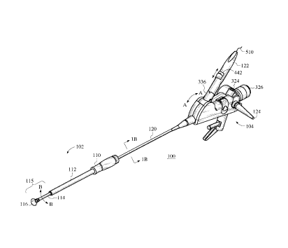
FIG. lA is a representative isometric view of a first embodiment of the
viewing
surgical scope apparatus of the present invention using articulated distal
section members.
FIG. 1B is a section view of viewing surgical scope apparatus shown in FIG.
lA.
7
CA 02263214 1999-02-26
FIG. 2A is an isometric view of the distal end of the viewing surgical scope's
introducer assembly shown in FIG. lA.
FIG. 2B is a section view of FIG. 2A.
FIG. 2C is an exploded view of the distal end of the viewing surgical scope
shown in
FIG. 2A.
FIG. 3A is an isometric view of the proximal end of the viewing surgical scope
apparatus shown in FIG. 1 A.
FIG. 3B is an exploded view of FIG. 3A.
FIG. 4A is an isometric view of the optical fiber advancement and control
handle
assembly of the viewing surgical scope apparatus shown in FIG. lA.
FIG. 4B is a section view of FIG. 4A.
FIG. 4C is an exploded view of FIG. 4A.
FIG. 5 is a representative side view of a piercing needle assembly used with
the
embodiments of the invention's viewing surgical scope apparatus.
FIGS. 6A, 6B, 6C & 6D are representative section views of viewing tubular
assemblies
that each have a clear distal tipped section with a working channel having
various orientations
at the clear distal tip.
FIG. 7 is a representative section view of a variation to the clear distal tip
tubular
member as shown in FIGS 6A-6D that has elements for controlling the working
device's
orientation at the viewing surgical scope's distal end.
FIGS. 8A is a second viewing surgical scope apparatus embodiment of the
invention
using a non-articulating viewing surgical scope that includes the clear distal
tip tubular
member shown in FIGs 6A-6D.
FIGS. 8B is a variation of the second viewing surgical scope embodiment of the
invention using a non-articulating viewing surgical scope that includes the
clear distal tip
tubular member shown in FIGs 6A-6D where the handle uses a sliding advance
mechanism for
the working device.
FIG. 9 is a perspective view of a patient illustrating first, second and third
minimally
invasively formed penetrations formed in the patient's chest, such as used for
access in TMR.
FIG. 10 is a perspective view of an interior of the patient's chest shown in
FIG. 9.
It will be understood that the invention's preferred embodiments have many of
the
individual elements whose functional aspects are similar. Thus, it will be
understood that
structural elements having similar or identical functions may have like
reference numerals
8
CA 02263214 1999-02-26
associated therewith. The appended drawings illustrate only typical
embodiments of this
invention and are therefor not to be limiting of its scope, for the invention
may admit to other
equally effective embodiments.
DETAILED DESCRIPTION
A minimally invasively formed penetration is a chest penetration that does not
entail
"open chest" surgery by gross spreading of the ribs or cutting through
excessive ribs and/or
the sternum. Minimally invasive surgery also involves formation of
penetrations that may be
performed intercostally or non-intercostally to access tissues and organs
without large incision
openings in a patient. Once devices have been introduced in this manner,
treatments may be
affected from within an organ outwards i.e. "inside-out," or in an "outside-
in" manner.
"Channels" refer to revascularization entries through the epicardium or
myocardium and
further includes entries that extend (i) through the endocardium from the
epicardium; (ii)
partially or fizlly through the myocardium; (iii) to form stimulation zones;
or (iv) to form drug
pockets. "Working devices" for treatment and diagnosis of affected
coronary/vasculature
tissue include devices configurable and extendable through a lumen within the
viewing surgical
scope's distal end such as: optical fiber elements capable of delivering laser
energy with or
without a piercing needle assembly at the distal end of the viewing surgical
scope, drug
delivery using a piercing needle assembly, RF tissue ablation devices,
ultrasound devices, or
mechanical coring devices.
FIG.1 A is a representative isometric view of the first embodiment of the
invention's
viewing surgical scope 100. The viewing surgical scope 100 is an articulating
bronchoscope
with a distal end introducer assembly 102 and a main body assembly 104. The
introducer
assembly 102 includes a handle portion 110 coupled to an essentially rigid
tube 112. Tube 112
surrounds a flexible member 114 with an attached suction cup 116 member.
Catheter 120
couples to the main body assembly 104 and is either rigid, semi-rigid or
flexible. A control
handle 122 provides control of an optical fiber advancement member 442 of an
optical fiber
element 510 which transmits laser energy from a remote laser energy source.
The
bronchoscope's catheter 120 has multiple conduits which are accessed through
the main body
assembly 104 via multiple portal openings such as a fiber optic waveguide
portal opening 124.
These conduits accomplish fiznctions such as illumination, aspiration or
irrigation of target
tissue at the scope's distal end at suction cup member 116. A hollow working
channel is
included with the catheter 120 for introducing implements such as a laser
energy delivery
9
CA 02263214 1999-02-26
optical fiber. The visualization scope shown can be a standard articulating
bronchoscope or
custom designed flexible endoscope made by Storz, Olympus or Pentax. The
visualization
scope's catheter 120 is within the bore 108 of the introduces assembly 102
shown in FIG. 2B.
FIG.1B is a section view at sectional line 1B-1B of the viewing surgical scope
100
shown in FIG. 1 A. The catheter 120 is a shaft of a bronchoscope with conduits
130 and
visualization lumen with internal fiber 132 & working channel 134 with
internal laser energy
optical fiber element 510 extending the length of catheter 120 that
communicates between the
main body assembly 104 and the end at cup member 116. In a typical
configuration, one or
more conduits 130 can be included within the catheter 120. An eyepiece 326
shown in FIG.1 A
observes target tissue at the distal end of the viewing surgical scope 100 via
the visualization
lumen with internal imaging fiber 132. Various types of ancillary viewing
capabilities such as
CCD monitoring can be attached at the eyepiece 326. A translatable laser
energy optical fiber
element 510 is translatable and is disposed within the working channel 134 to
deliver laser
energy at the distal cup member 116 to form TMR channels in the heart.
FIGS 2A, 2B & 2C show the introduces tubular assembly 102 of the viewing
surgical
scope 100 shown in FIG.1 A. Handle member 110 couples, either by threaded
member for
quick uncoupling or permanently coupled thereto, to an essentially rigid tube
112. A flexible
tubular member 114 is attached either permanently to or slidably disposed
within the tube 112.
Flexible tubular member 114 in turn is attached to the cup member 116. The
optional inner
tube 111 is attached to the flexible tubular member 114 and the inner tube 111
is slidably
disposed within the tube 112. The inner tube 111 is made integral with the
tube 112 when the
tubular member 114 is permanenetly attached to tube 112. Tube 111 when used
attaches to
the collet housing 202, otherwise the tube 112 is attached thereto. The distal
end of the
catheter 120, not shown in FIGS. 2A-2C, is disposed inside inner tube 111 and
flexible tube
114 in the bore 108. The catheter 120 is secured to the introduces tubular
assembly 102 at a
fixed location by manually tightening collet thumbscrew 200 into collet
housing 202, which
compresses grippes 204. A distal end lock ring 113 attaches the distal end of
the catheter 120
to the cup member 116 as shown in FIG. 2C. The flexible tubular member 114 can
be drawn
into the tube 112 by making the cup member 116 smaller then shown such that
when handle
110 can be decoupled from the collet housing 202 by twisting the handle 110
and then
pushing handle member 110 with tube 112 towards the distal end of the scope
100, the cup
member 116 collapses and resides within the tube 112 thereby providing ease of
scope 100
positioning through a minimally invasively formed penetration in a patient's
chest so that
CA 02263214 1999-02-26
entanglement with other instruments or internal body parts is minimized.
The flexible tubular member 114 and the suction cup member 116 form distal end
assembly 115. This articulating distal end assembly 115 is disconnectable and
interchangeable
with a essentially rigid non-articulating viewing tubular assembly 600
discussed below and
shown in FIGS 6A-6D for a viewing surgical scope apparatus. The introduces
tubular
assembly 102 with catheter 120 is for insertion into a patient's chest through
a minimally
invasive penetration using the handle 110 for emplacement, see U. S. Patent
Application S.N.
08/794,733, which teaches of a trocar used for initially providing a chest
wall penetration for
introducing instruments into a chest cavity.
Catheter 120 comprises the elongated shafting of a bronchoscope or flexible
endoscope tubing. The introduces tubular assembly 102 provides: a) stable
support for
emplacement within a patient's chest cavity and b) prevents unintended
rotation and axial
movement of the distal end of a working device such as the laser energy
delivery optical fiber
element 510. The flexible tubular member 114 allows deflection at the distal
end of the scope
100 by pivotal motions of the handle 122 which in turn causes a pivotal joint
indicated by
double arrow A-A in FIG.1 A to push or pull a control wire (not shown) or an
equivalent
translational member communicating between the bronchoscope's proximal body
assembly
104 and the distal end of the catheter 120. Tip deflection mechanisms in
bronchoscopes are
well known in the art. The flexible tubular member 114 can be made of flexible
silicon rubber
or other elastic material with flexural characteristics for providing the
necessary stability on a
beating heart. Cup member 116 can optionally communicate with a vacuum source
attached to
the proximal body assembly 104 through port 324 via one of the internal
conduits 130 to assist
in heart wall attachment. Cup member 116 provides a broad surface which locks
on the heart
when evacuated for stability during the procedure. Cup member 116 keeps the
optics clean
and provides a protective shield for sharp tools which can scratch adjacent
heart tissue. The
cup member 116 can equivalently be a flange member with a flexible grooved
annular surface
for locking onto a heart surface with or without vacuum assist or be a flange
member with a
gripping textured surface that attaches to tissue during the procedure.
FIGs 3A & 3B are views of the main body assembly 104 as shown in FIG. lA that
can
be mounted to the operating table or other structure using mounting shaft 306
that is attached
to the body mount 308. The body mount handle 310 allows manipulation of the
main body
assembly 104 when mounted to a fixture where the practitioner uses one hand to
hold the
introduces tube 112 at handle 110 and the other hand controls the handle 122
for optical fiber
11
CA 02263214 1999-02-26
510 translations and/or deflections of the distal end's cup member 116. Main
body assembly
104 in exploded view shown in FIG.3B has a right body housing 302 and a left
housing body
304. The right and left body housings 302 and 304 are configured as mating
halves of an outer
housing that encompass the proximal end of the visualization scope 342, which
is an
articulating-type bronchoscope in this embodiment of the invention. The
visualization scope
342 has at least two channels wherein a first working channel portal 322
communicates with
the working channel 134 and the visualization portal through eyepiece 326. A
CCD-camera
can optionally be used via the eyepiece 326. Portal opening 124 typically
provides
illumination at target tissue sites at the distal end cup member 116. Linkage
332 couples lever
330 via wheel linkage 334 to handle pivot member 336. Pivoting of handle 122
shown by
double headed arrow A-A in FIG. 1 A results in articulation of the flexible
member section 114
via control lever 330 action. The working channel port 322 optionally allows
introducing
procedural tools and instruments including but not limited to scissors,
graspers, fiber optic
tools, suture mechanisms without the pivot arm assembly as shown. Working
channel port 322
with the handle 122 feature as discussed above substantially aligns with and
allows free
movement of the handle pivot member 336 through a ball joint socket design
that couples to
the port 340 on visualization scope 342. Handle pivot member 336 allows
translation of the
working device such as an optical fiber element 510 therethrough.
FIGs 4A & 4B are partial component views of the handle 122 with the optical
fiber
element thumb slider 442 shown in FIG.1 A. FIG. 4B shows the handle 122
without the spring
biasing element 420 and an interposed triggering/retraction leaf spring member
and an internal
slider 444 for clarity. FIG. 4C is an exploded view showing the internal
components of the
handle 122. The thumb slider 442 advances and retracts the energy delivery
device such as the
optical fiber 510 independent of the triggered piercing needle member assembly
as shown in
FIG. S. The handle 122 as discussed above moves in unison with handle pivot
336 shown in
FIG. 1 A thereby providing articulation of distal tip cup 116. The
practitioner's hand can
control both the advancement of the optical fiber 510 and articulation of the
distal tip cup
member 116. The distal end 400 of handle 122 is inserted into pivot handle
member 336 and
retained in place by locking member 402. An end tube 404 sleeve enters the
handle 122 at its
proximal end 406 and another similar distal end tube 408 sleeve is disposed at
the distal end
400 and extends to the distal end of the scope 100. A mating right handle
portion 410 and a
left handle portion 412 are coupled together and enclose a needle piercing
spring loaded drive
assembly and energy delivery device advancement and control components. The
optical fiber
12
CA 02263214 1999-02-26
element 510 passes through the proximal and distal ends through tube 404 and a
needle
advance tube 408 which telescope with each other, the tube 404 is smaller than
the tube 408
and the tube 404 attaches to the optical fiber element 510, the tube 404
attaches internally to
the internal slider 444 and the tube 404 slides within the tube 408, thus
allowing translation of
the optical fiber 510 independent of the tube 408 movement. Movement of thumb
slider 442
in direction C disengages a ratchet 416 in mechanical cooperation with a
flexible latch 418
distal end locking member that disengages a piercing needle slider 422
resulting in needle
advance spring 420 to push the needle slider 422 forward causing the needle
advance tube 408
to move in direction C as well to advance the piercing needle distal end
assembly 500 as
shown in FIG. 5. Continued forward movement of thumb slider 442 advances the
fiber optic
element 510 through the needle advance end tube 408 which remains stationary.
Movement of
the thumb slide 442 is limited by fiber advance and depth stop button 424
slidably disposed
within slot 426 by either a threaded compression or a biased detent member
that cooperatively
engages the slot 426 at predetermined positions. Finally, retraction of
advance thumb slider
442 in the direction of arrow D causes the internal slider 444 to move
rearwardly and causes
the distal end of the triggering/retraction leaf spring member, which
cooperatively slides within
and engage internal slots in the slider 444, to engage the distal end face of
the slider 444 and
pull the piercing slider 422 rearwardly as well, thus resetting and latching
the needle slider
422 with spring 420 in relation to the latch 418 distal end face. The tube 408
is inserted into
the working channel of the inventions viewing surgical scope apparatus.
FIG. 5 is a representative side view of the piercing needle assembly's distal
end 500.
Piercing needle end portion 502 has a bevel cut end for piercing tissue and is
coupled to a
flexible section 504 which allows passage of the piercing needle distal end
assembly 500
through a working channel with bending such as a flexible catheter or pre-
shaped tubing. A
fiber optic element 510 or other energy delivery device 510 passes through a
lumen within
piercing needle assembly 500 as shown in FIGS. 4A, 4B and 5. Moreover, the
distal end
needle assembly 500 can be a flexible drug delivery conduit and be a working
device for the
invention's viewing surgical scopes. Similarly, the distal end piercing needle
assembly 500 can
be replaced with a piercing optical fiber element as taught in U. S. Patent
5,703,985 entitled
".Optical Fiber Device and Method for Laser Surgery Procedures," which is
hereby
incorporated by reference.
FIGS. 6A-D are representative section views of variations of a viewing tubular
assembly 600. The assembly 600 can be used with either a flexible or rigid
endoscope. In
13
CA 02263214 1999-02-26
particular, the assembly 600 as used with the viewing surgical scope 100
replaces the flexible
distal end assembly 115 as shown in FIG. 1 A; or alternatively and preferably
used with a rigid
shafted endoscope 200 discussed below and representatively shown in FIGs 8A &
8B. The
viewing tubular assembly 600 includes an optically clear or transparent end
tube cap 602
which fits over the visualization port distal end 604 of a scope's
visualization shaft and has a
working channel 606 (cut-off view). The distal ends 604 in FIGS 6A & 6B lie in
planes
essentially perpendicular to the central axis of the viewing tubular assembly
600 such that
optics provide essentially direct forward visualization with a predetermined
divergence
viewing angle E as shown. The end port 604 shown in FIG. 6C is at a 30°
angle with respect
to the central axis of the viewing tubular assembly 600. Distal end 604 can be
varied such that
the field of view is at an angle offset with respect to the central axis of
viewing tubular
assembly 600. The viewing tubular assembly 600 replaces the components of
flexible member
114 and cup member 116 in FIGs 2A-2C and cooperatively combines with the shaft
member
112 and connectively interfaces representatively with the working channel 134
with
appropriate tubing connectors with the working channel 606 shown in FIGS 6A-
6D. The end
cap 602 member is made from an acrylic or equivalent polycarbonate transparent
material and
coupled to a rigid tubular sleeve member 615. Moreover, the assembly 600 can
be a solid
object made of the same material as the end cap 602 member. The distal end of
the
visualization scope 604 terminates near the transparent end cap 602. The end
cap 602 can
made with desired optical light absorption/reflection characteristics.
Furthermore, the shape of
the end cap 602 can be conical, elliptical or include planar facets at various
angles with respect
to the viewing tubular assembly's 600 central axis. The end cap 602 is
designed and made in
accordance with required optical lens characteristics such as focus,
divergence, convergence,
directionability, collimation, polarization or diffusion.
The working channel 606 has various designs with differing bends that
cooperatively
are attached to the viewing tubular assembly 600. The working channel 606 as
shown is
external to the assembly 600, but can be incorporated into a lumen or be a
structural tube
either in the wall of the viewing tubular assembly 600 or conformably designed
to fit within the
inner wall surface of assembly 600 adjacent the distal end 604 of the
visualization scope 342
Qr an end shaft of a rigid or flexible endoscope. The working channel 606 is
shown attached to
the external wall of assembly 600 in FIGS. 6A-6D. Viewing tubular assembly 600
fi~nctions to
allow viewing of affected tissue while applying pressure to tissue for
stopping bleeding and
minimizing active tissue movement, e.g. a beating heart. The working channel
606 directs and
14
CA 02263214 1999-02-26
protects the operative working device such as the optical fiber element 510, a
drug delivery
needle or other energy delivery device that is controlled by handle 800 as
shown in FIGS. 8A.
The working channel 606 can be made of stainless steel, plastic or comparable
material. In the
preferred embodiment, the working channel 606 is clear to enable visualization
of fiber
movement. The working channel 606 in FIG. 6A has a curvature 608 such that the
fiber or
other working device is directed through the transparent end cap 602 in a
direction essentially
parallel with/or contiguous with respect to the central axis of the assembly
600. The working
channel 606 has a curvature 612 in FIG.6B which directs the working device
through the
transparent end cap 602 at approximately 45° with respect to the
central axis of the assembly
600. Likewise, the curvature 614 in the working channel 606 of FIG. 6C directs
the working
device through the transparent end cap 602 in a direction approximately
90° with respect to
the central axis of the viewing tubular assembly 600. Other orientations of
working channel
606 and/or distal end bends in tube 606 can be used to direct the working
device.
FIG. 7 is a representative section view of a variation of a movable distal
ended optical
ball viewing tubular assembly 700 that provides variable positioning of the
working device
such as the optical fiber 510 and can also be part of either viewing surgical
scope 100 or 200
as discussed below. The optically transparent rotatable member 706, which is
either a ball or
cylinder member, is at the distal end 708 and seats within a conformal shaped
end tube 701
that allows free rotation of the rotatable member 706. Upper steering wire 710
and a lower
steering wire 712 are coupled to the rotatable member 706. The steering wires
710 and 712
pass back to a proximal portion of the scope 100 or 200 to control mechanism
714. The
steering wires 710 and 712 are coupled to deflector knobs 716 for rotating the
rotatable
member 706 in a direction as shown by double headed arrow F. A guide channel
718 passes
through the rotatable member 706. A flexible coupling portion 720 extends
between the guide
channel 718 of the rotatable member 706 and the working channel 606, thereby
providing a
path for directing the working device such as an optical fiber 510
therethrough. Flexible
coupling portion 720 is a telescoping or an accordion-like interconnection
allowing
reorientation of the rotatable member 706 to direct the working device in a
direction G.
Tensioning steering wire 710 rotates the rotatable member 706 and re-directs
the guide
channel 718 in opposition to steering wire 712. Additionally, more control
wires can be
includes to provide multiple degrees of rotation of the rotatable member 706
for greater
controllability.
The articulating distal ended viewing tubular assembly 700 can replace the
components
CA 02263214 1999-02-26
of flexible member 114 and cup member 116, i. e. assembly 115 in FIGS 2A-2C
and
cooperatively slides on shaft member 112. The viewing tubular assembly 700
connectively
interfaces at least with the conduits 130 and 134 with appropriate tubing
channeling
connectors and by appropriate internal control wire connections within the
proximal end of
sleeve member 715 and to appropriate connections in the flexible catheter
shaft 120.
Moreover, the catheter 120 can be a stand alone viewing device whose distal
end which
representatively can be 604 in FIG. 7 and the work channel 606 would be tubing
attached to
the catheter 120 shafting. The viewing surgical scope 200 discussed below and
shown as FIG.
8A and 8B would have a control member 714 on the handle 800 with connecting
control wires
710 & 712. The assembly 700 would encompass the rigid endoscope shafting 601
as
discussed below.
The articulating assembly 700 of FIG.7 can have alternative designs such as an
assembly comprising an internal mechanical deflecting linkage mechanism for
changing the
orientation of the egression angle of the working channel 606. The transparent
surface
rotatable member 706 would be replaced with an essentially transparent cap
member
comparable to 602 with a flexible membrane to allow orientation displacement
of the working
channel 606 that is sealed within the membrane. Moreover, the deflecting
linkage mechanism
can be a light reflecting surface such that observations of tissue can be at
offset angles with
respect to the axial direction of the assembly tube 715 where the distal end
of the visualization
scope 604 has a normal surface with respect thereto.
FIG. 8A shows viewing surgical scope 200 with a handle assembly 800 using a
finger
trigger advance mechanism 804 and has the tubular viewing assembly 600. The
assembly 600
is non-articulating distal clear end cap 602 for visualizing and has a working
channel 606 for
directing the working device, e.g. an optical fiber 510 at a treatment site.
The visualization
scope is an endoscope whose distal end 604 is viewed through an eyepiece 806.
The distal end
604 of the endoscope can have different angular orientations as discussed
above for a required
distal end viewing field from the viewing tubular assembly 600. The viewing
surgical scope
200 for example can be a 10-mm sized rigid endoscope with a viewing tubular
assembly 600
that has a 12 mm-O.D. A smaller 5-mrn system endoscope, can also be used where
the
assembly 600 is about 10-12 mm O.D. that allows for additional space inside
the assembly 600
for additional working channels 606 that allow for drug delivery, lighting
etc. The handle
assembly 800 is ergonomically designed for hand gripping. The handle assembly
800 includes
a fiber advance mechanism using finger trigger 804 within the handle and
alignment retaining
16
CA 02263214 1999-02-26
members for attaching endoscope shafting 601 along with the viewing tubular
assembly 600.
The viewing tubular assembly 600 is user removable for quick disconnect from
the endoscope
shafting 601 for quick interchange of tubular assemblies 600 with dii~erent
working channel
606 egress angles for surgical procedures that occur at various aspects of the
heart surface,
such as the lateral, anterior, posterior or apexial walls when operating from
a single chest
penetration. The viewing tubular assembly 600 has a quick disconnect coupling
member 808
for connections of the working channel 606 for quick interchangeability of the
assembly 600.
Additionally, the articulated viewing tubular assembly 700 shown in FIG. 7 can
be used with
the necessary control features incorporated within the handle 800. This
feature allows access
to lateral, anterior or posterior locations of an organ where a practitioner
uses the same chest
wall penetration.
Finger trigger 804 controls translatable movement of the working device, e.g.
an
optical fiber element 510 with or without a piercing needle distal end
assembly 500 as shown
in FIG. 5. The finger trigger 804 actuates mechanical or electrically movement
of the working
device from the distal end of the viewing tubular assembly 600 shown by the
double arrow H,
preferably using incremental control. Mechanisms for the
advancement/retraction function
include rack and pinion components, a stepper motor with appropriate control,
pneumatic
driven mechanisms with incremental stepping functional components.
Alternatively, the handle can include a slide member 810 as shown in FIG 8B
which
can include a mechanism comparable to that discussed above in FIGS 4A & 4B
wherein a
triggering mechanism advances a needle piercing member 500 and cooperatively
works with
the optical fiber 510 through an adjustable range, e.g.1.5- 2.5 cm. The slide
member 810 can
include detents for a user to sense rate of advancement. The advancement
mechanism can also
be geared to provide advancement at translation ratios other than 1:1.
Retraction of the optical
fiber 510 can be accomplished by reversing the trigger button 812 that
cooperates with a
reversing rack mechanism inside handle 800) A stop setting member 814 can be
used to
position the optical fiber distal ending flush with the viewing tubular
assembly's 600 outer
surface. Alternatively, the mechanisms shown in FIGS 4A & 4B showing a slide
controlled
mechanism could be incorporated in handle 800 in lieu of the finger trigger
804. An
equivalent lever mechanism can be used in lieu of the finger trigger 804 which
would include
stops to limit optical fiber extension and retraction. In a TMR operation, the
optical fiber
element S10 would typically would be advanced in 1-mm increments.
FIG. 9 shows a perspective view of a patient 10 with first, second and third
minimally
17
CA 02263214 1999-02-26
invasive formed penetrations 12, 14 and 16 respectively. It will be
appreciated that the exact
location of penetrations 12, 14 and 16 is not limited to those illustrated in
FIG. 9.
Additionally, from 1 to N+1 numbers of penetrations may be made. The patient
is prepared for
the procedure and is positioned similarly to that used for a left thoracotomy.
The patient's left
arm is draped. A conventional double lumen endotracheal tube is used to
selectively deflate
one side or the other of the lungs. Preferably the left lung is collapsed
which allows access to
the chest cavity in the vicinity of the left lung. The other lung remains
inflated to provide
oxygenation.
The distal portion of either viewing surgical scope 100 or 200 is positioned
to reach a
desired aspect of a ventricular wall. A plurality of different
revascularization channels are
formed in the heart. A distal portion of the energy delivery device or other
working device can
be positioned against tissue of the wall of the heart through which the
channel is to be formed
while transmitting energy from a remote energy source through the optical
fiber element 510
or other energy delivery device. Additionally, the waveguide may be configured
to pierce the
epicardium, such as with a piercing needle as shown in FIG. 5, so that energy
is or can be
subsequently delivered to the myocardium. A revascularization channel can be
formed
through an epicardium into at least a portion of a myocardium or continue
through the
myocardium into all or only a portion of the endocardium.
In one method, penetration 12 is used for the introduction of either scope
100, 200 or
a separate rigid scope to provide global viewing capability of an internal
chest area of interest.
For standard TMR at the apex 20 region of the heart, a first penetration 12
can be formed in
the intercostal spaces, for example the fourth to sixth intercostal space that
is 10-12 mm in
diameter. A slight cut is made and a thoracic trocar is advanced through the
chest.
The scope 100, 200 or separate rigid visualization scope is used to visualize
the area,
look for larger coronary vessels, to inspect the condition of the pericardium,
and to check for
adhesions. The shape of the heart as well as its position is visualized.
Second penetration 14 is
formed inferior to penetration 12 and can be formed just above the diaphragm
and third
penetration 16 is formed superior to penetration 12. Penetrations 14 and 16
can be formed
substantially the same way as penetration 12 is formed or may be cut downs
only.
. For initial procedures a pair of thoracoscopic graspers may be introduced
through
penetration 14. Additional tools that can be introduced through penetration 14
include
scissors. The pericardial sac 18 shown in FIG. 10, if intact, is grabbed and
opened up using
standard surgical techniques. The pericardial sac is pulled away from the
heart and may be
18
CA 02263214 1999-02-26
suspended. Unwanted adhesions are removed.
After the tools are removed from penetration 14, either scope 100 or 200 with
a
working channel is introduced where the visualization scope, either a
bronchoscope 342 or an
endoscope can use a camera device attached to the eyepiece for viewing on a
monitor.
Additionally, additional viewing scope devices can be used during the
procedure as inserted in
the first penetration and the rigid scope can be inserted into second
penetration 14 after the
tools are removed from second penetration 14.
Third penetration 16 is formed, a trocar introduced and a pair of forceps
places an
absorbing medium, including but not limited to a piece of gauze, through the
third penetration
16. Third penetration 16 is created initially to open the pericardial sac and
subsequently may
be used as a treatment port, for visualization or for safety reasons. In the
event that a
structure, such as a coronary artery is nicked and bleeding is initiated,
direct pressure is
applied by placing the gauze on the area through third penetration 16 to stop
the bleeding. The
gauze is also useful for manipulating the heart and applying slight pressure
to TMR entrance
sites to avoid excessive bleeding. When using the scope 200, the tubular
member assembly 600
stops bleeding when applied to areas undergoing treatment.
Either of the viewing surgical scopes 100 or 200 shown in FIGs 1 A, 8A or 8B
is
initially positioned in penetration 14 and revascularization channels are
created at the desired
location, such as the apex 20. Preferably the working device such as the
energy delivery device
is inserted through the working channel of either of the scopes 100 or 200
adapted for the
procedure. The articulating-type scope 100 also may be initially positioned in
penetration 12
or 16. Once the desired number of revascularization channels are formed,
either of the scopes
100 or 200 can be removed and positioned in any of the other penetrations.
Graspers and
needle holders, or other instruments, are introduced through one of the
penetrations to stitch
back the pericardial sac as required. A check is made to ensure that there is
no bleeding,
trocars are removed and the penetrations closed. It will be recognized that
the procedure will
vary, depending upon the condition of the heart and the site of the procedure.
In the preferred use of the present invention, the distal portion of the
working device
such as the energy delivery device is positioned to reach a desired aspect of
a ventricular wall.
A plurality of different revascularization channels are formed in the heart. A
distal portion of
the energy delivery device can be positioned against tissue of the wall of the
heart through
which the channel is to be formed while transmitting energy from a remote
energy source
through the energy delivery device.
19
CA 02263214 1999-02-26
Suitable working devices that can be inserted in the working channels of
viewing
surgical scopes 100 or 200 include energy delivery devices which include laser
wave guides,
RF electrodes, microwave cutters, ultrasound transmitters, mechanical coring
devices or fluid
jets. Each energy delivery device is configured to be coupled to an energy
source including but
not limited to RF, laser, microwave, ultrasound, mechanical coring, fluid jet,
cryogenic fluid,
chemical ablation and the like. The distal portion of the working device such
as an energy
delivery device can be positioned next to the heart wall while energy is
delivered through the
energy delivery device. Alternatively, the energy delivery device can deliver
energy through a
gaseous medium to the heart wall. The scopes 100 or 200 distal end can include
a piercing
obturator member for initial entry between the pericardial sac and the
epicardium so that
energy is delivered into the myocardium with minimal tissue destruction. A
revascularization
channel can be formed through an epicardium into at least a portion of a
myocardium or
continue through the myocardium into all or only a portion of the endocardium.
Other surgical procedures that scopes 100 or 200 could be used include
gallbladder,
laparoscopy or laparotomy, colosectomy and other MIS operations that use other
working
devices for treatment of diseased tissue, such devices structurally configured
for a working
channel.
Unless defined otherwise, all technical and scientific terms used herein have
the same
meaning as commonly understood by one of ordinary skill in the art to which
this invention
belongs. Although any methods and materials similar or equivalent to those
described can be
used in the practice or testing of the present invention, the preferred
methods and materials are
now described.
While the principles of the invention have been made clear in illustrative
embodiments,
there will be immediately obvious to those skilled in the art many
modifications of structure,
arrangement, proportions, the elements, materials, and components used in the
practice of the
invention, and otherwise, which are particularly adapted to specific
environments and
operative requirements without departing from those principles. The appended
claims are
intended to cover and embrace any and all such modifications, with the limits
only of the true
purview, spirit and scope of the invention.