Note: Descriptions are shown in the official language in which they were submitted.
CA 02263273 1999-02-12
WO 98/08557 PCT/US97I13485
1
Combined Card~otomy Fluid and Venous BloO RP~Prvolr
BACKGRO '~N OF THE INVENT t nT~T
Field of the Invention
The present invention is in the field of reservoirs
used during surgery to both recover blood and other body
fluid from a surgical site (cardiotomy fluid), and to
receive blood from the circulatory system of the patient
(venous blood). The recovered cardiotomy fluid and blood
are each treated for re-infusion into the patient. For
example, the recovered cardiotomy fluid and blood may be
subjected to oxygenation, temperature control, and
circulatory pumping in a heart/lung apparatus in order to
sustain the life of the patient. The cardiotomy fluid
recovered from a surgical site additionally requires
treatment to remove foreign material that is picked up by
exposure to the ambient as well as because of the
surgical procedure itself {i.e., tissue particles or bone
fragments, as well as ambient bacteria, or particulates
from foreign body exposure, for example). Similarly, the
venous blood received from the circulatory system must be
treated to insure that any entrained bubbles of gas
created by extra-corporeal circulation are removed. The
collected blood and cardiotomy fluid after treatment are
returned to the patient via the heart/lung apparatus.
Related Technology
An example of conventional combined cardiotomy and
venous blood reservoirs is seen in each of United States
patent Nos. 4,737,139, issued 12 April 1988 to Paul F
CA 02263273 1999-02-12
WO 98/08557 PCT/US97/13485
2
Zupkas, et al., (the '139 patent); and 5,039,430, issued
13 August 1991 to Edmund R. Corey Jr., (the '430 patent).
The Zupkas '139 patent discloses a cardiotomy and
venous blood reservoir. According to the '139 patent the
device includes upper and lower chambers which are
vertically separated by a horizontal ring-like portion 69
of a support member. This support member ring-like
portion 69 serves to divide an upper cardiotomy chamber
from a lower venous blood chamber. The cardiotomy fluid
which is received by an upper inlet is subjected first to
a de-foaming step by a de-foamer 87, then to a filtering
step by a depth filter element 89. Finally the fluid is
again de-foamed by a de-foamer element 91 which is
surrounded by a mesh 93. The venous blood is de-foamed
by a lower portion of the de-foamer element, which is
shared with the cardiotvmy fluid in parallel but fluidly
separate flow paths. After flowing radially outwardly
through the de-foamer element, both cardiotomy fluid and
venous blood flow into a common chamber for return to the
patient via a lower outlet of the housing.
With the reservoir taught by the '139 patent, both
cardiotomy fluid and venous blood are subjected to a de-
foaming step but the cardiotomy de-foamer and the venous
blood de-foamer have substantially the same diameter.
However, because effective filter area for filtering
cardiotomy fluid is a controlling limitation in the
design of such reservoirs, the reservoir taught by the
'139 patent can be seen to impose a significant
limitation on the filter area available for this
function. With the device taught by the '139 patent, the
CA 02263273 1999-02-12
WO 98/08557 PCT/US97/13485
3
effective filter and de-foamer areas in each of the
venous blood and cardiotomy fluid flow paths are a
' function of the fractions of the height of the device
which are devoted to each flow path. Further, it is
recognized that the filtering element area provided for
cardiotomy fluid must be adequate to pass the necessary
fluid volume both at the beginning of use when this
filter is clean and unobstructed and entirely available
for fluid flow, and also after a period of use as this
cardiotomy fluid filter possibly becomes partially
clogged by debris. As a result, the ratio of cardiotomy
filter area versus volume for the reservoir taught by the
'139 patent is or can be unfavorable. The result of this
unfavorable ratio can be a reservoir that is limited in
cardiotomy filter area, or one which is overly tall in
order to achieve an acceptable cardiotomy filter area.
Such a tall filter may be overly large and unwieldy for
operating room personnel to work with, and may require an
excess volume of blood for initial filling.
In other words, because of the common diameters of
the filters and de-foamers in these conventional designs,
the area available to the cardiotomy filter is simply a
function of that part of the height of the device which
is devoted to treatment of cardiotomy fluid. Thus, the
device either has to be made very tall (with an attendant
excessive blood volume and size for the device), or most
of the height of the device must be allocated to the
cardiotomy flow path. The de-foamer area for the venous
blood flow path may be undesirably small as a result.
CA 02263273 1999-02-12
WO 98/08557 PCT/LTS97/I3485
4
Viewed another way, the device according to the '139
patent (and similar devices) are seen to provide a
branched tributary flow path with one branch receiving
cardiotomy fluid and typically effecting a de-foaming,
filtering, and de-foaming action on this blood. The
other branch of the flow path receives venous blood and
effects a de-foaming action only, with the de-foamer
being a separate section of the same physical element
which effects the final de-foaming of the cardiotomy
fluid. The two branches form a confluence, and lead
without further blood treatment to the outlet of the
device.
The Corey '430 patent effects a separation of filter
and de-foamer areas in the cardiotomy flow path as
compared to the de-foamer area provided for the venous
blood flow path by first filtering and de-foaming the
cardiotomy fluid and combining this cardiotomy fluid with
venous blood before passing the combined f luid through a
final de-foamer element. Thus, it is seen that the blood
flow path in this device is branched with a tributary
cardiotomy flow path having a filter element and a de-
foamer element. The venous blood flow path is a simple
tributary entering downstream of the de-foamer in the
cardiotomy branch. Downstream of the confluence of the
cardiotomy flow path with the venous flow path, the final
de-foamer element is situated so that both blood flows
pass through this element together.
In view of the above, it can be seen that in order
to achieve the most efficient use of space, economy of
use of materials of construction, and a desirable ratio
CA 02263273 1999-02-12
WO 98/08557 PCT/US97/13485
of filter and de-foamer areas to reservoir size and
volume (while also minimizing the volume of blood
' required to initially fill a combined cardiotomy and
venous blood reservoir), it would be desirable for the
5 filter element of the cardiotomy fluid flow path to also
serve a de-foaming function.
Additionally, an improvement could be realized if
the final de-foamer element were to be shared between the
cardiotomy fluid flow path and the venous blood flow path
without imposing the same-area restriction on the design
as is seen in the Zupkas '139 device (i.e., with the
filter/de-foamer being the same diameter for each flow
path with effective areas being a function of the
fractions of the height of the device which are devoted
to each flow path).
Further, it would be an advantage for purposes of
perfusionist convenience and facility in setting up the
apparatus if the venous and cardiotomy inlets to such a
device were independently rotatable for selection of
physical orientation, each with respect to a housing of
the device.
summary of the Invention
In view of the above, a primary object for this
invention is to avoid one or more of the deficiencies of
the conventional related technology.
Another objective for this invention is to provide a
combined cardiotomy fluid and venous blood reservoir in
which a filter element for the cardiotomy fluid flow may
also serve a de-foaming function.
CA 02263273 1999-02-12
WO 98/08557 PCT/US97/I3485
6
Additionally, it is an object for this invention to
provide a filter material for use in the cardiotomy flow
path which has an additional de-foaming function.
Accordingly, it will be seen that an object for this
invention is to provide a cardiotomy flow path in which a
filter element is not preceded by a de-foamer element,
and in which the filter element itself has a blood de
foaming property.
An additional object for this invention is to
provide a combined cardiotomy fluid and venous blood
reservoir in which a final de-foamer element may be
shared between the cardiotomy fluid flow path and the
venous blood flow path without imposing the same-diameter
restriction on the design as is seen in the conventional
technology (i.e., filter/de-foamer elements of about the
same diameter, with effective areas in each flow path
dependent upon the fraction of total device height
devoted to each flow path).
Yet another object for this invention is to provide
a combined cardiotomy and venous blood reservoir which
includes a vertically extending partition wall which
fluidly separates a surrounding de-foamer element into
separate portions, with each portion of the de-foamer
element flowing only one of cardiotomy fluid or venous
blood.
Another object for this invention is to provide such
a combined cardiotomy fluid and venous blood reservoir in
which a vertically extending partition wall serves to
support a blood-receiving cup within the reservoir.
CA 02263273 1999-02-12
WO 98/08557 PCT/US97/I3485
7
An object for this invention is to provide such a
combined cardiotomy fluid and venous blood reservoir in
which a vertically extending partition wall serves to
support a tubular member for conveying venous blood
vertically downwardly within a chamber of the reservoir,
and in which this tubular member is not required to
support a blood receiving cup which is disposed at an
opening of the tubular member to receive venous blood
therefrom and to prevent the reflux of air in the tubular
member.
Still further with respect to such a reservoir
having a vertically extending partition wall, it is an
object for this invention to use this wall to carry a
support grate within the de-foamer element, which support
grate supports the de-foamer element against radial
collapse.
Another object for this invention is to provide such
a combined cardiotomy fluid and venous blood reservoir in
which the vertically extending partition wall is
rotational within a chamber of the reservoir.
Further, an object for this invention is to provide
such a combined cardiotomy fluid and venous blood
reservoir in which a perfusionist may conveniently set up
the apparatus by independent rotation of the venous and
cardiotomy inlets in plan view independently of one
another. Thus, the perfusionist may select the physical
orientation of the blood flow conduits approaching the
apparatus for best convenience in view of the position of
the patient and the configuration of the surgical suite.
CA 02263273 1999-02-12
WO 98/08557 PCTII1S97/13485
8
Accordingly, one aspect of the present invention
provides a combined cardiotomy fluid and venous blood
reservoir having a housing defining a chamber therein and
a pair of upper inlets opening into the chamber, one of
the pair of upper inlets being a cardiotomy fluid inlet
while the other of the pair of inlets is a venous blood
inlet; an annular vertically-extending liquid-permeable
de-foamer element received in the chamber and having an
upper end into which the pair of upper inlets spill; a
l0 generally vertically extending partition wall portion
received within the de-foamer element between the pair of
upper inlets and effectively fluidly dividing the de-
foamer element horizontally into a first portion through
which flows only cardiotomy fluid, and a second portion
through which flows only venous blood; below the de-
foamer element the housing defines a basin portion of the
chamber for receiving and combining therein of both de-
foamed cardiotomy fluid from the first de-foamer element
portion and de-foamed venous blood from the second de-
foamer element portion; and an outlet leading from the
basin portion for flowing de-foamed cardiotomy fluid and
de-foamed venous blood out of the housing.
Another facet of the present invention provides a
method of treating both cardiotomy fluid and venous blood
including steps of flowing the cardiotomy fluid and the
venous blood each separately along a respective branch of
a branched flow path toward a confluence and an outlet;
flowing the cardiotomy fluid through a liquid-
permeable filtering element to provide filtered
cardiotomy fluid; providing a de-foamer
CA 02263273 1999-02-12
WO 98/08557 PCT/US97/13485
9
element in both branches of the flow path; de-foaming the
filtered cardiotomy fluid and the venous blood by flowing
' each separately through the de-foamer element; combining
filtered and de-foamed cardiotomy fluid and de-foamed
venous blood at the confluence of the flow path; and
flowing the combined cardiotomy fluid and venous blood
together from the outlet.
Still further, the present invention provides a
combined cardiotomy fluid and venous blood reservoir
having a housing defining a chamber therein and a pair of
upper inlets opening into the chamber, one of the pair of
upper inlets being a cardiotomy fluid inlet while the
other of the pair of inlets is a venous blood inlet; an
annular vertically-extending liquid-permeable de-foamer
element received in the chamber and having an open upper
end into which the pair of upper inlets spill; a
vertically extending partition wall portion received
within the de-foamer element between the pair of upper
inlets and effectively fluidly dividing the de-foamer
element horizontally into a first portion through which
flows only cardiotomy fluid, and a second portion through
which flows only venous blood; a vertically extending
tubular member carrying venous blood from the venous
blood inlet downwardly into the chamber, the tubular
member having an opening to the chamber adjacent to a
lower end thereof; and the partition wall carrying a
blood-receiving cup adjacent to the lower end of the
tubular member, the cup having a rim over which blood
received therein flow into the chamber and which is at a
higher elevation than the opening, whereby reflux of
CA 02263273 1999-02-12
WO 98/08557 PCT/US97/13485
gaseous fluid upwardly in the tubular member is prevented
by a fluid trap formed within the cup by venous blood
received therein.
An advantage of the present invention according to a
5 preferred embodiment thereof results from the ability to
effect a first de-foaming action on cardiotomy fluid
without having to provide a separate first de-foamer
element. That is, the cardiotomy fluid filter element
according to the preferred embodiment of the invention
10 itself serves as a first de-foamer for the cardiotomy
fluid. Further, an effective area for the cardiotomy
fluid filter element of the present invention can be
selected without being constrained to a diameter for this
element selected either for purposes of de-foaming of the
cardiotomy fluid, nor to a diameter of this element
selected for de-foaming of venous blood. That is, the
first cardiotomy filter/de-foamer according to one
embodiment of the invention is configured as a bag or
sack having effective filtering surface area completely
around its perimeter. An alternative embodiment of the
invention could provide a separate pre-filter de-foamer
for the cardiotomy fluid. If desired, the cardiotomy
fluid filter element may be pleated or corrugated to
increase its effective filter area. This first
filter/de-foamer according to the preferred embodiment of
the invention is in fluid flow series with a final de-
foamer element, so that fluid flow rates can be matched
by selection of effective areas.
Further, the de-foamer for the venous blood flow
path may also be dedicated only to this flow path,
CA 02263273 2001-09-20
11
although the de-foamer element itself may be shared by the two
flow paths in the preferred embodiment of the invention.
According to the preferred embodiment of the invention, the
portion of the de-foamer element dedicated to each one of the
two flow paths is selected by the location of a partition wall
which in plan view may have an offset to define a trough, and
which provides one way of selecting the effective parts of the
area of this de-foamer element which are exposed to each of
the two flow paths.
In accordance with an aspect of the invention, a combined
cardiotomy fluid and venous blood reservoir comprises:
a housing defining a chamber therein and a pair of
upper inlets opening into said chamber, one of said pair
of upper inlets being a cardiotomy fluid inlet while the
other of said pair of inlets is a venous blood inlet;
an annular vertically-extending liquid-permeable de-
foamer element received in said chamber and having an
upper end into which said pair of upper inlets spill;
a support member rotationally received within said
chamber, supporting said de-foamer element against radial
collapse;
a basin portion of said housing chamber defined
below said de-foamer element for receiving both de-foamed
cardiotomy fluid and de-foamed venous blood from said
second de-foamer element portion; and
an outlet leading from said basin portion for
flowing de-foamed cardiotomy fluid and de-foamed venous
blood out of said housing.
Additionally, a better understanding of the present
invention will be obtained from reading the following
description of a single preferred exemplary embodiment of the
present invention when taken in conjunction with the appended
CA 02263273 2001-09-20
11 a
drawing Figures, in which the same features (or features
analogous in structure or function) are indicated with the
same reference numeral throughout the several views. It will
be understood that the appended drawing Figures and
description here following relate only to one exemplary
preferred embodiment of the invention, and as such, are not to
be taken as implying a limitation on the invention. That is,
no limitation on the invention is implied by this reference,
and none is to be inferred.
Brief Description of the Drawing Figures
Figure 1 provides a perspective view of a combined
cardiotomy fluid and venous blood reservoir embodying the
present invention, and is generally an elevation view of the
apparatus;
CA 02263273 1999-02-12
WO 98/08557 PCT/LTS97/13485
12
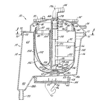
Figure 2 is a cross sectional elevation view of the
apparatus seen in Figure Z, and is taken at line 2-2 of
Figure 1 looking in the direction of the arrows;
Figure 3 is a cross sectional plan view taken at
line 3-3 of Figure 2 and looking in the direction of the
arrows;
Figure 4 provides an exploded perspective view of
the apparatus seen in Figures 1-3, and is taken from a
perspective similar to Figure 1. In this Figure the
dash-dot line indicates a continuation of the assembly
arrangement (as is indicated by the arrow head on this
line), and is used for convenience and economy of laying
out the drawing on the sheet;
Figures 5 and 6 are perspective views of a component
part of the apparatus, and are shown in a pre-assembly
configuration and in a use configuration, respectively,
in order to better illustrate features of the structure;
Figure 7 is an enlarged perspective view of an
indicated encircled portion of Figure 6, and is seen from
the side opposite to that seen in Figure 6 (i.e., from an
inner perspective looking radially outwardly generally
toward the viewer of Figure 6);
Figure 8 is an enlarged cross sectional view taken
at line 8-8 of Figure 5 looking in the direction of the
arrows; and
Figure 9 provides a schematic of a tributary
confluent blood fluid flow circuit provided by the
apparatus of the present invention.
CA 02263273 1999-02-12
WO 98/08557 PCTIUS97/13485
13
Detailed Description of an Exemplary
~ferred Embodiment of the Invention
Viewing Figures 1-4 in conjunction with one another,
and considering first Figure 1, a combined cardiotomy
fluid and venous blood reservoir 10 is seen in
perspective view providing primarily a side elevation
view and one-quarter top view. It will be understood
that by "cardiotomy fluid" is meant blood and body fluid
collected from a patient, usually at a surgical site by
means of suction lines, and which cardiotomy fluid is
mostly patient blood. This cardiotomy fluid may include
other body fluids in addition to blood, is not sterile
because of its exposure to the surgical site and ambient
air, and may contain tissue particles or bone fragments,
f or example .
The reservoir 10 includes a housing 12 having a
lower portion 14 with a side wall 16 and a lower wall 18
including a fluid outlet 20. The side wall 16 defines an
upper edge portion 22 with an upper edge 24 at which an
upper portion 26 (or top part) including a rotatable cap
or turret assembly (generally indicated with the numeral
28) is sealingly received. As will be seen, the cap part
28 is rotatable relative to the top part 26, as is
indicated by arrow 30. Near its outer periphery, cap
part 28 carries a plurality of circumferentially arrayed
cardiotomy fluid inlets (each referenced generally by the
arrowed numeral 32). Inlets 32 are disposed radially or
parallel with a radius of cap part 28. At its center,
cap part 28 carries a venous blood inlet 34, which is
also rotatable relative to the housing 12 and
CA 02263273 1999-02-12
WO 98/08557 PCT/US97/13485
14
independently of cap part 28, as is indicated by arrowed
numeral 36. Opposite to the cardiotomy inlets 32, the
turret 28 also carries a pair of parallel venous blood
inlets 34'.
The upper portion 26 of housing 12 also includes a
connection, indicated with the numeral 38, which may be
used as a vacuum connection or as an air vent.
Connection 38 provides for fluid flow both inwardly and
outwardly of the housing 12. As is indicated by the
arrowed numeral 40 on Figure 1, air flow is predominantly
outwardly of connection 38 when a vacuum is applied to
this connection. Connections 32, 34, and 38 are of hose-
barb configuration to allow convenient connection of the
flexible hoses used during a surgical procedure for fluid
flow of blood, body fluids, and other fluids, as will be
conventionally understood by those ordinarily skilled in
the pertinent arts.
Still viewing Figure 1, it is seen that the housing
12 defines a pair of opposite recesses 42 (only one of
which is visible) so that the apparatus can be supported
by a stirrup shaped bracket (not shown) extending from a
support column (also not shown). The reservoir 10 also
includes a lower hanging bracket 44 having a pair of
depending hook-shaped portions 46, and which is freely
rotatable through an angle of about 20 to 30 degrees, or
more, (as is indicated by arrowed numeral 48). The
bracket 44 and hooks 46 allow attachment of an
oxygenator/heat exchanger to the reservoir 10 for
convenient fluid flow interface therewith, while the
relative rotational freedom (i.e., arrow 48) of this
CA 02263273 1999-02-12
WO 98/08557 PCT/US97113485
bracket allows the perfusionist to place and route fluid
flow tubing and plumbing most desirably and conveniently.
Viewing now Figures 2-4, and keeping Figure 1 in
view as well, it is seen that the lower wall 18 of lower
5 housing portion 14 includes both a gently sloped wall
part 18a, and a more steeply sloped wall part 18b leading
into a basin 50. The basin 50 communicates outwardly of
the reservoir 10 via outlet 20, which is also of hose-
barb configuration. From the wall portion 18a depends a
10 circular lip 52 having a diametrically opposite pair of
resilient pawl-fingers 54. Carried on this circular lip
52 by the fingers 54 is the bracket 44, which includes a
circular central wall portion 56 carrying the hooks 46
and being carried by a circular upwardly extending
15 peripheral wall portion 58. This peripheral wall portion
58 circumscribes the lip 52 and defines a pair of
opposite outwardly opening slots 60 in which' the fingers
54 are movably received to allow about 20 to 30 degrees
of rotational freedom for the bracket 44, recalling arrow
48.
Centrally of the portion 18a (viewing Figure 2), the
lower wall 18 defines an upwardly protruding portion 18c,
which leads to a plateau 62 where a crown or arcuate
upward protrusion 64 of the wall portion 18 is disposed.
As will be seen, a support, filter, and de-foamer
assembly (generally indicated with the numeral 66) is
rotatably seated upon the crown 64. As is best seen in
Figure 2, this support/filter assembly 66 is disposed
within a chamber 68 cooperatively defined by the lower
CA 02263273 1999-02-12
WO 98/08557 PCTIITS97/13485
16
portion 14, upper portion 26, and cap part 28 of the
reservoir 10.
Still viewing Figure 2, and keeping Figures 1 and 4
at hand, it is seen that the upper edge portion 22 of the
lower portion 14 is rather thickened, and defines a
plurality of outwardly extending fingers 70. The top
part 26 includes a depending lip portion 72 which
circumscribes the portion 22 of the lower portion 14, and
which defines a plurality of outwardly opening apertures
74. The fingers 70 are received into the apertures 74 so
that the upper portion 26 is retained on the lower
portion 14. A sealing relation may be maintained between
the upper portion 26 and lower portion 14 either by
inclusion therebetween of a gasket material (not shown)
or by use of a sealing material between these components.
Thus, it will be appreciated that a vacuum communicated
into chamber 68 via port 38 may be effective to maintain
this chamber at a sub-ambient pressure, and to cause the
collection of cardiotomy fluid and body fluids via the
ports 32 and suction lines (not shown) attached thereto
during or after a surgical procedure. Ordinarily, during
a surgical procedure, the chamber 68 will be at a lower
level than the patient, so that venous blood flows into
the chamber by gravity and a vacuum is not necessary to
draw this blood into the reservoir 10.
As is best seen in Figure 4, the upper portion
(i.e., top) 26 includes a depending wall part 76 having a
radially inwardly disposed surface 78 and defining a
circular opening 80. The turret 28 is rotationally
received in opening 80, and includes a depending lip 82
CA 02263273 1999-02-12
WO 98/08557 PCT/US97/13485
17
which outwardly carries an O-ring type of sealing member
84. The O-ring 84 engages surface 78 to sealingly
separate the chamber 68 from ambient while still allowing
relative rotation of turret 28. The lip 82 of turret 28
includes two adjacent comparatively shorter pawl fingers
86 (only one of which is seen in the drawing Figures),
and two adjacent comparatively longer pawl fingers 88
(also only one of which is seen in the drawing Figures)
which are each diametrically opposite to the fingers 86.
As is best seen in Figure 2, and which can be
appreciated also by viewing Figure 4, the fingers 86 and
88 engage an upper ring portion 90 of a support member 92
forming a part of the support/filter and de-foaming
assembly 66, as will be further explained.
In order to complete this description of the turret
member 28 and its cooperation with the support/filter and
de-foamer assembly 66, it will be noted as is seen best
in Figure 4, that the depending lip 82 defines a pair of
notches 94 (only one of which is visible in Figure 4)
which at their side edges engage respective buttresses 96
defined inwardly of the ring portion 90 at its junction
with a transverse wall 98 extending diametrically from
side to side within the ring portion 90 of support member
92. Accordingly, the support/filter and de-foaming
assembly 66 is rotationally coupled to the turret member
28 so that as this turret is rotated to align ports 32,
the filter/de-foamer assembly 66 rotates within housing
12 and chamber 68.
Viewing Figures 5 and 6, it is seen that the support
member 92 is preferably formed as a plastic molding
CA 02263273 1999-02-12
WO 98108557 PCTlUS97/13485
18
having integral features as described above, and as are
further described below. Figure 5 illustrates the pre-
assembly configuration of this plastic molding. Viewing
Figure 5, it is seen that the support member 92 includes
both the transverse wall portion 98, which is offset at
parts 100 to define a vertically extending centrally
located trough 102, but also includes a vertically
extending short wall portion 104 extending from side to
side parallel to but spaced slightly from wall portion
98. The ring member 90 is of two differing depths in the
vertical direction, with the demarkation between these
depths being marked by the juncture of wall portion 104
with the ring portion 90. Stated differently, the wall
portion 104 defines a circular segment of the ring member
90 and cooperates therewith to define a D-shaped
depending portion of this ring member.
It further will be noted that the depth of the
trough 102 in the horizontal sense (i.e., the distance of
the offset in wall 98) effects the areas of the a final
de-foamer element which will be exposed to cardiotomy
fluid and venous fluid flows, respectively. In the
preferred embodiment illustrated, the offset of wall 98
is such that one-half of the final de-foamer element is
exposed to each flow path. However, this need not be the
case, and the offset can be selected to favor either flow
path with a larger portion of the final de-foamer element
area according to the wishes of the designer, as will be
seen.
Considered from the top downwardly, the ring portion
90 includes a pair of vertically spaced apart radially
CA 02263273 1999-02-12
WO 98/08557 PCT/US97/13485
19
outwardly extending ridges 106 and 108 which
cooperatively define a circumferentially continuous
groove 110 circumscribing the ring portion 90. Below the
ridge 108 is a second ridge 112 extending only about the
vertically deeper portion of ring portion 90 (i.e.,
terminating at wall 104), and cooperating with the ridge
108 to define a groove 114 extending about half way about
the ring portion 90. It will be noted in Figures 4, 5,
and 6, that line of sight communication between the
terminations of groove 114 can be had along the short
wall portion 104. This significance of this feature will
be explained below.
Further considering Figures 5 and 6, it is seen that
below the ring portion 90, the wall portion 98 of support
member 92 includes opposite side edges 116 leading
downwardly to an arcuate lower edge 118. Centrally of
the lower edge 118 is disposed a horizontally extending
plate-like portion 120 of the support member 92, which on
its lower side (seen in Figure 6) carries four
circumferentially arrayed and diametrically opposed
depending flange features 122. These flange features 122
straddle the crown 64 (indirectly, because other
structure is interposed) to effectively locate the lower
end of the support/filter and de-foaming assembly 66 in
the housing 12 while allowing relative rotation of this
assembly 66.
As is seen best in Figure 5, on the side of wall
portion 98 having the trough feature 102, a peripheral
wall portion 124 extends upwardly from the plate-like
portion 120 to define an upwardly opening blood-receiving
CA 02263273 1999-02-12
WO 98/08557 PCT/US97/13485
cup 126. Inwardly of this cup 126, three convergent
flanges (each generally referenced with numeral 128)
extend toward but short of intersection with one another
at the rotational axis (indicated with numeral 125 on
5 Figure 5) of the support/filter and de-foaming assembly
66. The two of these flanges 128 (indicated as 128a and
128b) which are spaced from wall portion 98 are stepped
to each define both a horizontally extending support edge
127 and a vertically extending locating edge 129 for a
10 central venous-blood inflow tube 130 (best seen in
Figures 2, 3, and 5). This venous blood inflow tube 130
is supported primarily from above by structure to be
described below. However, it should be appreciated that
the partition wall 98 may serve a support and locating
15 function with respect to the tube 130. The third flange
feature 128c extends from wall portion 98 into the cup
126 and simply provides a support edge 127 for the tube
130 (the tube 130 being trapped between edges 129 and
wall 98). Thus, it will be seen that the lower extent of
20 the tube 130 is trapped in alignment with cup 126 between
the wall 98 and the locating edges 129, with a lower
opening 131 of this tube within cup 126 and below an
upper edge 133 of wall 124. Thus, it is seen that the
cup 126 is entirely supported by the partition wall 98.
Still viewing Figures 5 and 6, it is seen that the
support member 92 includes a pair of opposite, somewhat
H-shaped, grate structures 132 extending laterally from
each one of the side edges 116. The grate structures 132
are offset from one another vertically. Interdigitated
with the grate structures 132 (in the pre-assembly
CA 02263273 1999-02-12
WO 98/08557 PCT/US97/13485
21
configuration shown in Figure 5) are opposite pairs of
grate arms 134 and 136, which are also offset from one
another vertically, but which align with the grate
structure 132 on the opposite side of the wall portion
98. The outer end portions of each structure 132 are
provided with T-shaped tabs 138, while the outer ends of
the arms 134 and 136 are provided with slots 140 (see,
Figure 7). Also, as is seen in Figure 8, at the juncture
of each structure 132, and of each arm 134 and 136, with
wall structure 98 (i . a . , at the side edges 116 ) , a notch
142 is provided to define an integral living hinge
portion (indicated with the numeral 142'). The positions
of the notches 142 are arranged so that the structures
132 and arms 134, 136 have a preferred bending direction,
as is indicated on Figures 5 and 8 with arrows 144.
Accordingly, the structures 132, and arms 134, and
136 may be folded perpendicularly to the wall portion 98
at their points of attachment thereto (i.e., at hinge
features 142') to bend circumferentially around, and to
be interconnected at the tabs 138 and slots 140, as is
best illustrated in Figure 7. This places the grate
structure generally in the use configuration seen in
Figure 6. However, it will be noted that the grate
structures 132 also include vertically extending grate
bar 146 which have a lower portion 146' provided with a
vertically extending slot 148. A vertical web 120' above
the plate-like portion 120, and the wall 124 of cup 126,
are each provided with a respective hook feature 150 onto
which the portions 146' are hooked at slots 150.
Consequently, the support structure 92 is thus converted
CA 02263273 1999-02-12
WO 98/08557 PCTlUS97/13485
22
from its pre-assembly configuration of Figure 5 to a use
configuration, as seen in Figures 2, 3, 4, and 6. In
this use configuration, the grate structure (132, 134,
136 with vertical bar parts 146) supports a de-foamer
element against radial collapse, as will be seen.
As is seen in Figures 2 and 4, on the side of
support structure seen in Figure 6, a bag 152 of depth
filter material is secured at its open upper end 154 to
the ring portion 90 and wall 104 by a tie strap 156
received about this bag 152 and into the groove 114.
This tie strap 156 and a portion of the bag I52 adjacent
to the open upper end 154 extend across wall 104 between
the terminations of the groove 114, as was alluded to
above. It will be appreciated that because the bag 152
is within the grate structure described above, this bag
is placed into position and secured with strap 156 before
the grate structure is completed into the configuration
seen in Figure 6. As will be explained below, the bag
152 also functions as a first cardiotomy fluid de-foamer
element. That is, the depth filter bag 152 is fabricated
of a depth filter element material which has preferably
been treated according to the teachings of this invention
in order to render it effective also as a first de-foamer
element acting on cardiotomy fluid and blood received
into the bag 152, as will be explained below.
After placement and securing of the bag 152, and
completion of the grate structure to the configuration
seen in Figure 6, a two-part de-foamer bag structure 158
is placed about the support structure 92 (i.e., around
the grate structure previously described). At its open
CA 02263273 1999-02-12
WO 98108557 PCTIUS97113485
23
upper end 160, the bag structure 158 is secured to ring
portion 90 by a tie strap 162 received in groove 110.
The bag structure 158 at its closed lower extent is
received slidably (i.e., rotationally} on the crown 64
and is conformal to this crown to allow the flanges 122
to effect rotational positioning of the support/filter
and de-foaming structure 66 at its lower end as was
mentioned above. The de-foamer bag structure 158 is
formed of an inner bag 164 of open-cell reticulated
polyurethane material, and an outer bag 166 of mesh
fabric material. As is known, the polyurethane material
is treated with a silicone or other effective de-foaming
agent.
It will be noted that the de-foamer bag structure
158 is also snug to vertical wall portion 98 from top to
bottom so that this wall portion 98 effectively divides
the de-foaming materials (i.e., inner and out bag members
164, and 166) of the outer de-foaming bag structure 158
horizontally into two separate portions so far as fluid
flow from within this bag structure outwardly is
concerned. That is, the portion of de-foamer bag
structure 158 which is disposed on the side of support
structure 92 seen predominantly in Figure S will effect
the final de-foaming of venous blood only. This is the
singular de-foaming step performed on the venous blood.
Conversely, the portion of the de-foamer bag structure
158 which is disposed on the side of support structure 92
seen predominantly in Figure 6 will effect the final de-
foaming of cardiotomy fluid. Importantly, it should be
noted that the cardiotomy fluid will have been filtered
CA 02263273 1999-02-12
WO 98/08557 PCT/US97/13485
24
and effectively de-foamed by the bag structure 152 before
flowing to de-foamer 158. Figure 9 will be discussed
below to further explain the fluid flow path circuit
effected by the reservoir 10.
Returning now to a reconsideration of Figure 4, it
is seen that the turret 28 defines a central opening 168
circumscribed by a downwardly extending wall 170, best
seen in Figure 2. The wall portion 98 includes a thinned
or recessed part 172 as is seen in Figure 5 to clear both
the lower end of this wall 172 and the upper end of the
tube 130, as will be seen. Rotationally received in the
opening 168 is a stem portion 174 of a fitting 176. This
fitting 176 defines the venous blood inlet 34, and
carries an O-ring type of sealing member 178. The O-ring
sealing member 178 engages the surface of wall portion
170 to allow relative rotation of fitting 176
independently of rotation of turret member 28, and
independently of the housing 12.
Because the tube member 130 is relatively
rotationally guided at its lower end in the cup 126 by
flanges 128, the support/filter and de-foaming assembly
65, and venous blood fitting 175 are each fully
rotational in plan view independently of housing 12 and
independently of one another. While rotation of the
turret member 28 will carry fitting 176 in rotation as
well, manual constraint or rotation of fitting 176 to a
selected rotational position is easily effected.
Inwardly of the housing 12 (that is, in chamber &8),
the fitting 176 defines a portion 180 on which the upper
end of tube 130 is received. As is best seen in Figure
CA 02263273 1999-02-12
WO 98/08557 PCT/US97/13485
2, the lower end of tube 130 is supported on the steps
127 of the stepped flanges 128 to be supported above the
floor of cup 126 (that is, above the upper surface of
plate-like portion 120, and at an elevation below the top
5 edge 133 of peripheral wall portion 124). The tube 130
at its lower end is also generally centered on the crown
feature 64 in plan view. Consequently, a liquid trap
feature is formed which prevents the reflux of air or
other gas upwardly into the open lower end of tube 130.
10 As a result, during use of the reservoir 10 the tube 130
normally runs full of venous blood and both splashing of
the blood as well as the possibility of the occurrence of
an air embolism from air reflux into the circulatory
system are prevented.
15 Having observed the structure of the combined
cardiotomy and venous blood reservoir 10 as described
above, its operation and use in a surgical procedure will
be apparent to those ordinarily skilled in the pertinent
arts. However, consideration now of Figure 9 will assist
20 the reader in understanding how the dual-function of
filter-de-foamer material 152 is realized and utilized in
the preferred embodiment of the invention. In Figure 9
it is seen that a branched flow path 182 extends from
inlets 32 and 34 to outlet 20. At the inlet 32, the
25 notation, "C.F. IN", means that cardiotomy fluid is
received at this inlet. Similarly, at inlet 34, the
notation, "V. IN", means the venous blood is received.
The flow path 182 is confluent, so that the path from
inlet 32 and the path from inlet 34 are tributaries to
the flow from outlet 20. Now, it is seen that in the
CA 02263273 1999-02-12
WO 98/08557 PCT/US97/13485
26
preferred embodiment of the invention the flow received
in inlet 32 is first exposed to depth filter material 152
which appears to include a filter element (indicated on
Figure 9 with numeral 152'), but which also includes an
effective de-foaming action (indicated with numeral 152"
on Figure 9). Consequently, the cardiotomy fluid is
effectively de-foamed and filtered simultaneously. Fluid
in the cardiotomy branch next flows to and through a
respective portion of the de-foamer element 158 (that is,
on the respective side of the wall portion 98), and is
subjected to another de-foaming step. Similarly, on the
venous blood side of the flow path 182, the blood is de-
foamed by a respective part of de-foamer element 158.
Next, the flow paths form a juncture at 182', and the de-
foamed fluid from both branches flows from outlet 20.
It will be recognized that instead of using a
combined filter/de-foamer element 152, it is possible
within the ambit of the present invention to provide a
separate de-foamer element for cardiotomy fluid upstream
of filter element 152. In this case, the ffilter element
152 would not need to be treated to act also as a de-
foamer itself. The de-foamer element upstream of filter
152 could simply be configured as another bag of de-
foamer material like that used for de-foamer 164. An
additional modification of the reservoir 10 would include
the inclusion of separate bags of elements of de-foamer
material for each of the cardiotomy fluid branch and for
the venous blood branch of the branched flow path
illustrated by Figure 9. In such an alternative
construction, the separation indicated by dashed line 98
CA 02263273 1999-02-12
WO 98/08557 PCT/US97/13485
27
in Figure 9 would be an actual separation because two
separate elements of de-foamer material would be used.
This construction would allow a further freedom of
selection of relative areas for the cardiotomy de-foamer
element versus the venous blood de-foamer element. Still
additionally, a modification to the reservoir disclosed
and described above could be effected by pleating or
corrugating the filter element 152. The use of pleated
or corrugated filter elements is known in the art, and
has the advantage of allowing a further increase in
effective filter area for the cardiotomy fluid flow path
without increasing the outward physical size of the
reservoir 10.
In order to realize the benefit of not having to
provide a separate cardiotomy fluid de-foamer element
upstream of filter 152 tas was conventional in the art
heretofore), and of also realizing concurrent de-foaming
and filtering of cardiotomy fluid at a first filter
element, the present invention provides a filter material
which preferably is a depth filter material, and which
also preferably is itself treated with an active de-
foaming agent. That is, depth filter material suitable
for use with human blood and body fluids is subjected to
a treatment to activate its surface as a de-foaming
surface as well. In other words, one method of achieving
this end is to immerse a depth filter material in a bath
of liquid carrier, such as a freon, which has a de-
foaming agent, such as a suspension of silicone or a
silicate material, therein. The filter material upon
withdrawal from the liquid bath and dried of the carrier
CA 02263273 1999-02-12
WO 98/08557 PCTIUS97113485
28
will retain sufficient quantity of the active de-foamer
agent to be effective as a first de-foamer for cardiotomy
fluid in the flow path 182, while also being effective
still as a filtering agent for this liquid. An example
of this de-foaming material is known in the art as "Tween
80", and is a surfactant. Heparin may also be added as a
coating on the material, which will inhibit clotting of
blood and resultant clogging of the filter 152.
Upon further consideration of the apparatus herein
depicted and described, it will be apparent that a
combined cardiotomy fluid and venous blood reservoir with
a fluid flow chamber which is horizontally divided into
two parallel and separate flow paths by a vertically
extending partition wall (i.e., the wall portion 98) is
provided. The final de-foamer material 158 is thus
effectively divided into separate portions for
respectively treating cardiotomy fluid and venous fluid,
while the surface area of the filter bag 152 for
cardiotomy fluid is not necessarily limited by a "same
diameter" design (i.e., substantially to an area
constrained to be about the "same" as a portion of the
venous blood final de-foamer), as are some prior designs.
This is the case because the filter/de-foamer 152 may be
made of a size sufficient to filter and de-foam the
cardiotomy fluid over all of its surface area while only
utilizing a sector (in plan view) of the final de-foamer.
On the other hand, the area of the final de-foamer
element 154 can be made as large as necessary while still
controlling volume (and blood capacity) of the apparatus
by selecting its diameter and vertical depth beyond that
CA 02263273 1999-02-12
WO 98/08557 PCT/US97/13485
29
of the first filter/de-foamer bag 152. Accordingly, an
uncommon flexibility is provided by the present invention
in selecting the relative sizes, areas, and fluid flow
capacities of the elements of the apparatus (i.e.,
filters and de-foamers) while still realizing an
apparatus of desirably small size and without excessive
blood capacity. Further, the present invention provides
a combined cardiotomy fluid and venous blood reservoir in
which the fluid flow connections for cardiotomy fluid may
be rotationally positioned in plan view independently of
the housing of the device. Also, the fluid flow
connection for venous blood receipt into the device may
be positioned rotationally in plan view independently of
the housing. Both cardiotomy and venous blood
connections may be rotationally positioned independently
of one another in plan view. Thus, the perfusionist
using a device according to the present invention has an
uncommon freedom of routing and connection possibilities
for the cardiotomy and venous blood flow conduits used
during a surgery or other procedure.
While the present invention has been depicted,
described, and is defined by reference to a single
particularly preferred embodiment of the invention, such
reference does not imply a limitation on the invention,
and no such limitation is to be inferred. The invention
is capable of considerable modification, alteration, and
equivalents in form and function, as will occur to those
ordinarily skilled in the pertinent arts. The depicted
and described preferred embodiment of the invention is
exemplary only, and is not exhaustive of the scope of the
invention. Consequently, the invention is intended to be
CA 02263273 1999-02-12
WO 98/08557 PCT/US97113485
limited only by the spirit and scope of the appended
claims, giving full cognizance to equivalents in all
respects.