Note: Descriptions are shown in the official language in which they were submitted.
CA 02265091 1999-03-08
APPARATUS FOR INTUBATION OF LACRIMAL DRAINAGE
PAT H WAY
TECHNICAL BACKGROUND
This invention relates to an apparatus for intubation of the
lacrimal duct (lacrimal drainage pathway) for treatments of lacrimal
duct obstruction and dry eye.
As shown in FIG.1, the lacrimal gland 14 secrete tears which
drain into the inferior nasal meatus 18 via the lacrimal duct after
moistening the ocular surface 17 having the cornea 15 and conjunctiva
16. The lacrimal duct consists of the upper punctum 1, lower punctum
2, vertical portion of the upper puctum 3, vertical portion of the lower
punctum 4, boundary portion between the upper vertical and horizontal
portions 5, boundary portion between the lower vertical and horizontal
portions 6, upper horizontal portion 7, lower horizontal portion 8,
common canaliculus 9, internal common punctum 10, lacrimal sac 11,
nasolacrimal duct 12. The lower end 13 of the nasolacrimal duct 12
opens into the inferior nasal meatus 18.
In patients with dry eye having hypofunction of the lacrimal
gland and deficiency of tears, tears which are very important for eye
immediately drain away via the lacrimal duct.
To suppress the tear drainage, occlusion of the upper punctum 1
and/or lower punctum 2 using electric cautery is performed. Occlusion
using punctal plug (mentioned later) which inserted into the upper
punctum 1 and lower punctum 2 is also performed.
1
CA 02265091 1999-03-08
By blocking the upper punctum 1 and lower punctum 2 like this,
tears are accumulated in the conjunctival sac and dry eye symptoms
disappear in many cases.
Dry eye symptoms include asthenopia, waking irritation,
grittiness, foreign body sensation, scratchiness, soreness, difficulty to
open the eyes in an air conditioning room, injection, burning and so on.
Recently, aggravation of dry eye symptoms by spending time in
front of a monitor is under consideration. This is due to the fact that
evaporation of tears is accelerated in individuals with tear deficiency
by decreased frequency of blinking which is induced by looking at
monitor.
Dropping of artificial tears is performed for the treatment of dry
eye. But the ingredients of artificial tears are far from those of
natural tears. It is best for eye to be wet with natural tears.
Therefore, the treatment of punctal occlusion is superior.
Unlike artificial tears, tears contain lysozyme, lactoferrin,
immunoglobulin, and so on which protect eye from bacterias and
viruses. And some of artificial tears contain preservative which is
harmful to eye.
As other roles of tears, there are an optical role wherein tears
make smooth the microscopically irregular surface of the cornea 15
improve eyesight, a role of lubricant wherein tears act as lubricant and
the movements of eyelids become smooth, and other roles. Artificial
tears can not be expected to play these various roles.
Therefore, occlusion of the upper punctum punctum 1 and/or
lower punctum 2 to wet eye with natural tears is superior. But
2
CA 02265091 1999-03-08
punctal occlusion by argon laser may induce epiphora postoperatively.
In such a case, punctal and canalicular surgery are needed to
reconstruct canaliculi and puncta.
The method using punctal plug is superior because punctal plug
can be removed easily in such cases.
From this point of view, in 1975 Freeman reported a punctal plug
as shown in FIG 2 for the treatment of dry eye. For example, see
Freeman, JM : The punctum plug : evaluation of a new treatment for
the dry eye. Trans Am Acad Ophthalmol Otolaryngol 79 : op 874-879,
1975.
The punctal plug shown in FIG 2 consists of the tip 21, shaft 22,
brim 23 and there is a hole 24 in the center of brim 23. The hole 24 is
continuous with a tubular lumen 25 of shaft 22 and the lumen 26 with a
closed end 27 of the tip 21. The puntal plug shown in FIG.2 measures
2.8mm in total length, in which 1.5~2.Omm in diameter of brim, 0.7mm
in height of brim, l.5mm in length of shaft and 0.7mm in diameter of
shaft.
The punctal plug in FIG 2 is used as shown in FIG 3. Punctal
plug is inserted into puncta 1, 2 and vertical portion of canaliculus 3,
4, and the total length of the puncta 1, 2 and vertical portions of
canaliculus 3, 4 is 2.5mm on the average. Therefore, the total length
2.8mm of the punctal plug is too long. Consequently, the brim 23
touches the cornea 28 and not infrequently induces foreign body
sensation.
FIG 4 shows a punctal plug of the FCI company. This is used for
the treatment of dry eye in Japan also. For example see, Junzo Hirano
3
CA 02265091 1999-03-08
& Miki Hirano : Experience of the treatment for a case with Stevens-
Johnson syndrome with severe keratoconus, Japanese Review of
Clinical Ophthalmology 91:41-44, 1997.
The punctal plug in FIG 4 is a miniaturized one. This punctal
plug measures l.7mm in total length, l.5mm in diameter of brim 23,
and is miniaturized as a whole. It measures O.lmm in thickness of
brim 23 which inclines 20' against the shaft 22.
The Punctal plug in FIG 4 also consists of tip 21, shaft 22 and
brim 23, and as in the punctal plug as shown in FIG 2, hole 24 is
continuous with the lumen 25 with closed end 27 of shaft 25.
In case of usage, the tip 29 of punctal plug is pushed into the
lacrimal duct until the boundary portion 5, 6 between the vertical
portion 3, 4 and horizontal portion 7, 8 of canaliculus or near the
boundary portion 5, 6 by a metal probe which is inserted from the hole
24 till the closed end 27.
FIG 5 shows a punctal plug with a tapered shaft form. This
plug is also miniaturized and consists of the tip 21, shaft 22 and brain
23. As in the punctal plug shown in FIG 2, the hole 24 is continuous
with lumen 25 with a closed end 27 of the shaft 22. The shaft 22
becomes gradually thinner as it tapers toward the brim 23.
Although corneal disorder is hard to be induced by
miniaturization of the punctal plug like this, the punctal plug
conversely migrates into the horizontal portion of canaliculus 7, 8 as
shown in FIG 6, and as shown in FIG 7 the punctal plug migrates into
the lacrimal sac 11 and nasolacrimal duct 12, resulting in canaliculitis
and dacryocystitis which sometimes need surgical interventions (For
4
CA 02265091 1999-03-08
example, see Rumelt S et al : silicone punctal plug migration resulting
in dacryocystitis and canaliculitis. Cornea 16 : 377-399, 1997.).
Let us do a little more explanation in this respect. For dry eye,
punctal plugs are inserted into puncta and left in place as shown in FIG
3. But the punctal plug is apt to extrude because of shallow insertion
under consideration.
And as shown in FIG 6, 7, the punctal plug migrating into the
lacrimal duct is under consideration.
Furthermore, as shown in FIG 2, FIG 4 and FIG 5, the edges of
the tip 29 of either punctal plug are angular and sometimes stimulate
canaliculus, resulting in the growth of pyogenic granuloma (For
example, see Rapoza PA & Ruddat MS . Pyogenic granuloma as a
complication of silicone punctal plug. Am J Ophthalmol 113 : 454-455,
1992).
And, stimulation of the tip 29 of punctal plug sometimes induces
canalicular abstruction between the vertical portion 3, 4 and horizontal
portion of canaliculus (For example, see Fayet B et al . Stenoses
canaliculaires compliquant la pose de bouchouns lacrimaux. Incidence
et mecanismus, J Fr Ophthalmol 15 : 25-33, 1992.)
Granuloma sometimes pushes the punctal plug out of the puncta.
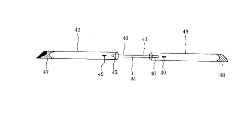
On the other hand, FIG 810 show the hithertofore various
nunchaku style silicone tubings which are invented by this inventor.
For example, see Patent No.2539325.
Any apparatus for intubation of the lacrimal duct shown in FIG
810 consists of thinner soft tube 40, 41 and thicker tube 42, 43 with
certain length, and the ends 47, 48 of the thicker tube are closed end.
CA 02265091 1999-03-08
Thinner tube 40, 41 exists between two thicker tube 42, 43, and
the middle point 44 of the thinner tube 40, 41 is marked.
The thinner soft tube 40, 41 is connected with the thicker tubes
42, 43. Two millimeter ends of the thinner tube 40, 41 are inserted
into the thicker tube 42, 43 for connection. Therefore, the jointed
portion 45, 46 are 2mm in length. The tips 47, 48 of the thicker tubes
are sharp pointed and closed. For example, 2mm tips of the tube are
completely sealed with silastic adhesive, and then diagonally cut to
taper the closed ends 47, 48. Small cuts 49 are applied to the thicker
tubes 42, 43 parallel to the tubes 42, 43.
The junctions 45 make steps in the case of FIG 8. As shown in
FIG 910, it is possible to make slopes 51 without making any steps.
And in the cases shown in FIG 9~ 10, the ends 53, 54 of the
thicker tube are in conical shape.
In the cases of FIG 8~9, it is very rare for the junctions 45 to
come off. However, as shown in FIG 10, a one piece tube without any
junctions which consists of the thinner tube 40, 41 and thicker tubes 42,
43 is made from the first.
1) In prior methods of monocanalicular intubation using the half
size nunchaku-style silicone tubing shown in FIGS 810 or a prior
silicone tube with the same thickness in its total length, it is necessary
to fix the tube at the puncta 1, 2 with suturing because they lack the
brim.
2) Prior punctal plugs shown in FIGS 2~7 are anglar, and its
stimulation sometimes induce granuloma.
6
CA 02265091 1999-03-08
3) Prior punctal plugs shown in FIGS 2~7, sometimes induce
canalicular obstruction between the vertical portion 3, 4 and the
horizontal portion 7, 8 of the canaliculus.
4) In prior punctal plugs shown in FIGS 2~7, the punctal plugs
sometimes migrate into the canaliculus, lacrimal sac and nasolacrimal
duct because the brim is circular and too small.
5) Prior nunchaku-style silicone tubings shown in FIGS 810 are
sometimes difficult to insert from the puncta 1, 2 because its closed
ends are not so sharp pointed.
6) Prior punctal plugs shown in FIGS 2~7, sometimes come off
because of its shallow insertion.
7) Dry eye symptoms are sometimes aggravated in patients with
both dry eye and dacryocystitis, after intubation using prior tubes
shown in FIGS 8~ 10 and/or dacryocystothinostomy.
8) Punctal plugs shown in FIGS 2~7, are not stable.
9) Tubes with the same thickness in its total length are not
stable even if the brim is attached to them.
10) Prior tubes shown in FIGS 810 sometimes induce slitting of
the puncta 1, 2 and canaliculi 3~8, as shown in FIG 11.
SUMMARY OF THE INVENTION
It is an object of the present invention to provide an apparatus
for intubation of the lacrimal duct which is stable in the lacrimal duct,
can be easily inserted into the lacrimal duct and be easily removed, is
not in danger of extrusion after leaving in place and growth of
granuloma.
7
CA 02265091 1999-03-08
It is another object of this invention to provide an apparatus for
intubation of the lacrimal duct which can be used for reconstruction of
lacrimal duct obstructions.
One apparatus according to this invention is an apparatus for
intubation of the lacrimal duct which is characterized by the presence
of, a thinner tube or rod, a prescribed length of thicker tube which is
joined with one end of the thinner tube or rod, and a stopper which is
joined with the other end of the thinner tube or rod in apparatus for
intubation of lacrimal duct inserted into the lacrimal duct.
Other apparatus according to this invention is an apparatus for
intubation of lacrimal duct which is characterized by presence of a
prescribed length of thicker tube and a stopper joined with the
posterior end of the thicker tube.
Other apparatus according to this invention is an apparatus for
intubation of lacrimal duct which is characterized by the fact that said
stopper is a punctal plug, brim, ring and so on.
The inventor has studied keenly for many years for the
treatment of lacrimal duct obstruction and dry eye apparatus for
intubation of lacrimal duct which can be used easily with decrease pain
to patients, can be quickly and correctly inserted into the lacrimal duct,
is not easily dislocated during intubation period, and can be easily
removed after accomplishment of treatment.
This invention is to provide a more improved apparatus for
intubation based on the apparatus which has been developed until now.
8
CA 02265091 1999-03-08
This invention especially improves the stableness of the apparatus for
intubation in the lacrimal duct.
In the present invention, an apparatus for intubation of the
lacrimal duct includes a stopper. As a stopper, a punctal plug, brim,
ring and other things are used.
Apparatus for intubation according to the present invention
consists of a prescribed length of thinner tube or rod, prescribed length
of thicker tube which is connected with one end of thinner tube or rod,
and punctal plug which is connected with the other end of thinner tube
or rod.
Other apparatus for intubation according to the present
invention omits the thinner tube or rod from said embodiment, consists
of a prescribed length of the thicker tube and the punctal plug which
connected with the posterior end of the thicker tube.
Other apparatus for intubation according to the present
invention wherein the punctal plug of said apparatus for intubation is
changed into a brim, consists of a prescribed length of thinner tube or
rod, a prescribed length of thicker tube which is connected wih one end
of the thinner tube or rod, and a brim which connected with the other
end of the thinner tube or rod.
Other apparatus for intubation according to the present
invention, wherein the punctal plug of said apparatus for intubation is
changed into a stopper, consists of a prescribed length of thinner tube
or rod, a prescribed length of thicker tube which connected with one end
of the thinner tube a rod and the stopper which is connected with the
other end of the thinner tube or rod.
9
CA 02265091 1999-03-08
As stated above, use of punctal plug, brim and ring as a stopper
brings about a great effect which each cannot be gained by prior arts.
In any said apparatus for intubation it is preferable that the tip
of the thicker tube is closed.
Furthermore, in a preferable apparatus for intubation according
to the present invention, its total length is 15~60mm including tube
and punctal plug, punctal plug is 1.5~2.5mm in length, thicker tube is
10~59mm in length and thinner tube is l~5mm in length.
In other preferable apparatus for intubation according to the
present invention, a thicker hard tube is connected with a punctal plug
via a thinner soft tube.
As a further other mode different from this, the thicker hard
tube is directly connected with the punctal plug without intervention of
the thinner soft tube to constitute an apparatus for intubation.
In yet another further other mode of the present invention,
various nunchaku-style silicone tubings are fixated with various
punctal plugs with silastic adhesive to be able to constitute an
apparatus for intubation.
In another further other mode of the present invention, central
segment is flexible, both ends of it are fixated to the punctal plug and
tube with silicone adhesive, the punctal plug and tube are thicker and
harder, and the central segment constitutes has constitution to be able
to pass through with forming a curve the boundary portion between the
vertical and horizontal portions of canaliculus.
And the thicker tube which is used in this invention has a closed
end and a small cut is applied to the part of the tube for probe to insert.
CA 02265091 1999-03-08
This makes for the apparatus to be inserted into the lacrimal duct
easily.
In the punctal plug used in this invention, the brim preferably
1.5~4.5mm in diameter prevents the punctal plug from migrating into
the lacrimal duct, and simultaneously prevents tears from flowing into
the puncta.
The apparatus for intubation of this invention has a great
stability in the lacrimal duct compared with conventional punctal plug
and it is very rare to extrude.
BRIEF DESCRIPTION OF THE DRAWINGS
FIG.1 is a schematic diagram of the lacrimal duct.
FIG.2 is a schematic diagram of the prior art.
FIG.3 is a schematic diagram showing how to use the punctal
plug in FIG.2.
FIG.4(A) is a schematic diagram showing a conventional other
plug.
FIG.4(B) is the bottom view of it.
FIG.4(C) is the mid-cross-sectional view of it.
FIG.S(A) is a schematic diagram showing conventional other
plug.
FIG.S(B) is the bottom view of it.
FIG.S(C) is the mid-cross-sectional view of it.
FIG.6 is a diagram showing a failure when the plug in FIG.2 is
used.
FIG.7is a diagram showing another failure when the plug in
11
CA 02265091 1999-03-08
FIG.2 is used.
FIG.8 is a diagram showing a conventional nunchaku-style
silicone tubing.
FIG.9 is a diagram showing other conventional nunchaku-style
silicone tubing.
FIG.10 is an explanatory diagram showing still other
conventional nunchaku-style silicone tubing.
FIG.11 is an explanatory diagram showing a failure in a
conventional nunchaku-style silicone tubing.
FIG.12 is a schematic diagram showing the apparatus for
intubation in the present invention.
FIG.13 is a schematic diagram showing other apparatus for
intubation in the present invention.
FIG.14 is a schematic diagram showing other apparatus for
intubation in the present invention.
FIG.15 is a schematic diagram showing other apparatus for
intubation in the present invention.
FIG.16 is a schematic diagram showing other apparatus for
intubation in the present invention.
FIG.17 is a schematic diagram showing other apparatus for
intubation in the present invention.
FIG.18 is a schematic diagram showing other apparatus for
intubation in the present invention.
FIG.19 is a schematic diagram showing other apparatus for
intubation.
FIG.20 is a cross sectional view showing other apparatus for
12
CA 02265091 1999-03-08
intubation.
FIG.21 is a perspective view showing the apparatus for
intubation in Fig.20.
FIG.22 is a cross sectional view showing other apparatus for
intubation.
FIG.23 is a perspective view showing the apparatus for
intubation in Fig.22.
FIG.24 is a cross sectional view showing other apparatus for
intubation .
FIG.25 is a perspective view showing the apparatus for
intubation in Fig.24.
FIG.26 is a perspective view showing the method of insertion of
the apparatus in Figs.17~25.
FIG.27 is a diagram showing monocanalicular intubation method
in Figs.17~25.
FIG.28 is a diagram showing other monocanalicular intubation
method in FIGS 1725.
FIG.29 is a diagram showing bicanalicular intubation method in
Figs.17~25.
FIG.30 is a sectional view showing other apparatus for
intubation in the present invention.
FIG.31 is a perspective view showing the apparatus for
intubation in Fig.30.
FIG.32 is an explanatory diagram showing the insertion method
of the apparatus for intubation in Figs.30~31.
FIG.33 is an explanatory diagram showing a monocanalicular
13
CA 02265091 1999-03-08
intubation method using the apparatus for intubation in Figs.30~31.
FIG.34 is an explanatory diagram showing other
monocanalicular intubation method using the apparatus for intubation
in Figs.30~31.
FIG.35 is an explanatory diagram showing bicanalicular silicone
intubation method using the apparatus for intubation in Figs.30~31.
FIG.36 is a cross sectional view showing other apparatus for
intubation in present invention.
FIG.37 is a perspective view showing the apparatus for
intubation in Fig.36.
FIG.38 is an explanatory diagram showing the method of
insertion of the apparatus for intubation in Figs.36~37.
FIG.39 is an explanatory diagram showing monocanalicular
silicone intubation method in Figs.36~37.
FIG.40 is an explanatory diagram showing other
monocanalicular silicone intubation method using the apparatus for
intubation in Figs.36~37.
FIG.41 is an explanatory diagram showing bicanalicular
intubation using the apparatus for intubation in Figs.36~37.
FIG.42 is a perspective view showing still other apparatus for
intubation in the present invention.
FIG.43 is a mid-cross sectional view showing the apparatus for
intubation in Fig.42.
FIG.44 is an explanatory diagram showing the method of
placement of the apparatus for intubation in Figs.42~43.
14
CA 02265091 1999-03-08
DETAILED DESCRIPTION OF THE EMBODIMENTS
The embodiments of this invention will be explained, referring to
figures.
FIG 1214 show three different embodiments of this invention.
In the embodiments of FIG 1214, the punctal plug P is attached
to an end of the thinner soft tube 40, 1~15mm in length, with silicone
adhesive. The posterior end of the thicker hard tube 42 is the other
end of the thinner tube 40. The tip of the thicker tube 42 is closed in a
conical shape.
The punctal plug P consists of the tip 21 which shape is the
frustum of the circular cone, brim 23 which shape is circular elliptical
and its whole body is a single piece. In the center of the brim 23, a
hole 24 is formed. The hole 24 is connected with the lumen of the shaft
22, and the lumen of the tip 21, and connected with the inner space of
the thinner tube 40 and the inner space of the thicker tube 42 with a
closed end 53.
In embodiments of FIG 12 and FIG 14, the axis of the thinner
tube 40 is coincident with the axis of the thicker tube 42 and the axis of
the plug. Whereas, although in embodiment of FIG 13, the axis of the
thinner tube 40 is coincident with the axis of the thicker tube 42, the
axis of the plug is not coincident with the axis of the thinner tube 40
and the axis of the thicker tube 42 to be formed at a prescribed angle
(for example 90150' ).
Although the plug without hole 24 can be used and a rod instead
of thinner tube 40 can be used in embodiment of FIGS 1214, the hole
24 of the plug P is connected with the closed end 53 via the lumen of the
CA 02265091 1999-03-08
shaft 22, the inner space of the thinner tube 40, the inner space of the
thicker tube 42 and the lumen of the tip 21. Its form will be explained.
Regarding Materials for tube 40, 42 and punctal plug P, it is
important to select one which is substantially unstimulating and non-
toxic to the tissue of the eye and a living body. From this point of view,
silicone is appropriate because its safety is already established as a
apparatus for treatment of lacrimal duct obstruction. Above all, the
combination of silicone tube 0.9~1.2mm OD and 0.5~0.7mm OD and
0.3~0.5mm ID is especially preferably used. Regarding punctal plug P,
silicone punctal plugs are preferable as shown in FIG 1214.
This composition will be concretely explained as follows. As
shown in FIG 1214, the right end of the thinner soft tube (0.5~0.7mm
OD, 0.3~0.5mm ID, 2~20mm in length, 40 is connected with the left end
of the thicker tube (0.9~1.2mm OD, 0.5~0.7mm ID, 5~50mm in length)
42. The tip 53 of the thicker tube 42 is sharp pointed and closed. For
example, 2mm tip of the thicker tube 42 is completely sealed with
silastic adhesive, and then diagonally cut to taper the closed end 53.
Small cuts 0.5mm in length 49 for probe 0.4mm in length to insert is
applied to the thicker tube 42 parallel to the tube 42. If the small cut
is applied perpendicularly to the tube, the tube may be broken during
operation. The preferable position of the small cut is 10 to 45mm from
the tip of the thicker tube 42. The tube 42 can be easily inserted into
the lacrimal duct by inserting the probe (not illustrated) from the small
cut 49. Making corresponding to the position of the small cut 49 make
it easier to discover the small cut.
Preferable total length of the apparatus of the present invention
16
CA 02265091 1999-03-08
is as follows.
Total length of 40~60mm apparatus are appropriate for adult-
nasolacrimal duct obstruction and total length of 30~50mm apparatus
for child-nasolacrimal duct obstruction. Total length of 10~60mm
apparatus are useful for reconstruction of canalicular obstruction and
for using as a punctal plug.
The length and thickness of the silicone tube depends on the
length and size of the inner space of the individual lacrimal duct. The
most used one being 51.7mm in total length consists of the thinner tube
40 0.64mm in thickness, l0mm in length, the thicker tube 42 0.94mm in
thickness, 40mm in length, and the punctal plug l.7mm in total length.
In order to be stable in the lacrimal duct, it is important that the
thinner tube is softer. As long as it is thinner and softer, for example
the soft rod without an opening 0.5~0.7mm in diameter can be used
instead of the thinner tube 40.
It is better to make a slope 51 without making any steps at the
junction.
If the tip 53 of the thicker tube is sharp pointed in a conical
shape, it is more easily inserted from the lacrimal puncta.
The embodiment in FIG 1214 are one piece without junctions
which consists of the thinner tube 40 and the thicker tube 42. As
shown in FIG 1718, it can be made by 2mm end of the thinner tube
inserted into the thicker tube for connection and fixation using silicone
glue. In the embodiment in FIG 18, the junction between the thinner
tube 40 and the thicker tube 42 has no steps, and in the case of FIG 17
the junction between the thinner tube 40 and the thicker tube 42 is a
17
CA 02265091 1999-03-08
step-like junction.
Although it is not illusrated, it is better for the tube to be
equipped with the probe from the first.
Constitutions of the embodiment in FIG 8~ 11 suit with
bicanalicular intubation in which a tube 40, 42, 41, 43 is introduced
into the lacrimal duct from the upper and lower puncta. Whereas,
constitutions of the embodiment in FIG 1216 suit with
monocanalicular intubation in which the apparatus for intubation is
inserted into the lacrimal duct from the upper puctum only (or the
lower punctum only).
In embodiment in FIG 15 and FIG 16, the punctal plug P is
attached to the posteinor end of the thicker hard tube 42 with silicone
glue without using a thinner soft tube. The tip 53 of the thicker tube
is in conical shape and closed. Punctal plug P is one piece which
consists of the tip 21 which is a shape of the frustum of the circular
cone, the shaft 22 which is tubular, and the brim 23 which is circular.
In the center of the brim 23, the hole 24 is applied. The hole 24 is
connected with the lumen of the shaft 22 and the lumen of the tip 21
which is connected with the inner space of the thicker tube 42 with a
closed end 53.
The tube 42 can be easily inserted into the lacrimal duct by
insertion of the probe (not illustrated) from the small cut 49 applied to
the thicker tube 42.
The embodiments of the constitution in FIG 1516 is superior for
the treatment of the lacrimal duct obstruction. As shown in FIG 1516,
only the thicker tube 42 is used and the thinner tube 40 is abbreviated.
18
CA 02265091 1999-03-08
The tip 53 of the thicker tube 42 is in a conical shape and closed.
When this is inserted into the lacrimal duct from the upper punctum 1,
the tube 42 is pushed into the lacrimal duct by the probe 61 (mentioned
later) which is inserted into the tube 42 till the tip 53. After insertion
of the tube 42, the probe 61 is removed.
In the embodiment in FIG 15, the axis of the thicker tube 42 is
coincident with the axis of the plug P. Whereas, in the embodiment in
FIG 16, the axis of the plug P is not coincident with the axis of the
thicker tube 42 to form at a prescribed angle (for example 90~ 150' ).
FIGS 1719 show other embodiments in this invention besides.
In the embodiment in FIG 17, the brim 23 is joined with one end
of the thinner soft rod 40 5~20mm in length with silicone glue. The
brim 23 is formed rather hard silicone.
In the embodiment of FIG 18, the brim 23 is joined with one end
of the thinner soft rod 40 5~20mm in length, The brim 23 is formed by
rather hard silicone. The posterior end of the thicker hard tube 42 is
joined with the other end of the thinner tube 40. The tip 53 of the
thicker tube 42 is in conical shape and closed. The thinner rod 40 and
the thicker tube 42 are sparately made and 2mm end of the thinner rod
is inserted into the thicker tube for connection with silicone glue.
This embodiment in FIG 18 has not any step in the junction between
the thinner rod 40 and the thicker tube 42.
In the embodiment in FIG 19, the brim 23 is joined with one end
of the thinner soft tube 40 with silicone glue. The brim 23 is formed by
rather hard silicone. The posterior end of the hard tube 42 is joined
with the other end of the thinner tube 40. The tip 53 of the thicker
19
CA 02265091 1999-03-08
tube 40 is in conical shape and closed. The thinner tube 40 is joined
with the thicker tube 42 with the shape of one body. In the
embodiment in FIG 19, the junction between the thinner tube 40 and
the thicker tube 42 has not any step.
In the embodiment in FIG 1719, the tube 42 can be inserted into
the lacrimal duct easily by a probe (not illustrated) which inserted into
the small cut 49 of the thicker tube 42.
FIG 2021 show other embodiments in the present invention
besides. The thinner soft rod consists of the segment 22 0.5mm OD
and 2~2.5mm in length, and the segment 40 0.5mm OD and 3~20mm in
length, and the segments 22, 40 form at angle of 90150 degrees. The
brim 23 is joined with an end of the thinner soft rod with silicone glue.
The brim 23 is circular, elliptic and others in shape, and formed by hard
silicone. The posterior end of the thicker hard tube 42 30~40mm in
length is joined with the other end of the thinner rod to form a shape of
single body. The tip 53 of the thicker tube 42 is in conical shape and
closed. The lumen 25 formed in the thinner rod 22 is connected with
the hole 24 of the brim 23. The lumen 25 extends into the thinner rod
till the corner which exists halfway in the thinner rod. The thicker
tube 42 1.0~1.2mm OD and 0.5mm ID has the inner space 55. The
junction between the thinner rod 40 and the thicker tube 42 is a slope
51 not to make a step. And, in this embodiment, the axis of the brim
23 is set to the axis of the thicker tube 42 at a prescribed angle (for
example 90150 degrees).
The embodiments in FIG 2021, the tube 42 can be easily
inserted into the lacrimal duct by the probe (not illustrated) which is
CA 02265091 1999-03-08
inserted into the inner space 55 from the small cut 49 applied to the
thicker tube 42.
FIG 2223 show still other embodiment of the present invention
besides. The thinner soft rod without a lumen consists of the segment
22 0.5mm OD, 1.5~2.5mm in length and the segment 40 0.5mm OD,
3~20mm in length, and at an angle of 90150 degrees between these
segments 22, 40. The brim 23 is joined with an end of such a thinner
soft rod with silicone glue. The brim 23 is circular elliptical and in
other shapes and is formed by hard silicone. The posterior end of the
thicker hard tube 42 30~40mm in length is joined with the other end of
the thinner rod to be a single body. The tip 53 of the thicker tube 42 is
sharp pointed and has a closed end. The thicker tube 1.0~1.2mm OD
and 0.5mm ID has the inner space 55 which extends to the closed end.
The junction between the thinner rod 40 and the thicker tube 42 is a
slope 51 not to make a step. And, in this embodiment, the axis of the
brim 23 is set at a prescribed angle (for example 90150 degrees) to the
axis of the thicker tube.
In the embodiments shown in FIGS 2021, the tube 42, can be
easily inserted into the lacrimal duct by the probe (not illustrated)
which is inserted from the small cut 49 applied to the thicker tube 42.
FIGS 2223 show still other embodiments according to the
present invention. The thinner soft rod without any opening, consists
of the segment 22 0.5mm OD and 1.5~2.5mm in length and the segment
40 0.5mm OD and 3~20mm in length, these segments 22, 40 form an
angle of 90150 degrees. The brim 23 is attached to an end of such a
thinner soft rod with silicone glue. The brim 23 is circular elliptic and
21
CA 02265091 1999-03-08
in other forms, and manufactured from hard silicone. With the other
end of the thinner rod, the posterior end of the thicker hard tube is
connected in the form of one body. The tip 53 of the thicker tube is
sharp pointed and closed. In the thicker tube 42 1.0~1.2mm OD and
0.5mm ID, the inner space 55 extends to the closed end. The junction
between the thinner rod 40 and the thicker tube 42 has the slope 51
without making any step. And, in this embodiment, the axis of the
brim 23 is at a prescribed angle (for example 90150 degrees) to the
axis of the thicker tube 42.
In the embodiments shown in FIGS 2223, the tube 42 can be
easily inserted into the lacrimal duct by the probe (not illustrated)
which inserted from the small cut 49 which applied to the thicker tube
42.
FIGS 2425 show still other embodiments according to the
present invention. The thinner soft rod consists of the segment 22
0.5mm OD and 2~2.5mm in length and the segment 40 and these
segments 22, 40 form an angle of 90150 degrees. The brim 23 is
attached to an end of the thinner soft rod with silicone glue. The brim
23 is circular, elliptical and in other forms, and manufactured from
hard silicone. The posterior end of the thicker hard tube 42 30~40mm
in length is connected with the other end of the thinner rod forming one
body. The tip 53 of the thicker tube 42 is sharp pointed and closed.
The lumen 25 created in the segment of the thinner rod 22 is connected
with the hole 24 of the brim 23. The lumen 25 extends in the total
length of the thinner rod. The thicker tube 42 1.0~1.2mm OD and
0.5mm ID has the lumen 55. The lumen 25 of the thinner tube 40 is
22
CA 02265091 1999-03-08
connected with the lumen 55. The junction is a slope 51 without
making any steps. And, in this embodiment, the axis of the brim 23 is
at a prescribed angle (for example 90150 degrees) to the axis of the
thicker tube 42.
In the embodiment shown in FIGS 2425, the thicker tube can be
easily inserted into the lacrimal duct by the probe (not illustrated)
which inserted into the small cut 49 applied to the thicker tube 42.
FIG 26 shows still another embodiment according to the present
invention. The brim 23 is attached to an end of thinner soft rod 40
with silicone glue. The brim 23 is circular, elliptical and in other
shapes and made of hard silicone. Into the posterior end of the thicker
tube, the thinner rod 40 is inserted to join them. The segment of the
thinner rod 22 forms a curve from junction 45 to the brim 23. The
junction 45 is formed so as to be a slope without making any step. And
in this embodiment, the axis of the brim 23 is at a prescribed angle (for
example, 90150 degrees) to the axis of the thicker tube 42.
The way of closing the embodiment shown in FIG 26 is
substantially the same as the above mentioned embodiments shown in
FIGS 2025, and the tube 42 can be easily pushed into the lacrimal duct
by the probe 61 which is inserted into the inner space 55 from the small
cut 49 applied in the thicker tube 42.
FIG 27 shows one of the post-operative state of placement of the
apparatus for intubation in the present invention inserted from the
lower canaliculus in the embodiments in FIGS 2026.
FIG 28 shows one of the post-operative state of placement of the
apparatus for intubation in the present invention inserted from the
23
CA 02265091 1999-03-08
upper canaliculus in the embodiments in FIGS 2026.
FIG 29 shows one of the post-operative state of placement of the
apparatus for intubation in the present invention inserted from the
upper and lower canaliculi as to the embodiments in FIGS 2026. This
is the most suitable placement for the treatment of dry eye.
FIGS 3031 show still other embodiment in the present
invention. The punctal plug P is attached to an end of the thinner soft
rod 40. The punctal plug P consists of the tip 21, intermediate portion
22 and the brim 23. The edge 20 is round not to induce granulation.
The brim 23 is circular, elliptical and in other shapes and made of hard
silicone. The posterior end of the thicker hard tube 30~40mm in
length in connected with the other end of the thinner rod to be a form of
one body. The tip 53 of the thicker tube 42 is sharp pointed and closed.
The lumen 25 created in plug P is connected with the hole 24. The
thicker tube 42 has the inner shape 55 and its outer diameter is
1.0~1.2mm, and inner diameter is 0.5mm. The junction is slope not to
make any steps. And, in these embodiments also, the axis of the brim
23 is at a prescribed angle (for example 90150 degrees) to the axis of
the thicker tube 42.
In the embodiments in FIGS 3031 also, the thicker tube 42 can
be easily inserted into the lacrimed duct by the probe 61 which is
inserted into the inner space 55 from the small cut 49 of the thicker
tube 42.
FIG 32 shows one of the way of insertion of the apparatus for
intubation in the present invention as to the embodiments shown in
FIGS 3031.
24
CA 02265091 1999-03-08
FIG 33 shows one of the post-operative state of placement the
apparatus for intubation in the present invention inserted from the
lower canaliculus in the embodiments in FIGS 3031.
FIG 34 shows one of the post-operative state of placement of the
apparatus for intubation in the present invention inserted from the
upper canaliculus as to the embodiments in FIGS 3031.
FIG 35 shows one of the post-operative state of placement of the
apparatus for intubation in the present invention inserted from the
upper and lower canaliculi as to the embodiments in FIGS 3031.
In many cases, one of the upper and lower puncta is sufficient to
prevent epiphona. Therefore it is superior to place the apparatus for
intubation in present intubation in the lower or upper canaliculus after
opening of obstructed portions) of the lacrimed duct.
FIGS 3637 show still other embodiments where the puntal plug
P is attached to an end of the thinner soft rod. The punctal plug P
consists of the tip 21 and the brim 23. The edge 21 a of the tip 21 is
round for granulation so as not to easily be induced. The brim 23 is
circular, elliptical, in other shapes and made of hard silicone. The
posterior end of the thicker hard tube 42 is connected with the other
end of the thinner rod 40 so as to form one body. The tip 53 of the
thicker tube 42 is sharp pointed and closed. The inner space 25
created in the plug P is connected with the hole of the brim 23. The
inner space 55 is present in the thicker tube 1.0~1.2mm OD and 0.5mm
ID. The junction between the thinner rod 40 and thicker tube 42 is the
slope 51 without making any step. And in this embodiments also, the
axis of the brim 23 is at a prescribed angle (for example 90150
CA 02265091 1999-03-08
degrees) to the axis of the thicker tube 42.
In the embodiments in FIGS 3637, the tube 42 can be pushed
into the lacrimal duct by a probe 61 which inserted into the inner space
55 from the small cut 49 of the thicker tube 42.
FIG 38 shows one of the way of insertion of the apparatus for
intubation in the present invention, as to the embodiments in FIGS
3637.
FIG 39 shows one of the post-operative states of placement of the
apparatus for intubation in the present invention inserted from the
lower canaliculas as to the embodiments in FIGS 3637.
FIG 40 shows one of the post-operative states of placement of the
apparatus for intubation in the present invention inserted form the
upper canaliculus as to the embodiments in FIGS 3637.
FIG 41 shows one of the post-operative states of placement of the
apparatus for intibation in the present invention inserted form the
upper and lower cacnaliculi as to the embodiment in FIGS 3637.
One of the upper and lower canaliculi is sufficient to prevent
epiphora. Therefore, it is superior to place the apparatus for
intubation in the present invention in the lower or upper canaliculus,
after opening of obstructed portions) of the lacrimal duct.
FIGS 4243 show still other embodiments in the present
invention.
The apparatus for intubation of the lacrimal duct shown in FIGS
4243, has a prescribed length of two thinner soft rods 40 and two
thicker tubes 42 to be inserted into the lacrimal duct. One end of each
thicker tube 42 is closed. Between the two thicker tubes 42, two
26
CA 02265091 1999-03-08
thinner rods 40 exist and between the two thinner rods 40, a thinner
rod 41 is present, and the midpoint of the thinner rod 41 has a marking
44. Junction between the thinner rod 40 and the thicker tube is a
slope 51 so as not to make any steps. The tip 53 of the thicker tube 42
is in a conical shape.
The punctal plug P is positioned between two thinner soft rods
40, 41. The punctal plug P consists of the tip 21, shaft 22 and brim 23.
The brim is circular, elliptical and in other shapes and made of hard
silicone. The plug P has not any inner space. The thicker tube has an
inner space 55 and its outer diameter is 1.0~1.2mm and inner diameter
is 0.5mm. The junction between the thinner rod 40 and the thicker
tube 42 has the shape 51 without making any steps.
In the embodiment in FIGS 4243, the tube 42 can be easily
pushed into the lacrimal duct by the probe 61 which is inserted into the
inner space from the small cut 49 of the thicker tube 42.
FIG 44 shows one of the ways of insertion from the upper and
lower canaliculi, of the apparatus for intubation in the present
invention, as to the embodiment sown in FIGS 4243.
Finally, the general method of surgery using the apparatus for
intubation according to the present invention will be explained.
Before insertion, the obstructed segments) of the lacrimal duct
is opened by insertion of probe 61. And in advance, the puncta are
dilated by punctal incision at their lateral wall or using a punctal
dilator. The tip 53 of the tube 42 enclosing the probe 0.4mm in
diameter 61 pushed into the inferior nasal meatus from the lower
punctum 2 via the lower canaliculus 4, 6, 8, common canaliculus 9,
27
CA 02265091 1999-03-08
lacrimal sac 11 and nasolacrimal duct 12. And then, only the
apparatus for intubation is left and the probe 61 is removed.
Next, if necessary, another apparatus for intubation is pushed
into the lacrimal duct from the upper punctum 1 and in advance, the
probe 61 0.5~1mm in diameter is inserted from the upper punctum 1.
The upper punctum is also dilated by punctal incision at the lateral
wall or using a punctal dilator. The tip 53 of the tube 42 is pushed into
the inferior nasal meatus by the probe 61 which inserted into the
thicker tube from the small cut in the same way.
It can be removed easily by holding and pulling the plug P, brim
23 or thinner rod 41 at the upper punctum 1 and the lower punctum 2
using forceps.
Although any of the apparatus for intubation in the present
invention is usually used under local anesthesia or general anesthesia
using an operating microscope, it can be used simply for many patients
under local anesthesia.
Silicone is preferable to make the apparatus for intubation
according to the present invention. Silicone is unstimulating and
non-toxic to the living body so it is possible to leave in place for a long
time.
Unlike the prior arts, the apparatus for intubation according to
the present invention, does not require difficult nasal procedure at all,
resulting in short operating time and a small burden on patients.
Unlike the prior tubes, it has the stopper and can be bent, to
make the tube stable in the lacrimal duct without easy migration.
Furthermore, patients feel very little pain.
28
CA 02265091 1999-03-08
Although it is easy to insert the apparatus for intubation
according to the present invention into the lacrimal duct and easy to
remove it, it is not easily dislocated during intubation period.
It is still better to combine the thicker tube with the thinner
tube or rod.
The small cuts applied to the thicker tube do not break the tube.
The tube with a sharp pointed tip in conical shape can be easily
intubated into the lacrimal duct after punctal dilation with the punctal
dilator only without punctal incision.
Using the apparatus for intubation, intubation into the lacrimal
duct can be performed more easily. This fact make it possible for
doctors to do intubation routinely before resorting to major surgical
interventions.
The apparatus for intubation is not easily dislocated compared to
the punctal plug when it is used for the treatment of dry eye.
Furthermore the following effects can be gained according to the
present invention.
1) It is unnecessary to fix it by sutures) in monocanalicular
intubation.
2) Incidence of granulation arise by the stimulation of the
angular portions in the prior punctal plug is decreased by rounding the
angular portion.
3) Obstruction between the vertical portion and horizontal
portion of canaliculus is not induced. Furthermore, it is useful for a
stmt of canalicular obstruction and nasolacrimal duct obstruction.
4) By making the brim of the punctal plug elliptical, the brim can
29
CA 02265091 1999-03-08
be enlarged so as not to stimulate the ocular surface and prevent the
plug from migrating into the canalicular.
By making the tip of the thicker tube sharp pointed, it can be
easily inserted from the punctum.
6) Tear fluid cannot enter into the punctum by the brim which
adhere to the puncta because the thicker tube is pulled into the
lacrimal duct due to the lacrimal drainage function.
7) Punctal occlusion and the treatment of lacrimal duct
obstruction and dacryocystitis can be performed simultaneously for
patients with dry eye, lacrimal duct obstruction and dacryocystitis.
8) Positioning of the thinner soft tube between the punctal plug
and the thicker tube makes the apparatus for intubation stable in the
lacrimal duct. Furthermore, making an angle of 90° - 150°
between
the axises of the punctal plug and the thicker tube makes it stabler.
9) In the apparatus which consists of the thinner and the thicker
tubes, attachment of the brim only to the thinner tube also makes it
stable.