Note: Descriptions are shown in the official language in which they were submitted.
CA 02266396 1999-03-22
W O 98/12969 PCTrUS97/17128
METHOD AND APPARATUS FOR INSERTING
A FIJFX~RLE MEMBlRANE INTO AN EYE
Fi~ of the ~nvf.nt~
The present invention pertains to a method and apl)a,~lus for inserting a flexible
intraocular lens or other flexible membrane into an eye.
rl~n..~.d of the Inv~nti~n
The natural crystalline lens of the eye plays a primary role in focusing light onto
the retina for proper vision. However, vision through the natural lens may become
impaired due to an injury, or due to the fi~rmation of a cataract caused by aging or
disease. To restore vision, the natural lens is typically replaced with an artificial lens.
An artificial lens may also be implanted to make a refractive correction.
Many surgical procedures have been developed for removing the natural lens.
Typically, a slender imrlement is inserted through a small incision in the eye to contact
the natural lens. The implement includes a c utting tip that is ultrasonically vibrated lo
emulsify the lens. The emulsified fr~mPnt~ of the lens are then aspirated out of the
eye through a passage provided in the cutting tip. The slender nature of the implement
enables extraction of the lens through a small incision in the eye. The use of a small
incision over other procedures requiring a large incision can lessen the trauma and
complications experienced during the surgery and postoperatively.
Rec~ 5e the incision required to remove the lens is small, the development of
intraocular implants to replace the lens has been in the direction of flexible implants
CA 02266396 1999-03-22
W O98112969 PCT~US97/17128
that do not require any enlargement of the incision. An intr~ocul~r lens commonly
includes a generally disk shaped optic which focuses light on the retina and an
outwardly PYt~nding haptic portion for proper positioning of the optic within the eye.
The flexible nature of the lens enables the lens to be folded and co~ ,ressed so as to
occupy a smaller cross-sectional area for passage through the narrow incision and into
the eye. Once inserted through the incision, the lens is permitted to expand to its
original size and shape.
A number of devices have been developed to insert a flexible intraocular lens
through a small incision in the eye. For eY~mple, U.S. Patent No. 4,681,102 to
Bartell uses a hinged cartridge which closes about a lens to fold the lens into a
narrower configuration. The cartridge is placed into an inserter mech~nicm whichadvances the folded lens into the eye. The inserter, however, requires several
co~llpollents to be manipulated and assembled during the operation. U.S. Patent No.
5,275,604 to l~h~ini~h et al. pushes the lens through a narrowing lumen formed with
grooves which act to fold the lens into a smaller size as it is pushed toward the eye.
The manufacture of spiraling grooves in a tapering lumen is difficult if not impossible
to accomplish in a practical manner. In U.S. Patent No. 5,304,182 to Rheini~h et al.,
a curling member is shifted laterally to fold the lens into a size small enough to pass
through the narrow incision. However, no locking arrangement is provided to ensure
completely closing of the curling member.
Moreover, while these devices function to reduce the cross-sectional size of thelens for insertion into the eye, they all require the opposing side edges of the lens to
be folded over on themselves in order to fit through the narrow incision. As a result,
CA 02266396 1999-03-22
W O 98/12969 PCTAUS97/17128
the lens must swing open within the eye to regain its origin~l shape and size. Such
unfolding causes the lens, and particularly the haptics, to be swung in an arc, and thus
risks ~l~m~ing the interior of the eye.
As the lens is released into the eye, lhe resiliency of the lens causes the lens to
5open and resume its natural shape. However, the folding and pressing of the lens
needed to pass the lens through the small incision places a ~ignific~nt amount of inward
pressure on the lens. As a result, the lens is frequently discharged from the inserter
with considerable force and velocity. This forceful, uncontrolled release of the lens
also places the interior of the eye at risk oi being injured.
10Further, many inserters do not ".~ control of the orientation of the lens as
the lens is advanced into the eye. Consequently, the lens may rotate or turn about a
lon~itll-lin~l axis as it is pushed through the inserter. Most lenses, however, are made
to be set within the eye in a specific ori~nt~tiQn. Accordingly, such turning of the lens
can result in the lens being placed in the eye in an ill-ploper orientation.
15~mnl~ry of th~ lnvPntin~.
The present invention pertains to a m~ethod and .~ s for inserting a flexible
intraocular lens or other flexible membrane into an eye without the above-noted risks
associated with inserter devices of the past. More specific~lly, the present inserter
the subst~nti~lly planar ~rient~tion of the opposing side edges of the lens as
20the lens is laterally con-pl~d into a smaller cross-sec~ion~l configuration for insertion
through a narrow incision in the eye Since the side edges of the lens are not folded
over on them~elves during coll,pression, the lens does not swing open within the eye
in order to regain its original shape. As a result, the risk of a part of the lens striking
CA 02266396 1999-03-22
W O 98/12969 PCTAUS97/17128
and injuring an interior portion of the eye after release of the lens from the inserter is
reAuced.
In the prer~l-ed construction, retainers in the form of troughs are formed alongthe interior of the inserter to receive and ~ in the side edges of the lens in asubstantially planar orientation during colllpless;on. The troughs further extend
through the inserter to hold the lens during adv~nce-ment toward the eye to prevent an
uncontrolled rotation of the lens. In this way, the lens is assured of being discharged
in its proper orientation.
In another aspect of the invention, the inserter ~ IllitS the lens to expand prior
to its release into the eye. In this way, the resilient force which works to expand the
coll"~sed lens is ~ i~t~d prior to the lens being discharged from the inserter. The
lens can thus be implanted into the eye in a controlled manner.
Rrief n~ n of the T)r~wir~c
Figure 1 is a perspective view of an instrument in accordance with the present
invention.
Figure 2 is a partial perspective view of the instrument with a co~ r~ssor in a
closed position.
Figure 3 is a partial perspective view of the instrument with the colllp.~ssor
removed.
Figure 4 is a lx;l~e~ e view of the compressor.
Figure S is a partial ~l~ e view of the instrument with an intraocular lens
at the free end of the instrument.
Figures 6A-6C are cross sectional views of the in~ nll~nt taken along line 6-6
CA 02266396 1999-03-22
W O 98/12969 PCTAUS97/17128
in Figure 1 with the co~ essor at different stages of collll)ressillg a lens.
Figure 6D is a partial cross s~lion~l view of the instrument taken along line 6-6
in Figure 1 with the coll,plessor in a closed position and the lens omitted.
Figure 7 is a cross-sectional view taken along line 7-7 in Figure 1.
Figure 8 is a partial cross-sectional view of a second embodiment of an
instrument in accordance with the present invention illustrating the co,l.plession of a
lens.
Figure 9 is a partial perspective view of a third embodiment of an instrument
in accordance with the present invention.
Figure 10 is a partial top plan view of the third embodiment of the instrument.
Figure 11 is a partial ~l~ e view of a fourth embodiment of an instrument
in accordance with the present invention.
Figure 12 is a partial perspective view of a fifth embodiment of an instrument
in accol~allce with the present invention.
Figure 13 is a partial cross-sectional view of the fifth embodiment of the
instrument.
nPt~ilP.l n~ 1;nn of the Pref.orrP-l F.mho~ nPr~
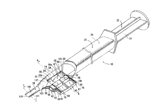
The present invention pertains to an inserter 10 (Figs. 1-7) for implanting a
flexible intraocular lens or other flexible membrane into an eye. An intraocular lens
typically includes an optic and a haptic portion, although the haptic portion isoccasionally omitted. The haptic portion can take many forms, but is usually
composed of plate or loop haptics. For illusl~ration purposes only, this application will
describe the use of inserter 10 with a lens 12 provided with a pair of loop haptics 16a,
CA 02266396 1999-03-22
W O 98/12969 PCTAUS97/17128
.
16b (Figs. 1, 5, 6, 8 and 10). Inserter 10, however, is usable with a wide variety of
lenses or other flexible membranes.
Lens 12 inrllld~s an optic 14 and a pair of loop haptics 16a, 16b (Figs. 1, 5, 6,
8 and 10). The haptics are thin, wire-like, resilient members which extend from
.li~",~ lly opposed sides 18a, 18b of optic 14 in opposite directions. Haptics 16a,
16b are arcuate in shape such that their free ends 20 point generally back toward optic
14.
In the preferred construction, inserter 10 includes a tubular member 22 for
receiving and directing the lens into an eye (Figs. 1-3 and 5-7). The tubular member
22 generally includes a body 24, a co~ ssing station 26, and a cannula 28 (Figs. 1-3
and 5). Body 24, cannula 28, and a support portion 29 of colllple~sing station 26 are
pr~eldbly formed as a unitar,v molded mel~lb~l, although an integral assembly of plural
parts could also be used.
At the proximal end of member 22, body 24 forms a ~wa~dly opening
passage which is adapted to receive a plunger 32 (Fig. 1). The plunger includes a base
34 m~tin~ly received in body 24 and a shaft 36 (Fig. 10) which extends forward to
engage and push lens 12 into an eye. As is known in the industry, the base of plunger
32 is shaped to prevent rotation of the plunger relative to tubular member 22. For
example, the base 34 and the passage may have complementary non-circular shapes or
a key and keyway construction. In addition, while plunger 32 is plerel~bly advanced
manually through body 24, a motor or other driving arrangement could be used to
move the plunger.
Coll-pn ssing station 26 includes an opening 38 in axial ~ nmPnt with the
CA 02266396 1999-03-22
W O 98/12969 PCTrUS97/17128
'7
passage of body 24 for receiving, col~essing and directing lens 12 into cannula 28
(Figs. 1-6D). Co",~lessing station 26 incll1des a support 29 molded with body 24 and
cannula 28, and a cu~pressor 40 which is mounted for movement in the support.
Support 29 inr1n(1e~ a generally U-shaped wing 42 provided with an elQng~te shelf 44
S and a pair of arms 46. The arms and shelf collectively define a lateral ch~nnel or
guideway 48 into which col,-plessor 40 is rnoveably received. A lip 50 formed along
the free end of each arm 46 retains co"l~l~s;~r 40 against shelf 44 and thereby restricts
the co",~l~;ssor to a lateral motion in channel 48. The inner end of each lip 50 defines
a shoulder 55 over which a latch 56 from co",pressor 40 is received to lock the
co",plessor in place for the operation. An additional abutting flange (not shown) or
other known construction may also be included to prevent complessor 40 from being
removed from çh~nn~l 48.
Co"~ ~r 40 inc.hldes a pair of sid~e faces 61 which are adapted to be m~tingly
received within ch~nn~l 48, and an inner sidewall 62 which is adapted to engage and
compress the lens 12. A cover flange 64 projects beyond sidewall 62 to overlie the
opposite side 58 of support 29 and enclose opening 38 when the co-,lpl~ssor is moved
inward (Figs. 2 and 5-6D). T~tr.hPs 56 are positioned along each side of compressor
40 above cover flange 64. Latches 56 have ramps 65 which ease the inward movement
of the coll-plessor, and abutting faces 68 which snap out to engage shoulders 55 and
lock coll.plessor 40 in its closed position with support 29. The con~ essor is
preferably irrevocably locked in place for a single use, but could be constructed to
permit release if desired. Ledges 70 underllie lips 50 to guide the lateral movement of
colllplessor 40 within ch~nnel 48 (Figs. 1 and 4).
CA 02266396 1999-03-22
WO 98tl2969 PCT/US97/17128
Comp,~ssor 40 is!aterally movable belw~n an open position wherein cover
flange 64 is spaced from side 58 of support 29 (Fig. 1), and a closed position wherein
cover flange 64 overlies side 58 and latches 56 engage shoulders 55 (Figs. 2, 5, 6C and
6D). In the open position, a gap 66 is defined between cover flange 64 and side 58 for
S the placing of a lens 12 into opening 38 (Fig. 1). The lens can be placed within tubular
member 22 prior to shipment or by medical pe,~onnel at the time of surgery. In the
closed position, sidewall 62 of compressor 40 is placed into an opposed relation with
a sidewall 60 of side 58, and in axial ~lignmPnt with the inner ends 52 of arms 46
(Figs. 2, ~, 6C and 6D).
10Each sidewall 60, 62 is provided with a retainer which receives and holds the
opposite side edges 18a, 18b of optic 14 to prevent the side edges from being folded
over or turning when co,l,pressor 40 is moved to its closed position (Figs. 4 and 6A-
6D). More specifically, the side edges 18a, 18b of the lens are ori~onted generally
along a central plane. The retainers function to hold and support the side edges of the
15lens in this generally planar rel~ion~hir, during co,.,~ ssion of the lens. Since the side
edges of the lens are not folded over on th~m~çlves, the lens expands laterally within
the eye without a swinging motion. This lateral shifting of the side edges for expansion
of the lens is safer and less likely to contact and damage the interior of the eye than a
swinging motion to unfold the lens. In the pr~fell~d construction, retainers are formed
20 as troughs 68, 70. Nevertheless, the retainers could have other constructions so long
as they m~in~in the sides of the lens in a subst~nti~lly planar orientation and permit
advancement of the lens into the eye.
Troughs 68, 70 are pltrerably flanked by inclined segm~nt~ 72-75 which
CA 02266396 1999-03-22
WO 98/12969 PCT/US97/17128
support and COIl-pleS5 the optic during inward movement of CO--~pl~ ssol 40, and which
help ~ ; in the sides of lens 12 in trougbls 68, 70 (Figs. 6A-6D). Sidewalls 60, 62
are spaced apart by upper and lower parallel surfaces defined by cover flange 64 and
shelf 44 to form an axial passage 76 through which the lens is advanced by plunger 32.
S As compressor 40 is moved inward, the side edges 18a, 18b of optic 14 are
received within troughs 68, 70 (Fig. 6A). Continued inward movement of the
co.llplessol causes the sides 18a, 18b to be snugly pushed into troughs 68, 70 in order
to prevent their release (Fig. 6B). This movement of co~ .ressor 40 also begins to
laterally co---~ the lens. Although the lens will have a tendency to crumple slightly
during co-llpftssion, side edges 18a, 18b of the lens are retained in troughs 68, 70 to
n.~ ;n the edges 18a, 18b in a generally planar relationship. Finally, when latches
56 are locked on shoulders 55, lens 12 is in a compressed configuration between
sidewalls 60, 62 (Fig. 6C). Inner ends 52 of arms 46 (Fig. 3) are also formed with
surfaces which are identical to sidewall 62 to form continuous walls for passage 76
(Pig. 6D).
While fl~nking segments 72-75 can be id~ntic ~l mirror images to one another
(see troughs 68a, 70a and segments 72a-75a of Fig. 8), they are preferably
asymmetrical to better orient the haptics for insertion (Fig. 6D). More specifically,
troughs 68, 70 are each partially defined b~y upper and lower faces 80-83. One face
80, 83 of each trough 68, 70 extends inward a greater ~ t~nce than the opposing face
81, 82. The longer faces 80, 83 merge with arcuate flanking segments 72, 75. The
shorter faces 81~ 82 terminate and interseclt flanking segments 73, 74 at points closer
CA 02266396 1999-03-22
W O 98112969 PCT~US97/17128
to the outer faces 92, 94 of troughs 68, 70. While the intersections of faces 81, 82
with fl~nking segment.s 73, 74 are preferably angular, they may also be rounded. In
this particular construction, side edge 18a with leading haptic 16a is placed adjacent
sidewall 60.
As co~ cssor 40 is moved inward, the side edges 18a, 18b will be received
into troughs 68, 70 (Figs. 6A-6D). As lens 12 com~ ses, the asymmetric faces 80-83
will cause the lens to dip slightly about the shorter faces 81, 82, and create a slight
twist in optic 14 so that leading haptic 16a tends to point in a dow.lw~rd direction.
This downward ori~n~ti~ln of leading haptic 16a will enable the surgeoll to more easily
place the haptic within the c~ps~ r bag of the eye. Similarly, the trailing haptic 16b
is shifted to incline slightly upward to avoid contact by plunger 32; that is, so that the
free end 77 of shaft 36 directly engages optic 14 (Fig. 10).
Alternatively, COlllylcS~ g station 26b inrhldes a support 29b molded with body
24b and cannula 28b, and a pair of opposed compressors 40b, 41b (Pigs. 9 and 10).
The compressors are supported in a pair of opposite slits 96b formed in the sides of
support 29b for lateral movement toward and away from each other. Conl~lessol~
40b, 41b have inner sidewalls 60b, 62b which are pre~ ~bly shaped as described above
for sidewalls 60, 62; nevertheless, the sidewalls could be symmetri~lly formed as with
sidewalls 60a, 62a. An opening 66b is formed in the top of support 29b to permit the
p~ mPnt of a lens 12. To prevent loss or outward bowing of the lens, a cover lOlb
is hinged to support 29b to overlie opening 66b before closure of co,l~r~ssors 40b,
41b. T.~CI1PS (not shown) are provided to lock the COIIIP1eSSO1S in their closedpositions.
CA 02266396 1999-03-22
W O 98/12969 PCTAUS97/17128
11
C~nn~ 28 projects forwardly from the distal end of co",pl~essing station 26 to
direct lens 12 into an eye (Figs. 1-3 anld 5). Cannula 28 preferably includes a
pluAil"al, funnel-shaped portion 103 which lapers to further colll~ess the lens, and an
elongate distal portion 105 which dirccts the co",plessed lens into an eye.
Nevertheless, the cannula could be formed to have a uniform taper across its length or
provided with no taper if, for eY~mrle, the co",presso.(s) has a longer stroke to
complete the desired co"~p,~s~ion of the lens.
An interior lumen 107, which extends through cannula 28, is axially aligned
with passage 76 of compressing station 26 to form a continuous duct through which
lens 12 is moved (Fig. 7). Lumen 107 is preferably de~med by sidewalls 109 provided
with troughs 111 and upper and lower fl~nklng segments 113, 115 to match sidewalls
60, 62 of co"",l~ ;ng station 26. At the rear end 117 of proximal portion 103,
troughs 111 are aligned with troughs 68, 70 (when compressor 40 is in the closedposition) to form a continuous retention of side edges 18a, 18b as the lens is advanced
into the eye. The sidewalls 109 of ~lUAilll~ll portion 103 plcrtl~bly converge fonvardly
at an angle of about 7~ to further compress the lens as it is advanced through cannula
28. As noted above, troughs 111 continue to hold the side edges 18a, 18b of optic 14
as the lens passes through cannula 28 to ~ in the generally planar orientation of the
side edge of the lens and to prevent turning of the lens during its adv~n~em~nt through
lumen 107.
Distal portion 105 of cannula 28 is an elongate, slender tube to permit entry ofthe inserter 10 through a narrow incision (not shown). While the sidewalls 109 in
distal portion 105 are angularly oriented to the sidewalls 109 in proximal portion 103,
CA 02266396 l999-03-22
W O 98/12969 PCT~US97/17128
12
they are id~-ntir~l with respect to the formation of the troughs 111 and fl~nking
seglllr~ 113, 115. Troughs and fl~nking segments therefore continue through distal
portion 105 to properly support and hold lens 12 throughout its passage through
cannula 28. Although the sidewalls 109 in distal portion 105 preferably convergeslightly for molding purposes, they could be formed with parallel walls.
The free end 119 of cannula 28 is preferably provided with a pair of opposed
longitu~in~l slits 121 in troughs 111 (Figs. 1-3, 5 and 7). Slits 121 are wide enough
to permit sides 18a, 18b of optic 14 to extend outward beyond the exterior sides 123
of cannula 28. The slits therefore permit lateral expansion of the lens prior to its
release into the eye. As a result, the natural resilient force which biases the lens to
assume its original unco~ essed shape is ~ ip~t~d in the controlled environment of
the cannula. The lens is thus not released with any velocity as in many prior art
inserters.
Further, since the lens is compressed without folding the side edges over on
themselves, eYp~n~ion of the lens re~uires only an outward, lateral movement of the
lens. The lens eXperipnces no swinging of the optic or haptics within the eye which
risks ~l~m~ging the interior of the eye. Slits 121 also continue to hold optic 14 and
prevent turning of the lens so that implantation of the lens in the proper orientation is
ensured. Accordingly, insertion of the lens with inserter 10 provides a safer
implantation procedure than heretofore realized.
A haptic guide 125 is optionally provided in the front of inserter lOb (or 10) to
ensure the proper positioning of the leading loop haptic 16a (see Figs. 9 and 10).
Haptic guide 125 inrlude~ a g~n~lly flat pull t~b 127 and a slender rod 129 projecting
,
CA 02266396 1999-03-22
W O 98/12969 PCTrUS97/17128
13
from the pull tab. Rod 129is sized to be r eceived rearwardly within lumen 107 from
free end 119. A hook 131 or other shoulder elemPnt is formed on the free end 133 of
rod 129. In use, rod 129is fully insertecl into lumen 107 so that hook 131is visible
through the gap 66b in co,l,pressiilg station 26b. As the lens is loaded into the
S opening, leading haptic 16a is looped over hook 131. Pull tab 127 is manually pulled
forward to remove rod 129 from inserter 10. Removal of haptic guide 125 can be
pelrolllled before or after closure of colllplessol~ 40b, 41b or cover lOlb. As the rod
moves forward, hook 131 engages and pulls haptic 16a forward so that its free end is
positioned into lumen 107. This pulling of the haptic tends to partially straighten
haptic 16a to point geneIally in the direction of the lens' movement. This positioning
of the haptic reduces the risk of the leading haptic 16a being drawn back and becoming
lodged around the optic during insertion. Ribs 135 or other gripping surface arepl~r~l~bly formed on pull tab 127 to enh~nce the manual grasping of the co,l,ponen~.
As is common with lens insertion procedures, a viscoelastic or other lubricant
m~tPri~l is injected into the inserter to ease lthe movement of the lens into the eye. The
lubricant can be injected prior to closure of col-lplessor 40 (or cover lOlb).
Altematively, cover flange 64c (or a wall of the tubular member) can be provided with
an aperture 137 through which the lubricant can be injected after the closing ofcolll~lwsor 40c (Fig. 11). Also, a lubricant pouch 139 filled with a lubricant 141 can
be attached to the exterior of cover flange 64d (Figs. 12 and 13). A barb can beprovided adjacent the aperture to puncture the plastic pouch to permit release of the
lubricant into the passage upon the application of pressure on the pouch. Alternatively,
pouch 139 inchld~Ps a frangible portion (e.g, by scoring) which is aligned with a small
CA 02266396 1999-03-22
W O 98/12969 PCTAUS97/17128
14
apc~llu~ 143 in cover flange 64. Once the co.l.l.lessor is moved to its closed position,
a user may apply pressure to lubricant pouch 139 to break open the pouch and ~ pen~e
the lubricant into opening 38 through aperture 143. Also, cover flange 64c can
cooperate with a fixed cutter (not shown) to open pouch 139 upon the closure of
S co~ 5sor4()c to permit the discharge of the lubricant through a~llure 143 and into
the passage.
The above ~ cuCsion concerns the prer~ d embodiments of the present
invention. Various other embo~liments as well as many changes and alterations may
be made without departing from the spirit and broader aspects of the invention as
defined in the claims. For example, the comLl~ssing station, with or without the
cannula, can be formed as a separable cartridge for colllple~sing the lens. The
cartridge can then be placed within an injector device for insertion of the lens into the
eye after the lens has been compressed. As is common with cartridges, flanges or
other structures could be included to f~ilh~te manipulation of the cartridge and prevent
turning of the cartridge in the injector device. Also, the central portion of the optic 14
could be manipulated into a U-shape, W-shape, or other folded configuration as
opposed to the direct co---p~ssion of the preferred embodiment. So long as the side
edges of the lens are ~ t~ ~A in a generally planar orientation the lens will still
expand with a lateral shifting motion which avoids the broad swinging of the outer
20 edges and haptics within the eye.