Note: Descriptions are shown in the official language in which they were submitted.
CA 02268483 1999-04-12
WO 98/16152 PCT/US97/18833
1
ELECTRODE ARRAY SYSTEM FOR MEASURING
ELECTROPHYSIOLOGICAL SIGNALS
Background of the Invention
This invention relates to physiological electrical signal monitors and more
particularly to a self-prepping multiple electrode array to connect to such
monitors.
Surgical procedures are becoming more non-invasive, and as a result the
use of non-invasive electrophysiological monitoring to evaluate global changes
of
a patient's condition during surgical procedures has increased significantly.
For
example, EEG monitors are now being used for monitoring cerebral function
during intra-operative procedures. Of particular interest are the assessment
of the
effects, of anesthetics, the evaluation of asymmetric activity between the
left and
right hemispheres of the brain in order to detect cerebral ischemia, and the
detection of burst suppression.
One of the greatest impediments to making intra-operative EEG monitoring
more widely practiced in the medical community is the traditional use of
multiple
electrodes in the standard International (10-20) Electrode Placement on the
head,
primarily in the scalp. Applying them takes considerable time and expertise,
requires multiple, separate and time consuming skin preparation steps, and
leaves
the patient's scalp and hair in disarray.
Various headsets and caps are studded with different style electrodes to
speed this process, but such headsets and caps are generally not disposable
(and
therefore must be cleaned), need to be adjusted to accommodate the widely
varying dimensions of the patients' heads, and require a considerable up-front
cost. Other problems are encountered in the present medical environment when
such headsets and caps are designed to be single-use disposable devices
because
such devices are on occasion re-used despite warnings, which results in the
spread
of infection. Such headsets and caps have also been used with equipment for
which it was not designed, which may be a well intentioned cost saving
practice,
but which could result in degraded performance of the device.
The most widely used electrodes are the reusable "gold cup" style
electrodes that are small, bare tin, silver, or gold plated metal cups on the
end of
CA 02268483 1999-04-12
WO 98/16152 PCT/US97/18833
2
unshielded wires that may be several feet long. Such electrodes may require
that
the multiple scalp and forehead electrode sites first be located by measuring
and
marking the head. Such sites must then be prepared before applying the
electrode
in order to get good electrical contact. This preparation is usually
accomplished
by abrading the electrode sites with a grit-impregnated solution or with some
other abrasive means to remove the outer layers of skin which cause the poor
electrical contact. The electrodes, up to 19 on the scalp for the full
International
(10-20) electrode placement, are then individually applied with adhesive to
the
prepared sites in contact with a blood-enriched skin layer, and are then
injected
with conductive electrolyte cream through the hole in the top of the
electrode,
thereby providing a relatively low electrical contact impedance. This process
leaves the patient with abraded spots, adhesive, and electrolyte cream
throughout
the scalp. Frequently, contact between the metal electrode and the skin
occurs,
causing a time-varying offset voltage that results in "baseline wander." The
electrodes also need to be placed with reasonable accuracy to achieve the
standard
placements or montages and to be able to repeat the same measurement at a
later
time.
The need to use multiple, separate preparation steps makes the set-up a
very time consuming process, taking perhaps up to half an hour of a medical
technician's time for even a small subset of the full International (10-20)
Electrode
Placement. The amount of expertise and time required to prepare a patient is
presently an impediment to intraoperative EEG monitoring being more widely
practiced. Also, care is needed to bundle the unshielded leads to reduce
electrical
noise interference. Additionally, after the procedure is over, the gold cup
electrodes and any placement harness need to be cleaned and sterilized since
they
are not intended to be disposable.
A number of prior art multiple electrode assemblies have been developed
for EEG monitoring. U.S. Patent Nos. 4,595,013 issued to Jones; 4,928,696
issued to
Henderson; 4,638,807 issued to Ryder; 4,072,145 issued to Silva; and 3,490,439
issued to Rolston are several examples. These multiple electrode assemblies,
however, all require some or all of the multiple, separate and time consuming
steps of skin preparation described above to reduce the contact impedance with
the skin before they are applied to the body. These separate skin preparation
CA 02268483 1999-04-12
WO 98116152 PCT/US97/18833
3
steps also make it difficult to improve contact impedance once the electrode
has
been applied to the patient or after the medical procedure is underway. If the
preparation was inadequate at the time the multiple electrode assembly is
applied,
it must be removed, the skin reabraded, and most likely a new electrode
assembly
would have to be reapplied, adding additional expense to the additional
preparation time. Too much abrasion can cause a skin injury, or bleeding,
leaving
the patient with a lasting wound. Separate devices required to abrade the skin
cause the risk to the applicator by potential contact with blood and by
possible
disease transmittal during preparation.
There are also a number of prior art multiple electrode assemblies that are
self prepping. U.S. Patent No. 4,709,702 and associated electrode U.S. Patent
No.
4,640,290, both issued to Sherwin, utilize an array of spring loaded metal
"tulip"
electrodes in a reusable headset that penetrates the outer dead layers of skin
to
achieve a low contact impedance. Also, U.S. Patent No. 4,770,180 and
associated
electrode U.S. patent No. 4,706,679 both issued to Schmidt utilize an array of
stiff,
bundled metal wires that contact and penetrate the patient's skin. The
drawback
with both of these assemblies is that the metal contact with the skin causes
highly
undesirable time-varying offset voltages that interfere with the sensitive
measurement of the small signal voltages of the body. Also, both of these
assemblies, and other assemblies that utilize a headset or cap such as the
assembly
described in U.S. Patent No. 4,967,038 issued to Gevins, need some adjustment
to
properly position the electrodes on the widely varying dimensions of the
patients'
heads, and require a high up-front cost and cleaning after use.
U.S. Patent No. 4,936,306 issued to Doty utilizes a spiral coil electrode that
may be metallic, and that uses cork-screws into patient's skin to achieve low
contact impedance. While this may achieve low contact impedance, it has the
significant drawbacks of discomfort to the patient and creating sites of
possible
infection because of the deep skin punctures made by the spiral coils. If made
of
metal, the spiral coils will also cause time-varying voltages. Lastly, these
electrodes are actually applied individually since they must be screwed into
the
patient's scalp, which adds time to the procedure.
U.S. Patent No. 4,683,892 issued to Johansson utilizes a headset with
multiple electrodes that are activated by compressed air, which impinge
against
CA 02268483 2005-06-08
69675-342
4
the patient's scalp, and that also dispense electrolyte
paste to improve contact. This is a complex and expensive
device, not intended for general, routine use in an
intraoperative environment.
It is therefore a principal object of the present
invention to provide a disposable, pre-gelled, self-prepping
multiple electrode array which easily and reliably prepares
the .skin to assume a relatively low contact impedance.
Another object of the present invention is to
provide a self-prepping multiple electrode array that does
not :require the use of more than one component to be handled
by the person applying the device, and fits most head sizes
in the general patient population.
Still another object of the present invention is
to provide a multiple electrode array that can monitor
cerebral function without the use of electrodes placed in
the scalp, and that is easily aligned on the head.
A further object of the present invention is to
prov_Lde a multiple electrode array that prevents its use
with monitoring equipment with which it was not intended to
be u;~ed.
Summarv of the Invention
Accordingly, in one aspect of the present
invention, there is provided an array of electrodes,
including only three electrodes, for monitoring
physiological electrical signals, said array comprising: a
flexible unitary body having a main portion, one satellite
portion, and a flexible portion located between said main
portion and said one satellite portion; two electrodes of
the three electrodes being permanently printed on said main
CA 02268483 2005-06-08
6967.5-342
4a
portion and a third electrode of the three electrodes being
printed on said satellite portion; and conductors printed on
said flexible unitary body to carry signals from said
electrodes.
In a second aspect of the present invention, there
is provided an array of electrodes, including only four
electrodes, for monitoring physiological electrical signals,
said array comprising: a flexible body having a main
portion, at least one satellite portion, and a flexible
portion located between said main portion and each of said
at least one satellite portion; at least two electrodes of
the :Four electrodes being positioned on said main portion
and one electrode of the four electrodes being positioned on
each of said satellite portions; and conductors printed on
said flexible body to carry signals from said electrodes.
In a third aspect of the present invention, there
is provided an array of electrodes for monitoring
phys_Lological electrical signals, said array comprising: a
flex_Lble body; at least two electrodes affixed to said
flex_Lble body; means for storing on said flexible body a
code unique to the array.
In a fourth aspect of the present invention, there
is pi=ovided a method of positioning electrodes for
monit:oring physiological electrical signals, said method
comprising the steps of: positioning a first electrode on a
forehead of a subject from whom the electrical signals are
to be monitored; positioning a second electrode on a first
temp7_e of the subject from whom the electrical signals are
to be monitored; and positioning a third electrode adjacent
said first electrode on the forehead of the subject, the
first: electrode and the second electrode being printed on a
main portion of a flexible unitary body and the third
CA 02268483 2005-06-08
6967.5-342
4b
electrode being printed on a satellite portion of the
flexible unitary body, the flexible unitary body having
conductors printed thereon to carry signals from the
elecvrodes .
An array of electrodes is constructed to allow the
user to easily adjust to the correct size of the patient's
head. The array is self-adhesive, pre-gelled and
disposable. The array fits easily over the temple and
forehead areas where EEG signals can be acquired by
specially designed monitors for purposes of monitoring a
numbe r of bodily phenomena, including but not limited to,
depth of anesthesia, and/or ischemia, and burst suppression.
The array is connected to the monitor via a tab connector
that is integral to the disposable device. The tab
connector is insertible into a reusable connector that is
part of a monitoring system.
The reusable connector is made of rigid contacts
posii~ioned side by side within a keyed cavity. The contacts
press against conductors of the disposable array when the
conductors are inserted into the cavity of the reusable
connector. The conductors of the disposable array are laid
on a flexible circuit constructed of a polyester substrate
that has a plastic clip as its backing and support. The
flex_Lble circuit when routed through this clip forms the tab
connector. This sensor tab connector, when inserted into
the reusable connector cavity, electrically
CA 02268483 1999-04-12
WO 98/16152 PCT/US97/18833
connects the electrodes to the monitor, allowing the acquisition of the
electrophysiological signals. The clip of the tab connector is self securing,
and
thus does not need any additional securing mechanism to keep the flexible
circuit
in place. The reusable connector and the disposable connector have
complementary locking mechanisms that provide for a secure connection.
Depending on the application and uniqueness of the array, a tab connector
may be used which includes a key that only fits to specific monitors. The
array
also can communicate with the monitor to indicate the type of application
utilizing
the electrodes and how many channels need to be configured.
The array contains two or more elements that when pressed against the
skin lower their contact impedance to the skin and thus provide better quality
signals. The elements contain built in blowout pockets that allow for the gel
to
adjust itself when pressure is applied to it. Such pockets also prevent the
gel from
getting blown into the adhesive areas or running into other element areas,
which
could cause channels to short circuit.
These and other objects and features of the present invention will be more
fully understood from the following detailed description which should be read
in
conjunction with the accompanying drawings in which correspondence reference
numerals refer to corresponding parts throughout the several views.
Brief Description of the Drawing-s
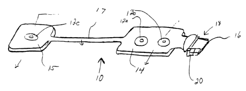
Figure 1 is a perspective view of the preferred embodiment of the electrode
array of the present invention;
Figure 2 is a side sectional view of the electrode array shown in Figure 3
taken along lines 2-2 of Figure 3;
Figure 3 is a top plan view of the electrode array shown in Figure 1;
Figure 4 is a bottom sectional view of the electrode array shown in
Figure 2;
Figure 5(a) through 5(c) are perspective views of a tab clip assembly
utilized by the electrode array shown in Figure 1 with a substrate is routed
through it;
CA 02268483 1999-04-12
WO 98/I6152 PCT/US97/18833
6
Figures 6(a) and 6(b) are top plan views of the EEG connector system used
with the electrode array shown in Figure 1 with Figure 6(a) showing the
connectors engaged and Figure 6(b) showing the connectors disengaged;
Figures 7(a) through 7(e) are elevational views of keys used in the EEG
connector system shown in Figures 6(a) and 6(b);
Figure 8 is a schematic diagram of the configuration coding utilized by the
EEG connector system shown in Figures 6(a) and 6(b) in its present
configuration;
Figure 9 is a flowchart of the steps taken to identify an electrode array
type;
Figure 10 is a bottom plan view of the electrode array shown in Figure 1;
Figure 11 is a diagram showing locations on the head where electrodes are
positioned for 2 channel monitoring;
Figure 12 is a perspective view of the gel blowout pockets and salt bridge
barriers utilized by the electrode array shown in Figure 1;
Figures 13(a) and 13(b) are representations of a human head showing the
locations of the placement of electrodes for one channel monitoring;
Figure 14 is an elevational view showing the sponge over tines construction
of the electrodes of the present invention;
Figure 15(a) is a top plan view of an alternate embodiment the electrode
array of the present invention which includes two elements for temple
connection;
Figure 15(b) is a bottom plan view of the electrode array shown in Figure
15(a);
Figure 16 is a representation of a human head with an alternate
embodiment of the electrode array locating the connector in an alternate
location,
being placed thereon;
Figure 17 is another representation of a human head on which another
alternate embodiment of the electrode array of the present invention is
positioned;
using the mastoid locations to place the two satellite electrodes.
Figure 18 is a side plan view of a female portion of an alternate
embodiment of the connector used in the present invention and a top plan view
of
the connector;
Figure 19 is a plan view of the components of a system utilizing the
electrode array shown in Figure 1.
CA 02268483 1999-04-12
WO 98/16152 PCT/ITS97/18833
7
Detailed Description of the Preferred Embodiments
Referring to Figures 1-4, an electrode array 10 is shown. In a preferred
embodiment the array 10 includes three electrodes 12 that are self adherent
and
self prepping to the forehead and temple areas and that are used to acquire
electrophysiological(EEG) signals. This array 10 comprises a flexible circuit
14
containing silver/silver-chloride (Ag/AgCI) conductors 16 on a polyester
substrate. These conductors are routed from specific montage locations to a
single
connecting tab 18. There can be up to eight (8) conductors 16 for providing up
to
eight signal lines of EEG data which can be captured simultaneously. This tab
18
contains a clip 20 which adds rigidity, a locking mechanism, self alignment,
polarity and a keying mechanism to the array. The clip 20 also adds a solid
contact area to the flexible circuit 14.
The array 10 comprises a main body 14 which in the embodiment shown
includes two electrodes 12a, 12b and a satellite body 15 which includes one
electrode 12c. The satellite body 15 allows the molutoying personnel to adjust
the
placement of the electrode 12c mounted on the satellite body 15 due to the
patient's head size. Extension 17, through which conductors 16 run, connects
the
main body 14 to the satellite body 15.
Referring to Figures 3 and 14, each of the three electrodes 12 mounted in
the array 10 contain a self prepping disk 30 which includes a set of flexible
tines
44 mounted with adhesive 45. The flexible tines 44 extended beyond the surface
of the gel 40 to contact the skin 32 as part of the normal application of the
electrode 12 to the skin 32. When pressure is applied to the electrodes 12,
the
flexible tines 44 are pushed through foam layer 42 against the skin 32, which
causes the tines 44 to part the high impedance outer layers of skin 32 to
expose
the low impedance, blood-enriched layers without scratching or abrading. This
prepping disk is made out of a plastic such as nylon constructed as hooks from
hook and loop fasteners of the type often said under the Velcro trademark.
These
hooks are then sheared to the correct height and stiffness. The electrodes 12
are
surrounded by an adhesive backed foam layer 43. The array contains markers 13
that indicate the correct locations that need to be pressed to achieve the
desired
skin impedance.
CA 02268483 2005-06-08
6967.5-342
8
Referring to Figures 4 and 12, the array contains
two blowout pockets 38, built into the basepad 39, that
allow the gel 40 to adjust its volume over a large area and
prevE=nt it from migrating to areas where it could cause
malf~~nction, such as short circuiting the two elements
adj absent to one another .
The blowout pockets 38 are formed by cutting
cylindrical shapes into the basepad 39 foam material. In
addii~ion to the blowout pockets 38, the array 10 also
contains two salt bridge barriers 46 which prevent
elect rolyte gel 40 from one electrode from contacting the
gel ~~0 of the other electrode which could cause the signals
to short circuit. The barriers 46 are also cut into the
adhesive basepad 39.
In the preferred embodiment a liquid hydrogel is
used that rests on the gel pockets 38 cut within the basepad
material 39. The gel 40 is retained within the pocket by a
polyurethane foam sponge 42. The sponge contains large
enough pores that allow the tines 44 to go through the pores
and contact the skin 32 during use. The tines 44 then work
in tile same manner as described in U.S. Patent No. 5,305,746
to separate the layers of skin to avoid the need to abrade
the skin to reduce impedance.
In a number of embodiments, the array 10 is
mount=ed over the forehead with its reference electrode 12b
over the center of the forehead. As shown in Figures 13(a)
and _L3(b), the ground electrode 12a is placed over the
forehead as well. The third electrode 12c in the satellite
body 15 is positioned over the temple area. In most cases,
eithe r the right or left temple is acceptable. Such an
array may also be used for EMG detection in the facial area.
CA 02268483 2005-06-08
69675-342
9
The tab connector of the present invention is
show:z in Figures 5 (a) -5 (c) . In Figure 5 (a) the conductors
16 which are mounted on a flexible material are inserted
into the clip 20 past the edge 46 of the clip 20. The clip
20 includes a hinge 47 which is folded back as shown in
Figure 5(b) until it is rotated a full one hundred eighty
degr~=es as shown in Figure 5(c). A slot 48 is provided on
each side of clip 20 for locking with extension 49 so that
the ~~lip 20 stays in a locked and closed position as shown
in Figure 5(c), so that it is ready to be used.
Referring to Figure 10, the tab connector 18 of
the <array 10 of the preferred embodiment has eight (8)
conductors. Out of the eight conductors, three are EEG
signal lines 16a, 16b, 16c, and four are logical signal
liner 16e, 16f, 16g, 16h used to identify the appropriate
array type being connected. In the embodiment shown, the
eight=h conductor 16d is not used. The unused conductor 16d
could be used in other embodiments as an additional EEG
sign<~1 line or as an additional means to identify an array
type. It is important that the sensor sends the
ideni~ification information to the monitor, so that the
monii~or can determine the number of active elements used as
well as their locations on the head. This way a monitor
will auto configure for a particular EEG monitoring session.
The preferred embodiment uses a three bit binary
code identification scheme such as the identification scheme
described in United States Patent Serial No. 5,813,404. In
such an identification scheme, the code is hard-wired in the
flex~_ble circuit of the particular array 10. A digital
signal converter in the monitor detects the array ID signals.
As shown in Figure 8, the code is set by selectively shorting
a common drive signal line [SEN DRV] 60 to the three code
CA 02268483 2005-06-08
69675-342
9a
signal lines [SEN 0:2] 62, 64, 66. These are the three array
identification signal lines. The [SEN DRV] line is pulsed
(driven) to a logic high at 8,192 Hz by the pulse generator
located on a monitor's digital signal converter. Pulsing the
line prevents a fault condition, such as a broken connection,
from injecting more than 50 micro amps of current into a
patient, as required by medical equipment standards, such as
IEC-o01-1.
The frequency of the pulse is chosen to be at the
Nyqu:ist frequency of the digitizers. These pulses will not
inte=rfere with the EEG signal because at this frequency it
will alias onto itself only in the first stage of
decimation, and will subsequently be filtered out completely
by the digital signal processor.
The patient interface connector code signal lines
are hulled down to a logic "0" by resistors 70, 72, 74
locai~ed in the digital signal converter 146 at the input to
the ~_eceiver circuit 76, which is a D-Flip-flop in a
preferred embodiment. As the common [SEN-DRV] line 60 is
driven high by the pulse generator, the patient interface
connector code lines [SEN 0:2] 62, 64, 66 are then read
(i.~, clocked in) by receiver circuit 76, which transmits
the binary code to the monitor 150. The patient interface
conne=ctor code signal lines that are shorted to the drive
signal will be read as a logic "1". The patient interface
connector code signal lines that are left open will be read
as a logic "0". Such a coding scheme allows for eight
different PIC cable types as follows:
CA 02268483 1999-04-12
WO 98/16152 PCT/US97/18833
# Code Cable Type
1 000 PIC not connected
2 001 2 channel Bipolar (5 signal wires
in use)
3 010 2 channel Referential (4 signal
wires in use)
4 011 1 channel electrode connection
5 100 1 channel sensor connection
6,7,8 Unassigned Spares
Referring now to Figures 9 and 19 the process for determining the
appropriate PIC will now be described. In step 82, a CPU in the monitor 150
periodically reads the PIC code, which in a preferred embodiment is read every
1.75 seconds. In step 84 the CPU in monitor 150 reads a PIC ID in the manner
described above with reference to Fig. 8. If the PIC ID is determined in step
86 to
be "000," (which indicates that a PIC is not connected) the system reiterates
the
process after each 1.75 second delay and continues to attempt to read a new
PIC
ID.
If the PIC ID is determined in step 88 to be "010," a two channel referential
EEG electrode set is detected and the monitor 150 is configured for 2-channel
referential EEG processing in step 90. The digital signal convertor is set to
referential mode in step 92. If, in step 94, the PIC ID is equal to "010," the
system
recognizes a two channel bipolar EEG electrode set and the monitor 150 is
configured for the appropriate EEG processing in step 96. The digital signal
convertor 146 is then set in step 98 to bipolar mode.
If the PIC ID is determined in step 100 to be equal to "011," the system has
detected a one channel EEG processing cable and the monitor 150 is configured
for 1-channel EEG processing in step 102. In step 106, digital signal
converter is
set to bipolar mode. If any other PIC ID is detected, error messages are
generated
and displayed in step 107 indicating that an illegal PIC ID was detected, and
that
no EEG processing should occur. After the CPU in monitor 150 determines that
the PIC ID is valid, the monitor checks if the PIC ID is a new PIC ID. If a
new
PIC ID is recognized the monitor initiates a self test in step 108 followed by
an
CA 02268483 1999-04-12
WO 98/16152 PCT/US97/18833
11
electrode impedance test in step 109. After this series of steps the system
again
returns after a 1.75 second delay to read additional PIC IDs in step 82.
In alternate embodiments where four pins are allocated for PIC IDs, the
digital signal convertor 146 can recognize up to 15 different combinations of
pigtail, PIC or connector type.
The current connector system allows either a single channel electrode array
or a dual channel electrode array. As shown in Figures 7(a)-7(e), it also
provides a
keying safeguard that allows for the connector to be selective as to what can
physically be plugged into it. By modifying the height of the connector rails
50
one can allow for a specific array to be a master key (Figure 7(a)) and other
arrays
to be specific to a mating connector. This keying mechanism can be used for
example to physically differentiate between array types. For instance, an
array
that allows single and dual channel monitoring, and one that allows only dual
channel monitoring. The master key is then available to connect to all
monitors
indiscriminately. For instance, it can be used to insert a test circuit to
service the
monitor, or used to insert a multipurpose array.
Referring to Figures 6(a) and 6(b), the tab connection on the array has a
locking mechanism, including extension 120 and receptor region 122 that
secures it
to the reusable connector 124. The locking action provides the user with
tactile
and audible feedback.
The reusable connector 124 includes a printed circuit board with contacts
and wires from a cable attached to it. The printed circuit board is then
inserted
into an assembly of two pre-molded housings secured together by ultrasonic
welding.
The electrode array 10 described above is used in connection with a new
non-standard electrode positioning (montage) for measuring the effects of
anesthetics on the brain as well as other cerebral phenomena.
Referring to Figures 13(a) and 13(b), one embodiment of this montage is
shown in which the reference electrode 12 is placed in the center of the
forehead
with the satellite electrode 12 being placed on the temple at eye level above
the
ear. This montage has several advantages over previously described montages,
as
it makes it easy to locate the electrodes on the patient, the electrodes are
easy to
CA 02268483 1999-04-12
WO 98/16152 PCT/US97/18833
12
apply to the patient and the EEG signal and the amplitude of such signal are
sufficient for the purposes for which they are used.
The location of the electrodes is important for monitoring the effects of
anesthetics. Prior art for monitoring the effects of anesthetics have
described EEG
systems using from 2 to 19 EEG channels, where the electrode locations have
been
identified by the international 10-20 systems. The electrode arrays described
above use 1 or 2 EEG channels. The specific electrode locations described in
this
patent are positioned in a unique anterior area of the subject's head from
which
EEG signals have not traditionally been taken. These anterior placed arrays
take
advantage of the global nature of the effects of anesthetics on the brain.
That is to
say that the global effects of anesthetics are reflected in the EEG detected
near the
anterior cerebral cortex. The electrode array described above provides a
rather
large EEG signal because of the inter-electrode spacing that has been
selected. The
electrodes, however, are not so widely spaced as to increase a noise signal
generated by the subject (e.g. EKG). In any signal processing system,
increases in
signal amplitude without an increase in the noise amplitude is desirable. This
is
particularly true with EEG monitoring because EEG is on the order of one
hundred times smaller than the electrocardiogram {EKG). The electrode array 10
facilitates the locating of the electrodes 12 at positions referenced to
easily
identified anatomical landmarks (i.e. center of the forehead, eye socket). In
addition, the electrode locations are entirely out of the subject's hair. This
allows
for easy application of the electrodes without the need to shave or otherwise
part
the subject's hair.
A system utilizing the electrode array of the present invention may be
configured in one or two channel monitoring modes. For the two channel mode
shown in Figures 15(a) and 15(b), one EEG channel measures from an electrode
location on the subject's forehead to the left of the lower temple area,
proximal to
the left eye socket (malar bone). The second EEG channel measures from the
same forehead electrode to the right lower temple area, proximal to the eye
socket.
A non-measurement ground electrode is also placed on the patient's forehead.
The two channel system has the advantages of signal redundancy (two channels
of
signal instead of one channel) and improved signal to noise ratio. The one
charulel configuration, an example of which is shown in Figure 1, uses the
center
CA 02268483 1999-04-12
WO 98/16152 PCT/US97/18833
13
forehead electrode plus either the left or right electrode described above
plus the
ground electrode. The one channel configuration has the advantage of using
less
space on the subject's head thereby making an operation on the head easier
since
there is a greater area over which to maneuver. The one channel configuration
being easier to apply because of the use of one less electrode.
Referring to Figures 15(a) and 15(b), an alternate embodiment of the present
invention is shown in which the array 10 of electrodes 12 includes two temple
electrodes 12c that allow for depth of anesthesia, burst suppression, ischemia
monitor, and EEG recordings as well as EMG detection. When a two channel
system is used, the signals could be averaged together or the second channel
could be used as a backup signal if the first channel signals are lost. The
placement of the electrodes on a human head in such a two channel system is
shown in Figure 11. Referring to Figure 10, in this configuration, conductor
16d is
used to provide the signal from the second temple.
Referring to Figure 16, the same array 10 described above in connection
with Figure 1 is used in a different manner with the center of the main body
14 of
the array 10 being placed over the temples and the electrode 12c on the
satellite
body 15 becomes the reference electrode. This configuration offers the
advantage
of keeping the cable away from the face of the patient.
As shown in Figure 17, another array 10 of electrodes 12 is shown with a
ground connection 12a two frontal connections and two mastoid connections that
can be used for depth of anesthesia, burst suppression, ischemia monitoring,
and
EEG recordings as well as EMG detection. As with the embodiments shown in
Figures 15(a) and 15(b), the configuration shown in Figure 17 can be used to
capture a hemisphere signal on each side of the head in order to produce
bipolar
readings.
In alternate embodiments, an array of electrodes will contain other passive
devices such as but not limited to resistors, capacitors, or jumpers, for
purposes of
generating a code for self configuration.
In another embodiment shown in Figure 18, the array 10 of multiple
electrodes 12 comprises of a flexible circuit with conductors that terminate
on a
tab connection that is double sided. The mating connector 124 has contacts 125
on
top and bottom. This allows an increase in the density of the circuit while
CA 02268483 1999-04-12
WO 98/16152 PCTIUS97/18833
14
keeping the size of the connector to a small profile. It also allows for the
separation of signals that are of digital nature from those of physioelectric
nature.
This reduces the amount of noise on the EEG signals.
Referring now to Figure 19, the electrode array 10 is shown in use with an
EEG monitor. The electrode array 10 is connected through corulector 20 to a
patient interface cable 142 which in turn is connected to a pigtail cable 144.
The
pigtail cable 144 is connected to a digital signal converter 146 which in turn
is
connected to monitor 150 through monitor interface cable 148. In another
embodiment, the digital signal converter may be embedded in the monitor
thereby
eliminating the need for cables 144, 148 or the electrode array 10 could also
be
connected to cable 144 thereby eliminating the need for cable 142.
While the foregoing invention has been described with reference to its
preferred embodiments, various alterations and modifications will occur to
those
skilled in the art. All such alterations and modifications are intended to
fall
within the scope of the appended claims.