Note: Descriptions are shown in the official language in which they were submitted.
CA 02269162 1999-04-16
METHOD FOR THE DETERMINATION OF ANALYTE
CONCENTRATION IN A LATERAL FLOW SANDWICH
IMMUNOASSAY EXHIBITING HIGH-DOSE HOOK EFFECT
Background of the Invention
Immunochromatographic strip formats have become increas-
ingly popular for quantitative and semi-quantitative assays
which use visual detection schemes. This type of immunoassay
involves the application of a liquid test sample suspected of
containing an analyte to be detected to an application zone of
an immunochromatographic test strip. The strip is comprised
of a matrix material through which the fluid test medium and
analyte suspended or dissolved therein can flow by capillarity
from the application zone to a capture zone where a detectable
signal, or the absence of such, reveals the presence of the
analyte. Typically, the strip will include means for immuno-
specifically binding the analyte to be detected with its spe-
cific binding partner which bears the detectable label. In
one such scheme, as disclosed in U.S. Patent 4,446,232; the
strip contains an enzyme labeled, mobile binding partner for
the analyte which is in a zone of the strip downstream from
the sample application zone. If analyte is present in the
test sample, it will combine with its labeled binding partner
to form a complex which will flow along the strip to a detec-
tion zone which contains a substrate for the enzyme label
which is capable of providing a colored response in the pres-
ence of the enzyme label. The strip contains another zone in
which analyte is immobilized, so that the labeled binding
partner which does not combine with analyte, due to the ab-
sence of sufficient analyte in the sample, will be captured
and thereby inhibited from reaching the detection zone. There
CA 02269162 1999-04-16
2
have been published various modifications of this technique,
all of which involve competitive specific binding systems in
which the presence or absence of analyte in the test sample is
determined by the detection or lack thexeof of labeled binding
partner in the capture zone.
An alternative to the above described immunometric assay
which detects the free labeled antibody is the so called sand-
wich format in which the capture zone contains immobilized an-
tibodies against an epitope of the analyte which is different
than the epitope to which the labeled antibody is specific.
In this format, there is formed a sandwich of the analyte be-
tween the immobilized and labeled antibodies and it is there-
fore an immunometric assay which detects the bound labeled an-
tibody species. This type of immunostrip format works well in
connection with the analysis of relatively low concentrations
of analyte, but can be of limited utility in the analysis of
fluids containing high analyte concentration. This adverse
effect is caused by the presence of excessive free analyte in
the sample that competes for binding with the immobilized an-
tibody in the strip's capture band with the analyte which has
become bound to the labeled antibody by interaction therewith
in a portion of the strip upstream from the capture zone.
This competition can result in less of the analyte/labeled an-
tibody conjugate being captured by the capture antibody and
consequently less signal being detected in the capture zone
than would be in the case if there were less analyte in the
test sample. A dose-response curve prepared using this type
of test strip will show increasing signal with increasing ana-
lyte up to the point where the analyte concentration begins to
block the interaction between the immobilized capture antibody
CA 02269162 1999-04-16
3
and the analyte/labeled antibody complex. Beyond this point,
increasing analyte in the test fluid results in a decrease in
the signal, so that the dose-response curve indicates decreas-
ing signal with increasing analyte. The slope of this sort of
dose-response curve somewhat resembles a hook which accounts
for this phenomena being known as the hook effect. Tradition-
ally, when the hook effect is observed or suspected, the fluid
sample is diluted to several dilutions to ensure the validity
of the results. The high dose hook effect may not occur if
sufficient labeled or capture antibody is present in the assay
medium. A complete dose-response curve (low to high analyte
concentration) is usually needed to verify the existence of
this effect. Accordingly, sample dilution is generally car-
ried out whenever there is reason to expect that the assay
might exhibit the hook effect. It is an object of the present
invention to provide a sandwich type assay method using an im-
munochromatographic strip whose efficacy is not affected by
high analyte concentrations in the test sample and, accord-
ingly, does not require sample dilution or reassaying of sam-
ples containing high analyte concentrations. This method in-
volves providing a strip with at least two capture bands and
optionally a collection band in which there is immobilized a
binding partner for labeled antibody which will bind labeled
antibody which has not formed a complex with analyte to
thereby facilitate its capture in one of the capture bands.
The collection band is optional since it is not needed for the
assay method to work in the sandwich format. However, by us-
ing a strip which contains a collection band, each sample
measurement will provide more information thereby improving
the assay's sensitivity and/or precision.
CA 02269162 1999-04-16
4
In EP 0 462 376 AZ there is disclosed a procedure in which
signal at the capture site and conjugate collection site of an
immunochromatographic strip are detected and the analyte con-
centration is determined by the intensity of the signal at the
capture site relative to the signal at the recovery site.
Also of interest in this regard is U.S. Patent 5,569,608.
In co-pending application [Serial No. 08/900,586] there
is disclosed an assay using an immunochromatographic strip
having multiple capture and/or collection sites in which the
signal from the detectable label in the capture zones) and
collection zones) is determined whereupon a final response
signal is determined using an algorithm and a number of sig-
nals which are chosen in a manner suited for a particular as-
say to provide a value for analyte concentration.
Summary of the Invention
The present invention is a method for determining the
concentration of an analyte in a fluid test medium. The
method comprises the steps of:
a) Providing a strip of a porous material through which
a test fluid suspected of containing the analyte can
flow by capillarity; the strip has at least two dis-
tinct capture regions in which there are immobilized
antibodies specific to a first epitope of the ana-
lyte. There are also provided antibodies specific
to a second epitope of the analyte which bear a de-
CA 02269162 1999-04-16
5
tectable label and are capable of flowing through
the strip along with the fluid test medium upon ap-
plying it to the strip up stream from the first of
the at least two distinct capture zones;
b) Applying the fluid test medium to the strip and al-
lowing it to flow along the strip carrying labeled
antibodies along with it to thereby contact the im-
mobilized antibodies in the distinct capture re-
gions. When sufficient analyte is present in the
fluid test medium to partial ly block binding of the
immobilized antibody with the first epitope of the
analyte in at least the first distinct capture re-
gion with which the fluid test medium comes into
contact as it flows along the strip, there is formed
a sandwich of the immobilized antibody, the analyte
and labeled antibody in the distinct capture regions
through which the fluid test medium carries analyte
the quantity of which sandwich is limited by the
partial blocking of the immobilized antibody;
c) Detecting, in a quantitative manner, the signal
emitted from the label on the labeled antibody in
each of the distinct capture regions in which the
sandwich has formed. This provides a pattern of
signals which is unique to the concentration of ana-
lyte in the fluid test medium; and
d) Mathematically combining the unique set of signals
to create a monotonous dose-response curve to factor
CA 02269162 1999-04-16
6
out the blocking of the binding between the immobi-
lized antibody and the first epitope of the analyte.
Brief Description of the Drawings
Fig. 1 is a representation of an assay device useful in
the present invention.
Fig. 2 is a dose response curve of the reflectance change
of an immunostrip having only one capture band.
Fig. 3 represents a complete dose response curve of the
reflectance change of an immunostrip having 3 capture bands
and one collection band which have been combined according to
the present invention.
Fig . 4 and 5 are partial dose-response curves of the re-
flectance change of an immunostrip having 3 capture bands and
one collection band which have been combined by multiple
mathematical methods.
Description of the Invention
The invention is practiced by first providing the test
matrix through which the fluid test sample can flow by capil-
larity. Typically, the matrix will be in the form of a strip
through which the test fluid flows horizontally. While the
matrix could be assembled in a layered format through which
the test fluid could flow vertically from top to bottom or
vice-versa, the following discussion is focused on the pre-
ferred strip format.
CA 02269162 1999-04-16
7
The strip can be prepared from any matrix material
through which the test fluid and an analyte contained therein
can flow by capillarity and can be of a material which is ca-
pable of supporting non-bibulous lateral flow. This type of
flow is described in U.S. Patent 4,943,522 as liquid flow in
which all of the dissolved or dispersed components of the liq-
uid are carried through the matrix at substantially equal
rates and with relatively unimpaired flow, as contrasted to
preferential retention of one or more components as would be
the case if the matrix material were capable of absorbing or
imbibing one or more of the components. An example of such a
material is the high density or ultra high molecular weight
polyethylene sheet material obtainable from Porex Technolo-
gies. Equally suitable for use as the matrix from which the
chromatographic strips can be fabricated are bibulous materi-
als such as paper, nitrocellulose and nylon.
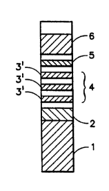
In a preferred embodiment of the present invention there
is provided a test device comprising a strip of nitrocellulose
having a region 2 (Fig. 1) which contains mobile specific
binding partner for the analyte which bears a detectable label
and can react with analyte present in the fluid test sample
applied to wicking pad 1 to form an analyte/labeled binding
partner complex upon applying the test sample to the wicking
pad and allowing it to flow up the strip to region 2 where
analyte in the test sample combines with labeled antibody spe-
cific to the analyte and flows to area 3 which contains two or
more capture bands 3' which contain immobilized antibodies
specific for an epitope of the analyte separate from that to
which the labeled antibodies are specific to form an immobi-
CA 02269162 1999-04-16
8
lized binding partner/analyte/labeled binding partner sandwich
in one or more of capture bands. When the analyte level in
the test fluid is low, the sandwich is formed in the first
distinct capture region without any interference, and the sig-
nal from the labeled antibody can be read without additional
steps. However, when excessive analyte is present, blocking
of some binding sites on the immobilized antibody occurs
thereby reducing sandwich formation in the first capture re-
gion. In this situation, unbound analyte-labeled antibody
conjugate flows through the first capture region and is cap-
tured in the second capture region . When the analyte concen-
tration is sufficiently high to block binding sites in the
second capture region, capture of the analyte-labeled antibody
conjugate takes place in a third or possible subsequent cap-
ture regions.
The number of capture bands is generally determined by
the analyte concentration range, the amount of immobilized an-
tibody in each band and the levels of differentiation re-
quired. Theoretically, there is no limitation on the number
of capture bands as long as they are needed and there is
enough space available on the test strip. Typically, a maxi-
mum of 3 capture bands 3' will be incorporated into capture
region 3 of the strip since this will provide enough capacity
for most assays. The labeled anti-analyte antibody is typi-
cally added to region 2 but not in excess of that which is
necessary to form conjugates with all of the analyte which
would be expected to be present in the test sample. Excess
labeled antibody/analyte conjugate is captured in the collec-
tion band of area 4 by a collection means for labeled antibody
such as immobilized IgG or it may all bind to the capture
CA 02269162 1999-04-16
9
bands at medium or high analyte concentration with no conju-
gate reaching the collection band. The collection band may
function as an internal control for the assay or participate
in the calculation of analyte concentration. In the latter
embodiment, it is possible to have two or more collection
bands on the strip for better analyte measurement to improve
the assay performance. The strip may optionally contain a
desiccated absorbant pad 5. This pad serves as a liquid sink
to facilitate the capillary flow of test fluid through the
strip. The desiccant in the pad enhances the process by ef-
fectively absorbing the liquid which reaches the top of the
strip.
Upon development of the strip by application thereto of
fluid test sample containing analyte, the signal produced by
the label in each of the capture bands and optional collection
bands is quantitatively detected, such as by use of a reflec-
tance spectrometer, to obtain a pattern of signals which is
unique to the concentration of analyte in the fluid test me-
dium. This pattern of signals is then mathematically combined
to create a monotonous (continuous increase with analyte con-
centration) dose-response curve. The curve is constructed to
factor out the hook effect blocking of the binding between the
immobilized antibody and the first epitope of the analyte
which is accomplished by providing a one-to-one relationship
between the assay response and the analyte concentration. The
problem caused by the hook effect is that the same assay re-
sponse can result from more than one analyte concentration. A
monotonous dose-response curve alleviates this problem, so
that unambiguous results can be obtained by a single sample
measurement. An immunostrip for the detection of C-reactive
CA 02269162 1999-04-16
protein (CRP) having multiple capture bands is disclosed in
Labmedica, April/May 1990, but there is no suggestion that the
signals from the capture bands be mathematically combined. It
appears that the multiple capture bands in the strip described
in this reference are used to indicate the analyte concentra-
tion level which is proportional to the number of visible
bands on the strip. These multiple bands are not designed to
measure the analyte in the range where hook effect occurs be-
cause this assay is based on sequential saturation of analyte-
labeled antibody complex in the capture zones without the
blocking of free analyte. This is in contrast to the mathe-
matical treatment of the pattern of signals in the present in-
vention. Unlike the immunostrip described in this reference,
the present assay system is designed to directly measure the
analyte concentration above the level where the hook effect
occurs. The mathematical treatment of the signal patterns
generated by the assay provides a unique revenus (a monotonous
dose-response curve) to evaluate the analyte concentration be-
yond the limit imposed by the hook effect.
In a preferred method of practicing the present invention
the antibody label is capable of reflecting light at a prede-
termined wavelength and there is provided a reflectance spec-
trometer having a detector of reflectance intensity with means
for moving the developed strip and detector relative to each
other such as a specimen table on which the strip is placed
which can be moved laterally under the read head of the detec-
tor. This technique assists in providing accurate quantita-
tion since the location of the strip relative to the detector
can be under microprocessor control, so that the reflectance
of the desired regions can be determined and then combined via
CA 02269162 1999-04-16
11
the use of pre-programmed' software to provide the monotonous
dose-response curve. Other labels, such as radio isotopes and
enzymes are suitable, since the only essential property of the
label is that it be capable of being quantitatively detected.
In the preferred embodiment of the present invention in
which the test strip contains 3 capture regions and a single
collection region, the unique pattern of signals is combined
by:
i) ratioing the signals of the second and third capture
regions against the signal of the first capture re-
gion;
ii) multiplying the two ratios by numbers that are
within the same range of magnitude as the signal
from the collection region; and
iii) subtracting the signal from the collection region
from the sum of the two products derived in step ii.
The method of practicing the present invention is further
illustrated by the following examples:
Example I
The multiple band immunostrip format of the present in-
vention was demonstrated using a lateral flow nitrocellulose
strip containing three test bands of monoclonal mouse anti-C
reactive protein (CRP) antibody and one control band of poly-
CA 02269162 1999-04-16
12
clonal donkey anti-goat antibody (IgG). The strip was pre-
pared as follows:
The assay was performed by mixing CRP calibrator contain-
ing affinity purified CRP of known concentrations in buffer at
pH 7.0 with an aqueous assay solution 0.2% (w/v) BSA; 0.05%
(w/v) Triton X-100; 0.75% (w/v) glycine; 5.85% (w/v) NaCl and
0.2% (w/v) NaN3 at pH 8.2 containing 0.04% (w/v) of a poly-
clonal goat anti-CRP antibody labeled with blue latex parti-
cles which was then pipetted onto a cassette containing the
nitrocellulose strip. After five minutes the reflectance
changes in the capture and collection bands were measured us-
ing a CLINITEK~ 50 reflectance spectrometer. By using differ-
ent decode calculations including 0R where OR = absolute back-
ground reflectance reading near the capture (test) or collec-
tion (control) bands minus absolute reflectance reading of the
test or control band. Both reflectance readings were obtained
minutes after the start of the assay. Two dose response
curves were obtained as set out in Figs . 2 and 3 . The first
curve was derived from the reflectance change of the first
test band, i . a . Decode 1 - 0R of first test band . The curve
of Fig. 3 was generated by a method of data computation util-
izing the change in reflectance from all 3 capture bands and
the collection band (the assay response) to calculate the ana-
lyte concentration, i.e.:
Decode 2 = 100 * T2/T1 + 50 . T3/T1 - CL
where: T1 = 0R of first capture band
T2 = ~R of second capture band
CA 02269162 1999-04-16
13
T3 = 0R of third capture band
CL = 0R of collection band
The calculated decode values (overall assay responses)
were plotted versus the known analyte concentrations and a
dose-response curve (Fig. 3) was constructed by curve fitting.
This curve is represented by the equation:
100*T2/T1 + 50*T3/T1-Cl = 31.005*I,n(Analyte concentration)-170.29
By solving this equation with assay responses obtained from
the capture and collection bands using test fluid containing
unknown concentrations of analyte, the unknown analyte concen-
tration was calculated as:
Analyte concentration = Bxp((100*T2/T1 + 50*T3/T1-CL + 170.29)/31.005]
As shown by the first dose response curve of Fig. 2, the
dynamic range of the assay in which only a single band is read
is limited by the high dose hook effect, i.e. the analyte con-
centration above a certain threshold cannot be measured with-
out sample dilution because of a decrease in the response sig-
nal. In contrast to the single band test, the multiple band
assay gives more than one result per test and displays a
unique pattern of band signals for each analyte concentration
level. These patterns can be used directly for assessment or
represented numerically, such as in the dose response curve of
Fig. 3, for quantitative or semi-quantitative evaluation. Dy-
namic range is the range between the maximum and minimum assay
responses. In Fig. 3, the dynamic range is approximately -60-
70.
CA 02269162 1999-04-16
14
Example II
In addition to analyte measurement with a single dose-
response curve, the multiple band assay provides the option of
multi-curve analyte calculation. This calculation method di-
vides the entire analyte concentration range into a few sec-
tions which are governed by dose-response curves derived from
various signal combinations. It is designed to utilize the
most sensitive portion of each dose-response curve in a speci-
fied concentration region so that any level of analyte (high,
medium or low) can be estimated with minimum error. An algo-
rithm based on the signal from each band is used to direct
each obtained assay response to the correct concentration re-
gion for data processing.
An example of a multi-curve analyte calculation using two
dose-response curves is illustrated by the following algo-
rithm:
Decode 3
Decode 9
Decode 4
No
Decode 3
CA 02269162 1999-04-16
This algorithm was derived empirically by comparing the
experimental data. The algorithm is designed to demonstrate
how a multicurve analyte calculation can be carried out in ac-
cordance with the present invention. The decision routine,
including the step CL>80?, ~T1-T2~<3CL?, T2>3CL?, is one
method of determining whether the analyte concentration of the
test sample was above or below 250 ng/mL which was the border-
line between the two regions governed by the dose-response
curves shown in Figs. 4 and 5. The decodes represent values
derived from the reflectance changes of capture and collection
bands to calculate the analyte concentration based on a dose
response curve. Decode is a number representing the reflec-
tance of color from the reagent as measured by the CLINITEK~
instrument.
This experiment was also carried out using calibrators of
known analyte concentrations. The assay responses were di-
vided into two groups based on their corresponding analyte
concentrations. For the group having low analyte concentra-
tions (0 ~ 250 mg/mL), the assay responses were mathematically
combined by the defined decode 3 equation decode 3 = Cl. The
dose response curve for decode 3 - CL is set out in Fig. 4.
This equation includes only the response from the collection
band because the capture bands did not exhibit a significant
level of differentiation at the low end of the concentration
range. The decode values were then plotted against the known
analyte concentrations and used to generate a dose-response
curve by curve fitting (Fig. 4). The mathematical equation
for the curve was represented by the equation:
CA 02269162 1999-04-16
16
Decode 3 = -O.d675*(Analyte concentration) + 13d.08
The dose response curve for decode 4 = T1/CL + 10 * T2/T1 + 100
* T3/T1 is set out in Fig. 5. The result and decode values
were plotted against the known analyte concentrations to ob-
tain a dose-response curve by curve fitting. Several signal
combinations (decode calculations) were then used to generate
a group of dose response curves (such as Figs. 4 and 5) that
are sensitive (significant change in assay response as the
analyte concentration varies) in different analyte concentra-
tion regions.
Decode s = 16.3oz*Bxp[0.0008*(Analyte concentration)]
which Was combined with the defined Decode 4 equation to give
T1/CI. + 10*T2/T1 + 100*T3/T1 = 16.302*Bxp[0.0008*(Analyte concentration)]
This equation was then transformed into
Analyte concentration = Ln[(T1/CL + 10*T2/T1 + 100*T3/T1)/16.302]/0.0008
which was used to determine the unknown analyte concentration
of test sample.
When a sample is assayed by the multiple-band immunofor-
mat, the reflectance change of each individual band is used to
estimate the analyte concentration. This is done by using a
screening algorithm (developed empirically or theoretically)
which suggests the proper dose-response curve (the one that is
most sensitive in the estimated concentration region) in the
CA 02269162 1999-04-16
17
established group for analyte evaluation. The established
group is the group of dose-response curves described in the
previous paragraph which are generated by various signal com-
binations (decode calculations) and are sensitive in different
analyte concentration regions. In the present example, the
established group is composed of the dose-response curves
shown in Figs. 4 and 5. The screening process is accomplished
by three sequential decision steps. In the first step, the
reflectance change of the collection band is evaluated. If it
is greater than 80, the analyte concentration is calculated
using the Decode 3 equation and the dose-response curve shown
in Fig. 4. Otherwise, the screening process continues to the
second step in which the absolute value of the difference be-
tween the reflectance changes of the first and the second cap-
ture bands is compared with the product of 3X the reflectance
change of the capture band. If the former is less than the
latter (~T1-T2~,3CL), the analyte concentration is by the de-
code 4 equation and the dose-response curve shown in Fig. 5.
For other cases (~T1-T2~>_3CL), the third screening step is
performed by examining whether the reflectance change of the
second capture band is greater than the produce of 3X the re-
flectance change of the collection band. If yes (T2>3CL), the
mathematical method involving the decode 4 equation is used
for analyte estimation. Elsewise, the decode 3 equation and
its corresponding dose-response curve are employed. The ad-
vantage of this method is to reduce error in analyte measure-
ment by using sensitive dose response curves across the entire
analyte concentration range. A single band immunoformat is
unable to provide this advantage because multiple dose re-
sponse curves cannot be created.
CA 02269162 1999-04-16
18
Example III
This is a generalized example in which the sandwich assay
for an analyte is carried out using an immunoformat having a
single capture band and using an immunoformat with multiple
capture bands. For a typical sandwich immunoformat with a
single capture band, the dose-response curve is established as
follows
Calibrators with Perform the Responses Plot ~
known analyte assay from the against ~X ~
concentrations capture band ~ ~
Obtain dose-response Mathematical equation for the
curve by curare fitting ~ dose-response curve (best fitted) is
~ ~ expressed as ~ = f(~
e.g., ~ = -0.3~ + 4~Jf - 2
The analyte concentration x is
catcutated by solving the equation
y ~ y = f(x)
e.g., y = -0.3x~ + 4x - 2
where the equation Y = f (X) is the best fitted dose-response
curve obtained by curve fitting.
The present invention involves the use of a sandwich im-
munoformat with multiple capture bands. More than one dose
response curve (one for each capture band) is prepared by per-
forming the assay with calibrators of known analyte concentra-
CA 02269162 1999-04-16
.
19
tion. The analyte concentration of the test sample is calcu-
lated by simultaneously solving multiple mathematical equa-
tions, each of which represents an individual dose-response
curve. One way to simplify this complex procedure is to
mathematically combine the responses of capture and collection
bands by a defined equation (referred to herein as a decode
equation) to form an overall assay response (referred to
herein as decode) which is similar to that of an immunoformat
with a single capture band. The data processing steps are il-
lustrated as follows:
1) Establishment of dose-response curve
Calibrators with Perform the Responses from Mathematically combine
known analyte assay capture and ~,, ~z, ~,, ....
concentrations collection bands
~,, ~z, ~a~ ....
Overall assay responses (or decodes) Plot y y
calculated by the decode equation against ~X '
e.g.,q=~,+~z-~z+....
where g is a mathematical function describing how Yl, Yz, Ys
... are combined, and
CA 02269162 1999-04-16
Obtain dose-response y Mathematical equation for the
curve by curve fitting dose-response curve (best fitted) is
' ~ expressed as y = h('~
e.g., y = -c7~ + 8~3f - 5
where h - the best fitted dose-response curve which is ob-
tained by curve fitting.
2) Calculation of unknown analyte concentration
Test sample Perform the Responses from Mathematically combb~e
with unimown assay capture and Y,. Ys. Ys, ....
concentration collection bands
x Y,. Yi~ Ya~ ....
Overall assay response (or decode) Use y and the dose-response
calculated by the decode equation curve to estimate x
~ = 9(Y,. Ys. Y~~ ....)
e.g., y= Y~ + yz - Ya + ....
The anaiyte concentration x is
calculated by solving the equation
g = h(x)
e.g., y = xz + 8x - 5
The decode equation Y - g ( yl, Yz. Y3 ~ ~ . ) and the dose-
response curve equation y = h(x) can be combined to give:
g ( Y~. Ys. Ya ~ ~ ~ ) - h ( x ) and the calculation of unknown
analyte concentration therefore becomes:
CA 02269162 1999-04-16
21
Test sample Perform the Responses from Use y,, yz, y,, .... and the
with unknown assay capture and combined equation to estimate x
concentration collection bands
x
Y,. Yz. Ya. ....
The analyte concxntration x is calculated
by solving the equation
9(Y,. Yz. Ya. ....) = h(x)
e.g., y, + yz - Ya + .... -xz + 8x - 5
These data processing steps, including the establishment
of the dose-response curve and calculation of the unknown ana-
lyte concentration, were followed in both Examples I and II.